Hinokiflavone Inhibits Growth of Esophageal Squamous Cancer By Inducing Apoptosis via Regulation of the PI3K/AKT/ mTOR Signaling Pathway - Frontiers
←
→
Page content transcription
If your browser does not render page correctly, please read the page content below
ORIGINAL RESEARCH
                                                                                                                                          published: 01 February 2022
                                                                                                                                       doi: 10.3389/fonc.2022.833719

                                              Hinokiflavone Inhibits Growth
                                              of Esophageal Squamous
                                              Cancer By Inducing Apoptosis
                                              via Regulation of the PI3K/AKT/
                                              mTOR Signaling Pathway
                                              Jida Guo †, Shengqiang Zhang †, Jun Wang , Pengfei Zhang , Tong Lu
                                              and Linyou Zhang *
                           Edited by:
                   Zhe-Sheng Chen,
                                              Department of Thoracic Surgery, The Second Affiliated Hospital of Harbin Medical University, Harbin Medical University,
  St. John’s University, United States
                                              Harbin, China
                        Reviewed by:
                           Shuang Fan,
 University Medical Center Göttingen,         Background: Globally, esophageal cancer ranks as the seventh most common cancer.
                               Germany        Esophageal squamous cell carcinoma (ESCC) is one of its major histological types. ESCC
                           Yanan Song,
 First Affiliated Hospital of Zhengzhou
                                              accounts for the vast majority of cases in China, and the mortality rate is high. Cisplatin,
                      University, China       the standard adjuvant chemotherapy drug for ESCC, has a modest response rate due to
                    *Correspondence:          the development of drug resistance. Hinokiflavone (HF) is a natural biflavonoid compound
                         Linyou Zhang
                                              with anti-melanoma activity. However, its anti-tumor effect on ESCC and the underlying
                lyzhang@hrbmu.edu.cn
     †
                                              mechanisms remain largely unknown.
      These authors have contributed
                 equally to this work         Methods: The ESCC cell lines KYSE150 and TE14 were used. The cell counting kit-8
                                              assay and flow cytometry analysis, along with colony formation, EdU, wound healing, and
                     Specialty section:
           This article was submitted to
                                              Transwell migration assays, were performed to assess cell characteristics (viability,
              Cancer Molecular Targets        migration, invasion, and apoptosis) following treatment with HF. Gene Ontology (GO),
                       and Therapeutics,
                                              Kyoto Encyclopedia of Genes and Genomes (KEGG), western blotting, and molecular
                 a section of the journal
                   Frontiers in Oncology      docking were used to investigate the pathways potentially modulated by HF. In vivo anti-
         Received: 12 December 2021           tumor effects of HF were also investigated using a mouse xenograft model.
           Accepted: 10 January 2022
          Published: 01 February 2022
                                              Results: Our findings revealed that HF inhibited ESCC cell proliferation. Hoechst 33342
                             Citation:
                                              staining, annexin V-FITC/PI staining, and western blotting confirmed that HF causes
  Guo J, Zhang S, Wang J, Zhang P,            caspase-dependent apoptosis. KEGG pathway enrichment analysis and western blotting
Lu T and Zhang L (2022) Hinokiflavone
                                              indicated that the PI3K/AKT/mTOR pathway played an important role in the process of
        Inhibits Growth of Esophageal
       Squamous Cancer By Inducing            HF-induced apoptosis. Furthermore, HF effectively impaired the migration and invasion
       Apoptosis via Regulation of the        abilities of KYSE150 cells and downregulated the expression of the matrix
 PI3K/AKT/mTOR Signaling Pathway.
             Front. Oncol. 12:833719.
                                              metalloproteinases (MMP) MMP2 and MMP9. HF inhibited tumor growth and exhibited
      doi: 10.3389/fonc.2022.833719           minimal toxicity in the organs of the KYSE150 xenograft model.

Frontiers in Oncology | www.frontiersin.org                                         1                                     February 2022 | Volume 12 | Article 833719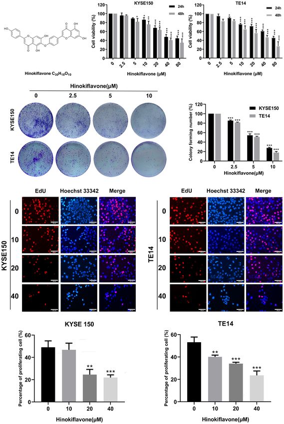
Guo et al.                                                                                                        Hinokiflavone Induces Apoptosis in ESCC

                                              Conclusion: This is the first study to demonstrate the inhibition of ESCC growth and
                                              progression by HF. The underlying mechanism is through blocking the PI3K/AKT/mTOR
                                              signaling pathway, thereby inhibiting cell proliferation and inducing apoptosis. HF can be
                                              used as a complementary/alternative agent for ESCC therapy.
                                              Keywords: esophageal cancer, hinokiflavone, apoptosis, KEGG analysis, molecular docking, PI3K/AKT/mTOR
                                              signal pathway

INTRODUCTION                                                                      effective in cancer therapies (20, 21). Accumulating evidence has
                                                                                  also shown that the PI3K/Akt signaling pathway is activated in
Esophageal cancer is the sixth leading cause of cancer-related                    multiple tumor types and contributes to tumor progression by
deaths and the seventh most common cancer worldwide (1). It is                    promoting tumor cell proliferation, apoptosis resistance, and
well known that there are two dominant histological types of                      distant metastasis (22, 23). Moreover, the PI3K/AKT signaling
esophageal cancer: esophageal adenocarcinoma and esophageal                       pathway plays a vital role in ESCC growth and metastasis
squamous cell carcinoma (ESCC). ESCC accounts for 90% of all                      (24, 25).
esophageal cancer cases in Asia and Africa (1). In recent years,                      In the current study, we extensively explored the anti-tumor
despite the development of multimodal treatment including                         effects of HF on human esophageal cancer cells in vitro and in
surgery combined with chemoradiotherapy and targeted                              vivo as well as the underlying molecular mechanisms. We found
therapy, the prognosis of esophageal cancer patients remains                      that HF exhibited a strong growth inhibitory effect on two ESCC
poor due to the strong malignancy of this cancer (2, 3).                          cell lines (KYSE150 and TE14) and the PI3K/AKT/mTOR
Therefore, the development of novel therapeutic approaches                        signaling pathway plays a crucial role in HF-induced apoptosis
for this disease is urgent and necessary.                                         in these cells. These results indicate that HF has potential anti-
    Natural products have a long history of use for the                           tumor activity, which can be exploited to develop effective
treatment of human disease, which is of great value for                           therapeutic strategies for ESCC.
drug discovery and development (4). Moreover, many
common chemotherapeutic compounds have been derived
from natural products (5, 6). Hinokiflavone (HF) (Figure 1A,                       MATERIALS AND METHODS
C30H18O10) has been extracted from several plants, including
Selaginella tamariscina, Juniperus phoenicea, and Rhus                            Reagents, Antibodies and Kits
succedanea, with high stability. It exhibits several biological                   HF of 98% purity was purchased from Chengdu Biopurify
activities including cytotoxicity (7), anti-HIV-1 reverse                         Phytochemicals, Ltd. (Chengdu, China). Dimethylsulfoxide
transcriptase activity (8), and antioxidant activity (9). Sui                     (DMSO), RPMI 1640 medium, and fetal bovine serum (FBS)
et al. (10) reported that the ethyl acetate extract of Selaginella                were purchased from Sigma Chemical Co. (St Louis, MO, USA).
doederleinii Hieron inhibits proliferation of A549 cell lines by                  Antibodies against b-actin, phosphorylated PI3K [p-PI3K
inducing apoptosis. Magne et al. (11) also identified natural                      (Tyr458 and Tyr199)], PI3K, phosphorylated AKT [p-AKT
flavonoids in HF that are useful therapeutic agents for breast                     (Ser473 and Thr308)], AKT, cleaved Caspase 3 (c-Caspase 3),
cancer. In addition, HF might also be a novel compound to                         phosphorylated mTOR [p-mTOR (Ser2448)], mTOR, matrix
treat melanoma by cell cycle arrest, inducing apoptosis and                       metalloproteinase-2 (MMP2), and MMP9 were purchased
blocking cell migration and invasion (12). However, the                           from Cell Signaling Technology, Inc. (Danvers, MA, USA).
antitumor effect of HF in ESCC and its specific targeted                           Antibodies against Bcl-2 and Bax were purchased from
signaling pathways have not been investigated.                                    Proteintech Group, Inc. (Chicago, IL, USA). RIPA lysis buffer,
    Multiple studies have demonstrated that phosphoinositides,                    phenylmethanesulfonyl fluoride (PMSF), and the bicinchoninic
the multiphosphorylated derivatives of phosphatidylinositol, are                  acid (BCA) protein assay kit were from Beyotime (Nanjing,
membrane-bound signaling molecules that play a crucial role in                    China). Enhanced chemiluminescence detection reagent was
a variety of cellular biological processes (13–15). Under general                 obtained from GE Healthcare Life Sciences Inc. (Marlborough,
physiological conditions, phosphatidylinositol-3-kinase (PI3K)                    MA, USA). SC-79 (AKT activator) was purchased from
activates AKT (protein kinase B) by phosphorylating Thr 308                       MedChemExpress (Shanghai, China). Cell Counting Kit-8
and Ser 473 (p-AKT) (16, 17). p-Akt then phosphorylates                           (CCK-8) was obtained from Dojindo Laboratories (Kumamoto,
several of its downstream effector proteins, such as tuberous                     Japan). Hoechst33342 and BeyoClick™ EdU Cell Proliferation
sclerosis complex 2 (TSC2) and glycogen synthase kinase 3                         Kit with Alexa Fluor 594 and the Annexin V-fluorescein
(GSK3a/b), and these activated proteins subsequently regulate                     isothiocyanate (FITC)/propidium iodide (PI) apoptosis
cell growth, survival, and apoptosis (18, 19). Whereas                            detection kit were purchased from Beyotime Biotechnology
mammalian target of rapamycin (mTOR) was shown to be the                          (Jiangsu, China).
major signaling molecule downstream of TSC2, targeting                                A stock solution of HF dissolved in DMSO (40 mM) was stored
regulation of the PI3K/Akt/mTOR pathway has been proven                           at -20°C and diluted with medium for experimental applications.

Frontiers in Oncology | www.frontiersin.org                                   2                                February 2022 | Volume 12 | Article 833719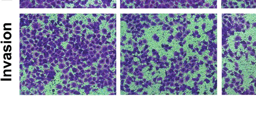
Guo et al.                                                                                                                  Hinokiflavone Induces Apoptosis in ESCC

                                 A                              B

                                 C

                                 D

 FIGURE 1 | Effect of hinokiflavone (HF) on the proliferation of esophageal squamous cell carcinoma (ESCC) cells. (A) Two-dimensional chemical structure of HF
 (C30H18O10). (B) KYSE150 and TE14 cell lines were incubated with different concentrations of HF for 24 and 48 h Cell viability was evaluated using the CCK-8 assay.
 (C) HF reduced colony formation of ESCC cells. KYSE150 and TE14 cells were treated with 0, 2.5, 5, and 10 mM of HF and cultured for 9 days to form colonies.
 Colonies were stained with crystal violet and counted. (D) EdU assay analyzed the antiproliferative effect of HF on ESCC cells. Hoechst 33342 cell nuclei dye
 presented as blue fluorescence cells represent total cells, and EdU positive cells presented as red fluorescence represent proliferating cells. Magnification, ×200;
 scale bars = 25 µm. All the above experimental data are presented as the mean ± SD of three independent experiments. *P < 0.05; **P < 0.01; ***P < 0.001
 compared to vehicle control (0 mM) group.

Cell Culture                                                                          Cell Proliferation Assay
KYSE150 and TE14 cell lines were purchased from the Cell Bank                         Cell viability was determined using the CCK-8 assay. KYSE150
of the Chinese Academy of Sciences. Cells were cultured in RPMI                       and TE14 cells were seeded at a density of 1×10⁴ cells/well in 96-
1640 medium with 10% FBS and 1% penicillin-streptomycin at                            well plates. After 24 h of incubation, the cells were treated with 0,
37°C in a humidified incubator with 5% CO2 and 95% humidity.                           2.5, 5, 10, 20, 40, and 80 mM of HF. Controls were treated with

Frontiers in Oncology | www.frontiersin.org                                       3                                     February 2022 | Volume 12 | Article 833719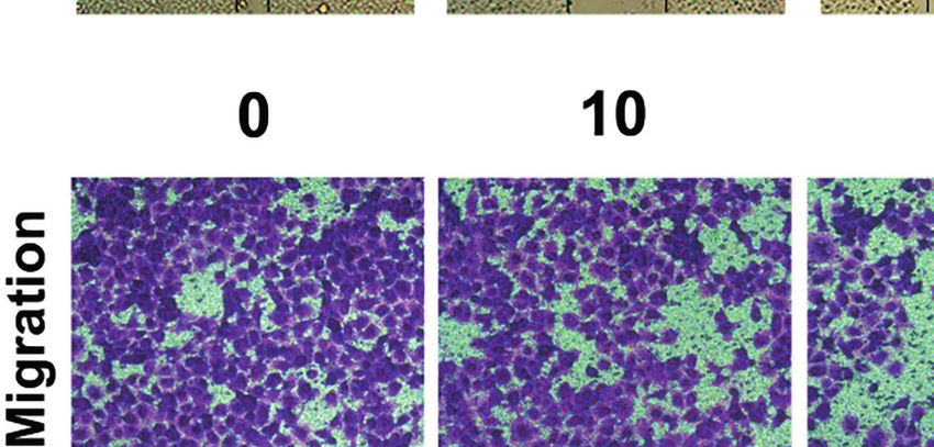
Guo et al.                                                                                                 Hinokiflavone Induces Apoptosis in ESCC

RPMI medium containing DMSO only. After 24 h and 48 h, old                 apoptosis rate was measured by flow cytometry (CytoFLEX,
medium was aspirated and 100 mL of CCK-8 working solution,                 Beckman Coulter, Brea, CA, USA).
diluted to 10% in culture medium, was added to each well and
further incubated for 4 h at 37°C in a humidified incubator. The            Gene Ontology and Kyoto
absorbance at 450 nm was measured using a microplate reader.               Encyclopedia of Genes and Genomes
IC50 values were calculated with Graphpad Prism 8 using data               Pathway Enrichment Analyses
obtained from three independent experiments.                               A total of 355 and 100 potential targets for HF were searched
                                                                           from the Pharma Mapper (http://www.lilab-ecust.cn/
Colony Formation Assay                                                     pharmmapper/) and Swiss target prediction (http://www.
KYSE150 and TE14 cells were plated in 6-well plates (1.0×10³               swisstargetprediction.ch/) databases, respectively. GO and
cells/well). After 24 h of incubation, the cells were incubated with       KEGG pathway enrichment analyses was performed using the
various concentrations (0, 2.5, 5, and 10 mM) of HF for 9 days.            DAVID database (26, 27).
Cells were fixed with 4% paraformaldehyde and then stained
with 0.5% crystal violet for 15 min, after which a dissecting              Molecular Docking
microscope was used to count colonies (> 50 cells).                        The 3D structures of PI3K (code 6pys) and AKT1 (code 4ejn)
                                                                           were downloaded from the Protein Data Bank (https://www.rcsb.
EdU Assay                                                                  org/). Chemical structure data of HF was obtained from
The EdU assay was performed to analyze proliferating cells by              PubChem (http://pubchem.ncbi.nlm.nih.gov/). Molecular
evaluating the incorporation of fluorescent labeled EdU into                docking was implemented using the LibDock module of
replicating DNA in the S phase of cell cycle. The BeyoClick™               the Discovery Studio 2016, and the results were evaluated
EdU Cell Proliferation Kit with Alexa Fluor 594 was used for this          using the LibDock score. A higher libdock score indicates
purpose, according to the manufacturer’s instructions. The                 a higher activity of the small molecule (such as HF) binding to
maximum excitation wavelength of Alexa 594 is 590 nm, and                  the target protein.
the maximum emission wavelength is 615 nm. Briefly, KYSE150
and TE14 cells were seeded in 96-well plates at a cell density of          Western Blot Analysis
5.0×10³ cells/well and treated with different concentrations of HF         KYSE150 and TE14 cells at a cell density of 5.0×10⁶ cells/well were
for 24 h. Edu was added to the culture medium and brought to a             treated with various concentrations of HF (0, 10, 20, and 40 mM)
final concentration of 10 mM, and the cells were cultured for an            for 24 h. Cells were washed twice using PBS and lysed by adding
additional 2.5 h at 37°C. Afterwards, cells were washed twice              RIPA cell lysis buffer containing PMSF. The cell lysate was
using phosphate-buffered saline (PBS) and fixed by 4%                       centrifuged at 12,000 × g for 20 min at 4°C, after which the
paraformaldehyde for 15 min at approximately 25°C. After co-               supernatant was gently aspirated using a pipette. The total protein
staining with Hoechst 33342, the cells were imaged using a                 content in the supernatant was measured using the BCA protein
fluorescence microscope (Leica, Wetzlar, Germany).                          assay kit. Equal amounts of protein (20 mg) were separated by
                                                                           SDS-PAGE (8%–12%). The separated proteins were then
Morphological Analysis by Hoechst                                          transferred onto a PVDF membrane. The membranes were
33342 Staining                                                             blocked with Quickblock blocking buffer (Beyotime
Alterations in cell morphology, including cell shrinkage and               Biotechnology, China) for 0.5 h and incubated with specific
apoptotic body formation, were analyzed after Hoechst 33342                primary antibodies overnight at 4°C on a shaker. Membranes
staining to identify apoptotic cells. KYSE150 and TE14 cells were          were washed three times for 10 min each with Tris-buffered saline-
seeded in 96-well plates at a density of 5.0×10³ cells/well and            Tween 20 (TBST), completed by incubation with horseradish
cultured for 24 h. After treatment with different concentrations           peroxidase (HRP)-conjugated secondary antibody for 1 h at
of HF (0, 10, 20, and 40 mM) for 24 h, the cells were washed twice         approximately 25°C. Our study used the following primary
using PBS and fixed in 4% paraformaldehyde for 15 min. Then,                antibodies: b-actin (1:1,000), phosphorylated PI3K (p-PI3K)
the cells were stained using Hoechst 33342 working solution                (Tyr458, 1:1,000 and Tyr199, 1:1,000), PI3K (1:1,000),
diluted well according to the instructions for 15 min at 25°C and          phosphorylated AKT (p-AKT) (Ser473, 1:1,000; Thr308,
analyzed under a fluorescence microscope (Zeiss, Axiovert 200,              1:1,000), AKT (1:1,000), c-Caspase 3 (1:1,000), p-mTOR
Oberkochen, Germany).                                                      (Ser2448, 1:1,000), mTOR (1:1,000), matrix metalloproteinase-2
                                                                           (MMP2) (1:1,000), MMP9 (1:1,000), Bcl-2 (1:2,000), and Bax
Quantification of Apoptosis                                                 (1:1,000). The secondary antibody was HRP-conjugated goat
Further verification of HF-induced apoptosis was carried out                anti-rabbit antibody (1:10,000; Cell Signaling Technology, Inc.).
using the Annexin V-FITC/PI apoptosis detection kit. After                 After three 10 min washes with TBST, the protein band was
treatment with HF (0, 10, 20, and 40 mM) for 24 h, KYSE150                 visualized using enhanced chemiluminescence detection reagent
and TE14 cells at a cell density of 1.0×10⁶ cells/well were                (GE Healthcare Life Sciences). Finally, protein bands were
trypsinized, collected by centrifugation, and washed twice with            quantified by densitometric analysis using ImageJ software, and
ice-cold PBS. The cells were then co-stained with 5 mL FITC-               the protein amounts were expressed relative to the corresponding
Annexin V and 5 mL PI for 15 min at 20–25 °C in the dark. The              reference protein.

Frontiers in Oncology | www.frontiersin.org                            4                                February 2022 | Volume 12 | Article 833719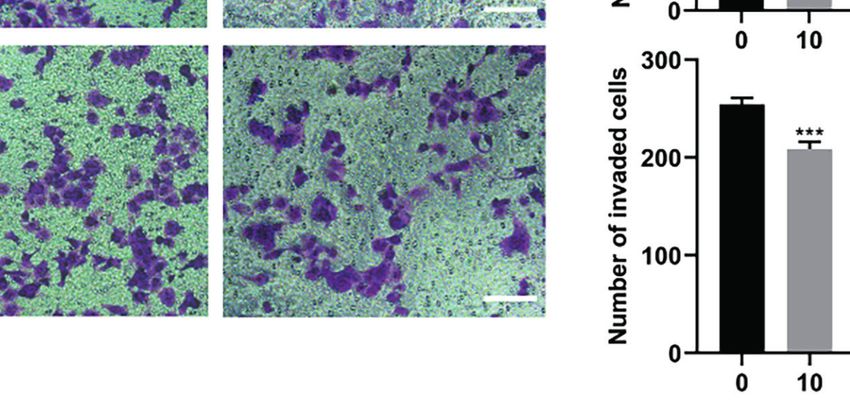
Guo et al.                                                                                                 Hinokiflavone Induces Apoptosis in ESCC

Wound-Healing Migration Assay                                             all animals were fed normal diet for another 3 days, after which
KYSE150 cells were grown to a density of approximately 80-90%             they were euthanized by cervical dislocation. The tumors were
in 6-well plates, after which the cell monolayer was scraped using        excised, weighed, and immunohistochemical staining was
a sterile 100-mL pipette tip to create “wounds”. Reduced serum            performed. The internal organs were collected for hematoxylin
medium (1 mL) containing HF (0, 10, 20, or 40 mM) was gently              and eosin (H&E) staining.
added to the wells along the lateral wall to ensure that it covered
the entire bottom. After incubation for 24 h in an incubator, the         Immunohistochemistry
cells were washed and fixed with 4% paraformaldehyde (1 mL/                Tumor tissues from mice were formalin fixed, then embedded in
well), and images were acquired under a microscope (Zeiss, Jena,          paraffin and sectioned at 4 µm thin sections were used for
Germany). Cell movement distances were measured by                        examination. For staining, tissue sections were deparaffinized
ImageJ (v1.8.0).                                                          and rehydrated following standard protocols. Antigen retrieval of
                                                                          tissues in our study was performed by boiling in sodium citrate
Cell Migration and Invasion Assay                                         buffer (10 mM, pH 6.0) for 10 min, after which the samples were
Transwell migration and invasion assays were implemented by               treated with 3% H2O2 for an additional 10 min. Each section was
using Boyden chamber (8 mm pore size, Corning, NY, USA). For              blocked with 10% goat serum albumin in a humidified chamber
migration assay, KYSE150 cells (2.0 ×10⁴ cells in 100 mL serum-           for 1 h at approximately 20–25°C. Then, tissue sections were
free medium) were added into the upper chamber, and 600 mL of             incubated with specific primary antibodies (p-AKT, p-mTOR,
medium with 10% FBS was added into the lower chamber. For                 Bax, and c-Caspase 3) overnight at 4°C in a humidified chamber.
invasion assay, the upper surface of the Transwell membrane was           After further washing, the sections were incubated with
coated with Matrigel (BD Biosciences, Franklin lakes, NJ, USA)            secondary antibody for 30 min at approximately 25°C,
diluted 1:8 with serum-free medium before adding cells. For both          followed by incubation with 3,3’-diaminobenzidine and
assays, various concentrations of HF (0, 10, 20, and 40 mM) were          counterstaining with hematoxylin. Stained sections were
added into the lower chamber. After incubation for 24 h, the              imaged under a microscope (Leica, DM4000B).
medium was discarded, the cells were gently removed from the
upper surface of the Transwell membrane with a cotton swab,               H&E Staining
and the migrated or invaded cells on the lower surface were fixed          Mouse organs (heart, liver, spleen, lung, and kidney) were fixed
with 4% paraformaldehyde, after which they were stained with              in formaldehyde, embedded in paraffin, and sectioned at 4 mm
0.5% crystal violet. Using microscopy, the number of stained cells        thickness. After deparaffinization and rehydration, tissue
on the lower surface was counted in five random fields per                  sections were stained with hematoxylin for 10 min, 1%
Transwell membrane (×100 magnification), and the Image J                   ethanol-hydrochloric acid for 30 s, and eosin solution for 3
software was used to enumerate the migrated/invaded cells.                min. The sections were dehydrated in graded alcohol, after which
                                                                          they were cleared with xylene and finally mounted using
Mouse Xenograft Model                                                     neutral balsam.
All experimental procedures and manipulations involving
animals were performed in compliance with the guidelines of               Statistical Analysis
the National Institutes of Health Guide for the Care and Use of           All data are presented using the mean ± SD of three independent
Laboratory Animals. Moreover, all animal experiments were                 experiments. Statistical analysis was performed using GraphPad
approved by the Institutional Animal Care and Use Committee               Prism version 8 (GraphPad Software, San Diego, CA, USA).
of the Second Affiliated Hospital of Harbin Medical University             Student’s t-test was used to compare two groups, and one-way
(SYDW2021-046). Female BALB/c-nu athymic mice, at 6 weeks                 analysis of variance was used for multiple comparisons.
and weighing 14-17 g, were purchased from Beijing Vital River             Statistically significant P-values are labeled as follows: *P <
Laboratory Animal Technology Co., Ltd. (Beijing, China). Mice             0.05, **P < 0.01, ***P < 0.001.
were maintained in a specific pathogen-free (SPF) grade rearing
environment using sterile water and food feeding. KYSE150 cells
(1.0×10⁷ cells in 100 µL PBS) were injected subcutaneously into           RESULTS
the right shoulders of the mice (n=18). Tumor size was measured
every 3 days using a caliper. The formula for calculating tumor           HF Inhibits the Proliferation of ESCC Cells
volume was as follows: V = 0.5×L×W2 (L, tumor longest                     The chemical structure of HF was drawn with ChemDraw
diameter; W, tumor shortest diameter). When the tumor                     software as shown in Figure 1A. KYSE150 and TE14 cell lines
volume reached 50 mm3, mice were randomized into three                    were treated with HF at different concentrations (0, 2.5, 5, 10, 20,
groups (n=6/group; control, 25 mg/kg HF, and 50 mg/kg HF).                40, or 80 mM) for 24 or 48 h, and cell proliferation was assessed
Next, HF (25 mg/kg and 50 mg/kg in 200 mL saline) was                     by CCK-8 assay, as described in the Methods. As shown in
administered by intraperitoneal injection to the two treatment            Figure 1B, HF significantly inhibited the cell viability of both
group mice daily for 21 days. The control mice received                   ESCC cell lines in a concentration- and time-dependent manner.
saline alone. The body weight and tumor volume of mice were               Furthermore, the IC50 values for KYSE150 cells treated with HF
measured every 3 days. After completion of drug administration,           for 24 h and 48 h were 27.92 mM and 24.91 mM, respectively,

Frontiers in Oncology | www.frontiersin.org                           5                                February 2022 | Volume 12 | Article 833719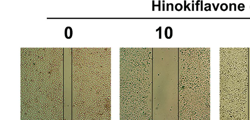
Guo et al.                                                                                                                   Hinokiflavone Induces Apoptosis in ESCC

while the respective values for TE14 cells were 26.21 mM and                          from 49.1% and 53.2% to 21.9% and 23.6%, respectively, after 40
22.07 mM. Long-term cell viability assays (colony formation                           mM HF treatment for 24 h. Taken together, these results indicate
assays) showed that KYSE150 and TE14 cells formed                                     that HF effectively inhibits ESCC cell proliferation.
significantly fewer colonies with increasing HF concentrations.
(Figure 1C). The anti-proliferative activity of HF was further                        HF Induces Apoptosis in ESCC Cells
verified using an EdU assay. As shown in Figure 1D, the                                We next explored whether HF induces apoptosis in ESCC cells.
proportion of proliferating KYSE150 and TE14 cells decreased                          Induction of apoptosis by HF was first evaluated by Hoechst

                                   A

                               B

                               C

                               D

 FIGURE 2 | Induction of apoptosis in ESCC cells by HF. (A) Hoechst 33342 nuclear dye was used to stain the KYSE150 and TE14 cells and utilized fluorescence
 microscopy to detect HF induced apoptotic nuclear morphological alterations (indicated by red arrows). Magnification, ×200; scale bars = 25 µm. (B) Percentage of
 KYSE150 and TE14 cells apoptosis after HF treatment was measured using Annexin V-fluorescein isothiocyanate (FITC)/propidium iodide (PI) double staining and
 analyzed by flow cytometry. (C, D) Expression levels of Bcl-2, Bax, and c-Caspase 3 were detected by western blot and quantitated via densitometric analysis using
 ImageJ software, while the expression level of b-actin was used as an internal control. The relative expression of experimental histones was calculated according to
 the protein scale set to 1 for 0 mM control group. All the above experimental data are presented as the mean ± SD of three independent experiments. *P < 0.05;
 **P < 0.01; ***P < 0.001 compared to vehicle (0 mM) group.

Frontiers in Oncology | www.frontiersin.org                                       6                                      February 2022 | Volume 12 | Article 833719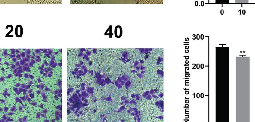
Guo et al.                                                                                                         Hinokiflavone Induces Apoptosis in ESCC

33342 staining assay. As shown in Figure 2A, HF-treated                        GO and KEGG Pathway
KYSE150 and TE14 cell cultures contained dense and heavily                     Enrichment Analyses
stained apoptotic cells, suggesting that HF might cause various                GO enrichment analysis of HF was performed on the 355
degrees of cell shrinkage, nuclear fragmentation, and condensed                putative targets of HF that were collected from the Pharma
nuclei formation. Furthermore, flow cytometric analysis of                      Mapper database to determine the relative significance with
Annexin V-FITC and PI staining revealed that the ratio of                      regards to biological processes, cell components, and molecular
early and late apoptotic cells increased in a dose-dependent                   functions. The top 20 significantly enriched GO terms are listed
manner after treatment with HF for 24 h. As shown in                           in Figures 3A–C and Table S1. KEGG pathway enrichment
Figure 2B, the mean apoptosis ratios (from three independent                   analysis of HF was performed, and the top 20 targets are listed in
experiments) were 11.0% (0 mM), 19.2% (10 mM), 24.2%                           Figure 3D and Table S2. The results show pathway enrichment
(20 mM), 40.6% (40 mM) for KYSE150 cells; 3.3% (0 mM),                         for the GO and KEGG terms “negative regulation of apoptotic
22.3% (10 mM), 29.9% (20 mM), and 55.6% (40 mM group)                          process,” “pathways in cancer,” and “PI3K-AKT signaling
for TE14 cells.                                                                pathway,” based on the Pharma Mapper database. Similarly,
    Next, apoptosis-related proteins Bcl-2 (anti-apoptotic) and                we obtained 100 putative targets of HF from the Swiss database.
Bax (pro-apoptotic) were analyzed by western blot. Caspase-3,                  The GO enrichment analysis of the Swiss database is shown in
which is a member of the caspase family and is cleaved during                  Figures S1A–C and Table S3, and the KEGG pathway
apoptosis (28–30), was also analyzed by western blot. Treatment                enrichment analysis is shown in Figure S1D and Table S4.
of KYSE150 and TE14 cells with HF significantly reduced the                     Interestingly, the results analyzed according to the Swiss
protein level of Bcl-2, while increasing the protein levels of Bax             database were similar to those analyzed by the Pharma Mapper
and c-Caspase 3 in a dose-dependent manner (Figures 2C, D).                    database. In summary, the analysis results of both databases
These results indicate that HF induced apoptosis of ESCC cells                 indicate that HF might target the PI3K-AKT signaling pathway,
involves the mitochondrial pathway.                                            leading to induction of apoptosis.

                            A                                                       B

                            C                                                       D

 FIGURE 3 | Gene Ontology (GO) and Kyoto Encyclopedia of Genes and Genomes (KEGG) pathway analyses of HF. Top 20 signaling pathways of HF in (A) GO-BP
 pathway analysis, (B) GO-CC pathway analysis, and (C) GO-MF pathway analysis. (D) Top 20 signaling pathways of HF in KEGG enrichment pathway analysis.

Frontiers in Oncology | www.frontiersin.org                                7                                   February 2022 | Volume 12 | Article 833719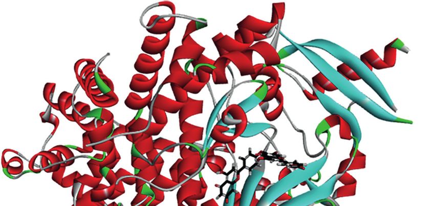
Guo et al.                                                                                                             Hinokiflavone Induces Apoptosis in ESCC

                          A                                               B

                          C                                               D

 FIGURE 4 | Schematic representation of three- and two-dimensional molecular docking models. (A) Three-dimensional view of HF and PI3K docking scenario.
 (B) Two-dimensional view of HF and PI3K docking scenario. (C) Three-dimensional view of HF and AKT1 docking scenario. (D) Two-dimensional view of HF and
 AKT1 docking scenario.

HF Is Predicted to Have Direct Interaction                                            HF Inhibits the PI3K/AKT/mTOR Pathway
With PI3K and AKT1                                                                    in ESCC Cells
To predict the possible mutual binding mode of HF with PI3K                           To verify the interaction of HF with PI3K/AKT, we analyzed HF-
and Akt, we further used molecular docking analysis. As shown                         treated KYSE150 and TE14 cells via western blotting. Our results
in Figure 4A, HF was predicted to have a strong interaction with                      showed that the protein levels of p-PI3K, p-AKT, and p-mTOR
PI3K (LibDock score 115.982). The molecular docking of other                          in KYSE150 and TE14 cells were significantly reduced by HF
conformations of HF with the protein PI3K is shown in Figures                         treatment at 10, 20, and 40 mM, whereas total expression levels of
S2A, B. The binding affinity was attributed to the following:                          PI3K, AKT, and mTOR were not significantly changed
hydrogen bonding with the ARG-770, GLN-895, SER-854, and                              (Figures 5A–C). To verify whether HF induces apoptosis
ASP-933 residues; the pi-pi T-shaped interaction with the TRP-                        through the inhibition of PI3K/AKT/mTOR pathway,
780 and TYR-836 residues; the pi-alkyl interactions with the ILE-                     KYSE150 cells were treated with HF in the presence or absence
800, ILE-848, VAL-850, and MET-922 residues of PI3K                                   of SC-79, a specific AKT activator (31). As shown in Figures 5D,
(Figure 4B). Moreover, the analysis results show that HF could                        E, SC-79 effectively enhanced the levels of phosphorylated
also interact strongly with protein AKT1 (LibDock score                               proteins including p-AKT and p-mTOR. Meanwhile,
1 43 . 7 52 , F i g u r e 4 C ) . M o l e c u l a r d o c k i n g o f o t h e r       pretreatment with SC-79 improved the inhibitory effect of HF
conformations of HF with AKT1 is shown in Figures S2C, D.                             on the protein expression of p-AKT, p-mTOR, and Bcl-2.
The binding affinity was attributed to the following: hydrogen                         Furthermore, the effect of HF to increase the expression
bonding with the SER-205, GLY-294, and ASP-274 residues; the                          amounts of apoptosis-related proteins (Bax and c-Caspase 3)
pi-alkyl interactions with the LYS-268, VAL-270, and LEU-264                          could be reversed by pretreatment with SC-79 as well. We also
residues, and pi-anion interactions with the ARG-273 and ASP-                         found an increased viability of KYSE150 cells in the HF and SC-
274 residues of AKT1 (Figure 4D). Therefore, in common with                           79 co-treatment group compared with HF alone treatment
the results of KEGG pathway enrichment analysis, HF showed                            (Figure 5F), suggesting that activation of AKT attenuated the
high binding activity with PI3K and AKT in this study.                                ability of HF to inhibit cell proliferation. These results indicate

Frontiers in Oncology | www.frontiersin.org                                       8                                February 2022 | Volume 12 | Article 833719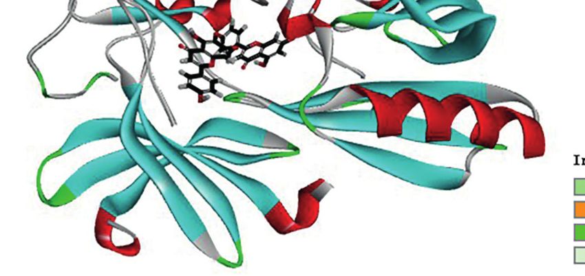
Guo et al.                                                                                                                Hinokiflavone Induces Apoptosis in ESCC

                             A                                                           D

                                   B                                                 E

                                   C                                                         F

 FIGURE 5 | HF induces apoptosis of ESCC cells by negatively regulating the PI3K/Akt/mTOR pathway. (A–C) KYSE150 and TE14 cells received various
 concentrations of HF intervention for 24 h, and the expression of pathway related proteins PI3K, p-PI3k, Akt, p-Akt, mTOR, and p-mTOR was detected by western
 blotting and quantified via densitometric analysis. (D, E) KYSE150 cells were pretreated with SC-79 (10 mM) for 24 h, and the expression of apoptosis related
 proteins Bcl-2, Bax and c-caspase 3 was determined by western blotting. (F) KYSE150 cells were first cultured with HF with or without addition of SC-79 (10 mM)
 for 24 h, and cell viability was detected using CCK-8 assays. The above experimental data are presented as the mean ± SD of three independent experiments.
 *P < 0.05; **P < 0.01; ***P < 0.001 compared to the control group.

that the anticancer effect of HF on ESCC involves regulation of                     control group, HF treatment significantly inhibited the Transwell
PI3K/AKT/mTOR pathway.                                                              migration and invasion of ESCC cells in a dose-dependent manner
                                                                                    (Figure 6B). Studies have shown that MMP family proteins play an
HF Suppresses Migration and Invasion in                                             important role in the regulation of cell migration and invasion
ESCC Cells                                                                          abilities, and that MMPs are always upregulated in invasive
Esophageal cancer patients with distant metastases have a very poor                 epithelial cancers (34, 35). Therefore, we explored whether the
prognosis (32), and the migration and invasion abilities of tumor cells             protein levels of MMP2 and MMP9 are associated with alterations
are vital factors affecting the process of tumor metastasis (33).                   in ESCC cell migration and invasion following treatment with HF. As
Therefore, we evaluated whether HF could inhibit the migration                      shown in Figures 6C, D, we found that HF significantly
and invasion of ESCC cells. As revealed by wound-healing assay                      downregulated the expression of MMP2 and MMP9 in ESCC cells.
(Figure 6A), the migratory ability of KYSE150 cells gradually became                Altogether, these results suggest that HF possesses an effective ability
weaker with higher HF concentrations. Moreover, compared with the                   to inhibit ESCC cell migration and invasion.

Frontiers in Oncology | www.frontiersin.org                                     9                                     February 2022 | Volume 12 | Article 833719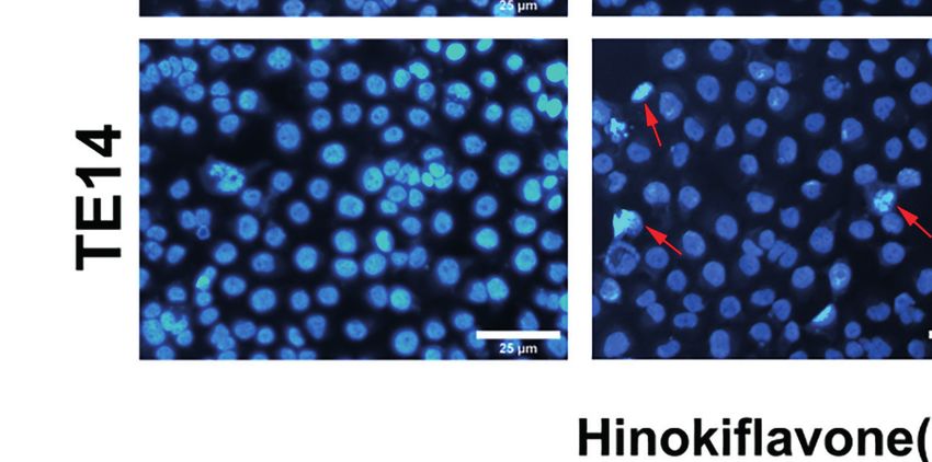
Guo et al.                                                                                                                   Hinokiflavone Induces Apoptosis in ESCC

                               A

                               B

                               C

                                D

 FIGURE 6 | HF inhibits ESCC cell migration and invasion. (A) Cell migration was measured in a scratch-wound assay. KYSE150 cells were cultured until reaching
 approximately 80-90% cell density, the culture was scratched as described in Methods, and further cultured with various concentrations of HF (0, 10, 20, and 40
 mM) for 24 h, after which the cells were fixed and photographed. Magnification, ×100; scale bars = 100 µm. The distance of cell migration from the same region was
 quantified using ImageJ. (B) KYSE150 cells were placed in the top chamber on the Transwell membrane with serum-free medium and the upper surface of the
 Transwell membrane was coated with/without Matrigel. Cells were treated using HF with a concentration gradient for 24 h, and then the cells were fixed and
 photographed using a microscope after crystal violet staining. Magnification, ×100; scale bars = 100 µm. Migrated and invaded cells were counted as described in
 the Methods. (C, D) KYSE150 cells and TE14 cells were treated with various concentrations of HF for 24 h, and then the cells were collected for protein extraction.
 The extracted proteins were later used for western blot analysis to determine the protein expression levels of MMP2 and MMP9. All the above experimental data are
 presented as the mean ± SD of three independent experiments. *P < 0.05; ***P < 0.001 compared to 0 mM vehicle group.

Antitumor Effect of HF in Mouse Xenograft                                             D). The mean size (or weight) of the tumors in HF 25 mg/kg, and
Model of ESCC                                                                         HF 50 mg/kg group was reduced to 348.9 mm3 (or 0.315 g) and
To investigate whether the antitumor effect of HF in vivo is                          126.9 mm3 (or 0.08 g), compared with 738.9 mm3 (or 0.525 g) in
consistent with its in vitro effects, ESCC xenografts were                            the control group (Figures 7B, D). As a crucial indicator of health,
established by subcutaneously transplantation of KYSE150 cells                        the average body weights of the control and HF-treated mice were
into BALB/c-nu mice, followed by treatment with HF (saline                            not significantly different at all time points (Figure 7E). Moreover,
control, 25 mg/kg HF, 50 mg/kg HF) for 3 weeks, as described in                       immunohistochemical analysis showed that HF suppressed the
the Methods (Figure 7A). Results from the animal experiments                          protein expression levels of p-AKT and p-mTOR, while
showed that transplanted tumors in the HF intervention groups                         upregulating those of Bax and c-Caspase 3 (Figure 7F). As
grew much slower than those in the control group (Figures 7B–                         shown in Figure 7G, the results of H&E staining experiments

Frontiers in Oncology | www.frontiersin.org                                      10                                      February 2022 | Volume 12 | Article 833719
Guo et al. Hinokiflavone Induces Apoptosis in ESCC
A
B C
D E
F G
FIGURE 7 | HF suppresses the growth of mouse ESCC xenograft tumors. (A) BALB/c-nu mice implanted with KYSE150 xenograft tumors were treated daily with
an equal volume of saline (control group) or HF (25 or 50 mg/kg) by intraperitoneal injection for total 21 days. (B) The tumor volume was measured every 3 days, and
the difference in tumor volume between HF treated and control mice is shown. ***P < 0.001. (C) After euthanasia of mice, subcutaneous xenografts were removed
and photographed. (D) The removed xenograft tumors were weighed and graphed for statistical analysis. ***P < 0.001 vs the control group. (E) Body weights of
tumor-bearing mice were measured every 3 days in the HF-treated and control groups. (F) Expression levels of p-AKT, p-mTOR, Bax, and cleaved-Caspase 3 were
detected by immunohistochemistry in the xenograft tumors. Magnification, ×400; scale bars = 50 µm. (G) H&E staining of the heart, liver, spleen, lung, and kidney of
experimental mice shows no pathological changes in the organ tissues of any group. Magnification, ×200; scale bars = 100 µm.
Frontiers in Oncology | www.frontiersin.org 11 February 2022 | Volume 12 | Article 833719Guo et al. Hinokiflavone Induces Apoptosis in ESCC
on the heart, liver, spleen, lung, and kidney of mice exhibited no blot analysis showed that the activation (phosphorylation) of
obvious pathological changes, such as necrosis, edema, or PI3K, as indicated by levels of p-PI3K, was inhibited by HF,
hemorrhage, indicating that HF possessed no major organ- whereas expression of PI3K protein itself was not significantly
related toxicity. Consistent with the experimental results in vitro, altered. This suggested that HF can regulate post-translational
these data suggest that HF inhibits tumor growth by inducing modification of PI3K by physically interacting with the protein,
apoptosis and does not have apparent adverse effects at the as shown by the results of the GO enrichment, KEGG
dose tested. pathway, and molecular docking analyses. It is plausible that
HF may directly attenuate the phosphorylation of PI3K
at tyrosine 199, as reported in another study (40). The PI3K/
Akt/mTOR pathway is a crucial signaling cascade in living
DISCUSSION organisms that has to be activated in a variety of cancers and
Despite progressive advances in the treatment of esophageal to regulate cell proliferation, invasion, and migration (20, 41).
cancer, its mortality rate remains high worldwide. Currently, We observed (Figures 5D–F) that SC-79 usage could activate the
surgery and chemotherapy remain the main treatment PI3K/AKT/mTOR pathway and partially counteract the pro-
modalities for esophageal cancer patients. However, since apoptotic and anti-proliferative effects of HF in ESCC cells.
chemotherapeutics have their own disadvantages, such as drug Overall, our results clearly show that the PI3K/AKT/mTOR
resistance and systemic toxicity, more effective therapeutic pathway was inhibited by HF, which induced apoptosis in
strategies with fewer side-effects are urgently needed. Many ESCC cells.
studies have demonstrated the promising antitumor activity of Metastasis is known to be a multistep biological process
some naturally active compounds (36). Our study is the first to wherein subsets of cancer cells spread from the primary tumor
indicate that HF inhibits the growth of ESCC cells. Furthermore, to distant tissues or other organs to form metastases affects the
we comprehensively and profoundly explored the detailed body’s normal physiological functions, which is the leading cause
molecular mechanisms underlying the antitumor effects of HF of cancer-related death (42). Moreover, for esophageal cancer
on ESCC in vitro and in vivo experiments. patients, approximately 50% have already developed distant
In this study, the CCK-8 and colony formation assays metastasis by the time of initial diagnosis (43, 44). Members of
demonstrated that HF had an effective anti-proliferative effect the MMP family of proteins, particularly MMP2 and MMP9,
on KYSE150 and TE14 cells, which was validated by the EdU play a crucial role in cancer metastasis (34, 45, 46). The results of
assay results. To investigate the anti-proliferation mechanism of the migration and invasion assay we performed showed HF
HF in ESCC cells, we used Hoechst 33342 staining assay to exhibited an obvious inhibitory effect on the metastatic potential
identify apoptotic features of cell shrinkage, nuclear of ESCC cells. We also found that HF significantly reduced
fragmentation, and condensed nuclei formation in HF-treated MMP2 and MMP9 expression in KYSE150 and TE14 cells. These
KYSE150 and TE14 cells. Flow cytometric analysis further results suggest that HF can significantly hinder the metastasis of
illustrated induction of apoptosis in ESCC cells by HF in a ESCC cells.
concentration-dependent manner. Apoptosis is an orderly Finally, to verify the antitumor effect of HF in vivo, an
enzymatic cascade that induces DNA fragmentation into esophageal cancer cell xenograft model was used in our
characteristic nucleosome fragments and culminates in cell study. Our experimental results showed that HF treatment
death (30, 37, 38). Previous study has shown that apoptosis via significantly inhibited xenograft tumor growth in mice. In
the mitochondrial pathway is commonly regulated by Bcl-2 addition, the body weight of the experimental group mice
family proteins, with representative proteins such as the anti- receiving HF treatment was not significantly different from
apoptotic protein Bcl-2 and the pro-apoptotic protein Bax (39). that of the control group mice. H&E staining also revealed
In this context, our experimental results proved that HF no significant histopathological alterations in the heart, liver,
intervention can cause a decreased expression of Bcl-2 protein lung, and kidney tissues of HF treated BALB/c-nu mice,
and an increase in Bax and c-Caspase 3 expression, suggesting indicating that HF did not induce significant systemic toxicity
that HF induced apoptosis of ESCC cells occurs through the at the tested doses.
mitochondrial pathway.
We next used two databases, the Pharma Mapper and Swiss
database, to screen the putative molecular targets of HF. The GO CONCLUSION
functional enrichment analysis showed that the predicted
potential targets of HF were significantly enriched in “negative Data from our in vitro, in silico, and in vivo experiments showed
regulation of apoptotic process.” Furthermore, KEGG pathway that HF exerts antitumor effects against ESCC by inhibiting
analysis revealed that HF might affect the PI3K-AKT signaling tumor growth and promoting apoptosis through the PI3K/AKT/
pathway. Our molecular docking analysis further indicated mTOR signaling pathway. HF may also possibly inhibit
that HF can dock with PI3K and AKT1. Western blot analysis metastasis by regulating MMPs. Overall, these findings reveal
demonstrated a progressive decrease in p-PI3K, p-AKT, and p- the underlying mechanisms by which HF inhibits the growth of
mTOR protein levels with increasing concentrations of HF human ESCC and provide new evidence supporting therapeutic
in KYSE150 and TE14 cells (Figure 5A). Interestingly, western potential of HF in ESCC.
Frontiers in Oncology | www.frontiersin.org 12 February 2022 | Volume 12 | Article 833719Guo et al. Hinokiflavone Induces Apoptosis in ESCC
DATA AVAILABILITY STATEMENT AUTHOR CONTRIBUTIONS
The datasets presented in this study can be found in online JG, SZ, and LZ contributed to the conception and design of this
repositories. The names of the repository/repositories and accession research. JG and SZ performed the experiments. JG and SZ analyzed
number(s) can be found in the article/Supplementary Material. the results. JG, SZ, JW, PZ, TL, and LZ wrote the paper. All authors
contributed to the article and approved the submitted version.
ETHICS STATEMENT SUPPLEMENTARY MATERIAL
The animal study was reviewed and approved by the Institutional The Supplementary Material for this article can be found online
Animal Care and Use Committee of The Second Affiliated at: https://www.frontiersin.org/articles/10.3389/fonc.2022.
Hospital of Harbin Medical University (SYDW2021-046). 833719/full#supplementary-material
14. Wymann MP, Schneiter R. Lipid Signalling in Disease. Nat Rev Mol Cell Biol
REFERENCES (2008) 9(2):162–76. doi: 10.1038/nrm2335
1. Sung H, Ferlay J, Siegel RL, Laversanne M, Soerjomataram I, Jemal A, et al. 15. Engelman JA, Luo J, Cantley LC. The Evolution of Phosphatidylinositol 3-
Global Cancer Statistics 2020: GLOBOCAN Estimates of Incidence and Kinases as Regulators of Growth and Metabolism. Nat Rev Genet (2006) 7
Mortality Worldwide for 36 Cancers in 185 Countries. CA Cancer J Clin (8):606–19. doi: 10.1038/nrg1879
(2021) 71(3):209–49. doi: 10.3322/caac.21660 16. Vanhaesebroeck B, Stephens L, Hawkins P. PI3K Signalling: The Path to
2. Bang YJ, Van Cutsem E, Feyereislova A, Chung HC, Shen L, Sawaki A, et al. Discovery and Understanding. Nat Rev Mol Cell Biol (2012) 13(3):195–203.
Trastuzumab in Combination With Chemotherapy Versus Chemotherapy doi: 10.1038/nrm3290
Alone for Treatment of HER2-Positive Advanced Gastric or Gastro- 17. Vicinanza M, D’Angelo G, Di Campli A, De Matteis MA. Phosphoinositides
Oesophageal Junction Cancer (Toga): A Phase 3, Open-Label, Randomised as Regulators of Membrane Trafficking in Health and Disease. Cell Mol Life Sci
Controlled Trial. Lancet (2010) 376(9742):687–97. doi: 10.1016/S0140-6736 (2008) 65(18):2833–41. doi: 10.1007/s00018-008-8353-2
(10)61121-X 18. Inoki K, Li Y, Zhu T, Wu J, Guan KL. TSC2 is Phosphorylated and Inhibited
3. Yang H, Liu H, Chen Y, Zhu C, Fang W, Yu Z, et al. Neoadjuvant Chemor- by Akt and Suppresses Mtor Signalling. Nat Cell Biol (2002) 4(9):648–57.
Adiotherapy Followed by Surgery Versus Surgery Alone for Locally Advanced doi: 10.1038/ncb839
Squamous Cell Carcinoma of the Esophagus (NEOCRTEC5010): A Phase III 19. Cross DA, Alessi DR, Cohen P, Andjelkovich M, Hemmings BA. Inhibition of
Multi-Center, Randomized, Open-Label Clinical Trial. J Clin Oncol (2018) 36 Glycogen Synthase Kinase-3 by Insulin Mediated by Protein Kinase B. Nature
(27):2796–803. doi: 10.1200/JCO.2018.79.1483 (1995) 378(6559):785–9. doi: 10.1038/378785a0
4. Newman DJ, Cragg GM. Natural Products as Sources of New Drugs Over the 20. Serra V, Markman B, Scaltriti M, Eichhorn PJ, Valero V, Guzman M, et al.
30 Years From 1981 to 2010. J Nat Prod (2012) 75(3):311–35. doi: 10.1021/ NVP-BEZ235, a Dual PI3K/Mtor Inhibitor, Prevents PI3K Signaling and
np200906s Inhibits the Growth of Cancer Cells With Activating PI3K Mutations. Cancer
5. Rane R, Karpoormath R. “Discovery and Development of New Anticancer Res (2008) 68(19):8022–30. doi: 10.1158/0008-5472.CAN-08-1385
Drugs Inspired From Natural Product Leads” Part 1. Anticancer Agents Med 21. Roulin D, Waselle L, Dormond-Meuwly A, Dufour M, Demartines N,
Chem (2015) 15(5):536. doi: 10.2174/187152061505150514130306 Dormond O. Targeting Renal Cell Carcinoma With NVP-BEZ235, a Dual
6. Rane R, Karpoormath R. “Discovery and Development of New Anticancer PI3K/Mtor Inhibitor, in Combination With Sorafenib. Mol Cancer (2011) 10
Drugs Inspired From Natural Product Leads” Part 2. Anticancer Agents Med (undefined):90. doi: 10.1186/1476-4598-10-90
Chem (2015) 15(8):932. doi: 10.2174/187152061508150817112012 22. Ko FC, Chan LK, Tung EK, Lowe SW, Ng IO, Yam JW. Akt Phosphorylation
7. Lin YM, Chen FC, Lee KH. Hinokiflavone, a Cytotoxic Principle From Rhus of Deleted in Liver Cancer 1 Abrogates Its Suppression of Liver Cancer
Succedanea and the Cytotoxicity of the Related Biflavonoids. Planta Med Tumorigenesis and Metastasis. Gastroenterology (2010) 139(4):1397–407.
(1989) 55(2):166–8. doi: 10.1055/s-2006-961914 doi: 10.1053/j.gastro.2010.06.051
8. Lin YM, Anderson H, Flavin MT, Pai YH, Mata-Greenwood E, Pengsuparp T, 23. Vivanco I, Sawyers CL. The Phosphatidylinositol 3-Kinase AKT Pathway in
et al. In Vitro Anti-HIV Activity of Biflavonoids Isolated From Rhus Human Cancer. Nat Rev Cancer (2002) 2(7):489–501. doi: 10.1038/nrc839
Succedanea and Garcinia Multiflora. J Nat Prod (1997) 60(9):884–8. 24. Li B, Tsao SW, Li YY, Wang X, Ling MT, Wong YC, et al. Id-1 Promotes
doi: 10.1021/np9700275 Tumorigenicity and Metastasis of Human Esophageal Cancer Cells Through
9. Wang G, Yao S, Zhang XX, Song H. Rapid Screening and Structural Activation of PI3K/AKT Signaling Pathway. Int J Cancer (2009) 125
Characterization of Antioxidants From the Extract of Selaginella (11):2576–85. doi: 10.1002/ijc.24675
Doederleinii Hieron With DPPH-UPLC-Q-TOF/MS Method. Int J Anal 25. Hong P, Liu QW, Xie Y, Zhang QH, Liao L, He QY, et al. Echinatin Suppresses
Chem (2015) 2015(undefined):849769. doi: 10.1155/2015/849769 Esophageal Cancer Tumor Growth and Invasion Through Inducing AKT/
10. Sui Y, Li S, Shi P, Wu Y, Li Y, Chen W, et al. Ethyl Acetate Extract From Mtor-Dependent Autophagy and Apoptosis. Cell Death Dis (2020) 11(7):524.
Selaginella Doederleinii Hieron Inhibits the Growth of Human Lung Cancer doi: 10.1038/s41419-020-2730-7
Cells A549 via Caspase-Dependent Apoptosis Pathway. J Ethnopharmacol 26. Huang DW, Sherman BT, Lempicki RA. Systematic and Integrative Analysis
(2016) 190(undefined):261–71. doi: 10.1016/j.jep.2016.06.029 of Large Gene Lists Using DAVID Bioinformatics Resources. Nat Protoc
11. Magne Nde CB, Zingue S, Winter E, Creczynski-Pasa TB, Michel T, (2009) 4(1):44–57. doi: 10.1038/nprot.2008.211
Fernandez X, et al. Flavonoids, Breast Cancer Chemopreventive and/or 27. Huang DW, Sherman BT, Lempicki RA. Bioinformatics Enrichment Tools:
Chemotherapeutic Agents. Curr Med Chem (2015) 22(30):3434–46. Paths Toward the Comprehensive Functional Analysis of Large Gene Lists.
doi: 10.2174/0929867322666150729115321 Nucleic Acids Res (2009) 37(1):1–13. doi: 10.1093/nar/gkn923
12. Yang S, Zhang Y, Luo Y, Xu B, Yao Y, Deng Y, et al. Hinokiflavone Induces 28. Pawlowski J, Kraft AS. Bax-Induced Apoptotic Cell Death. Proc Natl Acad Sci
Apoptosis in Melanoma Cells Through the ROS-Mitochondrial Apoptotic USA (2000) 97(2):529–31. doi: 10.1073/pnas.97.2.529
Pathway and Impairs Cell Migration and Invasion. BioMed Pharmacother 29. Kirsch DG, Doseff A, Chau BN, Lim DS, de Souza-Pinto NC, Hansford R,
(2018) 103(undefined):101–10. doi: 10.1016/j.biopha.2018.02.076 et al. Caspase-3-Dependent Cleavage of Bcl-2 Promotes Release of
13. Boss WF, Im YJ. Phosphoinositide Signaling. Annu Rev Plant Biol (2012) 63 Cytochrome C. J Biol Chem (1999) 274(30):21155–61. doi: 10.1074/
(undefined):409–29. doi: 10.1146/annurev-arplant-042110-103840 jbc.274.30.21155
Frontiers in Oncology | www.frontiersin.org 13 February 2022 | Volume 12 | Article 833719Guo et al. Hinokiflavone Induces Apoptosis in ESCC
30. Dolka I, Krol M, Sapierzynski R. Evaluation of Apoptosis-Associated Protein 41. Mi Y, Xiao C, Du Q, Wu W, Qi G, Liu X. Momordin Ic Couples Apoptosis
(Bcl-2, Bax, Cleaved Caspase-3 and P53) Expression in Canine Mammary With Autophagy in Human Hepatoblastoma Cancer Cells by Reactive
Tumors: An Immunohistochemical and Prognostic Study. Res Vet Sci (2016) Oxygen Species (ROS)-Mediated PI3K/Akt and MAPK Signaling Pathways.
105:124–33. doi: 10.1016/j.rvsc.2016.02.004 Free Radic Biol Med (2016) 90(undefined):230–42. doi: 10.1016/
31. Jo H, Mondal S, Tan D, Nagata E, Takizawa S, Sharma AK, et al. Small j.freeradbiomed.2015.11.022
Molecule-Induced Cytosolic Activation of Protein Kinase Akt Rescues 42. Chen YQ. Cancer and Metastasis Reviews. Introduction. Cancer Metastasis
Ischemia-Elicited Neuronal Death. Proc Natl Acad Sci USA (2012) 109 Rev (2013) 32(null):3–4. doi: 10.1007/s10555-012-9411-7
(26):10581–6. doi: 10.1073/pnas.1202810109 43. Enzinger PC, Mayer RJ. Esophageal Cancer. N Engl J Med (2003) 349
32. Tanaka T, Fujita H, Matono S, Nagano T, Nishimura K, Murata K, et al. (23):2241–52. doi: 10.1056/NEJMra035010
Outcomes of Multimodalitytherapy for Stage IVB Esophageal Cancer With 44. Horner MJ, Ries LAG, Krapcho M, Aminou R, Howlader N, Altekruse
Distant Organ Metastasis (M1-Org). Dis Esophagus (2010) 23(8):646–51. SF, et al. Seer Cancer Statistics Review, 1975–2006. Bethesda, MD:
doi: 10.1111/j.1442-2050.2010.01069.x National Cancer Institute (2009). Available at: http://seer.cancer.gov/csr/
33. Friedl P, Wolf K. Tumour-Cell Invasion and Migration: Diversity and Escape 1975_2006/.
Mechanisms. Nat Rev Cancer (2003) 3(5):362–74. doi: 10.1038/nrc1075 45. Liu F, Cao J, Wu J, Sullivan K, Shen J, Ryu B, et al. Stat3-Targeted Therapies
34. Chang C, Werb Z. The Many Faces of Metalloproteases: Cell Growth, Overcome the Acquired Resistance to Vemurafenib in Melanomas. J Invest
Invasion, Angiogenesis and Metastasis. Trends Cell Biol (2001) 11(11):37– Dermatol (2013) 133(8):2041–9. doi: 10.1038/jid.2013.32
43. doi: 10.1016/s0962-8924(01)02122-5 46. Xia Y, Lei Q, Zhu Y, Ye T, Wang N, Li G, et al. SKLB316, a Novel Small-
35. Huang W, Dong Z, Wang F, Peng H, Liu JY, Zhang JT. A Small Molecule Molecule Inhibitor of Cell-Cycle Progression, Induces G2/M Phase Arrest and
Compound Targeting STAT3 DNA-Binding Domain Inhibits Cancer Cell Apoptosis In Vitro and Inhibits Tumor Growth In Vivo. Cancer Lett (2014)
Proliferation, Migration, and Invasion. ACS Chem Biol (2014) 9(5):1188–96. 355(2):297–309. doi: 10.1016/j.canlet.2014.09.042
doi: 10.1021/cb500071v
36. Atanasov AG, Waltenberger B, Pferschy-Wenzig EM, Linder T, Wawrosch C, Conflict of Interest: The authors declare that the research was conducted in the
Uhrin P, et al. Discovery and Resupply of Pharmacologically Active Plant- absence of any commercial or financial relationships that could be construed as a
Derived Natural Products: A Review. Biotechnol Adv (2015) 33(8):1582–614. potential conflict of interest.
doi: 10.1016/j.biotechadv.2015.08.001
37. Ou L, Lin S, Song B, Liu J, Lai R, Shao L. The Mechanisms of Graphene-Based Publisher’s Note: All claims expressed in this article are solely those of the authors
Materials-Induced Programmed Cell Death: A Review of Apoptosis, and do not necessarily represent those of their affiliated organizations, or those of
Autophagy, and Programmed Necrosis. Int J Nanomed (2017) 12 the publisher, the editors and the reviewers. Any product that may be evaluated in
(undefined):6633–46. doi: 10.2147/IJN.S140526 this article, or claim that may be made by its manufacturer, is not guaranteed or
38. Elmore S. Apoptosis: A Review of Programmed Cell Death. Toxicol Pathol endorsed by the publisher.
(2007) 35(4):495–516. doi: 10.1080/01926230701320337
39. Cory S, Adams JM. The Bcl 2 Family: Regulators of the Cellular Life-or-Death Copyright © 2022 Guo, Zhang, Wang, Zhang, Lu and Zhang. This is an open-access
Switch. Nat Rev Cancer (2002) 2(9):647–56. doi: 10.1038/nrc883 article distributed under the terms of the Creative Commons Attribution License
40. Katase N, Nishimatsu SI, Yamauchi A, Yamamura M, Fujita S. DKK3 (CC BY). The use, distribution or reproduction in other forums is permitted, provided
Knockdown Confers Negative Effects on the Malignant Potency of Head and the original author(s) and the copyright owner(s) are credited and that the original
Neck Squamous Cell Carcinoma Cells via the PI3K/Akt and MAPK Signaling publication in this journal is cited, in accordance with accepted academic practice. No
Pathways. Int J Oncol (2019) 54(3):1021–32. doi: 10.3892/ijo.2018.4667 use, distribution or reproduction is permitted which does not comply with these terms.
Frontiers in Oncology | www.frontiersin.org 14 February 2022 | Volume 12 | Article 833719You can also read