Immune memory in convalescent patients with asymptomatic or mild COVID-19
←
→
Page content transcription
If your browser does not render page correctly, please read the page content below
Long et al. Cell Discovery (2021)7:18
                                    https://doi.org/10.1038/s41421-021-00250-9
                                                                                                                                                                                                        Cell Discovery
                                                                                                                                                                                                     www.nature.com/celldisc

                                     ARTICLE                                                                                                                                                    Open Access

                                    Immune memory in convalescent patients with
                                    asymptomatic or mild COVID-19
                                    Quan-Xin Long1, Yan-Jun Jia2, Xin Wang1, Hai-Jun Deng 1, Xiao-Xia Cao1, Jun Yuan1, Liang Fang3, Xu-Rong Cheng3,
                                    Chao Luo4, An-Ran He 4, Xiao-Jun Tang5, Jie-li Hu1, Yuan Hu1, Ni Tang1, Xue-Fei Cai1, De-Qiang Wang1, Jie Hu1,
                                    Jing-Fu Qiu5, Bei-Zhong Liu3, Juan Chen1 and Ai-long Huang 1

                                      Abstract
                                      It is important to evaluate the durability of the protective immune response elicited by primary infection with severe
                                      acute respiratory syndrome coronavirus 2 (SARS-CoV-2). Here, we systematically evaluated the SARS-CoV-2-specific
                                      memory B cell and T cell responses in healthy controls and individuals recovered from asymptomatic or symptomatic
                                      infection approximately 6 months prior. Comparatively low frequencies of memory B cells specific for the receptor-
                                      binding domain (RBD) of spike glycoprotein (S) persisted in the peripheral blood of individuals who recovered from
                                      infection (median 0.62%, interquartile range 0.48-0.69). The SARS-CoV-2 RBD-specific memory B cell response was
                                      detected in 2 of 13 individuals who recovered from asymptomatic infection and 10 of 20 individuals who recovered
                                      from symptomatic infection. T cell responses induced by S, membrane (M), and nucleocapsid (N) peptide libraries from
                                      SARS-CoV-2 were observed in individuals recovered from coronavirus disease 2019 (COVID-19), and cross-reactive T
                                      cell responses to SARS-CoV-2 were also detected in healthy controls.
                  1234567890():,;
1234567890():,;
                  1234567890():,;
1234567890():,;

                                    Introduction                                                                                        immune responses4–7 also present a prominent role in
                                      Coronavirus disease 2019 (COVID-19), caused by                                                    infection eradication, which is similar to the situation for
                                    severe acute respiratory syndrome coronavirus 2 (SARS-                                              other respiratory viral infections.
                                    CoV-2) infection1, is a global pandemic, with more than                                               Although SARS-CoV-2-specific antibodies and neu-
                                    102 million infections and more than 2,209,000 deaths as                                            tralizing antibodies develop rapidly after infection8,9,
                                    of 1 Feb 2021, according to the COVID-19 report of the                                              recent studies suggest that antibodies mounted against
                                    World Health Organization. The clinical manifestations                                              SARS-CoV-2 do not persist over time and decline several
                                    of SARS-CoV-2 infection range from asymptomatic dis-                                                weeks following the onset of symptoms10–13. Human
                                    ease or mild symptoms to severe pneumonia, acute                                                    memory B cell response is supposed to be long-lived, but
                                    respiratory distress syndrome (ARDS), and even death2.                                              different viral infections lead to variations in the duration
                                    Adaptive immunity3, including humoral and cellular                                                  of memory cells. Specific memory B cell responses to
                                    immune responses, has been proven to be a crucial step in                                           variola virus, varicella-zoster, measles, and mumps were
                                    viral infection control. In SARS-CoV-2 infection, adaptive                                          estimated to persist over 50 years14. Other viral infections,
                                                                                                                                        such as influenza15 and respiratory syncytial virus
                                                                                                                                        (RSV)16, confer a waning immunological memory
                                    Correspondence: Juan Chen (chenjuan2014@cqmu.edu.cn) or                                             response. For recovered SARS-CoV infection patients
                                    Ai-long Huang (ahuang@cqmu.edu.cn)
                                    1                                                                                                   followed up for 6 years, there was no peripheral memory
                                     Key Laboratory of Molecular Biology on Infectious Diseases, Ministry of
                                    Education, Chongqing Medical University, Chongqing 400016, China                                    B cell response17. To date, there is scattered evidence of
                                    2
                                     Department of Endocrinology and Metabolism, The Second Affiliated Hospital                          reinfection by SARS-CoV-218,19, and reinfections by nat-
                                    of Chongqing Medical University, Chongqing 400010, China
                                                                                                                                        ural infection occur for all four seasonal coronaviruses20.
                                    Full list of author information is available at the end of the article
                                    These authors contributed equally: Quan-Xin Long, Yan-Jun Jia, Xin Wang,                            It is unclear whether a potential anamnestic B cell
                                    Hai-Jun Deng, Xiao-Xia Cao

                                    © The Author(s) 2021
                                                        Open Access This article is licensed under a Creative Commons Attribution 4.0 International License, which permits use, sharing, adaptation, distribution and reproduction
                                                        in any medium or format, as long as you give appropriate credit to the original author(s) and the source, provide a link to the Creative Commons license, and indicate if
                                    changes were made. The images or other third party material in this article are included in the article’s Creative Commons license, unless indicated otherwise in a credit line to the material. If
                                    material is not included in the article’s Creative Commons license and your intended use is not permitted by statutory regulation or exceeds the permitted use, you will need to obtain
                                    permission directly from the copyright holder. To view a copy of this license, visit http://creativecommons.org/licenses/by/4.0/.
Long et al. Cell Discovery (2021)7:18                                                                           Page 2 of 14

response exists and whether this response is strong             chemiluminescent immunoassay (CLIA) (23.1% vs 10%,
enough to protect a person from reinfection after               P = 0.360). The virus-specific antibody levels and neu-
rechallenge with SARS-CoV-2.                                    tralizing antibody levels showed no difference at 6 months
  Across current studies, the basic observation in patients     after infection between the asymptomatic and sympto-
with COVID-19 is robust T cell activation and cycling           matic recovered groups (P = 0.074 and P = 0.870,
responding at a variably high frequency to epitopes across      respectively) (Supplementary Fig. S1a, b). The white blood
the majority of the viral proteome. Responding T cells          cell count, neutrophil count, and monocyte count were
show an activation phenotype, including the expression of       obviously lower in SARS-CoV-2-infected recovered indi-
Ki67, CD38, and human leukocyte antigen-DR (HLA-                viduals than in the healthy controls (Supplementary Fig.
DR)6,21,22, while elevated expression of exhaustion mar-        S2a–c).
kers, including programmed cell death protein (PD-1) and
T cell immunoglobulin mucin-3 (TIM-3), has also been            Memory B cell repertoire in individuals recovered from
reported in some studies23,24. Peripheral SARS-CoV-2-           SARS-CoV-2
specific memory CD4+ T cells and CD8+ T cells were                 Memory B cells are of great importance for long-term
detected in 100 and 70% of convalescent individuals fol-        humoral immunity. To define the memory B cell response
lowing the above 3-week infection, respectively25. Mem-         to SARS-CoV-2, peripheral blood mononuclear cells
ory T cell responses were detected in patients who              (PBMCs) from recovered patients and healthy controls
recovered from SARS followed up 1, 2 and even 4 years           were isolated and analyzed by flow cytometry. There was
later, with a clear decline over time26,27. The durability of   no difference in the percentages of naïve B cells
protective T cell memory following either SARS-CoV-2            (CD19+CD21+CD27-), total memory B cells (CD19+
infection or vaccination is a key question.                     CD27+), activated memory B cells (CD19+CD21-CD27+
  The objectives of the current study were to describe the      CD38-/low), and antibody-secreting B cells (CD19+CD21-
dynamics of antibodies, measure the antigen-specific B           CD27+CD38+/high) between the recovered individuals and
cell compartment, and evaluate the memory B cell and T          control subjects (Supplementary Fig. S3a–d). The fre-
cell response in patients recovered from SARS-CoV-2             quencies of SARS-CoV-2 virus-specific memory B cells
infection 6 months after infection. This study will provide     were further evaluated by fluorescent receptor-binding
insights into the protective capacity of immune memory,         domain (RBD) antigen (Fig. 1a). As shown in Fig. 1b, the
including humoral and cellular memory, that contributes         proportion of virus-specific memory B cells in individuals
to the duration of protection against reinfection after         recovered from symptoms and in asymptomatic patients
natural infection and the durability of vaccine protection.     was obviously higher than that in healthy controls (0.62 vs
                                                                0.33, P = 0.003; 0.54 vs 0.33, P = 0.035). However, there
Results                                                         was no significant difference in the proportion of virus-
Subject characteristics                                         specific memory B cells between individuals who recov-
  Recovered COVID-19 subjects were adults with a prior          ered from symptoms and asymptomatic individuals (0.62
positive COVID-19 PCR test and met the definition of             vs 0.54, P = 0.210) (Fig. 1b).
recovery based on the guideline from the Chinese Center           In addition, to define the functional properties of virus-
for Disease Control and Prevention. Healthy donors were         specific memory B cells, memory B cells in whole PBMC
adults with no prior diagnosis or recent symptoms con-          cultures were selectively activated and expanded with IL-2
sistent with COVID-19 and with negative SARS-CoV-2              and R848 as stimulation28, and the numbers of memory B
serological test results. Twenty individuals recovered from     cells that produce IgG and are able to bind the virus RBD
COVID-19 with symptoms (RS), 13 individuals recovered           were counted with a commercial ELISpot kit. As shown in
from asymptomatic infection (RA), and 10 healthy con-           Fig. 1c, the non-specific B cell responses, presented as the
trols (HC) were recruited in this study (Supplemental           total spots of B cells producing IgG, were not significantly
Table S1). The clinical parameters of these patients did        different in the three groups (RA vs HC, P = 1.000; RS vs
not show differences compared with healthy donors               HC, P = 0.878) (Fig. 1c). The SARS-CoV-2-specific B cell
(Supplemental Table S2). The median follow-up days of           responses were presented as RBD-specific IgG. As shown
symptomatic recovered individuals and asymptomatic              in Fig. 1d, e, 2 individuals (15.4%, 2 of 13) recovered from
recovered individuals were 169 (interquartile range (IQR):      asymptomatic infection were positive for RBD-specific
168–174) days and 170 (IQR: 164–174) days, respectively.        IgG production, and the number of spots per million
Six months after infection, SARS-CoV-2-specific IgG              PBMCs was 8 and 866, while 10 individuals (50%, 10 of
turned negative in 3 of the 13 individuals who recovered        20) recovered from symptomatic infection were positive
from asymptomatic infection, while 2 of the 20 individuals      for producing RBD-specific IgG, and the number of spots
who recovered from symptomatic infection showed ser-            ranged from 2 to 616 per million PBMCs (median 37, IQR
onegative characteristics, as measured by the current           12–129) (Fig. 1e). There was no RBD-specific blot in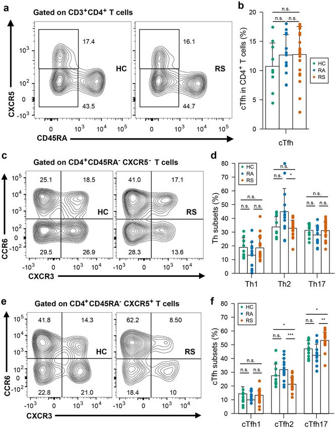
Long et al. Cell Discovery (2021)7:18                                                                                                      Page 3 of 14

 Fig. 1 Memory B cell repertoire in individuals recovered from COVID-19. a, b Flow cytometry analysis (a) of the percentages (b) of SARS-CoV-2
 RBD-specific memory B cells within the CD19+ CD27+ memory B cells of the PBMCs from recovered individuals and healthy controls (HC). Freshly
 isolated PBMCs were stimulated in vitro for 5 days with R848 and IL-2. After 5 days in culture, these cells were plated onto commercial ELISpot plates.
 c The counts of cells that produced total IgG in per million cultured PBMCs from recovered individuals and healthy controls. d Representative ELISpot
 dots in recovered individuals and healthy controls. e The counts of cells that produced RBD-specific IgG in per million cultured PBMCs from
 recovered individuals and healthy controls. f Correlation analysis between RBD-specific ELISpot dot number and virus-specific IgG levels in
 supernatant after the R848 stimulation on PBMC from individuals recovered from asymptomatic. g Correlation analysis between RBD-specific ELISpot
 dot number and virus-specific IgG levels in supernatant after the R848 stimulation on PBMC from individuals recovered from symptomatic. Each
 symbol represents an individual throughout. Data in b, c were analyzed using unpaired, two-sided Mann–Whitney U test, and data in f, g were
 analyzed using Spearman’s rank correlation test.

healthy controls (Fig. 1e). The SARS-CoV-2 IgG levels in                      specific blot numbers and virus-specific IgG levels in the
the supernatant after stimulation with IL-2 and R848 were                     supernatant was found (RA, Spearman’s rank correlation
also qualified by Enzyme-linked immunosorbent assay                            ρ = 0.632, P = 0.021; RS, Spearman’s rank correlation ρ =
(ELISA), and a correlation between the SARS-CoV-2-                            0.602, P = 0.005) (Fig. 1f, g). There was no correlation
Long et al. Cell Discovery (2021)7:18                                                                        Page 4 of 14

between spot number after memory B cell activation and         of 10 individuals), and a CD4+ T cell response to M and N
circulating SARS-CoV-2-specific IgG levels or virus-            was also detected in individuals recovered from symp-
specific memory B cells in peripheral blood from con-           toms, with 9 (45%) and 15 (75%) individuals recovered
valescent individuals (Supplementary Fig. S4a, b).             from COVID-19 with symptoms, respectively (Fig. 3b),
                                                               which were not significantly different than healthy con-
T cell subsets in COVID-19 convalescent individuals            trols (fisher’s exact P were 0.246 and 0.045 vs healthy
  Patients infected with SARS-CoV-2 exhibit disrupted T        controls, respectively). The magnitudes of the SARS-CoV-
cell homeostasis in the acute phase. To explore the cel-       2-specific CD4+ T cell responses measured were 0.26
lular immune responses to SARS-CoV-2, we first eval-            (IQR: 0.17–0.33), 0.10 (IQR: 0.02–0.18), and 0.16 (IQR:
uated T cell subsets in the peripheral blood isolated from     0.11–0.30) after stimulation with S, M, and N pool,
COVID-19 convalescent subjects and controls. There was         respectively (Fig. 3b). In individuals recovered from
no significant difference in the frequencies of total CD3+,     COVID-19 without symptoms, CD4+ T cell responses to
CD4+, or CD8+ T cells within the T cell population             S, M and N were detected in 11, 8 and 6 of 13 individuals
between convalescent patients and controls (Supplemen-         (Fig. 3b) (fisher’s exact P were 0.039, 0.090 and 0.669 vs
tary Fig. S5a–c). In addition, in either the CD4+ or CD8+      healthy controls) with magnitudes of 0.24 (IQR:
T pools from convalescent subjects, the distribution of        0.19–0.48), 0.16 (IQR: 0.03–0.34) and 0.08 (IQR:
naïve (CD45RA+CCR7+), central memory (CD45RA-                  0.01–0.14), respectively (Fig. 3b). The CD4+ T cell
CCR7+), and effector memory (CD45RA-CCR7-) T cells             response to N measured by AIM was higher in individuals
was similar to that in the T cell pools from healthy con-      recovered from symptoms than in individuals recovered
trols (Supplementary Fig. S5d–i). Circulating follicular       from asymptomatic symptoms (P = 0.020), whereas the
helper T (cTfh) cells facilitate the antibody response to      CD4+ T cell responses to S and M were not different
viral infections29, and cTfh cells in the circulation con-     (P = 0.782 and 0.346 for S and M, respectively) between
stitute a surrogate of cTfh cells in lymphoid tissues30. A     subjects recovered from symptomatic infection and
recent study showed that convalescent patients display an      recovered from asymptomatic infection (Fig. 3b). The rate
altered peripheral CD4+ T cell spectrum, especially for        of subjects with CD4+ T cell responses simultaneously to
cTfh cells31. As shown in Fig. 2a, b, we found that there is   S, M, and N was 0% (0/10), 30.8% (4/13), and 45% (9/20)
no difference in the frequency of cTfh between con-            in HC, RA, and RS, respectively, and individuals recovered
valescent patients and healthy controls. We also found         from SARS-CoV-2 infection have obviously higher rate of
that despite the similar proportions of circulating Th1,       CD4+ T cell response compared with HC.
Th2, and Th17 cells within CD4+ T cells between con-             SARS-CoV-2 S-, M-, and N-specific CD8+ T cell
valescent patients and healthy controls (Fig. 2c, d), cTfh2    responses (CD69+CD137+) were detected in 14, 19, and 6
frequency in individuals recovered from symptoms was           individuals recovered from COVID-19 with symptoms,
lower than those in healthy controls (with median 20.1%        and the magnitudes of the SARS-CoV-2-specific CD8+ T
vs 23.8%, P = 0.028), while cTfh17 population abundance        cell responses measured were 0.17 (IQR: 0.08–0.40), 0.40
was higher than those in healthy controls (with median         (IQR: 0.19–0.62) and 0.04 (IQR: 0.01–0.11), respectively
54.1% vs 47.0%, P = 0.016) (Fig. 2e, f).                       (Fig. 4a, b). In individuals recovered from COVID-19
                                                               without symptoms, CD8+ T cell responses to S, M, and N
Identification and quantitation of the SARS-CoV-2-specific       were detected in 8, 9, and 9 of 13 individuals (Fig. 4b)
T cell response in individuals recovered from COVID-19         (fisher’s exact P were 0.090, 1.000, and 0.222 vs healthy
  To delineate the memory response mediated by T cells,        controls), with magnitudes of 0.18 (IQR: 0.01–0.31), 0.41
PBMCs from recovered individuals and healthy controls          (IQR: 0.06–0.62) and 0.24 (IQR: 0.09–0.82), respectively
were stimulated with a spike (S), membrane (M), and            (Fig. 4b). The CD8+ T cell response to N was higher in
nucleocapsid (N) peptide libraries from SARS-CoV-2             individuals recovered from asymptomatic infection than
ex vivo. Phytohemagglutinin-L (PHA-L) was used as a            in individuals recovered from symptomatic infection (P =
positive control, while DMSO was used as the negative          0.007), and the CD8+ T cell responses to S and M were
control. As previously reported, the TCR-dependent             not different between them (P = 0.554 and 0.839 for S and
activation-induced marker (AIM) was used to identify           M, respectively) (Fig. 4b). The rate of subjects with CD8+
and quantify virus-specific CD4+ (co-expression of              T cell responses simultaneously to S, M, and N was 10%
OX40+CD137+) or CD8+ T (co-expression of                       (1/10), 38.5% (5/13), and 20% (4/20) in HC, RA, and RS,
CD69+CD137+) cells in recovered COVID-19 indivi-               respectively, and individuals recovered from SARS-CoV-2
duals25. As shown in Fig. 3a, b, a SARS-CoV-2 S-specific        infection have obviously higher rate of CD8 + T cell
CD4+ T cell response (OX40+CD137+) was detected in             response compared with HC.
17 of 20 individuals recovered from COVID-19 with                Regarding the distribution of the T cell response for
symptoms (fisher’s exact P = 0.030 vs healthy controls, 4       different epitopes evaluated from the AIM assay, nearly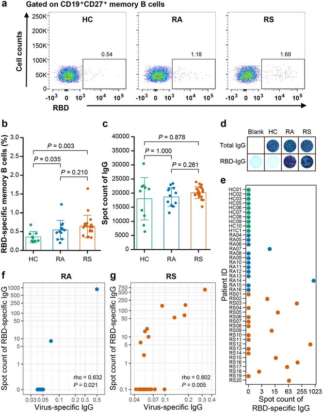
Long et al. Cell Discovery (2021)7:18                                                                                                     Page 5 of 14

 Fig. 2 T cell subsets in individuals recovered from COVID-19. Blood samples were collected from individuals recovered from COVID-19 and
 healthy controls, PBMC were isolated for analysis of T cell subsets. a Representative flow plots for the expression of CD45RA and CXCR5 within the
 CD4+ populations from healthy controls and recovered COVID-19. b Proportions of cTfh cells (CD45RA-CXCR5+) in CD4+ T cell populations from HC,
 RS and RA. c Representative flow plots for the expression of CXCR3 and CCR6 within the CD4+CD45RA-CXCR5-populations from healthy controls and
 recovered COVID-19. d Proportions of Th subsets within the CD4+CD45RA-CXCR5- populations from HC, RS, and RA. e Representative flow plots for
 the expression of CXCR3 and CCR6 within the cTfh populations from healthy controls and recovered COVID-19. f Proportions of cTfh subsets within
 the cTfh populations from HC, RS, and RA. Each symbol represents an individual throughout. RS, individuals recovered from COVID-19 with symptoms
 (n = 20); RA, individuals recovered from asymptomatic (n = 13); HC, healthy controls (n = 10). Data in b, d, f were analyzed using unpaired, two-sided
 Mann–Whitney U test. n.s., not significant.

40% of the CD4+ T cell response was against the S peptide                     proteins (Fig. 5a, b). Either CD4+ or CD8+ T cell subsets
library, while the M peptide library stimulated the                           might preferentially respond to different peptide libraries.
strongest CD8+ T cell response in individuals who                               Independently, to further confirm the functional
recovered from symptoms, suggesting that the distribu-                        repertoire of the SARS-CoV-2-specific T cell response,
tion of epitopes was different in different SARS-CoV-2                        PBMCs from recovered individuals and healthy controls
Long et al. Cell Discovery (2021)7:18                                                                                                  Page 6 of 14

 Fig. 3 SARS-CoV-2 specific CD4+ T cell response of recovered COVID-19 individuals. a Representative flow plots for the expressions of OX40
 and CD137 within CD3+CD4+ T cells of cultured PBMCs stimulated with SARS-CoV-2 peptide library in different subjects. b SARS-CoV-2-specific CD4+
 T cells measured as percentage of AIM+ (OX40+CD137+) after stimulation of PBMCs with peptide pools encompassing S, M, and N. Each symbol
 represents an individual throughout. RS, individuals recovered from COVID-19 with symptoms (n = 20); RA, individuals recovered from asymptomatic
 (n = 13); HC, healthy controls (n = 10). Data were background subtracted against DMSO negative control, and if the value is greater than 0.1%, the
 individual is considered as response to peptide library stimulation. The number of response individuals is shown in brackets, and the dotted line
 indicated the cut-off for positive responder. The comparisons between two groups were performed by using unpaired, two-sided Mann–Whitney U
 test. S, spike; M, membrane; N, nucleocapsid.

Long et al. Cell Discovery (2021)7:18                                                                                                  Page 7 of 14

 Fig. 4 SARS-CoV-2-specific CD8+ T cell response of recovered COVID-19 individuals. a Representative flow plots for the expressions of CD69
 and CD137 within CD3+CD4+ T cells of cultured PBMCs stimulated with SARS-CoV-2 peptide library in different subjects. b SARS-CoV-2-specific CD8+
 T cells measured as percentage of AIM+ (CD69+CD137+) after stimulation of PBMCs with peptide pools encompassing S, M, and N. Each symbol
 represents an individual throughout. RS, individuals recovered from COVID-19 with symptoms (n = 20); RA, individuals recovered from asymptomatic
 (n = 13); HC, healthy controls (n = 10). Data were background subtracted against DMSO negative control, and if the value is greater than 0.1%, the
 individual is considered as a response to peptide library stimulation. The number of response individuals is shown in brackets, and the dotted line
 indicated the cut-off for positive responder. The comparisons between two groups were performed by using unpaired, two-sided Mann–Whitney U
 test. S, spike; M, membrane; N, nucleocapsid.

were directly stimulated with different peptide pools for                   symptoms had a clear population of CD8+ T cells that
24 h, and the percentage of T cells producing interferon-γ                  produced IFN-γ, while CD4+ T cells producing IFN-γ
(IFN-γ) was determined with intracellular staining. As                      were detectable in 15 of 20 individuals stimulated with S
shown in Fig. 6, all individuals who recovered from                         peptide library. Thus, the majority of recovered COVID-
Long et al. Cell Discovery (2021)7:18 Page 8 of 14
Fig. 5 The composition of SARS-CoV-2 response in each individual is shown as a percentage of the total detected T cell response. a The
composition of SARS-CoV-2-specific CD4+ T cell response (OX40+CD137+) in individuals. b The composition of SARS-CoV-2-specific CD8+ T cell
response (CD69+CD137+) in individuals. The percentage of S, M, and N-specific CD4+ or CD8+ T cell response was shown as median (IQR), and were
performed by using two-sided Mann–Whitney U test. S, spike; M, membrane; N, nucleocapsid. RS, individuals recovered from COVID-19 with
symptoms (n = 20); RA, individuals recovered from asymptomatic (n = 13); HC, healthy controls (n = 10).
19 patients generated a specific T cell response against S and S N M O peptide pools, while in individuals recovered
protein of SARS-CoV-2 after rechallenge. from COVID-19 without symptoms, 9, 9, and 8 out of 13
individuals showed reactivity against the S1, S2 N, and S N
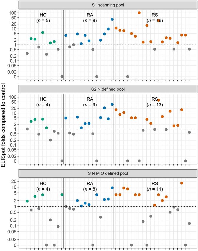
T cell ELISpot M O peptide pools. The fold changes of peptide stimulation
To further explore the virus-specific memory T cell are presented as the spot-forming unit ratio between
response, PBMCs from recovered individuals and healthy DMSO stimulation and peptide stimulation (Fig. 7). The S1
controls were stimulated with different peptide pools pool had higher reactivity in COVID-19 recovered indivi-
(Mabtech, S1 scanning pool, S2 N defined pool and S N M duals than in healthy controls (S1 scanning pool: RS vs HC,
O defined pool, peptide constitutions were listed in Sup- P = 0.028; RA vs HC, P = 0.154).
plementary Table S3), and virus-specific responses were
analyzed by commercial IFN-γ ELISpot assay. In individuals Discussion
recovered from COVID-19 with symptoms, 16, 13, and 11 The induction and duration of SARS-CoV-2-specific
out of 20 individuals showed reactivity against the S1, S2 N, memory T cells and B cells are important for long-termLong et al. Cell Discovery (2021)7:18 Page 9 of 14 Fig. 6 (See legend on next page.)
Long et al. Cell Discovery (2021)7:18                                                                                                Page 10 of 14

 (see figure on previous page)
 Fig. 6 Percentage of IFN-γ-producing T cell in response to different peptide library. Isolated PBMCs were stimulated with specific peptide
 library of SARS-CoV-2 for 24 h, and the proportions of IFN-γ-producing T cell within the CD4+ or CD8+ T cell population were determined with
 intracellular staining. a, b Representative flow plots for the expressions of IFN-γ within CD3+CD4+ T cells (a) or CD3+CD8+ T cells (b) of cultured
 PBMCs from healthy controls and recovered COVID-19. c Fold changes between percentage of IFN-γ+ producing CD4+ or CD8+ T cells stimulated
 with SARS-CoV-2 peptide library and with DMSO. Number of stimulation response individuals (fold change > 1) are depicted in brackets. The dotted
 line indicated the cut-off for positive responder. Each symbol represents an individual throughout. S, spike; M, membrane; N, nucleocapsid. RS,
 individuals recovered from COVID-19 with symptoms (n = 20); RA, individuals recovered from asymptomatic (n = 13); HC, healthy controls (n = 10).

protection and are also key issues in addressing reinfec-                   cTfh2 population. Interestingly, Jennifer A. Juno et al.
tion episodes and the duration of protection induced by                     recently reported that recovered patients (one month after
vaccines. There is a critical need to evaluate the capacity                 infection) exhibited robust cTfh responses to SARS-CoV-
of adaptive immune memory in individuals recovered                          2, and virus-specific cTfh cells were enriched for
from COVID-19. Here, we utilized PBMCs derived from                         cTfh17 subsets34. Thus, we speculate that abnormal dis-
recovered COVID-19 patients to determine the memory                         tribution of cTfh pools might be involved in the patho-
response mediated by B cells and T cells by a series of                     genesis or protective reaction of COVID-19. However, the
experimental techniques, including phenotype analysis,                      extremely low number of virus-specific circulating cTfh
functional measurement with ELISpot, intracellular                          cells in convalescent subjects, especially in this study with
cytokine staining and T cell AIM assessment. Consistent                     recovered individuals 6 months after infection, largely
with recent studies, a cross-reactivity in memory reaction                  impedes from further investigating the roles of specific
for coronavirus mediated by T cells was observed in                         cTfh subsets in virus immunity.
healthy controls7,25,32.                                                      SARS-CoV-2-specific T cell responses were detected in
  To the best of our knowledge, this is the first study to                   the majority of individuals recovered from SARS-CoV-2
evaluate the memory B cell response to SARS-CoV-2 in                        infection 6 months prior, similar to recent preprint35,36. In
individuals infected 6 months prior. Although virus-                        previous studies that investigated individuals recovered
specific memory B cells in different cohorts were all at low                 from SARS-CoV infection, memory T cells were shown to
levels, similar to a previous study33, the level of SARS-                   persist for many years7,17. In our study, AIM, ICS, and T
CoV-2-specific memory B cells in SARS-CoV-2-infected                         cell activation marker screening was conducted using
individuals was obviously higher than those in healthy                      large peptide mega pools to evaluate the T cell response in
controls. After stimulation with the combination of R848                    individuals infected 6 months prior to avoid the potential
and IL-228, memory B cells were selectively activated, and                  differences induced by different methods. In the CD4+ T
RBD-specific IgG was further detected as the ELISpot                         cell response, S protein accounted for nearly 40% of the
readout. In our study, we found that only 2 of 13 indivi-                   total CD4+ T cell response in individuals recovered from
duals who recovered from asymptomatic infection pre-                        symptomatic infection, which was obviously higher than
sented positive results, while 6 of 20 individuals who                      the CD4+ T cell response induced by M or N (39.1% vs
recovered from symptomatic infection presented positive                     26.3%, P < 0.001; 39.1% vs 33.6%, P = 0.004). Although the
results, suggesting that not every infected person can                      CD8+ T cell response was observed in both recovered
produce an effective humoral immune response to the                         cohorts with different peptide library stimulations, the
virus. This result is extremely important for vaccine                       positive rate was not significantly higher than that in
development and application. The magnitude of B cell                        healthy controls. This discrepancy in the T cell response
spot number was not correlated with the levels of virus-                    in SARS-CoV-2 infection convalescents may support
specific IgG in peripheral blood. These results indicated                    additional focus on eliciting the CD4+ T cell response
that memory B cell activation, differentiation, or                          using exogenous antigens in vaccine development, as is
antibody-secreting B cell (i.e., plasmablasts and plasma                    the case in herpes zoster vaccines37.
cells) formation may be deficient or out of balance in                         The difference of humoral and cellular immune
COVID-19-recovered individuals. cTfh cells indicated                        response in subjects recovered from different disease
maturation of the humoral immune response and were                          severity was also a hotspot. In virus-specific memory B
related to the establishment of specific memory B cells to                   cell functional test, the rate of individuals with positive
rapidly respond to possible reinfection. In the present                     B cell ELISpot result were higher in RS group com-
cohort, we found that despite cTfh frequency was similar                    pared with RA group (50.0% vs 15.4%) (Fig. 1f, g).
between recovered subjects symptoms and healthy con-                        Memory CD4+ or CD8+ T cell frequencies stimulated
trol, convalescent subjects displayed skewed cTfh subsets                   with S or M peptide library showed no difference
differentiation, with increased cTfh17 while decreased                      (Figs. 3b, 4b). Therefore, the long-term humoral
Long et al. Cell Discovery (2021)7:18                                                                                              Page 11 of 14

 Fig. 7 The SARS-CoV-2-specific T cell response measured by ELISpot in COVID-19 recovered individuals. ELISpot results shown as the fold
 changes of peptide stimulation were presented as the spot number ratio between peptide stimulation and DMSO stimulation. Number of
 stimulation response individuals (fold change > 1) are depicted in brackets. The dotted line indicated the cut-off for positive responder. RS,
 individuals recovered from COVID-19 with symptoms (n = 20); RA, individuals recovered from asymptomatic (n = 13); HC, healthy controls (n = 10).

immunity to SARS-CoV-2 was higher in individuals                           infection35 , this discrepancy might be due to the het-
who experienced a severe COVID-19 disease course,                          erogeneity in the study population39 .
while T cell memory did not show a similar pattern38.                        Importantly, cross-reactive T cell responses against S,
However, a recent preprint by Zuo J. et al. reported that                  M, or N peptide library were detected in 40.0% (4 of 10
symptomatic infection elicits a higher level of memory                     individuals), 20.0% (2 of 10 individuals), and 30.0% (3 of
T cells than asymptomatic infection at 6-month post-                       10 individuals) of healthy controls, which is consistent
Long et al. Cell Discovery (2021)7:18                                                                         Page 12 of 14

with previous studies reporting pre-existing immune            with 10% (wt/vol) fetal bovine serum (FBS), 1 mM sodium
responses potentially induced by other cor-                    pyruvate, 10 mM HEPES buffer solution, 100 μM non-
onaviruses7,25,32,40. In the H1N1 pandemic, the presence       essential amino acid solution, 50 μM β-mercaptoethanol,
of cross-reactive T cells was found to correlate with less     100 U/mL penicillin and 100 μg/mL streptomycin and
severe disease41,42, but a preprint study reported that pre-   stimulated with 1 μg/mL functional grade anti-CD28
existing humoral immunity to common coronaviruses              (eBioscience, clone 28.2) and 1 μg/mL SARS-CoV-2
does not confer cross-protection against SARS-CoV-2            Spike Glycoprotein, SARS-CoV-2 M or SARS-CoV-2 N
infection43. The possible effects of pre-existing T cells on   (Genscript, China) at 37 °C, 5% CO2 for 24 h. Stimulation
the susceptibility to SARS-CoV-2, differential modulation      controls were both conducted in the presence of 1 μg/mL
of SARS-CoV-2 infection severity, epidemiological models       anti-CD28 and separately conducted with equal con-
of herd immunity, and the performance of COVID-19              centrations of DMSO (Sigma) as vehicle control, or
candidate vaccines need to be carefully evaluated, espe-       2.5 μg/mL PHA-L Solution (eBioscience) with as positive
cially when SARS-CoV-2-specific T cell epitope is               controls.
available44.
  Specific memory B cell populations have the ability to        B cells stimulation
re-enter secondary germinal centers (GCs) to play roles          2 × 10⁶ PBMCs were cultured in 12-well plate (Corning)
upon recall immunization45. Therefore, virus-specific           at 37 °C, 5% CO2 in the presence of 1 μg/mL R848
memory B cells residing in other parts of the body, such       (Mabtech) and 10 ng/mL IL-2 (Mabtech) as previously
as the bone marrow and secondary lymphoid organs, need         described. After incubation for 5 days, cells were collected
to be inspected if possible. In addition, in the current       for analysis of spot numbers of producing IgG specific for
study, the longitudinal analysis of the dynamic kinetics of    SARS-CoV-2 RBD, and cell supernatant was used to
memory B cell and T cell responses was hampered by the         determine IgG levels.
lack of stored PBMC samples during acute infection.
Furthermore, our study did not recruit individuals who         Flow cytometry
recovered from severe conditions because severe COVID-           For analysis of surface marker, fresh PBMCs were
19 patients were scarce in local medical institutions. The     incubated for 30 min at 4 °C in PBS containing 2% FBS
lack of antigen-specific tetramers makes this study unable      with the fluorochrome conjugated antibodies titrated to
to directly analyze the proportion of antigen-specific          optimal concentrations. Fixable Viability Dye eFluor 780
memory T cell subsets. Finally, the relatively small cohort    (eBioscience) staining was used to exclude dead cells. The
size is also a limitation of the current study.                SARS-CoV-2-specific B cells were detected using bioti-
                                                               nylated S1-His recombinant protein (SinoBiological) and
Methods and subjects                                           Streptavidin-APC (Biolegend). For intracellular cytokine
Study subjects                                                 (IFN-γ) staining, surface stained cells were fixed and
  Subjects recovered from asymptomatic (n = 13) or             permeabilized with a Cytofix/Cytoperm kit (BD Bios-
symptomatic COVID-19 (n = 20) at least 6 months were           ciences) according to the manufacturer’s instruction. All
recruited in Wanzhou District (13/33) and Yongchuan            samples were acquired on BD FACSAriaTM II (BD Bios-
District (20/33), Chongqing, China (Supplementary Table        ciences) and analyzed using FlowJo software (Version
S1). Ten healthy controls were recruited to provide            10.0.8, Tree Star Inc., USA). All antibodies used in this
EDTA-K2 anticoagulant blood samples. All plasma was            study were listed in Supplementary Table S4. The specific
obtained by centrifuging blood samples at 3500 rpm for         gating strategies for each population are indicated in each
5 min and frozen at –80 °C for further analysis. The study     figure legend.
was approved by the Ethics Committee of Chongqing
Medical University. Written informed consent in accor-         T cell ELISpot
dance with the Declaration of Helsinki from all                  IFN-γ-secreting T cells were detected by Human IFN-γ
participants.                                                  SARS-CoV-2 ELISpotPLUS (HRP) kit (Mabtech) accord-
                                                               ing to the manufacture protocol. Briefly, 2.5 × 10⁵ cells
Cell isolation                                                 were incubated with 1 μg/mL SARS-CoV-2 S1 scanning
  PBMCs were isolated from EDTA-K2 anticoagulant               peptides, SARS-CoV-2 S N M O defined peptides or
whole blood using Ficoll-Hypaque (GE Healthcare, USA)          SARS-CoV-2 S2 N defined peptides (Mabtech) supple-
gradient centrifugation.                                       mented with 1 μg/mL anti-CD28 (eBioscience, clone 28.2)
                                                               for 24 h. Unstimulated controls were performed with
T cells stimulation                                            equal concentrations of DMSO, and positive control was
  2 × 10⁶ freshly isolated PBMCs were cultured in 48-well      incubated with 1 μg/mL CD3 (5 × 10⁴ cells, Mabtech,
plate (Corning) in RPMI 1640 medium supplemented               clone CD3-2) in the presence of 1 μg/mL anti-CD28.
Long et al. Cell Discovery (2021)7:18 Page 13 of 14
After 24 h treatment, spots were counted using an ELI- numbers (%), and the comparison between two groups
Spot Reader system (AID). Spots numbers were converted was assessed using χ² test or Fisher’s exact test. For the
into the number of spots per million cells. correlation analyses, Spearman’s rank correlation was
performed. The box plots show the medians (middle line)
B cell ELISpot and the first and third quartiles (boxes), and the bar plots
Numbers of B cells secreting IgG (total IgG) antibodies show the means ± SD. P value less than 0.05 was con-
or IgG specific for the SARS-CoV-2 RBD were conducted sidered statistically significant. All statistical analyses were
with Human IgG SARS-CoV-2 RBD ELISpotPLUS (HRP) performed using R software, version 3.6.0.
kit (Mabtech) according to the manufacture protocol.
Acknowledgements
Spots numbers were converted into the number of spots This work was supported by the Project from the Science & Technology
per million cells. Commission of Chongqing (cstc2020jscx-fyzx0053, cstc2018jcyjAX0027,
cstc2018jscx-msybX0376), Chongqing Municipal Education Commission
(KJQN201800422), the Emergency Project for Novel Coronavirus Pneumonia
ELISA from the Chongqing Medical University (CQMUNCP0301, CQMUNCP0304),
After stimulation mentioned above, the levels of IgG Venture and Innovation Support Program for Chongqing Overseas Returnees
specific for SARS-CoV-2 Spike S1 recombinant protein in (Cx2019070), the National Key R&D Project (2018YFE0107500) and the
Chongqing Talents Program. We acknowledge all patients involved in
cell supernatant were determined by ELISA according to the study.
the manufacturer’s instructions (SinoBiological).
Author details
1
Detection of IgG against SARS-CoV-2 Key Laboratory of Molecular Biology on Infectious Diseases, Ministry of
Education, Chongqing Medical University, Chongqing 400016, China.
All serum samples were inactivated at 56 °C for 30 min 2
Department of Endocrinology and Metabolism, The Second Affiliated Hospital
and stored at –80 °C before testing. SARS-CoV-2 specific of Chongqing Medical University, Chongqing 400010, China. 3Yongchuan
IgG against SARS-CoV-2 in plasma samples was tested Hospital Affiliated to Chongqing Medical University, Chongqing 402160, China.
4
Wanzhou District Center for Disease Control and Prevention, Chongqing
using magnetic chemiluminescence enzyme immunoassay 404100, China. 5School of Public Health and Management, Chongqing Medical
kits supplied by Bioscience Co. (approved by the China University, Chongqing 400016, China
National Medical Products Administration; approval
Author contributions
numbers 20203400183(IgG)), according to the manu- Q-X.L., Y-J.J., J.C., and A-L.H. conceived, designed, and supervised the study and
facturer’s instructions. Briefly, recombinant antigens wrote the manuscript. J.Y., L.F., X-R.C., C.L., A-R.H., X-J.T., N.T., Y.H., J-L.H., X-F.C.,
containing the nucleoprotein and a peptide D-Q.W., J-F.Q., and B-Z.L. coordinate the project. Q-X.L., Y-J.J., X.W., X-X.C.
performed B/T cell stimulation, flow cytometry, B/T ELISpot and ELISA. Q-X.L.,
(LQPELDSFKEELDKYFKNHTSPDVD) from the spike X-X.C., and J.H. performed in vitro neutralization assays. Q-X.L. and H-J.D.,
protein of SARS-CoV-2 were immobilized on magnetic processed data. All authors read and approved the contents of the manuscript.
particles. Antibody levels are presented as the measured
chemiluminescence values divided by the cutoff (S/CO). Conflict of interest
The authors declare no competing interests.
Neutralization detection
Neutralization detection using pseudovirus neu- Publisher’s note
tralizaion assay was carried out as previously described10. Springer Nature remains neutral with regard to jurisdictional claims in
published maps and institutional affiliations.
Briefly, a codon-optimized S protein that lacked the C-
terminal 19 amino acids was used to generate a luciferase- Supplementary information The online version contains supplementary
expressing pseudovirus, and the SARS-CoV-2 pseudo- material available at https://doi.org/10.1038/s41421-021-00250-9.
virus neutralization assay was carried out on 293 T cells
Received: 19 December 2020 Accepted: 9 February 2021
expressing ACE2 in a 96-well plate. All serum was diluted
at 1:160 and neutralization rate was calculated in the
previous work10.
References
Clinical parameters 1. Zhou, P. et al. A pneumonia outbreak associated with a new coronavirus of
All clinical parameters listed in Supplementary Table S2 probable bat origin. Nature 579, 270–273 (2020).
2. Huang, C. et al. Clinical features of patients infected with 2019 novel cor-
were determined by professionals in the clinical labora- onavirus in Wuhan, China. Lancet 395, 497–506 (2020).
tory followed Standard Operating Procedure. 3. Holly, M. K., Diaz, K. & Smith, J. G. Defensins in viral infection and pathogenesis.
Annu. Rev. Virol. 4, 369–391 (2017).
4. Liao, M. et al. Single-cell landscape of bronchoalveolar immune cells in
Statistical analysis patients with COVID-19. Nat. Med. 26, 842–844 (2020).
Continuous variables are presented as the median 5. Giamarellos-Bourboulis, E. J. et al. Complex immune dysregulation in COVID-
(IQR), and the comparison between two groups was 19 patients with severe respiratory failure. Cell Host Microbe 27, 992–1000
e1003 (2020).
evaluated using the two-tailed, non-parametric Mann- 6. Mathew, D. et al. Deep immune profiling of COVID-19 patients reveals distinct
Whitney U test; categorical variables are presented as immunotypes with therapeutic implications. Science 369, eabc8511 (2020).Long et al. Cell Discovery (2021)7:18 Page 14 of 14
7. Le Bert, N. et al. SARS-CoV-2-specific T cell immunity in cases of COVID-19 and 27. Yang, L. T. et al. Long-lived effector/central memory T-cell responses to severe
SARS, and uninfected controls. Nature 584, 457–462 (2020). acute respiratory syndrome coronavirus (SARS-CoV) S antigen in recovered
8. Liu, L. et al. High neutralizing antibody titer in intensive care unit patients with SARS patients. Clin. Immunol. 120, 171–178 (2006).
COVID-19. Emerg. Microbes Infect. 9, 1664–1670 (2020). 28. Pinna, D., Corti, D., Jarrossay, D., Sallusto, F. & Lanzavecchia, A. Clonal dissection
9. Long, Q. X. et al. Antibody responses to SARS-CoV-2 in patients with COVID- of the human memory B-cell repertoire following infection and vaccination.
19. Nat. Med. 26, 845–848 (2020). Eur. J. Immunol. 39, 1260–1270 (2009).
10. Long, Q. X. et al. Clinical and immunological assessment of asymptomatic 29. Crotty, S. T follicular helper cell differentiation, function, and roles in disease.
SARS-CoV-2 infections. Nat. Med. 26, 1200–1204 (2020). Immunity 41, 529–542 (2014).
11. Wang, K. et al. Longitudinal dynamics of the neutralizing antibody response to 30. Morita, R. et al. Human blood CXCR5(+)CD4(+) T cells are counterparts of T
SARS-CoV-2 infection. Clin. Infect. Dis. https://doi.org/10.1093/cid/ciaa1143 follicular cells and contain specific subsets that differentially support antibody
(2020). secretion. Immunity 34, 108–121 (2011).
12. Ibarrondo, F. J. et al. Rapid decay of Anti-SARS-CoV-2 antibodies in persons 31. Gong, F. et al. Peripheral CD4 T cell subsets and antibody response in COVID-
with mild Covid-19. N. Engl. J. Med. 383, 1085–1087 (2020). 19 convalescent individuals. J. Clin. Invest. 130, 6588–6599 (2020).
13. Huang, M. et al. Temporal antibody responses to SARS-CoV-2 in patients of 32. Sekine, T. et al. Robust T cell immunity in convalescent individuals with
coronavirus disease. Cell Discov. 6, 64 (2019). asymptomatic or mild COVID-19. Cell 183, 158–168 e114 (2020).
14. Amanna, I. J., Carlson, N. E. & Slifka, M. K. Duration of humoral immunity to 33. Vaisman-Mentesh, A. et al. SARS-CoV-2 specific memory B cells frequency in
common viral and vaccine antigens. N. Engl. J. Med. 357, 1903–1915 (2007). recovered patient remains stable while antibodies decay over time. medRxiv
15. Dushoff, J., Plotkin, J. B., Levin, S. A. & Earn, D. J. Dynamical resonance can https://doi.org/10.1101/2020.08.23.20179796 (2020).
account for seasonality of influenza epidemics. Proc. Natl. Acad. Sci. USA. 101, 34. Juno, J. A. et al. Humoral and circulating follicular helper T cell responses in
16915–16916 (2004). recovered patients with COVID-19. Nat. Med. 26, 1428–1434 (2020).
16. Ferguson, N., Anderson, R. & Gupta, S. The effect of antibody-dependent 35. Zuo, J. et al. Robust SARS-CoV-2-specific T-cell immunity is maintained at
enhancement on the transmission dynamics and persistence of multiple- 6 months following primary infection. bioRxiv https://doi.org/10.1101/
strain pathogens. Proc. Natl. Acad. Sci. USA. 96, 790–794 (1999). 2020.11.01.362319 (2020).
17. Tang, F. et al. Lack of peripheral memory B cell responses in recovered 36. Wise, J. Covid-19 and thrombosis: what do we know about the risks and
patients with severe acute respiratory syndrome: a six-year follow-up study. J. treatment? BMJ 369, m2058 (2020).
Immunol. 186, 7264–7268 (2011). 37. Heineman, T. C., Cunningham, A. & Levin, M. Understanding the immunology
18. To, K. K. et al. COVID-19 re-infection by a phylogenetically distinct SARS- of Shingrix, a recombinant glycoprotein E adjuvanted herpes zoster vaccine.
coronavirus-2 strain confirmed by whole genome sequencing. Clin. Infect. Dis. Curr. Opin. Immunol. 59, 42–48 (2019).
https://doi.org/10.1093/cid/ciaa1275 (2020). 38. Dan, J. M. et al. Immunological memory to SARS-CoV-2 assessed for up to
19. Duggan, N. M., Ludy, S. M., Shannon, B. C., Reisner, A. T. & Wilcox, S. R. Is novel 8 months after infection. Science 371, eabf4063 (2021).
coronavirus 2019 reinfection possible? Interpreting dynamic SARS-CoV-2 test 39. Crotty, S. & Ahmed, R. Immunological memory in humans. Semin. Immunol.
results through a case report. Am. J. Emerg. Med. 39, 256.e1–256.e3 (2021). 16, 197–203 (2004).
20. Edridge, A. W. D. et al. Seasonal coronavirus protective immunity is short- 40. Braun, J. et al. SARS-CoV-2-reactive T cells in healthy donors and patients with
lasting. Nat. Med. 26, 1691–1693 (2020). COVID-19. Nature 587, 270–274 (2020).
21. Kuri-Cervantes, L. et al. Comprehensive mapping of immune perturbations 41. Sridhar, S. et al. Cellular immune correlates of protection against symptomatic
associated with severe COVID-19. Sci. Immunol. 5, eabd7114 (2020). pandemic influenza. Nat. Med. 19, 1305–1312 (2013).
22. Diao, B. et al. Reduction and functional exhaustion of T cells in patients with 42. Wilkinson, T. M. et al. Preexisting influenza-specific CD4 T cells correlate with
coronavirus disease 2019 (COVID-19). Front. Immunol. 11, 827 (2020). disease protection against influenza challenge in humans. Nat. Med. 18,
23. Mazzoni, A. et al. Impaired immune cell cytotoxicity in severe COVID-19 is IL-6 274–280 (2012).
dependent. J. Clin. Invest. 130, 4694–4703 (2020). 43. Miyara, M. et al. Pre-COVID-19 humoral immunity to common coronaviruses
24. Zheng, M. et al. Functional exhaustion of antiviral lymphocytes in COVID-19 does not confer cross-protection against SARS-CoV-2. medRxiv https://doi.org/
patients. Cell Mol. Immunol. 17, 533–535 (2020). 10.1101/2020.08.14.20173393 (2020).
25. Grifoni, A. et al. Targets of T cell responses to SARS-CoV-2 coronavirus in 44. Mateus, J. et al. Selective and cross-reactive SARS-CoV-2 T cell epitopes in
humans with COVID-19 disease and unexposed individuals. Cell 181, unexposed humans. Science 370, 89–94 (2020).
1489–1501 e1415 (2020). 45. Mesin, L. et al. Restricted clonality and limited germinal center reentry
26. Li, C. K. et al. T cell responses to whole SARS coronavirus in humans. J. characterize memory B cell reactivation by boosting. Cell 180, 92–106
Immunol. 181, 5490–5500 (2008). e111 (2020).You can also read