Long-read sequencing and denovogenome assemblies reveal complex chromosome end structures caused by telomere dysfunction at the single nucleotide ...
←
→
Page content transcription
If your browser does not render page correctly, please read the page content below
3338–3353 Nucleic Acids Research, 2021, Vol. 49, No. 6                                                                         Published online 8 March 2021
doi: 10.1093/nar/gkab141

Long-read sequencing and de novo genome
assemblies reveal complex chromosome end
structures caused by telomere dysfunction at the
single nucleotide level
                                   1,2,†                     1,3,*,†                           4                               1,2,3,*
Eunkyeong Kim                              , Jun Kim                   , Chuna Kim                 and Junho Lee
1
 Department of Biological Sciences, Seoul National University, Gwanak-ro 1, Gwanak-gu, Seoul 08826, Korea,
2
 Institute of Molecular Biology and Genetics, Seoul National University, Seoul 08826, Korea, 3 Research Institute of

                                                                                                                                                                  Downloaded from https://academic.oup.com/nar/article/49/6/3338/6163097 by guest on 10 October 2021
Basic Sciences, Seoul National University, Seoul 08826, Korea and 4 Aging Research Center, Korea Research
Institute of Bioscience and Biotechnology, Gwahak-ro 125, Daejeon 34141, Korea

Received October 28, 2020; Revised January 28, 2021; Editorial Decision February 18, 2021; Accepted February 20, 2021

ABSTRACT                                                                                 INTRODUCTION
Karyotype change and subsequent evolution is trig-                                       DNA damage, such as double-strand breaks (DSBs) are the
gered by chromosome fusion and rearrangement                                             driving force for structural changes in chromosomes, and
events, which often occur when telomeres become                                          damaged telomeres caused by telomere erosion or stochas-
dysfunctional. Telomeres protect linear chromosome                                       tic loss can be recognised as DSBs at chromosome ends.
ends from DNA damage responses (DDRs), and                                               This telomere dysfunction sometimes extends further into
                                                                                         the chromosome, also generating structural changes in sub-
telomere dysfunction may result in genome instabil-                                      telomeric regions (1,2). The resulting telomere and sub-
ity. However, the complex chromosome end struc-                                          telomere damage can cause chromosome end-to-end fusion
tures and the other possible consequences of telom-                                      or structural rearrangements at chromosome ends, leading
ere dysfunction have rarely been resolved at the nu-                                     to karyotype evolution (3–5).
cleotide level due to the lack of the high-throughput                                       Chromosome ends affected by telomere dysfunction are
methods needed to analyse these highly repetitive                                        recognised as DSB sites and can be processed by a vari-
regions. Here we applied long-read sequencing tech-                                      ety of mechanisms. In the breakage-fusion-bridge (BFB) cy-
nology to Caenorhabditis elegans survivor lines that                                     cle, damaged chromosome ends fuse after telomeric-repeat
emerged after telomere dysfunction. The survivors                                        deletions, followed by breakage and more fusions during
have preserved traces of DDRs in their genomes and                                       subsequent cell division. Some subtelomeric regions are
our data revealed that variants generated by telom-                                      duplicated with inverted fragments, leading to copy num-
                                                                                         ber doubling of those regions, making them hallmarks of
ere dysfunction are accumulated along all chromo-                                        the BFB cycle (3,6). Fork Stalling and Template Switch-
somes. The reconstruction of the chromosome end                                          ing (FoSTeS) is another example, where a replicated DNA
structures through de novo genome assemblies re-                                         strand that is stalled by telomere dysfunction invades a dif-
vealed diverse types of telomere damage processing                                       ferent locus and continues replication to process the dam-
at the nucleotide level. When telomeric repeats were                                     aged chromosome end (2). This process adds diverse frag-
totally eroded by telomere dysfunction, DDRs were                                        ments from various loci in the genome, so results in odd-
mostly terminated by chromosome fusion events. We                                        number or stepwise copy number variation (CNV) and ei-
also partially reconstructed the most complex end                                        ther the same or the opposite directional replication.
structure and its DDR signatures, which would have                                          For the repair process to terminate, the DSB sites
been accumulated via multiple cell divisions. These                                      need protection against DSB recognition. Several path-
finely resolved chromosome end structures suggest                                        ways that provide this protection include end-to-end fu-
                                                                                         sion and telomerase-mediated or telomerase-independent
possible mechanisms regarding the repair processes                                       telomere maintenance, which leave their specific signatures
after telomere dysfunction, providing insights into                                      in the genomes. End-to-end fusion permanently conceals
chromosome evolution in nature.

* To
   whom correspondence should be addressed. Tel: +82 2 877 2663; Fax: +82 2 877 2661; Email: elegans@snu.ac.kr
Correspondence may also be addressed to Jun Kim. Email: dauer@snu.ac.kr
†
    The authors wish it to be known that, in their opinion, the first two authors should be regarded as Joint First Authors.

C The Author(s) 2021. Published by Oxford University Press on behalf of Nucleic Acids Research.
This is an Open Access article distributed under the terms of the Creative Commons Attribution-NonCommercial License
(http://creativecommons.org/licenses/by-nc/4.0/), which permits non-commercial re-use, distribution, and reproduction in any medium, provided the original work
is properly cited. For commercial re-use, please contact journals.permissions@oup.com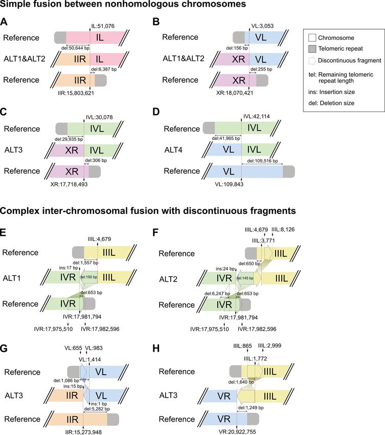
Nucleic Acids Research, 2021, Vol. 49, No. 6 3339

the damaged sites, and the fusion sites show specific features   lated only after telomere rearrangements were accumulated.
at the nucleotide level, such as comprehensive chromosome        Next, by reconstructing 60% of the all chromosome ends at
end losses and connected nonhomologous chromosomes.              the nucleotide level, we show that the nonhomologous chro-
Telomerase is the major player in most eukaryotic cells          mosome fusion events after deletions in both telomeric re-
that lengthens telomeric repeats and protects chromosome         peats and subtelomeres were a major way to conceal the ter-
ends to stop the repair process (7). Telomerase-independent      minal DSB sites. In addition, BFB cycles were induced when
telomere maintenance mechanisms, such as alternative             fusions occurred between sister chromatids. Moreover, we
lengthening of telomeres (ALT), can also reconstruct the         show a highly complex telomere structure that was recon-
protective chromosome ends by either recombination- or           structed using several subtelomeric regions as units, and at
replication-mediated mechanisms (8,9). Indeed, ALT is a          least one FoSTeS event for filling a gap between units. Fi-
major mechanism for lengthening and protecting chromo-           nally, we show that the remaining chromosome ends were
some end sequences in some species and acts as a backup          stabilised by ALT, and the TALT copies were duplicated
mechanism for telomerase in various eukaryotes (10,11).          with high accuracy and in the same direction. Resolving the

                                                                                                                                Downloaded from https://academic.oup.com/nar/article/49/6/3338/6163097 by guest on 10 October 2021
For example, in the free-living nematode Caenorhabditis el-      DDR consequences after telomere dysfunction in the C. el-
egans, some rare telomerase mutant worms survive telom-          egans lines studied here will shed light on how chromosome
ere dysfunction by replicating unique sequences, templates       end evolution proceeds in eukaryotes.
for ALT (TALTs), flanked with telomeric repeats to the dys-
functional telomeres. These TALT copies, along with the
remaining trace telomeric repeats, serve as a new protec-        MATERIALS AND METHODS
tive telomere sequence, so copy numbers of TALTs increase        Strain maintenance and accessions
dramatically in the worms (12,13). These worms, called
ALT survivors, arise within tens of generations after los-       All worms were maintained at 20◦ C under standard culture
ing their telomerase activity. Furthermore, the survivor lines   conditions. ALT survivors were isolated as previously
and their abnormal chromosomes can be stably maintained,         reported (12), and the trt-1(ok410) allele was used for
so the survivor lines are a reproducible model to examine        telomerase mutation. We also used the public PacBio long-
the consequences of telomere dysfunction in eukaryotes at        read data and genome assemblies for the CB4856, N2 and
the single nucleotide level.                                     VC2010 (a descendent of N2) strains. The CB4856 genome,
   Although telomere dysfunction, repair and consequent          ASM452629v1, was obtained from the NCBI (18); the
karyotype evolution have been studied in many species,           CB4856 raw read, SRR8599837, from NCBI (downsampled
the fusion sites and the new chromosome end structures           to 2.2 Gb, 260 000 reads using seqtk sample); the VC2010
at the nucleotide level are resolved rarely. It is because       genome: WS274 (19) was downloaded from WormBase
many molecular techniques, such as polymerase chain re-          (20,21)     (ftp://ftp.wormbase.org/pub/wormbase/releases/
action (PCR)-based, copy number-based and short-read             WS274/species/c elegans/PRJEB28388/); the VC2010 raw
sequencing-based methods are insufficient for resolving          read, SRR7594465, was from the NCBI (downsampled
the repetitive and complex structure of the chromosome           to 2.2 Gb, 260,000 reads using seqtk sample); and the N2
ends (1,2,14,15). Furthermore, because the lack of high-         genome, WBcel235 (ce11), was downloaded from Ensembl
throughput methods has prevented the analysis of genomic         (used for depicting chromosome end structures of the
regions outside chromosome ends after telomere dysfunc-          ALT3 and ALT4 survivor lines because the long-read-
tion events, genome instability caused by telomere dys-          based VC2010 genome assembly lacks telomeric repeats
function has been investigated by copy number changes,           in at least one chromosome). The seqtk tool was installed
rather than genome-wide structural variation and sequence        from https://github.com/lh3/seqtk.
changes (16,17). Long-read sequencing technologies over-
come these limitations, as the longer read length allows the
resolution of highly repetitive and complex structures at the    Genomic DNA preparation and whole genome sequencing
nucleotide level, and such regions can be covered by single      ALT survivor worms in mixed stages were collected from
reads. Recent technical advances have opened up opportu-         10-cm NGM plates and washed three times with M9 buffer.
nities to analyse genome-wide variants and chromosomal           Worms were lysed with worm lysis buffer (0.2 M NaCl, 0.1
rearrangements, as well as chromosome end structures, in a       M Tris–HCl (pH 8.5), 50 mM EDTA (pH 8.0), 0.5% SDS)
single reaction in organisms with small genomes.                 with proteinase K (0.1 mg/ml) at 65◦ C for 2 h. One volume
   Here, we analysed four C. elegans ALT survivors (ALT1,        of phenol/chloroform/isoamyl alcohol (25:24:1) was added
ALT2, ALT3 and ALT4) using Pacific Biosciences (PacBio)          and mixed gently for 15 min. The aqueous phase was sepa-
long-read sequencing technology to identify genome-wide          rated by spinning at 6000 g for 10 min at room temperature
variants at the nucleotide level and resolve complex chro-       and transferred to new tubes. Genomic DNA was precip-
mosome end structures after telomere dysfunction and re-         itated by adding two volumes of 100% ethanol and 0.2 M
pair. We found that ALT survivor lines accumulated thou-         NaCl and pelleted by centrifugation at 6000 g for 15 min.
sands of variants with variable numbers, indicating that         DNA pellets were washed with 70% ethanol three times
telomere dysfunction can generate genome instability. Fur-       and resuspended in water. Macrogen performed library
thermore, the C. elegans ALT survivor lines suffered from        preparation and sequencing steps using the PacBio Single
different degrees of genome instability, and DNA damage in       Molecule, Real-Time (SMRT) DNA sequencing technology
genomic regions away from telomeric regions was accumu-          (platform: PacBio RSII; chemistry: P6-C4).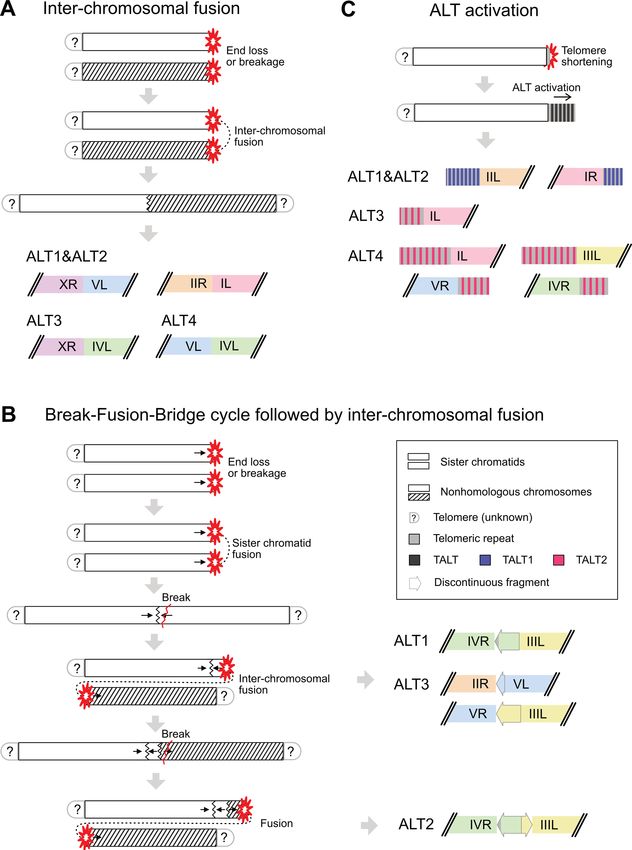
3340 Nucleic Acids Research, 2021, Vol. 49, No. 6

Genome assembly and polishing                                   -c 500) and called variants between each pair of assem-
                                                                blies using Assemblytics (33,34). We first compared variants
De novo genome assemblies of four ALT survivor strains
                                                                between ALT1 and CB4856 to variants between PD1074
were generated with ALT1 27×, ALT2 26×, ALT3 28×,
                                                                and CB4856, and also compared variants between ALT2
ALT4 32× long reads using Canu (version 1.6, genome-
                                                                and CB4856 to variants between PD1074 and CB4856. We
Size = 100m minReadLength = 1000 -pacbio-raw fil-
                                                                found overlapped indels between each pair using BEDtools
tered subreads.fastq.gz) (22). The assemblies were corrected
                                                                (bedtools intersect -wa -wb), and used these overlapped in-
with PacBio raw reads to increase base quality as fol-
                                                                dels with PD1074 and CB4856 to validate our variant call-
lows: First, PacBio raw fastq files were converted to
                                                                ing process as they came from the starting strain of ALT1
BAM files using Picard FastqToSam (version 2.18.7, de-
                                                                and ALT2 (35). The other variants in ALT1 and ALT2 were
fault option) (http://broadinstitute.github.io/picard/), and
                                                                also compared using bedtools to find shared variants gener-
then the BAM files were aligned to assemblies using
                                                                ated after telomere dysfunction and before ALT activation
Pbalign (version 0.4.1, default option) from the Genomic-
                                                                (35).
Consensus package (https://github.com/PacificBiosciences/

                                                                                                                                 Downloaded from https://academic.oup.com/nar/article/49/6/3338/6163097 by guest on 10 October 2021
                                                                   To assign templated insertions, we extracted inserted se-
GenomicConsensus). The aligned BAM files were indexed
                                                                quences from variant sets of our ALT lines and compared
and converted to SAM files using SAMtools (version 1.9,
                                                                the sequences to their flanking sequences or any genomic
index for indexing BAM files and view for converting BAM
                                                                sequences. We used 20-bp flanking sequences for ≥5- and
files to SAM files, default option) and pbindex (version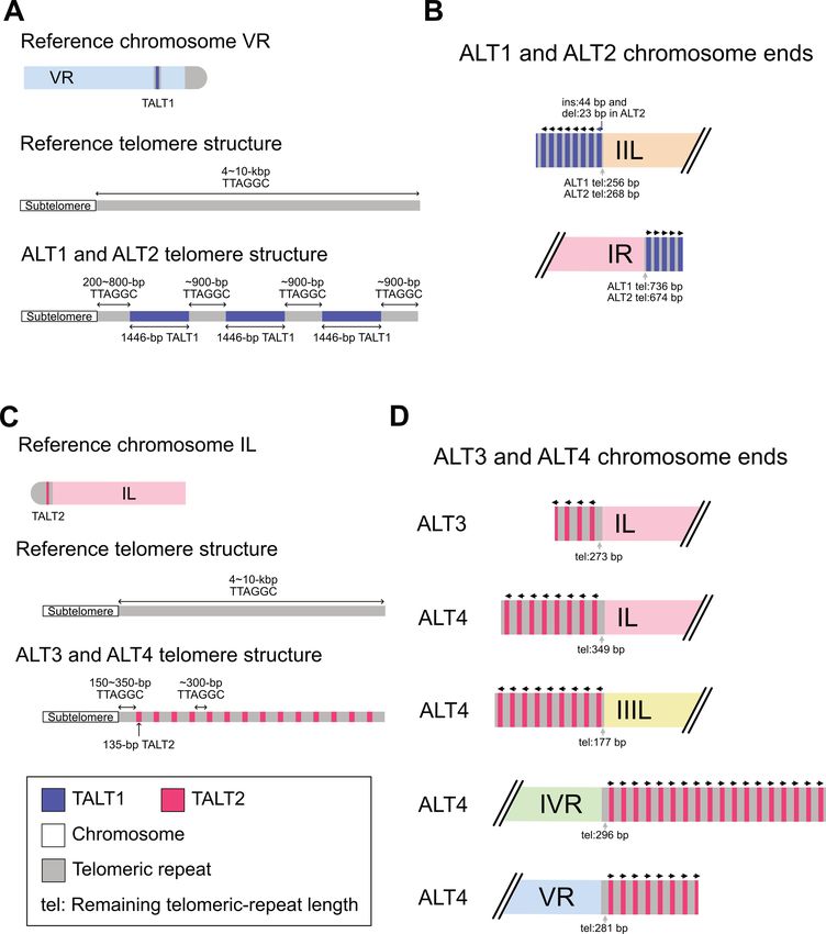
Nucleic Acids Research, 2021, Vol. 49, No. 6 3341

Local re-assembly of chromosome XL in the ALT1 survivor         Quality assessment of four ALT de novo genome assemblies
line
                                                                We used genome assembly to identify genomic variants be-
We extracted ALT1 read ids mapped to the chromo-                tween ALT lines and their respective reference genomes as
some XL ends of the reference or ALT1 genomes us-               our PacBio raw reads had relatively high error rates (∼5%),
ing SAMtools (samtools view alt1 on cb4856.bam X:0–             and first assessed qualities of our de novo genome assem-
78000 and samtools view alt1 on alt.bam tig00000439)            blies of the 4 ALT survivor lines. We obtained a total of
(23). The mapped reads with unique read ids were ex-            26–32× long reads for each strain (N50 = 8–11 kb; Sup-
tracted using seqtk (seqtk subseq), and then the FASTA          plementary Figure S1), and most of the reads were mapped
reads were assembled using Canu with some different             to the reference genomes with mapping quality ≥5 (86.6%,
parameters (version 1.6; canu genomeSize = 200k min-            82.4%, 89.5% and 82.6% for ALT1, ALT2, ALT3 and ALT4
ReadLength = 1000 corMhapSensitivity = high corMinCov-          survivor lines, respectively). We assembled these long reads
erage = 0 -pacbio-raw reads mapped to reference.chrXL.fa        of the four ALT survivor lines into contigs (N50: 236–395
and canu genomeSize = 300k minReadLength = 1000                 kb; longest contig length: 1.3–4.6 Mb; Supplementary Table

                                                                                                                                 Downloaded from https://academic.oup.com/nar/article/49/6/3338/6163097 by guest on 10 October 2021
corMhapSensitivity = high corMinCoverage = 0 -pacbio-           S1).
raw reads mapped to reference.chrXL and ALT1.chrXL.fa)             We then assessed and compared qualities of these four
(22). Finally, we manually merged unique contigs from the       de novo genome assemblies using the Benchmarking Uni-
two local assemblies.                                           versal Single-Copy Orthologs (BUSCO) analysis and ra-
                                                                tios of assembled repeat lengths to the total repeat lengths.
                                                                BUSCO uses the degree of fragmentation of known single-
                                                                copy ortholog genes to compare qualities of genome assem-
RESULTS                                                         blies. Because genome assemblies with poorer qualities have
Experimental design for analysing the consequences of telom-    shorter and more fragmented contigs, they also have lower
ere dysfunction                                                 BUSCO values. The four ALT genome assemblies had sim-
                                                                ilar BUSCO values, as about 80% of the genes are com-
We prepared and sequenced four C. elegans ALT survivor          plete single-copy and ∼20% are missing (Supplementary
lines using the PacBio RSII platform to examine the conse-      Figure S2A). Their complete single-copy BUSCO values
quences of telomere dysfunction at the genome-wide and          were lower than 99% complete single-copy orthologs in ref-
nucleotide levels. We first prepared two lines as internal      erence genomes, suggesting that some ALT genomic regions
controls because they shared known variants so we could         may be somewhat fragmented, but still most regions were
verify our variant calling process using long read-based        properly assembled and suitable to analyse the genomes at
genome assembly. These two lines, ALT1 and ALT2, had            the contig level.
been obtained from a common trt-1 telomerase mutant                In addition, many repetitive sequences are difficult to as-
mother whose genetic background is mostly CB4856 ex-            semble and can be missing in genome assemblies, so as-
cept for some N2 background. The N2 background mix-             sembled repeat lengths can be shorter than the real repeat
up is due to the process of introducing the trt-1 mutation      lengths. The real repeat length can be measured and es-
into the CB4856 strain by outcrossing. The starting trt-1       timated using the raw read depth as unassembled repeat
mutant strain had experienced substantial DDR events due        reads can still be mapped to the corresponding repetitive se-
to telomerase loss for several generations, manifested by a     quences in the genome assembly. Thus we can estimate the
severely reduced brood size, then individually separated into   true repeat length by mapping all raw reads to the genome
tens of plates. A few generations after separation, two sur-    assembly, measuring the raw read depth of the repetitive
vivor lines, ALT1 and ALT2, emerged with independently          sequences and normalising it with the average read depth
acquired ALT activation (Figure 1A). Thus the two lines         of the whole genome sequences. Following this logic, we
are expected to share both N2-type variants in the CB4856       counted the lengths of assembled repeats and also mea-
genetic background and DDR-mediated variants that were          sured and estimated the total repeat lengths in the four
generated during telomere dysfunction before line separa-       ALT assemblies. All of these lengths were comparable to
tion. They are also expected to contain independent DDR-        those of the reference genomes, except for the ribosomal
mediated variants that were generated after line separation.    RNA (rRNA) locus, verifying our assembly qualities (Sup-
We confirmed our variant calling process by examining how       plementary Figure S2B and Supplementary Tables S2 and
many N2-type variants are shared between variant sets of        S3). The measured rRNA length of ALT1 decreased to half
ALT1 and ALT2. We then investigated remaining non-N2-           and that of ALT4 increased 4–5 times than those of the ref-
type variants to compare patterns of genomic changes gen-       erence genomes.
erated during telomere dysfunction or just before ALT ac-
tivation by checking how many non-N2-type variants were
                                                                Validation of variant calling process based on genome assem-
shared or independently acquired in ALT1 and ALT2 and
                                                                bly
where they are located. We used the other two lines, ALT3
and ALT4, obtained from trt-1 mutant animals in the N2          We then assessed our assembly-based variant calling pro-
genetic background, to increase the number of replicates for    cess by testing how many internal control variants, the N2-
independent telomere dysfunction and DDR, as they were          type variants, were properly detected from the ALT1 and
separated before any telomere dysfunction was evoked (Fig-      ALT2 genome assemblies. We called ≥5-bp insertions and
ure 1B).                                                        deletions (indels) in ALT1 and ALT2 genome assemblies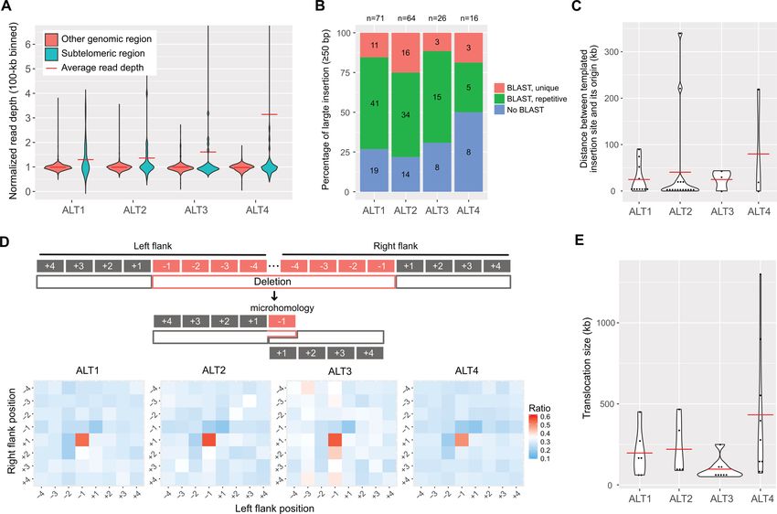
3342 Nucleic Acids Research, 2021, Vol. 49, No. 6

                                                                                                                                                           Downloaded from https://academic.oup.com/nar/article/49/6/3338/6163097 by guest on 10 October 2021
Figure 1. The experimental scheme to confirm the variant calling process used in this study and to understand genomic changes after telomere dysfunction
using the four Caenorhabditis elegans ALT survivor lines. (A, B) The four ALT survivor lines originated from telomerase mutant worms with two different
genetic backgrounds, CB4856 and N2. The left panel represents sequential events of telomere shortening, telomere dysfunction and chromosome fusion,
then ALT activation. (A) ALT1 and ALT2 originated from a telomerase mutant with a CB4856 genetic background and share some portion of N2-type
variants. Their common ancestor had experienced some level of same telomere damages and were separated before new ALT-mediated chromosome end
structures were stably maintained. These features gave us an opportunity to use the shared N2-type variants as internal controls to validate our genome
assembly-based variant calling analysis and to investigate patterns of genomic changes generated by telomere dysfunction. (B) ALT3 and ALT4 originated
from a telomerase mutant with an N2 genetic background that were separated when all telomeres were intact and were used to increase the number of
replicates for understanding telomere dysfunction and ALT activation events.

against the CB4856 genome and compared these variant                          Most of indels generated by telomere dysfunction were not
sets to the N2 variant set obtained by comparing N2 and                       shared by ALT1 and ALT2
CB4856 genomes. We found that the ALT1 and ALT2 vari-
                                                                              After excluding the N2-type variants from the ALT1 and
ant sets shared 506 and 391 insertions with the N2 variant
                                                                              ALT2 variant sets, we analysed remaining indels, which
set respectively, and 303 of the shared insertions were also
                                                                              were probably produced by telomere dysfunction rather
shared by ALT1 and ALT2 lines, with identical insertion
                                                                              than outcrossing process, to compare degrees of genome in-
sites. We also found that the ALT1 and ALT2 variant sets
                                                                              stability before and after line separation. These two lines
shared 622 and 500 deletions with the N2 variant set re-
                                                                              were separated after telomere dysfunction and before ALT
spectively, and 420 of those were shared between ALT1 and
                                                                              activation, so shared indels would be responsible for early
ALT2 variant sets (Supplementary Table S4). Therefore, we
                                                                              genome instability events after telomere dysfunction and
can conclude that high portions, though not all, of shared
                                                                              different indels would be responsible for late genome insta-
N2-type variants in ALT1 and ALT2 were correctly called,
                                                                              bility events by telomere dysfunction before or after ALT
validating our assembly-based variant calling process.
                                                                              activation. We found that ALT1 and ALT2 genome assem-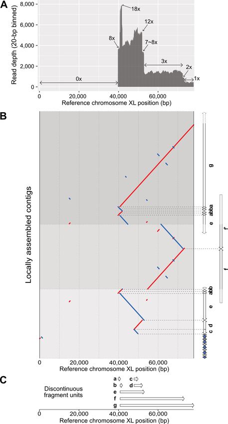
Nucleic Acids Research, 2021, Vol. 49, No. 6 3343

blies contained ∼6% overlapped indels only (Supplemen-             these sticky homologous ends can be annealed and then
tary Table S5), suggesting that the majority of indels were        connected (Figure 2D). We found significant enrichment of
acquired by the late genome instability caused by telomere         homology in the left flank −1, right flank +1 positions (Fig-
dysfunction after line separation. This scarcity of shared in-     ure 2D), which is consistent with the previous study showing
dels implies that telomere dysfunction can generate telom-         that TMEJ in C. elegans mainly uses 1-bp microhomology
ere damage and also genome-wide instability, but that they         (37).
might be induced at different time points, as ALT1 and                We also had an opportunity to explore possible translo-
ALT2 shared almost all telomere damage and chromosome              cation events as our genome assemblies had sufficiently
fusion events (see below).                                         long contigs to identify translocated fragments even longer
                                                                   than 50 kb. We found 28 translocated fragments, 22 of
                                                                   which were inter-chromosomal translocations (Supplemen-
CNVs, templated insertions, simple deletions with microho-
                                                                   tary Table S7). Our 4 ALT lines showed at least five large
mology and large translocations in the 4 ALT survivor lines
                                                                   translocated fragments that ranged 50–1300 kb in size, and

                                                                                                                                   Downloaded from https://academic.oup.com/nar/article/49/6/3338/6163097 by guest on 10 October 2021
We comprehensively investigated characteristics of the             ALT4, specifically, had remarkably larger translocated frag-
variants detected by long-read sequencing data and our             ments than other lines, including the three fragments of
assembly-based indel calling process. First, we compared           which sizes were 550 kb, 900 kb and 1.3 Mb, respectively
CNVs between subtelomeric regions and other genomic re-            (Figure 2E).
gions in the four ALT survivor lines and found that the               All of these lines of evidence suggest that telomere dys-
average read depths of subtelomeric regions were greater           function may not be restricted to telomeres, but lead to
than other genomic regions where average read depths were          whole genome instability and that the genome instability
nearly one (Figure 2A), indicating that subtelomeric regions       might be distinct from direct telomere damage in terms of
are susceptible to copy number changes. In contrast, indels        the time of action.
were not enriched in the subtelomeric regions (Supplemen-
tary Figure S3A and B), so CNVs and indels may have been
                                                                   Subtelomere deletions in chromosome fusion and break sites
produced through different mechanisms.
   All ALT lines contained >2000 indels (≥5 bp) of which           Our genome assemblies of the four ALT survivor lines gave
the majority of these insertions (>97%) and deletions              us an opportunity to understand telomere dysfunction and
(>64%) were not longer than 200 bp, and the ALT4 line had          end repair at the nucleotide level. We first labelled puta-
the largest number of indels, 3670 indels (Supplementary           tive end-containing contigs using end-specific sequences,
Table S6). We then thoroughly examined characteristics of          such as telomeric repeats, TALT copies and subtelomeric
sequences near indels to infer possible mechanisms that gen-       sequences. We recovered only 30 end-containing contigs (30
erated indels, such as random insertion, which is mainly ob-       out of 48 ends; Supplementary Table S8) because imper-
served in nonhomologous end joining or templated inser-            fect assembly quality and the repetitive nature of subtelom-
tion, which can be achieved by polymerase theta-mediated           eric regions restricted the full reconstruction and labelling
end joining (TMEJ). We first analysed whether inserted             of chromosome ends. The ALT1 assembly had nine end-
fragments had similar or same sequences near themselves to         containing ends out of 12 chromosome ends in the assem-
test if they were duplicated using other genomic regions as        bly, the ALT2 assembly had eight, the ALT3 assembly had
templates. We found that 12–15% of insertions had exactly          seven, and the ALT4 assembly had just six ends. Among the
full-matched sequences to their close flanking sequences           ends, 6, 6, 6 and 2 ends in ALT1, ALT2, ALT3 and ALT4,
(20-bp flank for 5–9 bp insertions, and 100-bp flank for 10–       respectively, were fused (20/30; Figure 3), and the other 3,
49 bp insertions; Supplementary Figure S3C). These small           2, 1 and 4 ends had TALT-containing end structures with
insertions, however, can have the same sequences by chance         no evidence of fusion (10/30; Figures 4 and 5). Intriguingly,
in some genomic regions, so we cannot extend search areas          ALT1 and ALT2 contained fusion sites that are identical at
beyond close flanking sequences. We thus used large inser-         the single nucleotide level, which validated that our assem-
tions with a size of 50 bp or longer to find their origins along   blies are suitable for analysing chromosome end structures.
whole genomes using BLAST and found that 50–80% of the             In addition, this also implies that ALT1 and ALT2 were in-
large insertions had at least one BLAST result somewhere           deed separated after severe telomere dysfunction, which had
in the reference genomes (Figure 2B). Among these large            led to chromosome fusion events.
templated insertions, we can calculate the distances between          Next, we resolved end-loss patterns and fusion sites in
their original sites and insertion sites for unique templates      the end-containing contigs at the nucleotide level. All 30
whose insertion sites were on the same chromosomes. The            ends exhibited telomeric-repeat deletions and 21 out of 30
templates were mainly located near the insertion sites, but        contigs exhibited additional subtelomere deletion (Figure
some portions were located as far as several hundreds of           3) in which the subtelomere deletion sizes varied from 156
kilobases (Figure 2C). Our data suggest that large insertions      to 109 516 bp (10 kb: 6 contigs, 29%, Figures 3 and 5 and Sup-
plated insertions, which is a signature of TMEJ.                   plementary Figure S4A). Interestingly, in all chromosome
   Next, we examined whether deletions have microhomol-            fusion sites, every chromosome end had deletions in telom-
ogy in junction sites, which is another evidence of TMEJ. If       eric repeats and even in subtelomeric sequences (20/20) and
polymerase theta was a major mechanism to repair DSBs,             was concealed by fusion between nonhomologous chromo-
then some deleted sequences on one side and remaining se-          somes (Figure 3 and Supplementary Tables S9 and S10).
quences on the other side should have microhomology, as            Specifically, ALT1 and ALT2 lines, which were separated
3344 Nucleic Acids Research, 2021, Vol. 49, No. 6
Downloaded from https://academic.oup.com/nar/article/49/6/3338/6163097 by guest on 10 October 2021
Figure 2. Genomic changes in the four ALT survivor lines and characteristics of CNVs, insertions, deletions and translocations. (A) Violin plots representing
CNV distributions in subtelomeric regions and other genomic regions are shown. Read depths were merged in 100-kb intervals and normalised by the
average whole genome depths. Subtelomeric regions were defined as 200-kb regions from each chromosomal end. (B) Ratios of templated insertions to the
insertions ≥50 bp. Blue bars represent insertions with no BLAST results, green bars represent those of repetitive BLAST results, and red bars represent
those of unique BLAST results. The numbers in the bars represent the actual number of large insertions, and those on top of the graph represent the total
number of large insertions. (C) Size distribution of distance between a templated insertion site and its origin is shown. Red horizontal bar represents the
average distance. (D) Microhomology between deletion junction sites. Top: Schematic representation of possible polymerase theta-mediated end joining
(TMEJ). Typical microhomology between left flank −1 and right flank +1 positions can be achieved by TMEJ. Bottom: Heatmaps represent the ratio of
the same sequences between each position pair in all ≥10-bp deletions. (E) Size distribution of >50-kb translocation. Red horizontal line represents the
average size of each line.
after the same telomere dysfunction and DDR, also shared some IVR, and this inverted fragment had short, ∼150 bp-
the same inter-chromosomal fusion sites between chromo- long, telomeric repeats (chromosome IVR 17 981 794, in-
some IIR and IL (reference position: chromosome IIR 15 verted chromosome IVR fragment [17 982 596–17 975 510]
803 621 and chromosome IL 51 076; Figure 3A) and chro- and IIIL 4679) (Figure 3E). ALT2 shared a similar chro-
mosome XR and VL (reference position: chromosome XR mosome fusion between the two chromosomes, but one
18 070 421 and chromosome VL 3053; Figure 3B). Indepen- additional discontinuous fragment from chromosome IIIL
dently generated ALT3 and ALT4 lines had an independent was inserted in the same direction in the fused chromo-
and distinct fusion pattern in chromosome IVL, where the some IIIL (chromosome VR 17 981 794, inverted chromo-
chromosome IVL was fused with chromosome XR in ALT3 some IVR fragment [17 982 596–17 975 510], chromosome
(reference position: chromosome IVL 30 078 and chromo- IIIL fragment [4679–8126] and chromosome IIIL 3771)
some XR 17 718 493; Figure 3C), but with chromosome VL (Figure 3F).
in ALT4 (chromosome IVL: 42 114 and chromosome VL The discontinuous fragments were found only in six out
109 843; Figure 3D). of 21 subtelomere-deleted ends, not in the ends with in-
Twelve out of 20 fused ends exhibited simple fusion be- tact subtelomere (zero out of nine ends). Their sizes var-
tween two subtelomere-deleted nonhomologous chromo- ied from 333 to 7110 bp, but most of them were 2–7 kb
somes, but the other eight fused ends had discontinuous (Supplementary Figure S4B). The fragments were aligned
fragments, such as partial inverted duplication (Figure 3E– to their subtelomere origins >99.9% in length and >98%
H and Supplementary Table S10). The chromosome IVR identity, indicating that BFB cycles, which produce dupli-
and IIIL fusion site in ALT1 contained an additional in- cated subtelomeric regions, may have inserted the fragments
verted duplication of the subtelomeric region in chromo- in the fusion sites, rather than microhomology-mediatedNucleic Acids Research, 2021, Vol. 49, No. 6 3345
Downloaded from https://academic.oup.com/nar/article/49/6/3338/6163097 by guest on 10 October 2021
Figure 3. A schematic representation of chromosome end structures (not to scale) is shown. (A–D) Simple fusion between nonhomologous chromosomes.
(A, B) Inter-chromosomal fusion sites of the Caenorhabditis elegans ALT1 and ALT2 survivor lines. References represent CB4856 chromosomes. (A)
Chromosome IL and IIR ends were fused to each other after a 50 644-bp deletion in the chromosome IL subtelomeric region and a 6387-bp deletion
in the chromosome IIR subtelomeric region. (B) Chromosome VL and XR ends were fused to each other after a 156-bp deletion in the chromosome
VL subtelomeric region and a 255-bp deletion in the chromosome XR subtelomeric region. (C) Inter-chromosomal fusion sites of the ALT3 survivor
line. References represent N2 chromosomes. Chromosome IVL and XR ends were fused to each other after a 29,935-bp deletion in the chromosome
IVL subtelomeric region and a 306-bp deletion in the chromosome XR subtelomeric region. (D) Inter-chromosomal fusion sites of the ALT4 survivor
line. References represent N2 chromosomes. Chromosome IVL and VL ends were fused to each other after a 41,965-bp deletion in the chromosome IVL
subtelomeric region and a 109 516-bp deletion in the chromosome VL subtelomeric region. (E–H) Complex inter-chromosomal fusion with discontinuous
fragments. (E) The fusion site of chromosome IIIL and IVR ends in the Caenorhabditis elegans ALT1 survivor line contained a discontinuous fragment,
which would have originated from the chromosome IVR end, but was located in the opposite direction. The origin of the discontinuous fragment was
supposed to be chromosome IV:17 975 510–17 982 596 in the reference CB4856 genome. The fused chromosome IIIL end had a 1557-bp subtelomere
deletion, and the chromosome IVR end had a 653-bp subtelomere deletion. References represent corresponding CB4856 chromosomes. (F) The fusion
site of chromosome IIIL and IVR ends in the ALT2 survivor line contained not just the same discontinuous IVR fragment in the ALT1 survivor line,
but also another discontinuous fragment, which originated from the chromosome IIIL end and was located in the same direction. The origin of the
discontinuous IIIL fragment was supposed to be chromosome III:4679–8126 in the reference CB4856 genome. The fused chromosome IVR end had
almost the same subtelomere deletion, but the chromosome IIIL end had a much shorter 650-bp subtelomere deletion than that of the ALT1 survivor
line. References represent the corresponding CB4856 chromosomes. (G) The fusion site of chromosome IIR and VL ends in the ALT3 survivor line
contained a discontinuous fragment, which would have originated from the chromosome VL end, but was located in the opposite direction. The origin
of the discontinuous fragment was supposed to be chromosome IV:655–983 in the reference N2 genome. The fused chromosome IIR end had a 5282-bp
subtelomere deletion, and the chromosome VL end had a 1086-bp subtelomere deletion. References represent the corresponding N2 chromosomes. (H)
The fusion site of chromosome IIIL and VR ends in the ALT4 line. A discontinuous fragment would have originated from chromosome III:865–2999 in the
reference N2 genome and was located in the opposite direction. The fused chromosome IIIL end had a 1640-bp subtelomere deletion, and the chromosome
VR end had a 1249-bp subtelomere deletion. References represent corresponding N2 chromosomes.3346 Nucleic Acids Research, 2021, Vol. 49, No. 6
Downloaded from https://academic.oup.com/nar/article/49/6/3338/6163097 by guest on 10 October 2021
Figure 4. Schematic representation of chromosome end structures that were reconstructed by one-way replication of TALT (Template for ALT) and telom-
eric repeats (not to scale). All subtelomeres were intact and short telomeric repeats remained at the junctions between a subtelomeric region and a TALT
copy. (A) The origin of the TALT1 (upper), typical telomere structure in the reference genome (middle) and new telomere structure of the Caenorhabditis
elegans ALT1 and ALT2 survivor lines (lower). The original TALT1 (blue bar) is located in the right arm of chromosome V and consists of a 1446-bp
genomic sequence flanked by short telomeric repeats (grey bars) (B) Assembled and confirmed TALT1-mediated chromosome end structures in the ALT1
and ALT2 lines. (C) The origin of the TALT2 (upper), typical telomere structure in the reference genome (middle) and new telomere structure of the ALT3
and ALT4 survivor lines (lower). The original TALT2 (red bar) is located in the far left subtelomeric region of chromosome I and consists of a 135-bp
genomic sequence flanked by short telomeric repeats (gray bars). (D) Assembled and confirmed TALT2-mediated chromosome end structures in the ALT3
and ALT4 survivor lines.
and error-prone replication (Figure 3E–H and Supplemen- One-way replication of TALTs in ALT-mediated chromo-
tary Table S11). In addition, short inserted sequences, a sig- some ends
nature of nonhomologous end joining, were found in fusion
The 10 end-containing contigs that did not exhibit inter-
sites of chromosome IIR and chromosome VL in ALT3, as
chromosomal fusion had new chromosome end structures
well as in the inverted duplicated fragment of chromosome
constituted by TALT copies and short telomeric repeats,
IVL in ALT1 and ALT2, further supporting the possibility
which suggests a possible role of telomeric repeats in TALT
that BFB cycles generated discontinuous fragments (Sup-
replication (Figures 4 and 5). Subtelomere deletion was not
plementary Table S10).
observed in nine out of 10 contigs, and their telomeric re-Nucleic Acids Research, 2021, Vol. 49, No. 6 3347
peats were shortened, but still remained in 200–800 bp,
which is one tenth of the estimated telomeric-repeat length
of 4–10 kb (38). The new chromosome ends of ALT1 and
ALT2 were composed of 1.4-kb TALT1 (Figure 4A and B)
and those of ALT3 and ALT4 were composed of 135-bp
TALT2 (Figure 4C and D), as previously reported (12), and
these TALT copies were flanked with similar-length canon-
ical telomeric repeats (∼900 bp for TALT1 and ∼300 bp for
TALT2; Figure 4B and D). This unit structure of the new
chromosome ends, each TALT with flanking telomeric re-
peats at its both ends, was identical to that of the original
TALTs, which also have flanking canonical or degenerated
telomeric repeats and are located in internal chromosomal
Downloaded from https://academic.oup.com/nar/article/49/6/3338/6163097 by guest on 10 October 2021
regions (Figure 4A and C). Moreover, the direction of all
these TALT copies and their flanking canonical and degen-
erated telomeric repeats was always the same, except only
one TALT on the XL end of ALT1: from the internal chro-
mosomal region to the end (9/10 at the very first TALT of
ends, 71/72 at all the TALTs found in end contigs; Figure
4B and D).
Complex chromosomal rearrangements in chromosome XL of
ALT1
In this study, all the subtelomere-deleted ends in the iden-
tified chromosome ends underwent inter-chromosomal fu-
sion, and all telomeric-repeat-remaining ends resulted in
TALT-mediated telomeres. One exception was the chro-
mosome XL of ALT1, which has extreme rearrangements
among the end-containing contigs. The chromosome XL
of ALT1 had a subtelomere deletion, but was covered with
TALT copies and telomeric repeats. This chromosome was
also the only end that has an inverted TALT copy and
telomeric repeats at the beginning of the TALT-mediated
telomere. We used the ALT1 reads mapped to chromosome
XL of the reference genome to understand the cryptic end
structure in detail and to avoid errors from possible mis-
assembly and low contiguity in the ALT1 assembly. Their
mapping pattern showed that 40-kb subtelomere deletion
occurred and the read depth changed 18×, 8×, 12×, 8×,
3×, 2× and 1× along the 30-kb region of the remaining sub-
telomere (Figure 5A). This complex read-depth change and
Figure 5. Complex chromosomal rearrangement events found in the chro-
mosome XL end of the Caenorhabditis elegans ALT1 survivor line. (A) odd-number CNV are representative characteristics of FoS-
Complex and stepwise read-depth changes in the chromosome XL end of TeS, and the current state-of-the-art assemblers have limited
the ALT1. The x-axis represents reference CB4856 chromosome XL po- ability to deal with such complex structures, so we locally
sitions and the y-axis represents average read depths of raw ALT1 reads and manually re-assembled the subtelomere structure using
(20-bp binned). (B) Dot plot representing alignment between the refer-
ence chromosome XL end (X:0–78 000) and locally, manually re-assembled
the reads mapped on chromosome XL of ALT1 and the ref-
three contigs of the ALT survivor line. Each contig was aligned to the ref- erence genomes.
erence CB4856 genome, and units were identified and classified based on We partially, but more finely, assembled the structure
their alignments (white arrows, units a–f). Different background colours and confirmed that high read-depth regions in the refer-
represent different contigs. Red: forward strand matches; blue: reverse ence chromosome XL were resolved as repetitively dupli-
strand matches; horizontal grey dots: boundaries of each contig; horizon-
tal dashed grey lines: boundaries of each unit; blue arrows: TALT; gray cated fragments in several contigs (Figure 5B), and that the
arrows: telomeric repeats. (C) The list of discontinuous fragment units and end can be partitioned into some repetitive units, which
genomic positions where they originated based on the reference CB4856 have unique start and end positions (Figure 5C, single ar-
genome. White arrows are placed as their reference positions (a: 39 683–40 rows a–g). The start and end positions of the repetitive units
727, b: 40 833–41 912, c: 47 882–49 705, d: 47 420–51 990, e: 40 591–52 899,
f: from 40 kb to 72 825, g: from 40 591 to the end of chromosome X).
overlapped with the regions of complex read-depth changes,
suggesting that the repetitive units were used for replica-
tion and were subsequently recovered in our locally assem-
bled subtelomere (Supplementary Table S12). The repetitive
units were mainly replicated by inverted duplication, but
when the unit a and b were connected as a-b-b or a-b-b-a,3348 Nucleic Acids Research, 2021, Vol. 49, No. 6
duplications were not in an inverted manner, but in the same genome assemblies of four ALT survivor lines depicted
direction, which cannot be generated by the BFB cycle. In chromosome fusion and breakage sites and new end struc-
addition, units a and b had 66-bp homology sequences at tures at the nucleotide level and allowed us to model these
the connected region (Supplementary Figure S5A–C), and sites as specific types of traces that DDR leave behind. Thus,
two copies of unit f, denoted as f and f that were connected our long-read sequenced ALT survivor lines can serve as
in the opposite direction, also contained 110-bp homol- reproducible resources to investigate telomere dysfunction
ogy sequences in their junction site (Supplementary Fig- and DDR, and can model karyotype evolution events at the
ure S5D-F). This subsequent length of homology sequences nucleotide level.
is another characteristic of FoSTeS; therefore, we speculate
that at least one or two FoSTeS events may have occurred Telomere dysfunction causes telomere damage and genome
and partially contributed to reconstructing this subtelom- instability at different time points
ere. In contrast, other duplications contained very short ho-
Several types of variants generated after telomere dysfunc-
mology or insertions at the junction sites, suggesting that
tion were widespread throughout the genome of C. elegans
Downloaded from https://academic.oup.com/nar/article/49/6/3338/6163097 by guest on 10 October 2021
nonhomologous end joining was mainly used to connect re-
ALT survivor lines, indicating that telomere dysfunction
maining units.
can generate a subtle level of genome instability in addition
to telomere-specific DNA damage. Intriguingly, ALT1 and
DISCUSSION ALT2, which had commonly experienced severe telomere
damage events, including chromosome fusions, shared only
Chromosome ends are susceptible to karyotype evolution.
a small portion of indels generated after telomere dysfunc-
Telomeres provide a protective structure at chromosome
tion. In other words, DNA damage in other genomic re-
ends but can suffer from gradual telomeric-repeat shorten-
gions away from telomeric regions was accumulated only af-
ing or stochastic telomere deletion and following DDR may
ter telomere rearrangements accumulated. Our finding that
fuse the ends resulting in a novel karyotype. For example,
there is a distinct time lapse between telomere rearrange-
the haploid chromosome number of humans contains one
ments and chromosomal damage caused by telomere dys-
fewer than that of their close relative apes (39–41). Some
function is both unprecedented and unexpected, partly be-
wild mice exhibit a 20%–50% reduction in haploid chro-
cause genome instability generated by telomere dysfunction
mosome numbers compared to that of the laboratory stan-
has rarely been studied at the genome-wide and nucleotide
dard mouse (42–44), and these karyotype reduction events
levels as well as in a time series.
are likely the consequences of chromosome fusions between
This time lapse phenomenon, unless only specific to
two acrocentric chromosomes (45). However, because their
ALT1 and ALT2 survivors, suggests that either it may re-
ancestral genome sequences were not available, it was diffi-
quire some time for DNA damage caused by telomere dys-
cult to elucidate how telomere dysfunction was terminated,
function to spread from telomeric regions to other genomic
for example, which DDR was involved or how fusions oc-
regions, or that genome-wide accumulation of indels may
curred. In this report, we used ALT survivor lines in C. ele-
start just before ALT activation as ALT turn-on needs adap-
gans as a model to dissect the consequences of telomere dys-
tation to DNA damage. In yeasts, severe telomere dysfunc-
function and karyotype evolution. Our ALT survivor lines
tion results in delayed cell cycle and cellular senescence, and
had overcome telomere dysfunction using several DDR and
this cellular senescence can be overcome by adaptation to
ALT mechanisms, such as reconstructing TALT-mediated
DNA damage that allows cells to bypass the cell cycle ar-
telomeres and chromosome fusion events, which result in
rest and increases mutation accumulation (46). Although
fewer chromosome numbers than their ancestral lines (12).
we cannot directly test these hypotheses because our exper-
Because their ancestral lines were fully sequenced, includ-
imental design does not contain any other lines that were
ing the chromosome ends (18,19), we applied a long-read
separated with tight time intervals, the increased mutation
sequencing technology to the ALT survivor lines to un-
rates in our lines may represent the adaptation to DNA
derstand the molecular consequences of telomere dysfunc-
damage, similar to the yeast, as these lines were separated
tion by comparing whole genome sequences of the ances-
before serious cellular senescence.
tral and ALT survivor lines. We independently assembled
genomes of two different ALT survivor lines that share com-
TMEJ is a main repair mechanism for the genome-wide
mon fusion and breakage events to validate our long-read
DSBs generated by telomere dysfunction
sequencing-based methodology. Their genome assemblies
share the same fusion and breakage sites at the single nu- The genome-wide DSBs are thought to be repaired mainly
cleotide level, suggesting that even genome assemblies with by TMEJ because they have known TMEJ signatures.
moderate sequencing depths can resolve genomic changes TMEJ repairs DSBs by replication fork stalling, but this
after karyotype evolution. repair is error-prone, resulting in short-length indel gener-
The high-resolution maps drawn using long-read se- ation (37). In addition to these short-length indels, TMEJ
quencing of ALT survivor lines enabled a detailed exami- exhibits other characteristics, including microhomology be-
nation of chromosomes at the single nucleotide level. Anal- tween two DSB ends to connect them and templated in-
ysis of CNVs and genome-wide variants generated after sertions, which replicate other genomic sequences into the
telomere dysfunction suggested that DNA damage caused deletion sites (37). All of these signatures were found in the
by telomere dysfunction is not limited to the chromosome majority of our indels; therefore, the genome instability and
ends, but also spreads to other chromosomal regions to gen- DSBs generated after telomere dysfunction were mainly re-
erate subtle genome-wide instability. In addition, de novo paired by TMEJ in our ALT lines.Nucleic Acids Research, 2021, Vol. 49, No. 6 3349
ALT lines suffered from different degrees of telomere dys- chromosome ends by BLAST using a unique sequence fur-
function ther inside the subtelomeric regions.
The simplest terminating mechanisms are simple fusion
Degrees of genome instability were different among the
sites without any BFB cycle (Figure 6A). All of these six
ALT lines. In particular, ALT4 exhibited a higher number
fusion sites involved subtelomere deletion at all 12 chro-
of indels (Supplementary Table S6) and also larger translo-
mosome ends and only occurred between nonhomologous
cation fragments (Figure 2E), which implies that this line
chromosomes (Figure 3A-D). This situation may arise from
may have suffered a higher level of genome-wide DSBs.
the holocentric chromosomes in C. elegans (53). Fusion
How telomere dysfunction can cause genome instability and
between monocentric nonhomologous chromosomes may
why different ALT survivor lines suffered from different de-
generate dicentric chromosomes, which leads to chromo-
grees of DSBs still remain elusive. Variations in the num-
some breakage and new DSB sites after segregation errors
ber of rRNA were also observed in the ALT survivor lines
during cell division. In contrast, holocentric nonhomolo-
(Supplementary Table S3). Although the total estimated
gous chromosomes do not suffer from chromosome fusion
lengths of other repetitive elements in the ALT survivor
Downloaded from https://academic.oup.com/nar/article/49/6/3338/6163097 by guest on 10 October 2021
followed by segregation, because their whole chromosomal
lines were still comparable to those of reference genomes, es-
regions can act as centromeres, resulting in a stable fusion
timated rRNA lengths were reduced in ALT1 and increased
chromosome.
in ALT4. These change are likely due to the characteris-
Our results show that all simple fusion sites underwent
tic of C. elegans genome where the rRNA genes are clus-
subtelomere-subtelomere fusions, but are different from a
tered near the end of the chromosome IR (47,48). The sus-
previous study, where ∼40% of fusion sites had telomere-
ceptibility of subtelomeric regions to the telomere dysfunc-
subtelomere fusion (1). One possible explanation for this
tion may result in fluctuation in rRNA copy numbers. The
discrepancy is that the previous study used a PCR-based
copy number may therefore be reduced through DNA dam-
method that checked whether the nearest subtelomeric re-
age and nonhomologous end joining and may be increased
gions still remained after the fusion events. The authors di-
by microhomology-mediated replication mechanisms, such
rectly amplified some fusion sites, but could not amplify the
as break-induced replication. We cannot resolve the possi-
others because of the repetitive nature of subtelomeric re-
ble mechanisms because the long-read sequencing technol-
gions. Thus, they moved primers far from telomeric repeats
ogy we used in this study still has limitations, such as the
and reasoned that if the possible nearest primers work, then
∼10-kb read length and ∼5% error rate. The exact struc-
subtelomere deletion did not occur. This elegant approach
ture of rRNA clusters can be resolved by error-free and
worked well, but the repetitive nature of subtelomeric re-
ultra-long sequencing technologies, as human centromeres
gions may have limited the detection of short, ∼100-bp sub-
of chromosomes 6 and X were resolved by merging the
telomere deletions. Indeed, all PCR-amplified fusion sites
two state-of-the-art technologies (49,50); Oxford Nanopore
were identified as subtelomere-subtelomere fusion sites. An-
Technologies ultra-long reads that have >1-Mb read length
other possible reason for the discrepancy between our re-
and PacBio high-fidelity (HiFi) reads that have 1% or less
sults and the previous report is that different methods of
error rate (51,52).
generating chromosome fusion lines may give different re-
sults. The previous study used brood size reduction as a sig-
nature of chromosome fusion, but we passed more genera-
All simple fusion sites exhibited total telomere erosion and
tions after brood size reduction to turn on ALT mechanisms
subtelomere-subtelomere fusion
(12). Thus our ALT survivor lines had suffered from telom-
Our de novo genome assemblies of four C. elegans ALT sur- ere dysfunction for more generations, possibly resulting in
vivor lines revealed possible repair or terminating mecha- more subtelomere deletion events. In addition, the previous
nisms of DSBs at the chromosome ends. Substantial conti- study used a double mutant harbouring mutations in both
guity of our genome assemblies allowed us to identify chro- telomerase and a major component of the canonical non-
mosome end structures at the nucleotide level, but unfortu- homologous end joining genes, while we used a telomerase
nately, only 30 out of 48 chromosome ends were revealed. single mutant worm. A third possibility is that the remain-
This may be due to the limitation of our end-searching pro- ing chromosome ends that we could not assemble may have
cess that listed the chromosome end by BLAST of telom- telomere-subtelomere fusions, but this is unlikely because
eric repeats, the TALT sequence or subtelomeric sequences almost all fusion sites are assembled in our ALT survivor
to the assemblies. The repetitive nature of subtelomeric lines, which have karyotypes of 2n = 6–8 (Supplementary
regions and our end-searching process resulted in huge Figure S6).
BLAST outputs, which restricted accurate detection of the
chromosome ends. Moreover, the repetitiveness also inhib-
BFB cycles worked after sister chromatid fusion
ited the de novo genome assembly process, thus some chro-
mosome ends that were partially assembled and had no We successfully assembled four other fusion sites that con-
unique sequences, would not be included in our search pro- tained discontinuous fragments, which remained between
cess. For example, chromosome IIIR, which was not found nonhomologous chromosomes, suggesting that a BFB cy-
in any of the four lines, and chromosome XL, which was not cle between sister chromatids had occurred (Figure 3E-H).
found in three of the lines, may have no searchable unique These fragments had >97% identity with the original, in-
sequences. If the contiguity can be further increased by in- tact sister chromatids and four out of five fragments were
creasing the readdepth or read length, and by reducing the inverted at the end of the sister chromatid. This suggests
read error rate, our end-searching process may retain more that the sister chromatids were fused and broken, leaving3350 Nucleic Acids Research, 2021, Vol. 49, No. 6
Downloaded from https://academic.oup.com/nar/article/49/6/3338/6163097 by guest on 10 October 2021
Figure 6. Model for chromosome end repair or reconstruction processes after telomere dysfunction. (A) Simple inter-chromosomal fusion between
subtelomere-deleted chromosome ends. (B) Break-Fusion-Bridge (BFB) cycle followed by inter-chromosomal fusion. Black arrows represent the orien-
tation of the broken, discontinuous fragments. (C) ALT-mediated chromosome end reconstruction. Short remaining telomeric repeats may facilitate the
replication of TALTs and telomeric repeats to the new ends.
an inverted, high-identity fragment at the end (Figure 3E, G the same breakpoints and different orientations of discon-
and H). Unlike nonhomologous chromosome fusion, sister tinuous fragments: the chromosome IVR fragment was in-
chromatid fusion causes a chromosome segregation prob- verted, but the chromosome IIIL fragment was duplicated
lem during the cell cycle, so the fusion chromosome must in the same orientation. The two sister chromatids of chro-
be broken, generating a discontinuous fragment. mosome IV were likely fused and broken, then the broken
Among the four BFB sites, three were stabilised after end fused with chromosome III in the common ancestor
nonhomologous chromosome fusion events, but the BFB of ALT1 and ALT2. After this nonhomologous chromo-
site in the ALT2 genome had an additional fusion event, some fusion, ALT1 and ALT2 would have followed differ-
leaving two discontinuous fragments between nonhomol- ent paths. The chromosome IV end in ALT1 was stabilised,
ogous chromosomes (Figures 6B and 3F). We can specu- but in ALT2, chromosome IV may have suffered from ad-
late on the fusion and breakage events at this site based on ditional breakage and fusion, leaving the two discontinu-You can also read