Multiplexed engineering glycosyltransferase genes in CHO cells via targeted integration for producing antibodies with diverse complex type N glycans
←
→
Page content transcription
If your browser does not render page correctly, please read the page content below

www.nature.com/scientificreports
OPEN Multiplexed engineering
glycosyltransferase genes in CHO
cells via targeted integration
for producing antibodies
with diverse complex‑type
N‑glycans
Ngan T. B. Nguyen, Jianer Lin, Shi Jie Tay, Mariati, Jessna Yeo, Terry Nguyen‑Khuong &
Yuansheng Yang*
Therapeutic antibodies are decorated with complex-type N-glycans that significantly affect their
biodistribution and bioactivity. The N-glycan structures on antibodies are incompletely processed
in wild-type CHO cells due to their limited glycosylation capacity. To improve N-glycan processing,
glycosyltransferase genes have been traditionally overexpressed in CHO cells to engineer the
cellular N-glycosylation pathway by using random integration, which is often associated with large
clonal variations in gene expression levels. In order to minimize the clonal variations, we used
recombinase-mediated-cassette-exchange (RMCE) technology to overexpress a panel of 42 human
glycosyltransferase genes to screen their impact on antibody N-linked glycosylation. The bottlenecks
in the N-glycosylation pathway were identified and then released by overexpressing single or
multiple critical genes. Overexpressing B4GalT1 gene alone in the CHO cells produced antibodies with
more than 80% galactosylated bi-antennary N-glycans. Combinatorial overexpression of B4GalT1
and ST6Gal1 produced antibodies containing more than 70% sialylated bi-antennary N-glycans.
In addition, antibodies with various tri-antennary N-glycans were obtained for the first time by
overexpressing MGAT5 alone or in combination with B4GalT1 and ST6Gal1. The various N-glycan
structures and the method for producing them in this work provide opportunities to study the glycan
structure-and-function and develop novel recombinant antibodies for addressing different therapeutic
applications.
The Fc region of IgGs possesses two identical N-glycans which are composed of a core heptasaccharide with three
mannoses and two N-acetylglucosamine (GlcNAc). The core structure is further decorated with various sugar like
fucose, bisecting GlcNAc, galactose and sialic acid to form a complex-type bi-antennary glycan. N-glycosylation
is a critical quality attribute for therapeutic monoclonal antibodies (mAbs), since it affects the drug efficacy,
safety, and pharmacokinetic properties1,2. The fucosylation of N-glycans is known to negatively impact the
antibody dependent cellular cytotoxicity (ADCC) of IgG3,4. It has been shown that afucosylated antibodies have
100-fold increased ADCC compared to the fucosylated o nes3. The galactose moiety of N-glycan is less critical to
ADCC than the fucose residue, while its impact on complement dependent cytotoxicity (CDC) is s ignificant5.
Several studies have demonstrated that increasing galactosylation of antibodies boosted CDC for IgG1 and IgG3
subclasses especially6,7. On the contrary, the presence of terminal sialic acid residues on N-glycans neutralized
the enhanced CDC activity of the galactosylated mAbs8. Sialylation also significantly impaired ADCC of the
fucosylated mAbs while it did not adversely change ADCC of the fucose-free I gGs9–11. Though removing sialic
acid seems to be beneficial for enhancing the Fc effector functions, de-sialylated antibodies displayed poorer pK
with faster serum clearance and shorter half-life, potentially dampening drug efficacy12. Bas et al. have provided
Bioprocessing Technology Institute, Agency for Science, Technology and Research (A*STAR), Singapore, Singapore.
*
email: yang_yuansheng@bti.a-star.edu.sg
Scientific Reports | (2021) 11:12969 | https://doi.org/10.1038/s41598-021-92320-x 1
Vol.:(0123456789)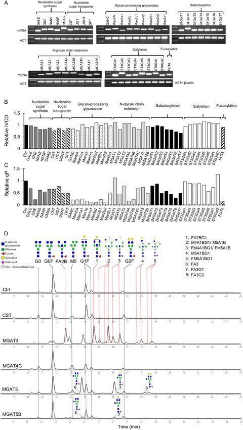
www.nature.com/scientificreports/
evidence that hyper-sialylated N-glycans significantly increased the IgG serum persistence in the animal m odel13.
The terminal sialic acids could also enhance the anti-inflammatory function of antibodies up to 10-fold14,15. Thus,
sialylated N-glycans on antibodies are also desirable in many therapies like intravenous immunoglobulin (IVIG).
It has been recognized that a specific N-glycan profile is favourable over others, depending on the therapeutic
applications of antibodies. Being able to engineer a diverse range of N-glycan structures on antibodies will be of
great advantage for different therapeutic purposes.
Chinese Hamster Ovary (CHO) cell is the most commonly used host cell for industrial production of thera-
peutic IgGs due to their capability to perform proper protein folding, assembly of complexes and human-like
glycosylation16,17. However, wild-type CHO cells generally produce antibodies with incomplete N-glycans that
are often highly fucosylated but severely lacking galactose, bisecting GlcNAc and terminal sialic acids18,19. To
improve complex N-glycan processing, it is necessary to engineer the CHO N-glycosylation pathway and this is
often done by overexpression of glycosyltransferase genes in CHO cells. Several studies have shown that over-
expression of α-2,6-sialyltransferase 1 (ST6Gal1) gene alone20,21 or in combination with galactosyltransferase
gene18,19,22 in CHO cells could produce antibodies carrying more sialylated N-glycans. However, the achieved
sialylation improvement in these studies has been limited. Overexpression of other glycosyltransferase genes that
regulate precursor b iosynthesis23,24, nucleotide sugar t ransport25 and branching e xtension26 alone in CHO cells
have been shown to increase sialylated N-glycan structures on erythropoietin (EPO) and IFNγ proteins. Since
glycoproteins differ in the positions and numbers of N-glycosylation sites, it is unclear whether the above strate-
gies, which are effective in producing complex N-glycans on recombinant proteins, will work for antibodies as
well. In addition, some glycosyltransferase enzymes in the N-glycan biosynthesis pathway have multiple isoforms
that could contribute differently to the N-glycan processing in both protein- and site-specific manner27–29. Bet-
ter understanding of the distinct functions of different isoenzymes will provide new opportunities to engineer
proteins with more diverse types of N-glycan structures.
Engineering the N-glycosylation pathway by overexpression of glycosyltransferase genes in CHO cells has
often been achieved via random integration18,20,24–26. The generated stably transfected cell pools exhibit highly
heterogeneous gene expression and clonal v ariation30,31, which has limited the success of achieving desirable
glycosylation outcomes. Targeted integration approach could overcome such issues by directing transgenes
integration into specific loci in genome. As all targeted integration cells have the same genetic background, the
obtained stably transfected cells exhibited high levels of phenotypic and transcriptional homogeneity32. Targeted
integration of transgenes has often been achieved by using recombinase-mediated-cassette-exchange (RMCE).
The technique works through the action of recombinases which recognize the specific recombination sites that
are pre-determined in the genome and replace the sequence between the two non-compatible sites with a new
transgene33,34. Among different recombinases, the Flp/FRT system has been extensively applied in CHO cells to
express monoclonal antibodies35 and recombinant proteins34,36 probably due to its high specificity, integration
efficiency and low cytotoxity.
In this study we utilized a Flp/FRT-based RMCE system to first overexpress a panel of 42 human glycosyl-
transferase genes in CHO cells to screen their impact on the N-glycosylation of a model IgG antibody, rituximab.
These genes are involved in the different steps of the entire core N-glycosylation pathway and have diverse func-
tions, ranging from sugar precursor synthesis, glycan processing, branching, galactosylation to terminal capping
(Table 1). The critical genes that significantly influenced the IgG N-glycosylation were identified and further
overexpressed in combination to achieve an array of complex-type N-glycan structures. We also demonstrated
that the enzyme expression level was critical in determining glycosylation outcomes and combinatorial engineer-
ing of multiple enzymes was required for obtaining highly complex N-glycan structures on mAbs.
Results
Design of RMCE to screen for functional roles of human glycosyltransferase genes in N‑gly‑
cosylation of antibodies in CHO cells. In order to study the impact of overexpressing human glyco-
syltransferase genes on IgG glycosylation, we co-expressed the individual glycosylation gene together with the
antibody gene in a CHO K1 master cell line (MCL) via RMCE-based targeted integration (Fig. 1A). The MCL
was confirmed to have one single integrant of a landing pad vector by Southern blot and targeted locus amplifi-
cation (TLA) analysis (data not shown). The landing pad vector contained a hygromycin resistant gene (HYG)
flanked by a wild-type flippase recognition target (FRT) and its mutant FRT3. An impaired puromycin resistant
gene lacking a start codon ((ATG-)Puro) was placed downstream of FRT. The transcription of HYG gene was
driven by a chimeric promoter (ChiP) and terminated by a SV40 polyadenylation signal (pA) upstream of FRT.
As a result, the impaired (ATG-)Puro was not expressed before activation by RMCE. Each targeting vector was
designed to be promoter-less and carry the rituximab light chain (LC) and heavy chain (HC) genes, together
with one or two specific human glycosyltransferase genes and the DsRed gene linked through multiple wild-
type encephalomyocarditis virus (EMCV) internal ribosome entry site (IRES) (Fig. 1B). An additional wild-type
EMCV IRES was placed downstream of DsRed for activating the expression of (ATG-)Puro upon replacing the
HYG cassette in the MCL through RMCE. The targeting vector carrying only rituximab and DsRed genes served
as the control. Rituximab is a therapeutic IgG1 mAb for treating rheumatoid arthritis and B-cell non-Hodgkin’s
lymphoma37.
RMCE was carried out by co-transfection of a specific targeting vector and the vector expressing an enhanced
recombinase flippase (FLPe) followed by puromycin selection to obtain stably transfected pools. As the target-
ing vectors did not have promoter and polyadenylation signal, they could not express the carried genes if they
randomly integrated into the chromosomes. The combination of promoter-trap and ATG-trap ensured that only
cells with correct RMCE expressing all genes carried on the targeting vectors can survive the puromycin selection.
As all targeted cells in a stably transfected pool share the same integration site, variation caused by position effect
Scientific Reports | (2021) 11:12969 | https://doi.org/10.1038/s41598-021-92320-x 2
Vol:.(1234567890)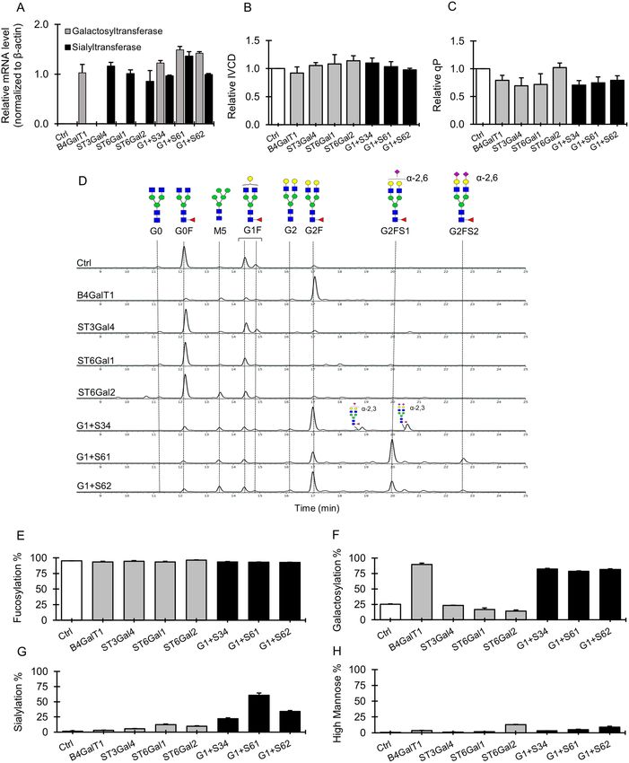
www.nature.com/scientificreports/
Group Name Accession No Gene
GALE NM_000403 UDP-galactose 4-epimerase
GNE NM_005476.5 UDP-N-acetylglucosamine-2 epimerase (Human)
NANS NM_018946.3 Sialic acid synthase
Nucleotide sugar synthesis
NANP NM_152667.2 N-acetylneuraminic acid phosphatase
CMAS NM_018686.5 Cytidine monophospho-sialic acid synthase
CST NM_006416.4 CMP-sialic acid transporter
UGT NM_005660.2 UDP-galactose transporter
UGNT NM_032826.4 UDP-N-acetylglucosamine transporter
GFT NM_018389.4 GDP-fucose transporter
Nucleotide sugar transporter GANC NM_198141.2 Neutral α-glucosidase C
MANEA NM_024641 Endo-α mannosidase
MAN1A1 NM_005907 Mannosyl-oligosaccharide 1,2-α-mannosidase IA
MAN1B AF_027156 Mannosyl-oligosaccharide 1,2-α-mannosidase IB
MAN1C1 AF_261655 Mannosyl-oligosaccharide1,2-α-mannosidase IC (isoform 1)
MAN2A2 NM_006122.2 α-mannosidase 2A member 2
MAN2B1 NM_000528.3 α-mannosidase, class 2B, member 1
MAN2B2 NM_015274.2 α-mannosidase, class 2B, member 2
Glycan- processing glycosidase
MAN2C1 NM_006715.3 α-mannosidase, class 2C, member 1
MGAT1 NM_001114618.1 α-1,3-mannosyl-glycoprotein 2-β-N-acetylglucosaminyltransferase
MGAT2 BC_006390 α-1,6-mannosyl-glycoprotein 2-β-N-acetylglucosaminyltransferase
MGAT3 NM_002409.4 β-1,4-mannosyl-glycoprotein 4-β-N-acetylglucosaminyltransferase
MGAT4A NM_012214.2 α-1,3-mannosyl-glycoprotein 4-β-N-acetylglucosaminyltransferase A
MGAT4B AB_000624 α-1,3-mannosyl-glycoprotein 4-β-N-acetylglucosaminyltransferase B
MGAT4C BC_064141 α-1,3-mannosyl-glycoprotein 4-β-N-acetylglucosaminyltransferase C
MGAT5 NM_002410.4 α-1,6-mannosyl-glycoprotein 6-β-N-acetylglucosaminyltransferase
N-Glycan chain extension
MGAT5B NM_144677.2 α-1,6-mannosyl-glycoprotein 6-β-N-acetylglucosaminyltransferase B
B4GALT1 NM_001497.3 β-1,4-Galactosyltransferase 1
B4GALT2 NM_030587.2 β-1,4-Galactosyltransferase 2
B4GALT3 NM_001199873.1 β-1,4-Galactosyltransferase 3
B4GALT4 NM_212543.1 β-1,4-Galactosyltransferase 4
B4GALT5 NM_004776.3 β-1,4-Galactosyltransferase 5
B4GALT6 NM_004775.3 β-1,4-Galactosyltransferase 6
Galactosylation B4GALT7 NM_007255.2 β-1,4-Galactosyltransferase 7
ST3GAL1 NM_174963.3 β-Galactoside -α-2,3-sialyltransferase 1
ST3GAL2 NM_006927.3 β-Galactoside -α-2,3-sialyltransferase 2
ST3GLA3 NM_174963.3 β-Galactoside -α-2,3-sialyltransferase 3
ST3GAL4 NM_001254757.1 β-Galactoside -α-2,3-sialyltransferase 4
ST3GAL5 NM_003896.3 β-Galactoside -α-2,3-sialyltransferase 5
Sialylation ST3GAL6 NM_006100.3 β-Galactoside -α-2,3-sialyltransferase 6
ST6GAL1 BC_040009 β-Galactoside -α-2,6-sialyltransferase 1
ST6GAL2 NM_032528.2 β-Galactoside -α-2,6-sialyltransferase 2
Fucosylation FUT8 NM_178155.2 α-1,6-Fucosyltransferase
Table 1. List of human glycosyltransferase genes used in the study.
and gene copy number could be potentially minimized32,38, which enabled studying gene functions in a pool of
transfected cells without the need of isolating single cell clones. In contrast to using multiple promoters for co-
expression of multiple genes, using IRES has advantage of allowing independent expression of multiple genes
in one transcript without causing interference to each o thers39. Inclusion of the DsRed gene in each targeting
vector permitted easy analysis of the homogeneity of gene expression in a population of targeted cells by FACS.
Identification of critical human glycosyltransferase genes which overexpression affect anti‑
body N‑glycosylation in CHO cells. In total, a panel of 42 human glycosyltransferases in the core N-gly-
cosylation pathway were selected in this study (Table 1). They were categorized into eight groups based on
their known functions: nucleotide sugar synthesis, nucleotide sugar transport, glycan-processing glycosidases,
N-glycan chain extension, galactosylation, sialylation and fucosylation. The impact of each gene on cell growth,
antibody productivity and N-glycan profiles was studied by generating 42 stably transfected pools expressing
individual genes through RMCE (Fig. 1). The correct integration of each targeting vectors into the landing pad
Scientific Reports | (2021) 11:12969 | https://doi.org/10.1038/s41598-021-92320-x 3
Vol.:(0123456789)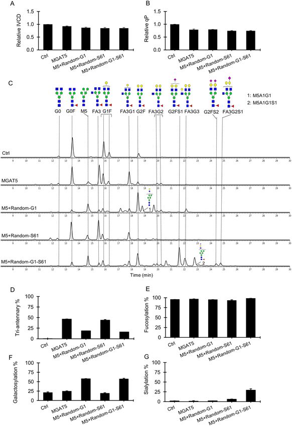
www.nature.com/scientificreports/

                                            Figure 1.  Overview of RMCE and plasmid vectors for the generation of stably transfected CHO cell pools that
                                            co-express IgG and human glycosyltransferase genes. (A) Schematic representation of RMCE. (B) Schematic
                                            representation of targeting plasmid vectors carrying IgG rituximab LC and HC genes together with one or two
                                            human glycosyltransferase genes and DsRed gene. (C) Schematic representation of plasmid vectors expressing
                                            B4GalT1 and ST6Gal1 individually or in combination for random integration. RMCE, recombinase-mediated-
                                            cassette-exchange; MCL, master cell line; GOI, gene of interest; ChiP, a chimeric promoter consisting of murine
                                            cytomegalovirus (CMV) enhancer, human CMV core promoter and human CMV intron A; mCMV, murine
                                            CMV enhancer and promoter; FRT and FRT3, wild-type and mutated flippase recognition targets; Flpe,
                                            enhanced recombinase flippase; IRES, wild-type encephalomyocarditis virus (EMCV) internal ribosome entry
                                            site (IRES); pA, simian virus 40 polyadenylation signal; LC, light chain cDNA; HC, heavy chain cDNA; G.E.1,
                                            glycosyltransferase gene 1; G.E.2, glycosyltransferase gene 2; DsRed, cDNA encoding red fluorescent protein;
                                            (ATG-)Puro, start-codon ATG-deleted puromycin resistence gene; Bla, blasticidin-S resistance gene; B4GalT1,
                                            β-1,4-Galactosyltransferase 1 gene; ST6Gal1, β-Galactoside -α-2,6-sialyltransferase 1 gene.

          Scientific Reports |   (2021) 11:12969 |              https://doi.org/10.1038/s41598-021-92320-x                                                    4

Vol:.(1234567890)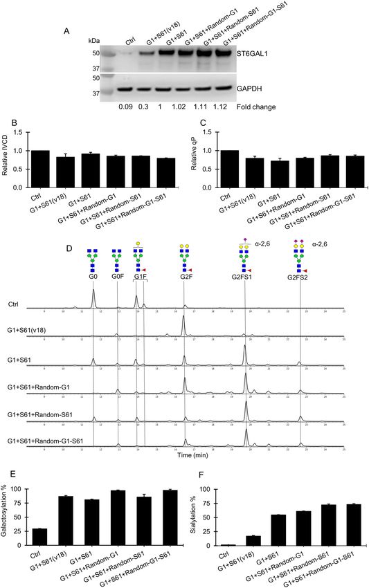
www.nature.com/scientificreports/
was verified by the 5′ and 3′ junction PCRs (Supplementary Fig. S1) using two pairs of primers specifically bind-
ing to the region in the landing pad vector and the targeting vector, respectively (Table S1). Analysing mRNA
by RT-PCR confirmed successful overexpression of all human glycosyltransferase genes in the stably transfected
pools (Fig. 2A). Moreover, FACS analysis of the stable pools indicated that the targeted cells had homogenous
DsRed expression levels (Supplementary Fig. S2), which certified the use of targeted pools for studying gene
functions and avoided the need of isolating clones. Each stable pool was characterized for growth, antibody
expression level and N-glycosylation in 7-day fed-batch cultures. Adding feed medium and harvesting samples
at exponential growth phase excluded the possible effect of nutrients depletion on antibody expression and
glycosylation. Overexpressing each glycosylation gene caused little change in cell growth as indicated by the
integrated viable cell density (IVCD) compared to the control culture (Fig. 2B). In contrast, significant decreases
in specific antibody productivity (qP) were observed in stable pools overexpressing many glycosylation genes
(Fig. 2C). All glycan-processing glycosidases decreased qP ranging from 0.1 to 0.5-fold of the control culture.
Majority of the genes involved in other parts of the glycosylation pathway exhibited less effect on qP except for a
few particular genes, such as GNE, MGAT4A, MGAT4B and FUT8 which overexpression resulted in decreased
qP to about 0.2-fold of the control culture. Since folding and assembly of antibody LC and HC occur in the
Endoplasmic reticulum (ER), overexpression of ER-resident enzymes like mannosidases may negatively affect
folding of antibodies, and possibly the secretion rate. Further studies are needed to have a clearer understanding
of how glycan-processing glycosidases and other genes affect antibody secretion.
The N-linked glycan profiles of Rituximab produced in stable pools overexpressing various human glyco-
syltransferase genes were analyzed by hydrophilic interaction liquid chromatography (HILIC). The different
N-glycosylation components were further quantified as percentage of fucosylation, galactosylation, tri-antennary
branching, high-mannose and sialylation. Among the 42 tested genes, only 10 resulted in dramatic shifts in
antibody N-glycosylation. The chromatograms of N-glycans for these ten genes together with the control were
presented in Fig. 2D,E, while genes with no or little effect on N-glycan profiles were not shown. Consistent with
previous studies18,19, we observed that the control culture produced antibodies with N-glycans that were mostly
G0F, a fucosylated bi-antennary species lacking both galactose and sialic acid (Fig. 2D). The targeted integra-
tion pool overexpressing MGAT3 produced IgGs carrying many bisecting glycans such as FA2B and FA2BG1,
fucosylated bisecting species without and with galactose, respectively (Fig. 2D). The antibody fucosylation in
MGAT3-expressing cells also reduced to 65% as compared to 98% in the control antibody (Fig. 2F). Similarly,
overexpressing B4GalT1 produced antibodies containing mainly G2F, bi-galactosylated glycans and overexpress-
ing ST6Gal1 slightly increased G2FS1 and G2FS2, fucosylated bi-antennary species containing one and two
α-2,6-sialic acids, respectively (Fig. 2E). Interestingly, we also observed 6 other genes, which impacts on antibody
N-glycosylation have not been emphasized in previous studies, significantly changed the N-glycan profiles of
antibodies. B4GalT1 has six galactosyltransferase isoenzymes. Only overexpression of B4GalT2 led to signifi-
cant conversion of G0F species to G1F and G2F, a mono- and bi-galacosylated species (Fig. 2E), although the
galactosylation increment was lower compared to that achieved by overexpressing B4GalT1 (Fig. 2H). ST6Gal2
is the only isoenzymes of ST6Gal1, which overexpression also resulted in a 9% increase in the α-2,6-sialic acid
content (Fig. 2I). Six isoenzymes of α-2,3-sialyltransferase, that are responsible for incorporating α-2,3-sialic
acids into the glycans, were tested. It has been reported that all these isoenzymes are naturally expressed in CHO
cells29 although sialic acids on antibodies produced in the wild-type CHO cells are minimal18. Overexpressing
human ST3Gal4 gene resulted in slightly enhanced α-2,3-sialic acids while the other five human sialyltransferase
isoenzymes had no effect on antibody N-glycosylation (Fig. 2I). MGAT4 and MGAT5 have been reported to
enhance branching of N-glycans on E PO40. Interestingly, among three MGAT4 and two MGAT5 isoenzymes,
overexpression of MGAT4C enhanced high-mannose while MGAT5 and MGAT5B resulted in production of
antibodies containing many tri-antennary glycans (Fig. 2D,G). This is interesting as it is well known that antibody
glycans are bi-antennary. Most genes acting in the earlier steps of the N-glycosylation biosynthetic pathway like
nucleotide sugar synthesis and transporter showed little impact on the IgG fucosylation, branching, galactosyla-
tion and sialylation (Fig. 2F,G,H,I). High-mannose structures were slightly enhanced in a number of cultures
overexpressing CST, MGAT4C, MGAT5B, ST3Gal1 and ST6Gal2, though the magnitude was small, ranging
from 4 to 10% (Fig. 2J). The increases in high mannoses could be due to different mechanisms. For instance,
it is known that ST3Gal1 plays a key role in adding α2,3-linked sialic acid to substrates in O-glycans 41,42. A
previous study observed that inhibition of O-glycosylation pathway increased N-glycan levels, suggesting that
the O-glycosylation and N-glycosylation pathways could be interacted and influenced with each a nother43. We
speculate that overexpression of ST3Gal1 in CHO cells might enhance the O-glycosylation pathway, which in
turn negatively affect the N-glycosylation, thus resulting in increased proportion of high mannose and decreased
proportion of galactose. Regarding the increased high mannose by overexpressing MGAT4C and MGAT5B, one
possible explanation is that the branching reaction is competing with the later steps of N-glycan extension reac-
tions. The reason for the increased high mannose by overexpression of other enzymes is hardly understood. We
speculate that overexpression of glycosylation enzymes at high abundance may result in their relocation in the
ER and Golgi, thus resulting in unexpected functions. Further studies are needed to elucidate the roles of these
genes in high-mannose glycan formation.
Combinatorial engineering of complex‑type bi‑antennary N‑glycans on antibodies. Over-
expression of B4GalT1 gene alone in the wild-type CHO cells converted majority of G0F to G2F glycans on
IgGs (Fig. 2E). However little to no sialylated species were observed in the B4GalT1 overexpressing pools. To
obtain highly sialylated bi-antennary glycans, we co-expressed each of the three isoenzymes ST3Gal4, ST6Gal1
and ST6Gal2, which were identified to slightly enhance mAb sialylation when being overexpressed individu-
ally in CHO cells (Fig. 2E,I), with B4GalT1 in the MCL using targeting vectors and RMCE as described in
Scientific Reports | (2021) 11:12969 | https://doi.org/10.1038/s41598-021-92320-x 5
Vol.:(0123456789)www.nature.com/scientificreports/
Figure 2. Impact of overexpressing individual human glycosyltransferases on growth, productivity and
N-linked glycosylation of antibodies produced in stably transfected CHO cell pools. All stable pools were
generated via RMCE. The control (Ctrl) pool expressed only IgG1 rituximab LC, HC and DsRed genes. Each
of the other 42 stable pools co-expressed IgG1 rituximab genes, a specific human glycosyltransferase gene
and DsRed. (A) RT-PCR analysis of human glycosyltransferase transcripts in different stable pools. β-actin
(ACT) was used as an internal control. (B–C) Relative change in the integrated viable cell density (IVCD) and
specific productivity (qP) in each stably transfected pool to the control. (D–E) Aligned HILIC chromatograms
of N-linked glycans on antibodies in the control and representative stable pools which exhibited changes in
the N-glycan profiles. Symbolic representation of the N-glycan structures was depicted for dominant peaks.
Red dotted line indicated the N-glycan structures that were only found in MGAT3 sample. (F–J) Relative
distribution of fucosylation, galactosylation, tri-antennary, sialylation and high-mannose on antibodies
produced in different stable pools.
Scientific Reports | (2021) 11:12969 | https://doi.org/10.1038/s41598-021-92320-x 6
Vol:.(1234567890)www.nature.com/scientificreports/
Figure 2. (continued)
Fig. 1A,B. Across all the combinatorial pools, the transcript levels of the two glycosyltransferase genes were
relatively comparable (Fig. 3A), indicating no transcriptional interference between them. Compared to the sin-
gle B4GalT1 overexpressing pools, combinatorial overexpression of B4GalT1 with either ST3Gal4 (G1 + S34),
ST6Gal1 (G1 + S61) or ST6Gal2 (G1 + S62) did not generate negative impacts on cellular growth IVCD (Fig. 3B)
and specific productivity qP (Fig. 3C). mAbs produced in the single B4GalT1 overexpressing pools contained
predominantly G2F glycans, while mAbs produced in all the three ST3Gal4, ST6Gal1 and ST6Gal2 single gene
overexpressing pools contained mostly G0F and small proportion of G1F glycans (Fig. 3D). Upon co-expressing
Scientific Reports | (2021) 11:12969 | https://doi.org/10.1038/s41598-021-92320-x 7
Vol.:(0123456789)www.nature.com/scientificreports/
Figure 3. Impact of co-expressing B4GalT1 and sialyltransferase isoenyzmes on the growth, productivity
and glycosylation of antibodies in stably transfected CHO cell pools. All stable pools were generated through
RMCE. The control pools expressed IgG rituximab LC, HC and DsRed genes. Other stable pools expressed IgG
rituximab LC and HC genes, B4GalT1 and ST6Gal1 either individually or in combination, and DsRed. G1 + S34,
G1 + S61, G1 + S62 are combinatorial stable pools co-expressing B4GalT1 and either of ST3Gal4, ST6Gal1
and ST6Gal2 gene, respectively. All stable pools were characterized in 7-day fed-batch cultures. (A) Relative
galactosyltransferase/ sialyltransferase transcript levels in different stable pools to the internal control, β-actin
(ACT) as determined by quantitative real time-PCR (qRT-PCR). (B–C) Relative change in the integrated viable
cell density (IVCD) and specific productivity (qP) of each stably transfected pool to the control. (D) Aligned
HILIC chromatograms of N-linked glycans on antibodies in different stable pools. Chromatograms shown are
one of biological replicates with similar results. (E–H) Relative distribution of fucosylation, galactosylation,
sialylation and high-mannose on antibodies produced different stable pools. Each point represents the average
and standard deviation of measurements from two independent stably transfected pools.
Scientific Reports | (2021) 11:12969 | https://doi.org/10.1038/s41598-021-92320-x 8
Vol:.(1234567890)www.nature.com/scientificreports/
the B4GalT1 gene and one sialyltransferase gene, we observed a significant increase in the sialylated complex
N-glycans, G2FS1 and G2FS2, the mono- and di-sialylated species in the combinatorial pools (Fig. 3D). The
overall galactosylation distribution in the combinatorial pools was nearly 80%, similar to that in the single
B4GalT1 overexpressing pools (Fig. 3F). The G1 + S61 pools produced the highest sialylation increment up to
60% (Fig. 3G), with majority of N-glycans being G2FS1, followed by a small proportion of G2FS2 species. In
contrast, co-expressing the B4GalT1 gene and any one of the three sialyltransferase genes had no significant
impact on the levels of fucose and high mannose compared to the control (Fig. 3E,H).
There was still a sizeable proportion of the G2F glycans on antibodies produced in the combinatorial G1 + S61
pools, suggesting that the expression level of sialytransferase was probably insufficient to add more sialic acids to
G2F. In order to obtain further increased sialylation, we further overexpressed B4GalT1 and/or ST6Gal1 in the
G1 + S61 targeted pools through random integration of three vectors expressing B4GalT1 and ST6Gal1 genes
either individually or in combination, respectively (Fig. 1C). This resulted in increased B4GalT1 and/or ST6Gal1
transcripts as shown via RT-PCR (Supplementary Fig. S3A) and elevated overall ST6Gal protein levels (Fig. 4A)
in the G1 + S61 + Random-G1, G1 + S61 + Random-S61 and G1 + S61 + Random-G1-S61 stable pools. Compared
to the G1 + S61 targeted pools, the newly generated stable pools did not exhibited decrease in the cellular growth
IVCD (Fig. 4B) and specific antibody productivity qP (Fig. 4C). The G1 + S61 + Random-G1 pools with additional
overexpression of B4GalT1 showed a moderate increase in galactosylation from 80 to 98% and a small increment
in sialylation from 60 to 62% (Fig. 4D,E,F). Stacking up ST6Gal1 expression alone in the G1 + S61 + Random-S61
pools increased the sialic acid content to nearly 75% despite no additional enhancement in the total galactose
level compared to the G1 + S61 targeted pools. The increased expression levels of both B4GalT1 and ST6Gal1
in the G1 + S61 + Random-G1-S61 pools elevated both galacosylation and sialylation levels. However, the total
sialylation in the G1 + S61 + Random-G1-S61 pools was similar to that in the G1 + S61 + Random-S61 pools
in spite of the increased galactosylation. These findings suggested that sialylation had become the bottleneck
for maximizing sialic acids in G1 + S61 targeted pools. To verify this hypothesis, we generated G1 + S61(v18)
targeted pools using a targeting vector which was same as the one for generating G1 + S61 targeted pools except
that the ST6Gal1 gene was controlled by a mutant IRESv18 with redueced translation efficiency39. Compared to
the G1 + S61 targeted pools, the ST6Gal1 expression in the G1 + S61(v18) pools was reduced by 70% (Fig. 4A)
while the B4GalT1 gene expression remained similar (Supplementary Fig. S3A). With decreased ST6Gal1 protein
level, we observed a corresponded decrease in the total sialylation from 60 to 17% in the G1 + S61(v18) pools
while the galactose content remained similar compared to the G1 + S61 targeted pools (Fig. 4E,F). This further
supported our hypothesis that the expression level of ST6Gal1 gene was the limiting factor in the G1 + S61 pools
for obtaining further enhanced sialic acid content on antibodies.
Combinatorial engineering of complex‑type tri‑antennary N‑glycan on antibodies. Inspired
by the novel finding of tri-antennary N-glycans on mAbs produced by overexpressing MGAT5, we explored
the possibility of engineering tri-antennary N-glycans with high galactosylation and sialylation by introducing
B4Gal1 and ST6Gal1 genes into the MGAT5 over-expressing pools. To avoid incomplete integration through
RMCE due to the large size of the targeting vectors (unpublished data), we utilized the same three vectors
expressing B4GalT1 and ST6Gal1 individually or in combination for random integration (Fig. 1C). RT-PCR
analysis confirmed successful expression of the newly introduced B4GalT1 and ST6Gal1 transcripts (Supple-
mentary Fig. S3B). Compared to the MGAT5 targeted integration pools, additional expression of B4GalT1 and/
or ST6Gal1 in the combinatorial pools resulted in a 10% reduction in IVCD (Fig. 5A) while the specific pro-
ductivity qP of remained unchanged (Fig. 5B). Antibodies produced in the control pools contained mainly bi-
antennary N-glycans, of which more than half were G0F followed by lesser abundance of G1F and G2F species
(Fig. 5C). Besides having a similar trended distribution of G0F, G1F and G2F as seen on the control antibodies,
antibodies produced in the MGAT5 overexpressing pools also carried a high proportion of tri-antennary N-gly-
cans that were largely agalactosylated (FA3), followed by moderate level of tri-antennary N-glycans containing
one galactose (FA3G1) and a small proportion of tri-antennary N-glycans containing two (F3AG2) and three
galactoses (FA3G3) (Fig. 5C). The relative distribution of total tri-antennary N-glycans in the MGAT5 pools
reached about 50% (Fig. 5D). Further overexpression of B4Gal1 gene in the MGAT5 pools converted most G0F
and G1F into G2F glycans. Interestingly, significant decrease in FA3 level was observed but without accompany-
ing the corresponded increase in the galactose content in tri-antennary N-glycans (Fig. 5C). Only slight glyco-
sylation shifts toward tri-antennary glycans that were bi-galactosylated (FA3G2) and tri-galactosylated (FA3G3)
were observed in the M5 + Random-G1 pools. As a result, the total tri-antennary N-glycans decreased by half
as compared to the MGAT5 pools (Fig. 5D). Compared to the MGAT5 N-glycan distribution, there were little
changes to the glycan profile in the M5 + Random-S61 pools whereby ST6Gal1 was further introduced (Fig. 5C).
When co-expressing ST6Gal1 and B4GalT1 gene in the MGAT5 overexpressing pools, the peaks for G2F and
G2FS1 bi-antennary glycoforms were most prominent. The higher complex tri-antennary N-glycans decorated
with two galactoses and terminal sialic acids (FA3G2S1) were also detected although the magnitude was low.
Overall, stably co-expressing B4GalT1 and ST6Gal1 genes in the MGAT5 overexpressing pools had no effect on
fucose levels while enhanced galactosylation level to 55% and sialylation to 30% (Fig. 5E,F,G). The increment was
a lot less than that observed in the G1 + S61 targeted pools (Fig. 3F,G). This could be in part due to the low gene
overexpression and clonal variation effect caused by the random integration, highlighting the necessity of using
targeted integration technology for cell engineering.
Scientific Reports | (2021) 11:12969 | https://doi.org/10.1038/s41598-021-92320-x 9
Vol.:(0123456789)www.nature.com/scientificreports/
Figure 4. The relationship between the expression level of B4GalT1 and ST6Gal1 and the galactose and sialic acid contents
on the antibodies produced in the stably transfected CHO cell pools. The targeted pools of control (Ctrl), G1 + S61 and
G1 + S61(v18) were generated using different targeting vectors through RMCE. The control targeting vector expressed IgG
rituximab LC, HC and DsRed genes. The two targeted vectors for generating G1 + S61 and G1 + S61(v18) pools were the same,
which carried IgG rituximab LC and HC genes, DsRed, B4GalT1 and ST6Gal1, except that the ST6Gal1 in the former vector
was controlled by a wild-type IRES while in the latter vector was controlled by a mutant IRESv18 with reduced translation
efficiency. The G1 + S61 targeted pools were further transfected with three plasmid vectors expressing B4GalT1 and ST6Gal1
individually or in combination, respectively, followed by selection with blasticidin to generate three sets of stably transfected
pools, G1 + S61 + Random-G1, G1 + S61 + Random-S61 and G1 + S61 + Random-G1-S61, for further enhancing B4GalT1
and/or ST6Gal1 expression levels. All stable pools were characterized for growth, productivity and antibody glycosylation
in 7-day fed-batch cultures. (A) Western Blot analysis of ST6Gal1 protein levels in different stable pools. The housekeeping
protein GAPDH was used as a loading control. (B–C) Relative change in the integrated viable cell density (IVCD) and specific
productivity (qP) to the control pool. (D) Aligned HILIC chromatograms of N-glycans on antibodies produced in different
stable pools. Chromatograms shown are one of biological replicates with similar results. (E–F) Relative distribution of
galactosylation and sialylation level of N-glycans on antibodies produced in different stable pools. Two stably transfected CHO
cell pools were generated for each vector. Each point represents the average and standard deviation of measurements from two
independent stably transfected pools.
Scientific Reports | (2021) 11:12969 | https://doi.org/10.1038/s41598-021-92320-x 10
Vol:.(1234567890)www.nature.com/scientificreports/
Figure 5. Combinatorial engineering of tri-antennary N-glycans on antibodies for enhanced galactosylation
and sialylation. The control (Ctrl) and MGAT5 targeted pools were generated through RMCE. The control
targeted pools expressed IgG rituximab LC, HC and DsRed genes. The MGAT5 targeted pools expressed IgG
rituximab LC and HC genes, MGAT5 and DsRed. The targeted pools overexpressing MGAT5 were further
transfected with three plasmid vectors carrying the B4GalT1 and ST6Gal1 gene individually or in combination,
followed by blasticidin selection to generate three sets of stable pools, M5 + Random-G1, M5 + Random-S61 and
M5 + Random-G1-S61. All stable pools were characterized for growth, productivity and antibody glycosylation
in 7-day fed-batch cultures. (A–B) Relative change in the integrated viable cell density (IVCD) and specific
productivity (qP) to the control pool. (C) Aligned HILIC chromatograms of N-glycan profile on antibodies
produced in different stable pools. Chromatograms shown are one of biological replicates with similar results.
(D–G) Relative distribution of the tri-antennary, fucosylation, galactosylation, and sialylation levels of N-glycans
on antibodies produced in different stable pools. Each point represents the average and standard deviation of
measurements from two independent stably transfected pools.
Scientific Reports | (2021) 11:12969 | https://doi.org/10.1038/s41598-021-92320-x 11
Vol.:(0123456789)www.nature.com/scientificreports/
Discussion
To expand the diversity of N-glycan structures on antibodies, we developed the CHO targeted integration tech-
nology, which permits overexpression of glycosyltransferases, to produce antibodies with complex N-glycan
structures. By overcoming position effects and thus minimizing clonal variations, our technology enabled the
study of gene functions in stably transfected CHO cell pools without the need of isolating clones. Together with
the use of multi-cistronic vectors, we were able to engineer both single and multiple genes simultaneously in
the glycosylation pathway to produce antibodies with various N-glycan structures. By overexpressing B4GalT1
gene alone, we produced IgGs that were more than 80% bi-galactosylated, which is known to be beneficial for
enhancing both ADCC and CDC of the therapeutic antibodies6,7. We also demonstrated that combinatorial
overexpression of B4GalT1 and ST6Gal1 produced antibodies containing bi-antennary complex N-glycans with
more than 70% sialic acids. Highly sialylated glycans are reported to be valuable for increasing antibody clear-
ance time13 and anti-inflammatory functions14,15. In addition, antibodies with various tri-antennary N-glycans,
which have not been reported before, were obtained by overexpressing MGAT5 alone or in combination with
B4GalT1 and ST6Gal1. Further investigation of how these novel glycan structures affect antibody functions is in
progress. The diverse range of N-glycan structures obtained in this study can be used for optimizing the glycan
designs on recombinant antibodies to suit different therapeutic applications.
Cellular demand for sugar precursors and glycosyltransferase levels vary as the N-glycan elongating. Limited
substrates or modifying enzymes at any steps of the N-glycosylation pathway will create bottlenecks in N-gly-
can processing44,45. Identifying and mitigating these bottlenecks are crucial for producing higher complex-type
N-glycans such as sialylated structures. Via systematic screening of 42 glycosyltransferase genes, we found that
overexpression of most genes involved in the nucleotide sugar synthesis and transporter steps had little impacts
on the IgG glycan profile, suggesting that early steps of the glycosylation pathway were not limiting the forma-
tion of complex-type N-glycans on antibodies. Recent study by Sumit et al. identified that temporal bottlenecks
occurred at the galactosylation and sialylation steps in CHO cells by analysing the intermediate metabolites and
glycosylated species in antibody production45. Our study supported their finding and further demonstrated
that co-expression of galactosyltransferase and sialyltransferase was required for obtaining high mAb sialyla-
tion. Moreover, our work suggested that the bottlenecks in the glycosylation pathway shifted as the expression
of different glycosylation genes was perturbed. The antibodies produced in the wild-type CHO MCL had low
galactose and sialic acids. Overexpression of B4GalT1 in the CHO MCL increased galactose but not sialic acid,
suggesting galactosylation was the first bottleneck for producing complex glycans in the wild-type CHO cells.
In the G1 + S61 targeted pools overexpressing both B4GalT1 and ST6Gal1, further introduction of ST6Gal1 but
not B4GalT1 resulted in additional sialylation improvement, suggesting once the galactosylation bottleneck was
released, the expression level of downstream sialyltransferase became the limiting factor for further increasing
IgG sialylation. Therefore, multiplexed engineering glycosyltransferase genes is required for better control of
the N-glycosylation dynamics.
In contrast to some glycoproteins that naturally carry multi-antennary glycoform46,47, the number of N-glycan
branches on antibodies are often limited to two, partly due to the embedment of the Fc N-glycans within a cavity
formed by the two CH2 d omains2,48. Such restricted configuration also makes it increasingly difficult for enzymes
acting on later steps of the glycosylation pathway, such as MGAT4 and MGAT5 to access the growing N-glycans
during transition of IgG proteins through the Golgi apparatus. Interestingly, we observed in this study that tri-
antennary structure in IgG could be produced by overexpression of MGAT5 or MGAT5B. Consistent with our
study, Fukata et al. has previously demonstrated that overexpression of MGAT4 and/or MGAT5 genes could
increase multi-antennary sugar chains in human interferon (IFN)-γ49. We speculate that the access to glycosyla-
tion sites on proteins in the ER is related to the abundance of enzymes in the Golgi. As Golgi-residing enzymes
can continuously recycle back to the ER50, enriching MGAT5 level in Golgi may also potentially increase its
distribution in ER, thus allowing them to readily access the glycosylation sites on the folding proteins.
We observed a low incorporation level of galactose into the tri-antennary but not bi-antennary N-glycans
when stacking expression of B4GalT1 in the MGAT5-overexpressing cells. This suggested that galactosylation
might be directed more favourably to the bi-antennary structures than the tri-antennary ones. In addition, the
proportion of tri-antennary structures are reduced with increased B4GalT1 protein levels. Hence, we suspect a
potential competition between tri-antennary formation and galactosylation. The presence of galactose residues
on the bi-antennary branches may inhibit the addition of the third sugar chain catalysed by MGAT5. A compu-
tational simulation of N-glycosylation pathway predicted galactosyltransferase activity could control the final
level of antennarity of glycoproteins51. The study later demonstrated the enrichment of tri- and tetra-antennary
glycans of human chorionic gonadotropin only in the absence of B4GalT4 isoenzyme. The relationship between
galactosyltranferases and branching enzymes remains to be investigated. Such understanding will enable better
designs of glyco-engineering strategies in the future.
Our work gained insights into the contribution of individual isoenzymes in forming complex N-glycans on
antibodies. Single knockout of the B4GalT1 gene resulted in only partially reduction of galactosylation in EPO
and Fc-fusion EPO27, suggesting a potential involvement of other family members. In our study, we identified
that overexpression of B4GalT2 gene in CHO cells could moderately produce more galactosylated antibodies.
This provides a new opportunity to utilize B4GalT2 gene for N-glycan-engineering purposes in the future as
well as further investigate the involvement of other isoenzymes. In addition, increasing evidence has suggested
that the isoenzymes regulate N-glycan formation in a protein specific manner. For example, a study by Qi et al.
demonstrated that sialylation of EGFR was highly affected by ST3Gal6 level, but not other i soenzymes52. On the
other hand, single knockout of three α-2,3 sialyltransferase isoenzymes in CHO cells revealed both ST3Gal4 and
ST3Gal6 were important for EPO protein sialylation29. In contrast, our study indicated that ST3Gal4 played more
important roles than ST3Gal3 and ST3Gal6 in IgG sialylation. We also observed that both α-2,6 sialyltransferase
Scientific Reports | (2021) 11:12969 | https://doi.org/10.1038/s41598-021-92320-x 12
Vol:.(1234567890)www.nature.com/scientificreports/
enzymes were more efficient in incorporating sialic acid into the growing glycans than α-2,3 sialyltransferases,
although they are not expressed in CHO cells naturally. This finding suggested that α-2,6 sialyltransferase family
members may exhibit higher specificity toward antibodies.
Methods
Cell culture and media for maintenance of CHO K1 master cell line (MCL). The CHO K1 master
cell line (MCL) was generated by nucleofection of a landing pad vector into CHO K1 cells (ATCC), followed
by screening clones for single copy integration by southern blotting. The landing pad vector expressed a hygro-
mycin resistant gene (HYG) using a chimeric promoter (ChiP) which consisted of the murine CMV enhancer
(M11788), the hCMV core promoter and the hCMV intron A (M60321). The HYG expression cassette was
flanked by FRT3 and FRT. An impaired puromycin resistant gene lacking start codon ((ATG-)Puro) followed
by the simian virus 40 (SV40) polyadenylation signal (pA) was placed downstream of FRT for selecting cor-
rect cassette exchange by RMCE (Fig. 1A). It was confirmed that the MCL contained only one copy of landing
pad vector at a single integration site by southern blotting and targeted locus amplification (TLA) analyses
(Cergentis). The MCL was grown in a protein-free medium (maintenance media) consisting of 50% HyQ PF
(GE Healthcare Life Sciences) and 50% CD CHO (Thermo Fisher Scientific) supplemented with 1 g/L sodium
carbonate (Sigma), 6 mM glutamine (Sigma) and 0.1% Pluronic F-68 (Thermo Fisher Scientific) in a humidified
Kuhner shaker (Adolf Kühner AG) with 8% CO2 at 37 °C. Routine subculture was conducted every 3 to 4 days
by seeding cells at density of 3 × 10⁵ cells/mL in 15 mL of fresh medium in 125 mL shake flasks (Corning). Cell
density and viability were determined by trypan blue exclusion method on Vi-Cell XR viability analysers (Beck-
man Coulter).
Constructing targeting plasmid vectors for expression of mAb and human glycosyltransferase
genes. The control targeting vector carrying an expression cassette of FRT3-LC-IRES-HC-DsRed-IRES-FRT
was constructed by Genscript. LC and HC represented the rituximab light chain cDNA and heavy chain cDNA,
respectively. IRES represented the wild-type encephalomyocarditis virus (EMCV) internal ribosome entry site.
Three unique restriction sites, MfeI, XhoI and PmeI were included between HC and IRES-DsRed for insertion
of more expression units. To construct targeting vectors expressing various single or double human glycosyl-
transferase genes, two basic vectors were first constructed by inserting one synthesized unit IRES-GE1 using
MfeI and XhoI followed by inserting the second synthesized unit IRES-GE2 using XhoI and PmeI. GE1 and
GE2 represented specific human glycosyltransferase genes. The IRES element used upstream of GE1 and GE2
was either the wild-type EMCV IRES or a mutated IRESv18 variant with reduced strength for expressing a
gene. Two restriction sites, ClaI and NruI were created by mutating the six bases in front of the 10th ATG in the
EMCV IRES upstream of GE1 and GE2, respectively during gene synthesis. Targeting vectors expressing other
single or double human glycosyltransferase genes were subsequently constructed by inserting the synthesized
human glycosylation genes between ClaI and XhoI or NruI and PmeI. Synthesis of IRES-GE1, IRES-GE2, other
glycosyltransferase genes and inserting them into the control vector or the two basic vectors for expressing single
or double glycosylation genes were all done by Genscript. The sequences of the wild-type IRES, IRESv18, FRT
and FRT3 were described in previous studies53,54. The sequences of DsRed gene and pA were cloned from the
pIRES2-DsRed vector (Clonetech) and the pcDNA3.1 (+) vector (Thermo Fisher Scientific), respectively. The
rituximab LC cDNA and HC cDNA were designed based on the amino acid sequences published in the inter-
national ImMunoGeneTics information system (IMGT). The sequences of human glycosyltransferase genes
were retrieved from NCBI (Table 1). The vector expressing enhanced FLP recombinase (FLPe) was synthe-
sized by Genscript (Fig. 1A). The sequence of murine CMV enhancer and promoter (mCMV) (M11788) was
described in a previous study55. The sequence of FLPe gene was from the pCAGGS-FLPe vector (Gene Bridges).
The sequence of SpA was from the pcDNA3.1 (+) vector (ThermoFisher). The three plasmid vectors for over-
expressing B4GALT1, ST6GAL1 and the combination of B4GALT1 and ST6GAL1 in stable transfections were
constructed using a previously described tricistronic vector56. This tricistronic vector expressed LC, HC and
Blasticidin (Bla) resistant gene in one transcript through the use of two wild-type EMCV IRES. The LC gene is
under the control of the chimeric promoter (ChiP). Expression of HC and Blasticidin (Bla) resistant gene were
driven by the wild-type EMCV IRES. The two vectors expressing B4GALT1 or ST6GAL1 were constructed by
replacing the region LC-IRES-HC in the tricistronic vector with B4GALT1 or ST6GAL1. The vector expressing
the combination of B4GALT1 and ST6GAL1 was constructed by replacing the LC with B4GALT1 and the HC
with ST6GAL1, respectively. All restriction enzymes used were purchased from New England Biolabs. Plasmids
were all propagated using chemically competent DH5α E.Coli cells (Thermo Fisher Scientific).
Generating stable mAb‑producing cell lines via recombinase‑mediated‑cassette‑exchange
(RMCE) and random integration. The MCL was co-transfected with an appropriate targeting vector and
a vector expressing FLPe using Amaxa SG Cell Line 4D-Nucleofector X Kit and program FF-137 (Lonza). In
each transfection, 1 × 10⁷ cells were transfected with 5 µg of targeting plasmid vector and 5 µg of FLPe plasmid
vector in circular format. The transfected cells were then re-suspended in 2 mL of maintenance media preloaded
in 6-well suspension culture plates (NUNC) and incubated in the static IncuSafe incubators (Sanyo). At 24 h
post-transfection, they were collected by centrifugation (100 × g, 5 min) and re-suspended in 15 mL of protein-
free maintenance medium in 125 mL shake flasks in the humidified Kuhner shaker (Adolf Kühner AG) with 8%
CO2 at 37 °C. Four days later, the transfected cells were subjected to selection in the maintenance media contain-
ing Puromycin (InvivoGen) at 20 µg/mL. Selection was continued for two weeks by passaging in the selection
medium every 3 to 4 days. Stably transfected cell pools were deemed established when cell viabilities recovered
over 95%.
Scientific Reports | (2021) 11:12969 | https://doi.org/10.1038/s41598-021-92320-x 13
Vol.:(0123456789)www.nature.com/scientificreports/
The protocol for further transfection of the three multi-cistronic vectors expressing B4GALT1, ST6GAL1 and
the combination of these two genes respectively into the targeted pools already expressing either the combina-
tion of B4GALT1 and ST6GAL1 or MGAT5 was the same as that described for RMCE with slight modifications.
In each transfection 5 µg of linearized plasmids were transfected to 1 × 10⁷ cells. After incubating in 2 mL of
protein-free medium in the 6-well suspension culture plates (NUNC) overnight, the transfected cells were col-
lected by centrifuge at 100 × g for 5 min and then resuspended in 15 mL of protein-free medium supplemented
with blasticidin (Thermo Fisher Scientific) at 20 µg/mL. Passaging in selection medium was subsequently carried
out every 3 to 4 days until cell viabilities recovered over 95%.
Characterization of growth and productivity of stable pools. Stable cell pools were subjected to
seven-day fed-batch production by seeding 30 mL of cultures at viable cell density of 3 × 10⁵ cells/mL in 50 mL
tube spin (TPP) in the humidified Kuhner shaker (Adolf Kühner AG) with 8% C O2 at 37 °C. 3 mL of Ex-Cell
Advanced CHO Feed 1 (with glucose) (Sigma) and 400 µL 45% (w/v) D-glucose (Sigma) were added at day 5.
Cell density, viability and antibody titer were monitored at day 3, 5 and 7 using the Vi-Cell XR viability ana-
lyzer (Beckman Coulter) and an IMMAGE 800 immunochemistry system (Beckman Coulter), respectively. The
IMMAGE 800 immunochemistry system utilized anti-human Fc region antibodies for IgG quantification. The
specific mAb productivity (qP) in the exponential phase of cultures was calculated as the difference in mAb con-
centration between day 5 and 7 divided by the integrated viable cell density (IVCD) which was determined based
on the trapezoidal method. Two sets of ten million cells were collected from each culture at day 5 for analysis of
mRNA and protein levels, respectively. Flow cytometry was performed at day 5 on BD FACSCalibur to deter-
mine the homogenous expression of the DsRed protein in the stable pools. Flow cytometry data were analyzed
using FlowJo software. Culture supernatant was harvested at day 7, by centrifuging at 5000 × g over 10 min to
remove cells and use for N-glycan analysis.
Analysis of genomic DNA, mRNA and protein levels. To confirm correct cassette recombination,
crude genomic DNA (gDNA) was extracted from cell pellets using PureLink Genomic DNA Mini Kit (Thermo
Fisher Scientific) according to the manufacturer’s protocols. 100 ng of gDNA was used as template for PCR using
2X REDiant Master Mix (1st Base) with PCR condition according to the manufacturer’s protocols. PCR prim-
ers for 5′ and 3′ junction PCRs are listed in the Supplementary Table S1. PCR products were visualized on 1%
agarose gels stained with ethidium bromide.
Total RNA was isolated using RNeasy Mini Kit (Qiagen) from ten million cells collected from each fed-batch
culture. Analysis of the mRNA levels for human glycosyltransferase genes (Table 1) and β-actin was done using
either reverse transcription polymerase chain reaction (RT-PCR) or quantitative real-time PCR (qRT-PCR)
as described p reviously53. Primer sequences used for mRNA analysis were listed in Supporting Table S2. For
RT-PCR, 1 µg of total RNA from each sample was used as template for cDNA synthesis. The PCR products cor-
responding to each human glycosyltransferase gene were further analyzed by electrophoresis on 2% agarose gel
and visualized by ethidium bromide staining.
To carry out Western blotting for protein level analysis, ten million cells were first homogenized in the Cel-
Lytic M (Sigma) supplemented with Halt Protease and Phosphatase Inhibitor Cocktail (Thermo Fisher Scientific).
10 µg of each protein sample quantified by Pierce BCA protein assay (Thermo Fisher Scientific) was fraction-
ated on 4–12% gradient PAGE gel (Thermo Fisher Scientific), followed by transfer to a PVDF membrane using
iBlot (Thermo Fisher Scientific). The membrane was then blocked for 1 h with a blocking buffer comprising of
1 × Tris buffered saline with 0.1% Tween20 (1st Base) with 5% non-fat milk. The membrane was probed with
primary mouse anti-ST6Gal1 antibody (1:500) (R&D Systems), or GAPDH (1:1000) (Abcam) overnight at 4 °C.
After washing with TBST, the membrane was incubated with 1:5000 corresponding diluted secondary antibody
(Promega) in blocking buffer for 1 h at room temperature. After washing, the membrane was visualized using
Amersham ECL Western blotting detection reagents and analysis system (GE Healthcare Life Sciences) accord-
ing to the manufacturer’s instructions.
Antibody purification and N‑ glycosylation analysis. mAb in the culture supernatant was purified
using MabSelect SuRe Protein A column (GE Healthcare Life Sciences) on a GE AKTA explore 100 (GE Health-
care Life Sciences). The purified mAbs were analyzed for the N-glycosylation using hydrophilic interaction chro-
matography (HILIC) with fluorescence detection. The protocols for protein A purification and N-glycosylation
analysis have been described in a previous s tudy53. The data obtained from N-glycosylation analysis was ana-
lyzed with the UNIFI Biopharmaceutical software platform (version 1.8). The N-glycan structures were assigned
to peaks based on the alignment of observed and expected glucose unit (GU) values. The peak assignment fol-
lowed the method described in a previous s tudy53. The abundance of each structure was expressed as percentage
of total peak area. The level of galactosylation, fucosylation were calculated as described in a previous study57.
Galactosylation is the percentage of the number of galactose residues in G1, G1F, G2 and G2F in the total num-
ber of galactose residues in G0, G0F, G1F, G1, G2 and G2F if they are fully galactosylated. Fucosylation is the
percentage of fucosylated species (G0F, G1F, G2F) in the total sum of fucosylated and afucosylated species (G0,
G1, G2). Sialylation was the sum of the relative abundance of mono- and bi-sialylated species. The high mannose
was the relative abundance of M5 structure on mAb.
Scientific Reports | (2021) 11:12969 | https://doi.org/10.1038/s41598-021-92320-x 14
Vol:.(1234567890)You can also read