Murine glial progenitor cells transplantation and synthetic PreImplantation Factor (sPIF) reduces inflammation and early motor impairment in ALS mice
←
→
Page content transcription
If your browser does not render page correctly, please read the page content below

www.nature.com/scientificreports
OPEN Murine glial progenitor cells
transplantation and synthetic
PreImplantation Factor (sPIF)
reduces inflammation and early
motor impairment in ALS mice
Karolina A. Ambrożkiewicz1, Urszula Kozłowska1,2, Valerie Haesler3, Eytan R. Barnea4,5,
Martin Mueller3,6* & Maciej Kurpisz1,6*
Amyotrophic lateral sclerosis (ALS) is a progressive motor neuronal disorder characterized by neuronal
degeneration and currently no effective cure is available to stop or delay the disease from progression.
Transplantation of murine glial-restricted precursors (mGRPs) is an attractive strategy to modulate
ALS development and advancements such as the use of immune modulators could potentially extend
graft survival and function. Using a well-established ALS transgenic mouse model (SOD1G93A), we
tested mGRPs in combination with the immune modulators synthetic PreImplantation Factor (sPIF),
Tacrolimus (Tac), and Costimulatory Blockade (CB). We report that transplantation of mGRPs into the
cisterna magna did not result in increased mice survival. The addition of immunomodulatory regimes
again did not increase mice lifespan but improved motor functions and sPIF was superior compared to
other immune modulators. Immune modulators did not affect mGRPs engraftment significantly but
reduced pro-inflammatory cytokine production. Finally, sPIF and CB reduced the number of microglial
cells and prevented neuronal number loss. Given the safety profile and a neuroprotective potential of
sPIF, we envision its clinical application in near future.
Amyotrophic lateral sclerosis (ALS) is a progressive motor neuronal disorder characterized by degeneration
of the upper and lower motor neurons of the cortex, the brain stem, and the spinal cord. Although the start is
focal, the heterogeneous manifestations and disseminating patterns suggest the spreading among advancement
motor neuronal pools with glial partners1. The end stage of ALS is the respiratory failure (about 3–4 years after
the onset) and currently no effective cure is available to stop or delay the disease from progression. Therefore,
testing of new therapeutic approaches is essential and one well-defined animal model is the SOD1G93A transgenic
mouse model2. In this model, mice express a G93A mutant form of human SOD1 and develop progressive loss of
upper and lower motor neurons. Typically, within 4–5 month of symptom onset mice express muscle denerva-
tion, weakness, and atrophy resulting in subsequent d eath3.
The importance of glial cells in the degeneration of motor neurons emerged after synthesis of SOD1 mutants
and consecutive selective silencing1. The activation of innate immune cells such as microglia and excessive extra-
cellular production of superoxide contribute substantially to ALS development and progression. Not surprisingly,
the transplantation of murine glial-restricted precursors (mGRPs) is an attractive strategy to modulate ALS
development4. Notably, GRPs are oligodendrocytic and astrocytic cell lineage precursors and recent advance-
ments support its clinical application for ALS t reatment5. However, the optimal therapeutic window, the ideal
cell type and dose, and delivery site or method are still non optimized. The recently updated Cochrane review
underlines the need of further research to explore combination of cellular therapy and novel therapeutics6. Such
1
Institute of Human Genetics, Polish Academy of Sciences, Poznan, Poland. 2Institute of Bioorganic Chemistry,
Polish Academy of Sciences, Poznan, Poland. 3Department of Obstetrics and Gynecology, Department of
Biomedical Research, University Hospital Bern, University of Bern, Bern, Switzerland. 4The Society for the
Investigation of Early Pregnancy (SIEP), Cherry Hill, NJ, USA. 5BioIncept LLC, Cherry Hill, NJ, USA. 6These
authors contributed equally: Martin Mueller and Maciej Kurpisz. *email: martin.mueller@insel.ch; kurpimac@
man.poznan.pl
Scientific Reports | (2022) 12:4016 | https://doi.org/10.1038/s41598-022-08064-9 1
Vol.:(0123456789)
www.nature.com/scientificreports/
adjuvant therapeutics may include synthetic PreImplantation Factor (sPIF), Tacrolimus (Tac), and Costimula-
tory Blockade (CB).
PIF is a small 15-amino acid pregnancy derived peptide, which is secreted by the trophoblast/embryo7,8.
Besides immunomodulatory properties in and outside pregnancy7,9, a synthetic version of PIF (PIF analog:
sPIF) was successfully tested in animal models of multiple immune d isorders10–15 and received a Fast-Track FDA
approval (autoimmune diseases of non-pregnant subjects clinicaltrials.gov, NCT02239562). sPIF was able to
reverse and prevent paralysis and restore myelination through inhibiting neuro-inflammation in murine models
of experimental autoimmune e ncephalomyelitis16. The neuroprotective property of sPIF was further underscored
by its ability to mitigate neuronal loss and microglial activation in murine model of immature brain injury as
well9,12,13,17. Tac is a calcineurin inhibitor and a first-line immunosuppressive drug used after organ transplant18,
which additionally may enhance neuronal regeneration19. Finally, the T cells feature crucial immune response
toward allografts and co-stimulatory molecules involved in T-cell activation may be targeted as well20. The so
called costimulatory blockade (CB) includes CTLA4 (Cytotoxic T-lymphocyte-associated antigen 4) and MR-1
(anti-CD154 antibody/CD40L monoclonal antibody) targeting CD40/CD154 a xis21,22.
Together, immune-modulatory therapeutics in combination with stem cell transplantation possess the poten-
tial to enhanced cell protection/regeneration in ALS. We aimed to evaluate the potential of sPIF, Tac and CB in
combination with mGRPs transplantation using the S OD1G93A transgenic mouse model.
Results
mGRPs and sPIF improve motor function but not survival. Currently, GRPs transplantation in
ALS is a promising therapeutic approach and clinical trials are in preparation (ClinicalTrials.gov Identifier:
NCT02478450). We hypothesized that mGRPs transplantation in combination with immune modulatory thera-
peutics will impact the graft function and therefore modulate the course of ALS5,23. To test this hypothesis we
transplanted allogenic mGRPs into the cisterna magna using a well-defined murine ALS model of transgenic
SOD1G93A mice in combination with immunomodulatory drugs: sPIF, Tac, and CB (Fig. 1A). Surprisingly, the
mGRPs transplantation alone did not result in increased mice survival, which is in contrast to previous reports24.
The addition of immunomodulatory regimes improved mice survival but did not reach statistical significance,
while sPIF seemed to be superior compared to other immune modulatory drugs (Fig. 1B compare sPIF to other
groups). We therefore focused on functional outcomes next.
Functional outcomes such as the hanging wire test are an excellent tool to detect early motor impairment,
which impart quality of l ife3. The mGRPs transplantation alone did not impact mice performance measured by
hanging wire test (Fig. 1C compare mGRPs to Sham). The additional application of immune modulatory drugs
resulted in a significant delay of early motor impairment in the sPIF group only (Fig. 1C compare sPIF to other
groups). This difference was especially significant in the first 60 days (Fig. 1C red line at 0.6 threshold) suggesting
that sPIF predominantly impacted the early phase of the injury25. We aimed to investigate the grafts` survival
and inflammatory responses in the brain next.
Immunomodulatory therapeutics reduce peripheral immune response but not graft sur-
vival. We measured the graft survival using Bioluminescent imaging first. We detected a rapid decline of graft
viability during the first days with only marginal signal after day 14 (Fig. 2A and B compare mGRPs alone to
other groups). This observation is in line with the lack of mGRPs` effect (Fig. 1). As the application of immune
modulators did not impart the grafts` survival (Fig. 2A and B), we focused on immune responses next. Nota-
bly, GRPs modulate immune responses and result in neuroprotection24. As shown in Fig. 2C the SOD1G93A
transgenic mice showed expectantly increased peripheral pro-inflammatory IL-1α and IL-12 cytokines. The
transplantation of mGRPs resulted in lower cytokines peaks but only with the additional application of immune
modulators (Fig. 2C; compare sPIF, Tac and CB to Injury and mGRPs groups) which decreased IL-1α peak
significantly. Interestingly, in the sPIF group IL-12 plateaued at day 28 (Fig. 2C; compare sPIF to other groups),
which may be due to the different immune regimes. We reported the detailed effect of mGRPs on cytokines
production recently26. We turned our attention towards the central nervous system next.
mGRPs + sPIF reduce inflammation and preserve neuronal cells. Given the functional improve-
ments and peripheral modulation of cytokines by additional application of sPIF, Tac, and CB (Figs. 1C and 2C),
we aimed to evaluate changes in the central nervous system next. We focused on Iba1 positive cells, as these are
marker of microglial activation in the brain12,27,28. Expectantly, we detected increased number of Iba1 + cell in
both motor nuclei regions (Fig. 3A and B; compare Ctr to Injury). The transplantation of mGRPs reduced the
number of Iba1 + cells but the addition of sPIF or CB resulted in significant prevention of neuroinflammation
(Fig. 3A and B; compare Injury to sPIF or CB). Notably, these results are in line with previous sPIF`s immune
modulatory effect on the brain12,28. We evaluated the neuronal cells next and detected a reduced number of
mature neurons in both motor nuclei regions (Fig. 4A and B compare Ctr to Injury). The transplantation of
mGRPs restored the loss partially and again the application of both sPIF and CB resulted in significant protec-
tion of neuronal cells (Fig. 4A and B; compare Injury to sPIF or CB). These results are in line with both the
modulation of peripheral (Fig. 2) and central (Fig. 3) immune responses, which we postulate to be responsible
for the functional improvements (Fig. 1C).
mGRPs + sPIF impart mGRPs progeny. We detected a functional improvement in mice despite the rapid
decline of graft viability during the first days (Fig. 1C, Fig. 2A, B) compare mGRPs alone to other groups). There-
fore, we decided to detect mGRPs progeny in the brains next26. Notably, glial precursors differentiate into astro-
cytes and oligodendrocytes and sPIF may affect their differentiation capacity17,29,30. Transplantation of mGRPs-
Scientific Reports | (2022) 12:4016 | https://doi.org/10.1038/s41598-022-08064-9 2
Vol:.(1234567890)
www.nature.com/scientificreports/
Figure 1. Experimental setup and clinical outcomes. (A) Cartoon summarizing the experimental setup and
treatment groups in the ALS mouse model (n = 6 each group). The timing and duration of immunomodulatory
drug applications are shown in green and GRPs transplantation in red. All animals received GRPs except
Injury group, which received a sham transplantation. (B) Kaplan–Meier curves indicating survival by group.
sPIF transplantation seems superior but did not reach statistical significance. (C) Functional assessments using
hanging wire test by group. sPIF treatment resulted in significant improvement of motor outcome. mGRPs: glial-
restricted precursors; sPIF: synthetic PreImplantation Factor; Tac: Tacrolimus, CB: co-stimulatory blockade.
Differences between groups were analyzed using a repeated measure ANOVA model or using a log-rank test.
GFP+
cells in the ALS transgenic mouse model (SOD1G93A) resulted in few GFP + astrocytes and oligodendrocytes
in the brain parenchyma and this was evident in the sPIF group only (Figs. 5, 6, 7 and 8 compare mGRPs group
to other groups). This observation is in line with the detected functional improvement (Fig. 1C compare sPIF to
other groups) but whether sPIF administration alone and/or in combination with mGRPs transplant is beneficial
warrant further investigation.
Discussion
The transplantation of mGRPs alone (using our therapeutic regime) didn`t result in extended mice survival
or functional improvement (Fig. 1). However, we here provide the first evidence that the additional use of
immune modulatory drugs in ALS and especially sPIF can be promising. The combination of mGRPs and sPIF
modulated not only the peripheral and central immune responses (Figs. 2 and 3) or neuronal survival (Fig. 4)
but importantly improved functional outcomes as well (Fig. 1C). Given the lack of clear effect on graft viability
(Fig. 2A and B) and its progeny (Figs. 5, 6, 7 and 8), the role of sPIF (alone or synergistic with mGRPs) is cur-
rently unclear. However, the neuroprotective effects of sPIF in both neonatal and adult murine models of brain
injury open the possibility that sPIF alone may be neuroprotective in the ALS model as w ell12,13,16,17. The second
successful immune modulatory drug was CB. Although CB is promising, the co-stimulatory molecules are
expressed prominently in mice and therefore especially mice are vulnerable to immunological co-stimulatory
Scientific Reports | (2022) 12:4016 | https://doi.org/10.1038/s41598-022-08064-9 3
Vol.:(0123456789)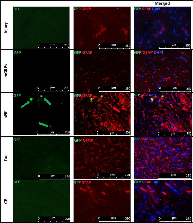
www.nature.com/scientificreports/
Figure 2. Graft survival and peripheral immune responses. (A) Representative bioluminescent images of
the transplanted mGRPs and (B) graft survival over time. The immune modulators didn`t impact the graft
survival. (C) Peripheral cytokines measured over time by groups with expected reduction of pro-inflammatory
cytokine levels by additional use of immune modulators. mGRPs: glial-restricted precursors; sPIF: synthetic
PreImplantation Factor; Tac: Tacrolimus, CB: co-stimulatory blockade. *p < 0.05; **p < 0.01; ***p < 0.001. Data
are presented as mean ± SD (one-way analysis of variance (ANOVA) followed by a post–hoc test (Bonferroni
test).
blockade31. The use in humans is therefore not possible. Besides the modulation of innate immunity, the question
of allogeneic versus semi-allogeneic transplantation and the principal role of GRPs implantation and potential
histocompatibility discordance has to be c onsidered26. In such case, sPIF, as a pregnancy derived peptide, may
have an advantage and further ameliorate an autoimmune syndrome.
The lack of neuroprotection after mGPRs` transplantation without immune modulatory drugs is unclear. The
transplantation regime including optimal cell graft source, phenotype, dose, and method of transplantation need
further investigations6. We have to consider that GRPs source and phenotype differ depending on the species31
and the therapeutic agents need to be delivered and distributed broadly. The change of transplantation technique
(for example use of MRI) or site (for example lumbar puncture) in combination with hydrogel-embedded vehicle
may be b eneficial32,33. The increase of cell number or viability to support cell migration to distant brain areas or
the co-grafting of mesenchymal stem cells or its microvesicles may further increase the e fficiency34,35. Impor-
tantly, however, GRPs injected to CNS and analyzed in post mortem tissues indicated pro-astrocyte (Fig. 6) and
pro-oligodendrocyte (Fig. 8) differentiation exclusively in sPIF regime. Finally, preconditioning of mGRPs prior
transplantation may affect the graft function as well.
We acknowledge that the chosen sPIF regime (starting day − 1 until day 14 after mGRPs transplantation) may
not be sufficient. We speculate that longer sPIF treatment would have a more pronounced effect on long-term
functional outcomes and perhaps on mice survival. We base this speculation on the early inflammation control
(until day 14) and potential correlation with decreased functional performance afterwards (Fig. 1C). The longer
application as in case of Tac or preconditioning of the cell graft can be envisioned as well. This longer (or even
permanent) approach may maintain the GRPs transplant and/or suppress the autoimmune anti-SOD1 protein
mutant reaction. Finally, we acknowledge that we didn’t take sex differences into account as the number of
Scientific Reports | (2022) 12:4016 | https://doi.org/10.1038/s41598-022-08064-9 4
Vol:.(1234567890)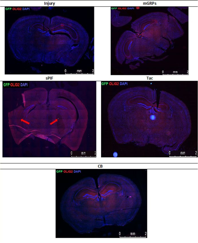
www.nature.com/scientificreports/
Figure 3. Central immune responses. (A) Representative images of microglial cells (Iba-1 positive cells)
in two regions of the brain. (B) All therapeutic groups reduced the number of microglial cells significantly.
mGRPs: glial-restricted precursors; sPIF: synthetic PreImplantation Factor; Tac: Tacrolimus, CB: co-stimulatory
blockade. *p < 0.05; **p < 0.01; ***p < 0.001. Differences are calculated using one-way analysis of variance
(ANOVA) followed by a post–hoc test (Bonferroni test).
included animals was not sufficient and may impact the discordant results24,25. Further studies are ongoing but
beyond the scope of this manuscript.
Altogether, the application of mGRPs imparts the course of ALS partially but the additional application of
immune-modulatory therapeutics improved the neuroprotective effects significantly. The question whether sPIF
alone would have been sufficient or if the mGRPs/sPIF would produce a synergistic effect needs further investi-
gation. Given that sPIF is neuroprotective, crosses the blood–brain barrier, and already received a FAST-Track
FDA approval (autoimmune diseases of non-pregnant subjects clinicaltrials.gov, NCT02239562), the clinical
romising12,13,17,28,36,37.
use could be very p
Materials and methods
Animals and treatments. B6.Cg-Tg (SOD1G93A)1Gur/J (Animalab, The Jackson Laboratory, Bar Harbor,
ME, USA, RRID: IMSR_JAX:004435) and SCID (Animalab, Charles River Laboratory, Wilmington, MA, USA,
RRID: IMSR_JAX:001303) mice were obtained from JaxMice. SOD1G93A animals were divided randomly into 5
groups and SCID animals were used as control. We defined animals (SOD1G93A n = 6) with sham transplantation
(surgery but no transplantation) as Injury, (SOD1G93A n = 6) with mGRPs transplantation as (GRPs), (SOD1G93A
n = 6) with mGRP + sPIF as (sPIF), (SOD1G93A n = 6) with mGRP + Tac as (Tac), and (SOD1G93A n = 6) with
mGRP + CB (MR1 + CTLA4) as (CB).
mGRP cells were received as a gift from the Department of Radiology and Radiological Science, Johns Hop-
kins School of Medicine (Baltimore, MD, USA). Cells were isolated as previously described from spinal cords
dissected from Luc + /PLP/GFP + mice between E12.5 and E14 stage35. Briefly, cells were cultured in DMEM/F12
medium (Thermo Fisher, Waltham, MA, USA), supplemented with N2 and B27 (Life Technologies, Carlsbad, CA,
USA), 1% BSA (Abcam, Cambridge, UK), Penicilin-streptomycin (Life Technologies, Carlsbad, CA, USA) and
20 ng per mL bFGF (Peprotech, Rocky Hill, NJ, USA). Cell culture flasks were covered with poly-L-lysine and
laminin (Sigma, Saint Louis, MO, USA). The cells were cultured until monolayer was achieved, detached from
culture flasks and characterized as previously described in terms of neural and glial cell markers, pro-neurogenic
phenotype and their potential to differentiate into astrocytes31. mGRP cells were detached from the culture flask
with TrypLE (Thermo Fisher, Waltham, MA, USA) enzymatic digestion. Reaction was disrupted by adding PBS
in 1:3 proportion. Cells were centrifuged 5 min with 1000 RPM. The viability of the mGRPs was assessed with
trypan blue exclusion and only suspensions with over 70% of cell viability were used for transplantation. The
suspension was prepared out of 0.5 mln mGRP cells in 10 µl of PBS. Prior to surgery, we assured acclimatiza-
tion of all animals to the laboratory environment and we provided animals with food and water ad libitum. We
used controlled conditions of light (12 h light/12 h dark) and temperature (23–25 °C) for animal housing. We
Scientific Reports | (2022) 12:4016 | https://doi.org/10.1038/s41598-022-08064-9 5
Vol.:(0123456789)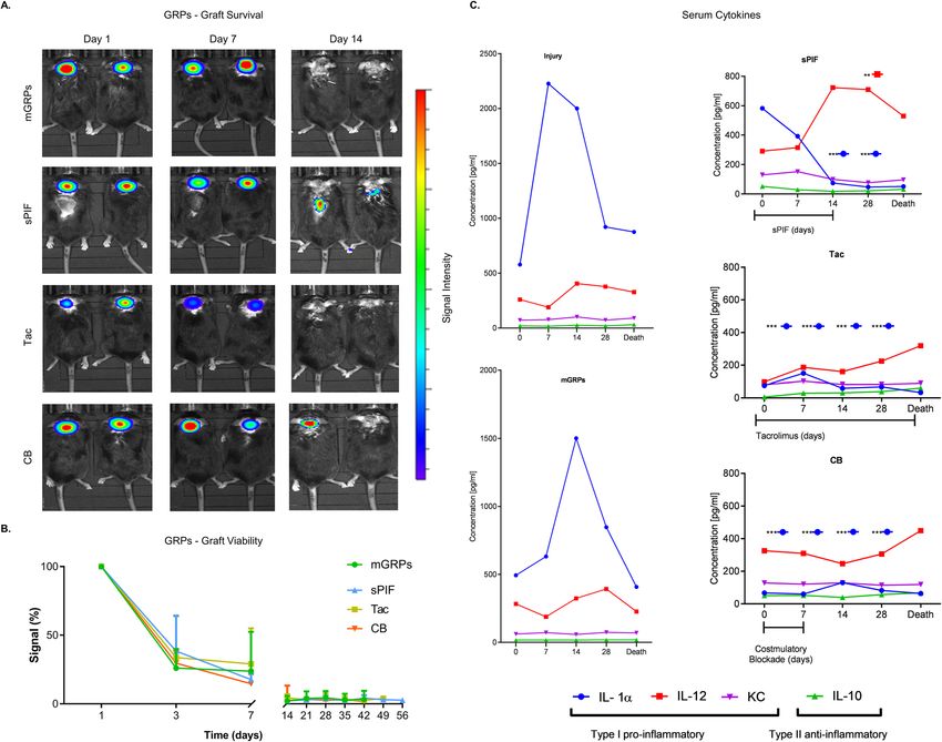
www.nature.com/scientificreports/
Figure 4. Neuronal survival. (A) Representative images of mature neurons cells (NeuN positive cells) in two
regions of the brain. (B) All therapeutic groups reduced the number of microglial cells significantly. mGRPs:
glial-restricted precursors; sPIF: synthetic PreImplantation Factor; Tac: Tacrolimus, CB: co-stimulatory
blockade. *p < 0.05; **p < 0.01; ***p < 0.001. Differences are calculated using one-way analysis of variance
(ANOVA) followed by a post-hoc test (Bonferroni test).
followed aseptic rodent survival surgery guidelines. Prior to cell transplantation mice were anesthetized with
5% (induction) and maintained in 2% isoflurane. Animals were immobilized in stereotaxic device (Leica, Wet-
zlar, Germany). 10 µl of cell suspension was administered into cisterna magna 2 µl/min via infusion pump (KD
Scientific, Hollston, MA, USA).
We synthesized synthetic P IF15 (MVRIKPGSANKPSDD) by solid-phase peptide synthesis (Peptide Syn-
thesizer, Applied Biosystems) employing Fmoc (9-fluorenylmethoxycarbonyl) chemistry at Bio-Synthesis, Inc.
(Louisville, TX, USA) as previously published38. Briefly, we carried out final purification by reversed-phase HPLC
and we verified the identity by matrix-assisted laser desorption/ionization time-of-flight mass spectrometry
(amino acid analysis at > 95% purity). sPIF was applied in amount of 1 mg/kg body weight starting at day -1 prior
to graft administration, and administered for 2 weeks after mGRPs transplantation (Fig. 1A).
Tacrolimus (Sigma Aldrich, St.Louis, MO, USA) was applied in amount of 1 mg/kg body weight starting at
day − 1 prior to grafting and administered every day until the end of observation (day 56; Fig. 1A). Costimulatory
Blockade (CB) included MR1 (CD40L, Hozel Diagnostika, Bio X Cell, Lebanon, NH, USA, RRID: AB_1107601)
and CTLA4 (Orencia, Bristol-Myers Squibb Company, New York, NY, USA), which were administered at amount
25 mg/kg at days 0, 2, 4 and 6 (day 0 defined as day of transplantation; Fig. 1A).
The immune modulatory regimes were chosen based on previous reports of successful immune modulation
and neuroprotective property11,17,39–41.
All used procedures followed the requirements of Commission Directive 86/609/EEC, which concern the
protection of animals used for experimental and other scientific purposes. We acquired Local Ethical Committee
for Animal Experimentation, Poznan University of Life Sciences approval on preclinical studies (No. 12/2017”
Application of glial progenitors for treatment of ALS”). We should like also to confirm that all animal experi-
ments were performed in accordance with relevant guidelines and regulations. Additionally, the reporting in the
manuscript follows the recommendations in the ARRIVE guidelines.
Serum collection and cytokine analysis. We collected samples every week, starting from day of graft
transplantation (day 0), 100 µl of blood from mouse tail. Blood was stored 1 h in room temperature, and over-
night in + 4 °C. Serum was gently aspirated, and centrifuged 10 min with 2000 RPM to separate blood mor-
photic bodies. Purified serum was stored in − 80 °C until further analysis. Multiplex ELISA 23-cytokine kit
(M60009RDPD) was obtained from Bio-Rad (Bio-Plex Pro Mouse Cytokine 23-plex Assay #M60009RDPD,
Hercules, Bio-Rad, CA, USA, RRID: AB_2857368) and performed according to instructions. 50 µl of diluted in
sample diluent, 1:4 serum samples and diluted at range 1:10–1:20. Measurement standards were used, diluted
9 times according to the protocol and added at template plate. Vortexed beads were added to the assay 96-well
Scientific Reports | (2022) 12:4016 | https://doi.org/10.1038/s41598-022-08064-9 6
Vol:.(1234567890)
www.nature.com/scientificreports/
Figure 5. mGRPs progeny in brain parenchyma. Representative immunofluorescence images of astrocytes
(GFAP + red marker) and mGRPs (GFP + green marker) in the diencephalon on day 56. Green arrows indicate
positive cells in the sPIF group. mGRPs: glial-restricted precursors; sPIF: synthetic PreImplantation Factor; Tac:
Tacrolimus, CB: co-stimulatory blockade.
plate. Plate was again washed 3 times with wash buffer, then from template plate, 50 µl of samples, standards and
blank were transferred to measurement plate with multichannel pipette. Plate was sealed with tape and incu-
bated 30 min. After this time, plate was washed 3 times. 25 µl of detection antibody was added to the wells with
multichannel pipette. Plate was sealed and incubated for 30 min. and next washed 3 times using wash buffer.
50 µl per well Streptavidin-PE was added, with multichannel pipette. The plate was sealed, and incubated for
10 min. Wells were washed 3 times with wash buffer. Beads were resuspended with 125 µl of assay buffer and
analysis was performed with Bio Plex 200 reader.
Functional testing and bioluminescent imaging (BLI) of graft survival. To assess the motor capac-
ity of our treatment groups, we performed motor skills assays3. Starting at day -7 SOD1G93A mice (n = 30) were
trained in hanging wire. Animals were trained 3 times a week in 2 days interval. We performed weekly meas-
urement starting at day 0 until the end of the experiment. Hanging wire test was used to test neuromuscular
Scientific Reports | (2022) 12:4016 | https://doi.org/10.1038/s41598-022-08064-9 7
Vol.:(0123456789)
www.nature.com/scientificreports/
Figure 6. Immunofluorescence staining of mGRP cells (GFP + green marker) and astrocytes (GFAP + red
marker) on whole cross section of the mouse diencephalon in the study groups. There can be seen a significantly
larger area of astrocytes in the group of mice with sPIF administration (indicated by red arrows). mGRPs: glial-
restricted precursors; sPIF: synthetic PreImplantation Factor; Tac: Tacrolimus, CB: co-stimulatory blockade.
strength and c oordination3. Briefly, mice were hung on a wire and had to catch the wire both by upper and lower
limbs as well as tangle it with tail in order to move the whole distance towards a stick (repeated three times for
each mouse and obtaining an average). Maximum points (1.5 point) were given when mice performed the task.
Medium score (1 point) were given when mice were able to accomplish the whole task, but catching wire with
both limbs and tail took more than 5 s. Low score (0.66 point) was given when mouse was able to accomplish the
task only using upper limbs. Poor score (< 0.66 points) was given when mouse was unable to move the whole dis-
tance and we counted the seconds, which mice were able to hang on the wire (1 s = 0.016 points). All functional
testing was performed in a blinded manner.
We performed imaging at days 1, 3, 7, and then weekly. We performed measurements using IVIS Lumina
IT series III (Perkin Elmer, Waltham, Massachusetts, USA) and Xeno Light Luciferin (Perkin Elmer, Waltham,
MA, USA) 150 mg per 1 kg. We defined time optimal enzyme activity at 10 min.
Scientific Reports | (2022) 12:4016 | https://doi.org/10.1038/s41598-022-08064-9 8
Vol:.(1234567890)www.nature.com/scientificreports/
Figure 7. mGRPs progeny in brain parenchyma: representative immunofluorescence images of
oligodendrocytes (OLIG2 + red marker) and mGRPs (GFP + green marker) in the diencephalon on day 56.
Green arrows indicate positive cells in the sPIF group. mGRPs: glial-restricted precursors; sPIF: synthetic
preimplantation factor; Tac: Tacrolimus, CB: co-stimulatory blockade.
Tissue collection and immunohistochemical staining. At the end of experiment, mice were subjected
to full body perfusion with 20% sucrose and 10% buffered formalin suitable for histology assays (Sigma Aldrich,
St.Louis, MO, USA). Mice were decapitated and incubated in formalin for 24 h. Following the tissues (brain and
spinal cord) were isolated, paraffin fixed for further analyses. After brain removal we fixed the tissue in formalde-
hyde solution (4%) for 2–4 h at RT followed by 4 °C for a total time of 24–48 h. Fixed tissues were embedded in
paraffin and finally sectioned into 7 µm slices. After deparaffinization of the slides (xylol 3 × 3 min, EtOH 100%
2 × 3 min, EtOH 95% 1 × 3 min, EtOH 70% 1 × 3 min and dest. H2O), the target was retrieved in Citrate Buffer
(10 mM, pH = 6.0) in a pressure cooker for 12 min and then they were let cool down for another 20 min. Slides
were washed in 0.1% Tween-20/TBS (pH7.6) and blocked 1 h in 10% goat serum/1% bovine albumin/1%TritonX
in TBS (pH7.6). One part of the slides was incubated overnight with a rabbit monoclonal Neuronal antibody
(NeuN, Abcam Cat# ab177487, RRID:AB_2532109) at 1:5000. Other slides were washed in 0.1% Tween-20/
TBS (pH7.6) and blocked one hour in 10% rabbit serum/1% bovine albumin/1%TritonX in TBS (pH7.6). Iba1
Scientific Reports | (2022) 12:4016 | https://doi.org/10.1038/s41598-022-08064-9 9
Vol.:(0123456789)www.nature.com/scientificreports/
Figure 8. Immunofluorescence staining of mGRP cells (GFP + green marker) and oligodendrocytes
(OLIG2 + red marker) on whole cross section of the mouse diencephalon in the study groups. There can be seen
a larger area of oligodendrocytes in the group with sPIF administration (indicated by red arrows). mGRPs: glial-
restricted precursors; sPIF: synthetic PreImplantation Factor; Tac: Tacrolimus, CB: co-stimulatory blockade.
antibody (Abcam Cat# ab5076, RRID:AB_2224402) at 1:100 concentration was applied on these slides during
24 h. Following first antibody incubation, slides were washed in 0.1% Tween-20/TBS (3 × 5 min) and incubated
in endogenous peroxidase blocking solution at RT for 15 min. Peroxidase-labeled polymer (DAKO anti rabbit or
anti goat) was applied to the slides for one hour at RT. Slides were washed in in 0.1% Tween-20/TBS (3 × 5 min),
followed by application of DAB + chromogen in buffer substrate for 10–30 min, according to the manufacturer’s
instructions (DAKO EnVision + System-HRP (DAB), K4007). Slides were rinsed in ddH2O, counterstained in
hematoxylin–eosin and dehydrated in a series of ethanol baths (95 > 100%) and xylene, and mounted with Eukitt
(Medite Service AG).
Assessment of neuronal and microglial positive cells were performed in the region of interest (ROI). We
defined ROI as the brainstem (facial and hypoglossal) motor nuclei as these were reported to be affected in
SOD1G93A model p reviously42. All images of immunohistochemical stainings were obtained with a Zeiss based
microscope (Pannoramic, 3CCD camera Hitachi HV-F22CL 1,4MP and Zeiss AxioCam MRm monochrome
camera) equipped with a digital camera. We used 40 × or 60 × objective to acquire images for ROI evaluation.
Consecutive coronal sections per animal and for each specific immunostaining were acquired by an independent
Scientific Reports | (2022) 12:4016 | https://doi.org/10.1038/s41598-022-08064-9 10
Vol:.(1234567890)www.nature.com/scientificreports/
observer blinded to the experimental conditions. We analyzed and reconstructed the images using Image J (US
National Institutes of Health).
Immunofluorescence staining of the brain tissues. Paraffin slides of the brain tissue after heated in
1 h in 60 °C were deparaffinized in: 1 × 10 min. Xylen I, 1 × 10 min Xylen II, 1 × 10 min EtOH 100%,1 × 10 min
EtOH 70%,1 × 10 min EtOH 40%, 1 × 10 min dest.H2O. The target was retrieved in Citrate Buffer + Tween
20(10 mM, pH = 6.0) in a water bath for 20 min in 97 °C and then they were let cool down for another 20 min.
Slides were washed in PBS three times and blocked 1 h in 10% Goat serum + 1% bovine albumin serum. All
slides were incubated overnight at 4 °C with a first primary mouse Anti-GFP antibody 1:500 (Abcam Cat#
ab1218, RRID:AB_298911). Washed three times with PBS, then secondary antibody Goat Anti-Mouse IgG H&L
(Alexa Fluor® 488) in concentration 1:700 (Abcam Cat# ab150117, RRID:AB_2688012) was applied for 40 min
in RT. Then one part of the slides was washed three times in PBS and the second primary Rabbit Anti-GFAP anti-
body (Abcam Cat# ab7260, RRID:AB_305808) was applied 1:1000, incubated overnight at 4 °C. After that the
secondary antibody Goat Anti-Rabbit IgG H&L (Alexa Fluor® 594) (Abcam Cat# ab150080, RRID:AB_2650602)
was applied for 40 min RT in concentration 1:700. The second part of the slides was incubated with Rabbit
Recombinant Anti-Olig2 antibody (Abcam Cat# ab109186, RRID:AB_10861310) 1:100 incubated overnight in
4 °C. Next day the secondary antibody Goat Anti-Rabbit IgG H&L (Alexa Fluor® 594) (Abcam Cat# ab150080,
RRID:AB_2650602) was applied for 40 min RT in 1:700 concentration. All slides were washed three times in
PBS and cell nuclei were stained with DAPI (VECTASHIELD® Antifade Mounting Medium with DAPI, Vec-
tor Laboratories, Burlingame, CA, USA, Cat# H-1200, RRID: AB_2336790) for 30 min in RT. The slides were
washed in PBS, mounted with DAKO mounting medium (Agilent Technologies, Santa Clara, CA, United States)
and analyzed under Leica DM5500 fluorescent microscopy with LAS X software.
Quantification and statistical analysis. We performed all quantifications including the manual cell
counts in a blinded manner. We counted NeuN and Iba1 positive cells in the ROI (see above) as previously
reported43–45. We determined the number of cells by unbiased counting of positive c ells43. We presented the
results as the mean ± standard deviation (mean ± SD). We used one-way analysis of variance (ANOVA) followed
by a post–hoc test (Bonferroni test) for IHC and ELISA Analyses. Considering the effect of therapy on physical
activity across time we first described the temporal variation of the physical activity of each mouse group by
calculating, at each time point and for each group the mean physical activity and the associated 95%-confidence
interval. For the 5 groups, we then investigated the effect of the therapy on the physical ability of the mice. Differ-
ences between groups were analysed using a repeated measure ANOVA model with a fixed effect for the group,
of the time and a random intercept for the mouse. In a second, more explorative, approach we determined,
for each group the time when the physical activity was first measured below a certain threshold. This “time to
reduced activity” was then compared between groups using an ANOVA model with a fixed effect for group. The
clinically relevant threshold was determined according to previous observations and defined as value 0.9 for
the Wire Test. Finally, to assess difference in survival between groups, we used Kaplan–Meier curves by group.
Differences between survival curves were tested using a log-rank test. We determined statistical significance at
p < 0.05.
Received: 18 June 2021; Accepted: 21 February 2022
References
1. Taylor, J. P., Brown, R. H. Jr. & Cleveland, D. W. Decoding ALS: from genes to mechanism. Nature 539, 197–206. https://doi.org/
10.1038/nature20413 (2016).
2. Gurney, M. E. Transgenic-mouse model of amyotrophic lateral sclerosis. N. Engl. J. Med. 331, 1721–1722. https://doi.org/10.1056/
NEJM199412223312516 (1994).
3. Olivan, S. et al. Comparative study of behavioural tests in the SOD1G93A mouse model of amyotrophic lateral sclerosis. Exp.
Anim. 64, 147–153. https://doi.org/10.1538/expanim.14-0077 (2015).
4. Kruminis-Kaszkiel, E., Wojtkiewicz, J. & Maksymowicz, W. Glial-restricted precursors as potential candidates for ALS cell-replace-
ment therapy. Acta Neurobiol. Exp. 74, 233–241 (2014).
5. Goutman, S. A., Savelieff, M. G., Sakowski, S. A. & Feldman, E. L. Stem cell treatments for amyotrophic lateral sclerosis: A critical
overview of early phase trials. Expert Opin. Investig. Drugs 28, 525–543. https://doi.org/10.1080/13543784.2019.1627324 (2019).
6. Abdul Wahid, S. F., Law, Z. K., Ismail, N. A. & Lai, N. M. Cell-based therapies for amyotrophic lateral sclerosis/motor neuron
disease. Cochrane Database Syst. Rev. 12, CD011742. https://doi.org/10.1002/14651858.CD011742.pub3 (2019).
7. Barnea, E. R. et al. Immune regulatory and neuroprotective properties of preimplantation factor: From newborn to adult. Phar-
macol. Ther. 156, 10–25. https://doi.org/10.1016/j.pharmthera.2015.10.008 (2015).
8. Moindjie, H. et al. Preimplantation factor (PIF) promotes human trophoblast invasion. Biol. Reprod. 91, 118. https://doi.org/10.
1095/biolreprod.114.119156 (2014).
9. Spinelli, M. et al. Synthetic preimplantation factor (sPIF) reduces inflammation and prevents preterm birth. PLoS ONE 15,
e0232493. https://doi.org/10.1371/journal.pone.0232493 (2020).
10. Weiss, L. et al. Preimplantation factor (PIF) analog prevents type I diabetes mellitus (TIDM) development by preserving pancreatic
function in NOD mice. Endocrine 40, 41–54. https://doi.org/10.1007/s12020-011-9438-5 (2011).
11. Azar, Y. et al. Preimplantation factor reduces graft-versus-host disease by regulating immune response and lowering oxidative
stress (murine model). Biol. Blood Marrow Transpl. J. Am. Soc. Blood Marrow Transpl. 19, 519–528. https://d oi.o
rg/1 0.1 016/j.b
bmt.
2012.12.011 (2013).
12. Mueller, M. et al. PreImplantation factor promotes neuroprotection by targeting microRNA let-7. Proc. Natl. Acad. Sci. USA 111,
13882–13887. https://doi.org/10.1073/pnas.1411674111 (2014).
13. Mueller, M. et al. PreImplantation factor bolsters neuroprotection via modulating protein kinase A and protein kinase C signaling.
Cell Death Differ. 22, 2078–2086. https://doi.org/10.1038/cdd.2015.55 (2015).
Scientific Reports | (2022) 12:4016 | https://doi.org/10.1038/s41598-022-08064-9 11
Vol.:(0123456789)www.nature.com/scientificreports/
14. Chen, Y. C. et al. PreImplantation factor prevents atherosclerosis via its immunomodulatory effects without affecting serum lipids.
Thromb. Haemost. 115, 1010–1024. https://doi.org/10.1160/TH15-08-0640 (2016).
15. Shainer, R. et al. PreImplantation factor (PIF) therapy provides comprehensive protection against radiation induced pathologies.
Oncotarget https://doi.org/10.18632/oncotarget.10635 (2016).
16. Weiss, L. et al. Preimplantation factor (PIF*) reverses neuroinflammation while promoting neural repair in EAE model. J. Neurol.
Sci 312, 146–157 (2012).
17. Spinelli, M. et al. Preimplantation factor modulates oligodendrocytes by H19-induced demethylation of NCOR2. JCI Insight https://
doi.org/10.1172/jci.insight.132335 (2021).
18. Yu, M., Liu, M., Zhang, W. & Ming, Y. Pharmacokinetics, pharmacodynamics and pharmacogenetics of tacrolimus in kidney
transplantation. Curr. Drug Metab. 19, 513–522. https://doi.org/10.2174/1389200219666180129151948 (2018).
19. Saffari, T. M. et al. Exploring the neuroregenerative potential of tacrolimus. Expert. Rev. Clin. Pharmacol. 12, 1047–1057. https://
doi.org/10.1080/17512433.2019.1675507 (2019).
20. Marino, J., Paster, J. & Benichou, G. Allorecognition by T lymphocytes and allograft rejection. Front. Immunol. 7, 582. https://doi.
org/10.3389/fimmu.2016.00582 (2016).
21. Lenschow, D. J. et al. Long-term survival of xenogeneic pancreatic islet grafts induced by CTLA4lg. Science 257, 789–792. https://
doi.org/10.1126/science.1323143 (1992).
22. Webber, A. B. & Vincenti, F. An update on calcineurin inhibitor-free regimens: The need persists, but the landscape has changed.
Transplantation 100, 836–843. https://doi.org/10.1097/TP.0000000000000872 (2016).
23. Filipi, T., Hermanova, Z., Tureckova, J., Vanatko, O. & Anderova, A. M. Glial cells-the strategic targets in amyotrophic lateral
sclerosis treatment. J. Clin. Med. https://doi.org/10.3390/jcm9010261 (2020).
24. Lepore, A. C. et al. Focal transplantation-based astrocyte replacement is neuroprotective in a model of motor neuron disease. Nat.
Neurosci. 11, 1294–1301. https://doi.org/10.1038/nn.2210 (2008).
25. Alves, C. J. et al. Early motor and electrophysiological changes in transgenic mouse model of amyotrophic lateral sclerosis and
gender differences on clinical outcome. Brain Res. 1394, 90–104. https://doi.org/10.1016/j.brainres.2011.02.060 (2011).
26. Kozlowska, U. et al. Assessment of immunological potential of glial restricted progenitor graft in vivo-is immunosuppression
mandatory?. Cells https://doi.org/10.3390/cells10071804 (2021).
27. Brettschneider, J. et al. Microglial activation correlates with disease progression and upper motor neuron clinical symptoms in
amyotrophic lateral sclerosis. PLoS ONE 7, e39216. https://doi.org/10.1371/journal.pone.0039216 (2012).
28. Migliara, G. et al. PIF promotes brain re-myelination locally while regulating systemic inflammation clinically relevant multiple
sclerosis M. smegmatis model. Oncotarget 8, 21834–21851. https://doi.org/10.18632/oncotarget.15662 (2017).
29. Sandrock, R. W. et al. Isolation, characterization and preclinical development of human glial-restricted progenitor cells for treat-
ment of neurological disorders. Regen. Med. 5, 381–394. https://doi.org/10.2217/rme.10.24 (2010).
30. Walczak, P. et al. Human glial-restricted progenitors survive, proliferate, and preserve electrophysiological function in rats with
focal inflammatory spinal cord demyelination. Glia 59, 499–510. https://doi.org/10.1002/glia.21119 (2011).
31. Klimczak, A. et al. Immunological characteristics and properties of glial restricted progenitors of mice, canine primary culture
suspensions, and human QSV40 immortalized cell lines for prospective therapies of neurodegenerative disorders. Cell Transpl.
28, 1140–1154. https://doi.org/10.1177/0963689719848355 (2019).
32. Schoeberlein, A. et al. Homing of placenta-derived mesenchymal stem cells after perinatal intracerebral transplantation in a rat
model. Am. J. Obstet. Gynecol. 205(277), e271-276. https://doi.org/10.1016/j.ajog.2011.06.044 (2011).
33. Malysz-Cymborska, I. et al. MRI-guided intrathecal transplantation of hydrogel-embedded glial progenitors in large animals. Sci.
Rep. 8, 16490. https://doi.org/10.1038/s41598-018-34723-x (2018).
34. Lepore, A. C. et al. Long-term fate of neural precursor cells following transplantation into developing and adult CNS. Neuroscience
139, 513–530. https://doi.org/10.1016/j.neuroscience.2005.12.043 (2006).
35. Srivastava, A. K., Bulte, C. A., Shats, I., Walczak, P. & Bulte, J. W. Co-transplantation of syngeneic mesenchymal stem cells improves
survival of allogeneic glial-restricted precursors in mouse brain. Exp. Neurol. 275(Pt 1), 154–161. https://doi.org/10.1016/j.expne
urol.2015.10.008 (2016).
36. Weiss, L. et al. Preimplantation factor (PIF*) reverses neuroinflammation while promoting neural repair in EAE model. J. Neurol.
Sci. 312, 146–157. https://doi.org/10.1016/j.jns.2011.07.050 (2012).
37. Hayrabedyan, S. et al. Synthetic PreImplantation Factor (sPIF) induces posttranslational protein modification and reverses paralysis
in EAE mice. Sci. Rep. 9, 12876. https://doi.org/10.1038/s41598-019-48473-x (2019).
38. Di Simone, N. et al. Synthetic PreImplantation Factor (PIF) prevents fetal loss by modulating LPS induced inflammatory response.
PLoS ONE 12, e0180642. https://doi.org/10.1371/journal.pone.0180642 (2017).
39. Lepore, A. C. et al. Human glial-restricted progenitor transplantation into cervical spinal cord of the SOD1 mouse model of ALS.
PLoS ONE 6, e25968. https://doi.org/10.1371/journal.pone.0025968 (2011).
40. Lunn, J. S. et al. Intraspinal transplantation of neurogenin-expressing stem cells generates spinal cord neural progenitors. Neurobiol.
Dis. 46, 59–68. https://doi.org/10.1016/j.nbd.2011.12.044 (2012).
41. Oh, B. et al. Split tolerance in a murine model of heterotopic en bloc chest wall transplantation. Plast. Reconstr. Surg. Glob. Open
5, e1595. https://doi.org/10.1097/GOX.0000000000001595 (2017).
42. Kadoyama, K., Funakoshi, H., Ohya, W. & Nakamura, T. Hepatocyte growth factor (HGF) attenuates gliosis and motoneuronal
degeneration in the brainstem motor nuclei of a transgenic mouse model of ALS. Neurosci. Res. 59, 446–456. https://doi.org/10.
1016/j.neures.2007.08.017 (2007).
43. Ofengeim, D. et al. N-terminally cleaved Bcl-xL mediates ischemia-induced neuronal death. Nat. Neurosci. 15, 574–580 (2012).
44. Jellema, R. K. et al. Cerebral inflammation and mobilization of the peripheral immune system following global hypoxia-ischemia
in preterm sheep. J Neuroinflammation 10, 13. https://doi.org/10.1186/1742-2094-10-13 (2013).
45. Sizonenko, S. V., Kiss, J. Z., Inder, T., Gluckman, P. D. & Williams, C. E. Distinctive neuropathologic alterations in the deep layers
of the parietal cortex after moderate ischemic-hypoxic injury in the P3 immature rat brain. Pediatr. Res. 57, 865–872. https://doi.
org/10.1203/01.PDR.0000157673.36848.67 (2005).
Author contributions
M.M., M.K., designed the study. K.A.A., U.K., V.H., and M.M. carried out the experiments. E.R.B., provided
intellectual inputs. K.A.A., M.M., M.K and E.R.B. wrote the manuscript. M.K. secured funding of the study. All
authors reviewed and contributed substantially to the manuscript.
Funding
This study was sponsored by The National Centre for Research and Development, Poland, governmental agency,
Grant No. STRATEGMED1/233209/12/NCBR/2015.
Scientific Reports | (2022) 12:4016 | https://doi.org/10.1038/s41598-022-08064-9 12
Vol:.(1234567890)www.nature.com/scientificreports/
Competing interests
The authors declare that the research was conducted in the absence of any commercial or financial relationships
that could be construed as potential conflict of interest. M.M. received an unrestricted grant from BioIncept.
E.R.B. is the founder of BioIncept and sPIF is BioIncept proprietary.
Additional information
Correspondence and requests for materials should be addressed to M.M. or M.K.
Reprints and permissions information is available at www.nature.com/reprints.
Publisher’s note Springer Nature remains neutral with regard to jurisdictional claims in published maps and
institutional affiliations.
Open Access This article is licensed under a Creative Commons Attribution 4.0 International
License, which permits use, sharing, adaptation, distribution and reproduction in any medium or
format, as long as you give appropriate credit to the original author(s) and the source, provide a link to the
Creative Commons licence, and indicate if changes were made. The images or other third party material in this
article are included in the article’s Creative Commons licence, unless indicated otherwise in a credit line to the
material. If material is not included in the article’s Creative Commons licence and your intended use is not
permitted by statutory regulation or exceeds the permitted use, you will need to obtain permission directly from
the copyright holder. To view a copy of this licence, visit http://creativecommons.org/licenses/by/4.0/.
© The Author(s) 2022
Scientific Reports | (2022) 12:4016 | https://doi.org/10.1038/s41598-022-08064-9 13
Vol.:(0123456789)You can also read