Potential Role of Fluoride in the Etiopathogenesis of Alzheimer's Disease
←
→
Page content transcription
If your browser does not render page correctly, please read the page content below

International Journal of
             Molecular Sciences

Review
Potential Role of Fluoride in the Etiopathogenesis of
Alzheimer’s Disease
Marta Goschorska 1, * , Irena Baranowska-Bosiacka 1 , Izabela Gutowska 2 , Emilia Metryka 1 ,
Marta Skórka-Majewicz 2 and Dariusz Chlubek 1
 1    Department of Biochemistry and Medical Chemistry, Pomeranian Medical University in Szczecin,
      Powst. Wlkp. 72, 70-111 Szczecin, Poland; irena.bosiacka@pum.edu.pl (I.B.-B.);
      emilia_metryka@o2.pl (E.M.); dchlubek@pum.edu.pl (D.C.)
 2    Department of Biochemistry and Human Nutrition, Pomeranian Medical University in Szczecin,
      Broniewskiego 24, 71-460 Szczecin, Poland; izagut@poczta.onet.pl (I.G.); marta_skorka@o2.pl (M.S.-M.)
 *    Correspondence: rcmarta@wp.pl; Tel.: +48-91-466-1515; Fax: +48-91-466-1516

 Received: 19 November 2018; Accepted: 6 December 2018; Published: 9 December 2018

 Abstract: The etiopathogenesis of Alzheimer’s disease has not been fully explained. Now, the disease
 is widely attributed both to genetic and environmental factors. It is believed that only a small
 percentage of new AD cases result solely from genetic mutations, with most cases attributed
 to environmental factors or to the interaction of environmental factors with preexistent genetic
 determinants. Fluoride is widespread in the environment and it easily crosses the blood–brain
 barrier. In the brain fluoride affects cellular energy metabolism, synthesis of inflammatory factors,
 neurotransmitter metabolism, microglial activation, and the expression of proteins involved in
 neuronal maturation. Finally, and of specific importance to its role in Alzheimer’s disease, studies
 report fluoride-induced apoptosis and inflammation within the central nervous system. This review
 attempts to elucidate the potential relationship between the effects of fluoride exposure and the
 pathogenesis of Alzheimer’s disease. We describe the impact of fluoride-induced oxidative stress and
 inflammation in the pathogenesis of AD and demonstrate a role for apoptosis in disease progression,
 as well as a mechanism for its initiation by fluoride. The influence of fluoride on processes of AD
 initiation and progression is complex and warrants further investigation, especially considering
 growing environmental fluoride pollution.

 Keywords: Alzheimer’s disease; fluoride; neuroinflammation;                                             reactive oxygen species;
 cyclooxygenases; antioxidant enzymes; apoptosis

1. Introduction
     Alzheimer’s disease (AD) is a progressive, irreversible neurodegenerative disease and one of
the most common causes of dementia. Its clinical symptoms, including the impairment of memory
and cognitive functions, are caused by neuronal loss, primarily in the hippocampus and neocortex [1].
Characteristic pathomorphological signs of Alzheimer’s disease include neurofibrillary tangles (NFTs)
and amyloid plaques (AβPs), also known as senile plaques. Additionally, pathomorphological studies
on AD brains reveal the presence of amyloid neuropathy, granulovacuolar degeneration, synaptic
pathology, white matter rarefaction, transactive response DNA-binding protein 43 (TDP-43) pathology,
and neuroinflammation [2,3].
     Although the etiopathogenesis of AD has not been fully explained, a distinction has been made
between its sporadic (sAD) and familial forms (fAD), and the disease is now widely attributed both to
genetic and environmental factors [4].

Int. J. Mol. Sci. 2018, 19, 3965; doi:10.3390/ijms19123965                                                   www.mdpi.com/journal/ijms

Pobrano z Repozytorium Pomorskiego Uniwersytetu Medycznego w Szczecinie / Downloaded from Repository of Pomeranian Medical University in Szczecin 2022-06-15
Int. J. Mol. Sci. 2018, 19, 3965                                                                                                        2 of 23

2. The Role of Environmental Factors in AD Etiopathogenesis
     At present, it is believed that only a small percentage of new AD cases result solely from
genetic mutations, with most cases attributed to environmental factors or to the interaction of
environmental factors with preexistent genetic determinants [5]. Studies of AD risk factors have
been largely inconclusive; however, they have succeeded in generating a list of potential risk
factors and demonstrating that the role of these environmental factors in the development of AD
is equally important as that of previously established associated genetic mutations [5,6]. Like other
neurodegenerative diseases, risk factors include hypertension, hyperhomocysteinemia, hyperlipidemia,
and exposure to pesticides and certain metal ions. Additionally, growing attention has highlighted the
combination of certain factors. For example, high-fat/high energy diets have been shown to lead to
increased aluminum (Al) concentrations in plasma and result in increased concentrations in the brain
due to the ability of Al to cross the blood–brain barrier (BBB) [6]. Al has also been shown to alter BBB
functions, increasing permeability for nonmetals, such as fluoride [7]. Furthermore, fluoride is known
to spontaneously form complexes with trace quantities of aluminum in aqueous environments [8,9].
The roles of Al and F, mainly as AlFx and NaF, have been the subject of extensive investigation in the
etiopathogenesis of AD and other human and animal diseases [8,10].

3. Neurobiological Processes Leading to AD
    Several theories have endeavored to explain the development of AD-related pathomorphological
changes in the brain, such as the cholinergic hypothesis (including altered glutamatergic transmission),
amyloid aggregation theory, tau protein theory, and oxidative stress theory, of which amyloid β
aggregation and tau hyperphosphorylation are currently considered to play the most crucial roles [4].

3.1. Cholinergic Hypothesis
     Altered cholinergic function, implicated in age-related memory loss since the 1980s [11], is
one of the most salient changes observed in AD development [12]. The cholinergic hypothesis
was one of the earliest explanations for the etiopathogenesis of AD [4] and serves as the basis for
most current AD treatment strategies [13], suggesting that dysfunction of acetylcholine-containing
neurons results in the disruption of cognitive processes [13]. This led to the definition of AD as
primarily a neurodegenerative process resulting from selective destruction of cholinergic neuronal
aggregations within brain structures (including the hippocampus, frontal cortex, amygdala, nucleus
basalis, and medial septum, as well as within the areas responsible for memory, learning, and
mnemonic processes) [4,13]. This selective depletion of cholinergic neurons in affected brain areas
presents as a reduction in cholinergic markers, i.e., acetylcholinesterase and acetyltransferase [14,15].
These changes consequently lead to a reduction in the number and density of nicotinic acetylcholine
receptors in AD patients, as well as a reduced expression of subunits α3, α4, and α7 in the cerebral
cortex and hippocampus, increased choline uptake, reduced expression of muscarinic acetylcholine
receptors, impaired acetylcholine secretion, abnormal axonal transmission, and impaired neurotrophin
support [4]. Importantly, in AD patients cholinergic receptors bind amyloid β, disrupting receptor
function [16]. Based on brain biopsies of AD patients investigating acetylcholinesterase activity, choline
uptake, and acetylcholine synthesis activity, it was confirmed that cholinergic denervation already
occurs in the early stages of the disease. Post-mortem studies have confirmed a positive correlation
between cholinergic denervation and advancement of memory disorders [17].

3.2. Glutamatergic Hypothesis
     The glutamatergic hypothesis is an alternative to the cholinergic hypothesis [17], whereby
cholinergic dysfunction in AD is explained by abnormalities in related glutamatergic transmission [18].
     Glutamate, the anionic form of the amino acid glutamic acid, is the most abundant
neurotransmitter in the human brain;—it mediates most excitatory neurotransmission and plays a key

Pobrano z Repozytorium Pomorskiego Uniwersytetu Medycznego w Szczecinie / Downloaded from Repository of Pomeranian Medical University in Szczecin 2022-06-15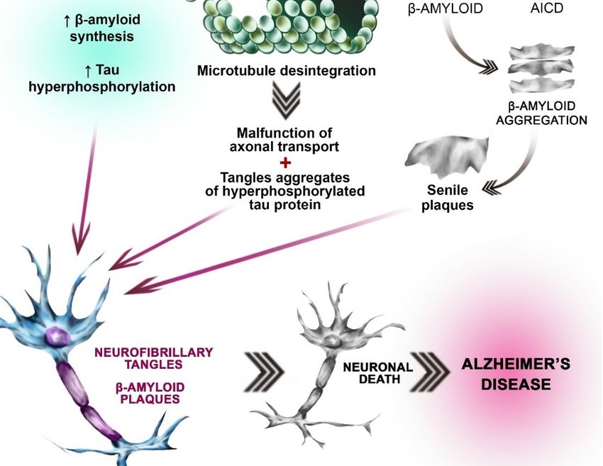
Int. J. Mol. Sci. 2018, 19, 3965                                                                                                        3 of 23

role in the processes of memory creation and learning. It also displays neurotoxic properties in animal
studies, wherein it leads to the formation of neurodegenerative lesions similar to those observed in the
human brain in AD [17,19]. Under normal conditions, glutamatergic transmission in the hippocampus
is associated with the generation of a cytosolic calcium ion signal responsible for various synaptic
plasticity phenomena including the consolidation of learning and memory processes [4]. However,
in pathological situations neurodegeneration occurs as a result of N-methyl-D-aspartate (NMDA)
glutamate receptor hyperactivation causing a sustained increase in intracellular calcium, chlorine
and sodium ions, thereby leading to excessive depolarization of the postsynaptic membrane [4,18,20].
In AD, alterations in glutamatergic signaling lead to prolonged neuronal exposure to extracellular
glutamate, which in turn results in the aforementioned receptor hyperstimulation and excitotoxicity [4].

3.3. Oxidative Stress Hypothesis
      Reactive oxygen species (ROS) are produced in all living organisms as byproducts of normal
metabolic reactions and as a result of xenobiotic exposure [21]. Under physiological conditions, they
play an important role in cellular signaling throughout the body [22], but in excess they are harmful to
all cell types, including nerve cells [21]. Oxidative stress caused by an imbalance between production
and elimination of ROS is linked to the pathogenesis of numerous diseases [23,24]. Oxidative stress is
not considered to be a phenomenon that itself initiates AD pathogenesis. However, it has been shown
to facilitate the progression of the disease and worsen prognosis [24] (Figure 1).

3.4. Amyloid β Aggregation Hypothesis
     Amyloid β (Aβ) aggregation plays a key role in the etiopathogenesis of AD. The misfolding of
Aβ leads to the formation of β pleated sheet-rich aggregates and impairs neuronal function [25].
     Aβ is a peptide consisting of 39 to 42 amino acids, some of which form a hydrophobic
transmembrane domain. Aβ occurs in several isoforms, of which the most hydrophobic and toxic is
1–42 [24]. It forms extracellular aggregates [26] of varying size dependent on the balance between its
synthesis and degradation [27].
     Aβ is a peptide product derived from the amyloid precursor protein (APP), a transmembranous
protein whose primary function is not known. APP expression increases in cells under increased oxidative
stress [4]. Production of Aβ from APP occurs through a sequence of proteolytic cleavages, mediated by
secretase enzymes of the disintegrin and metalloproteinase family (ADAM) [4]. In AD patients, initial
cleavage leads to the formation of an extracellular soluble fragment (APPsβ) and a longer carboxylic
fragment (C99) [4]. This process is mainly catalyzed by β-site-APP-cleaving enzyme (BACE1), whose
expression is modulated by oxidative stress, ischemia, trauma, inflammation, and hypoxia—situations
common in aging and neurodegenerative disease [4]. γ-secretase (consisting of presenilin, nicastrin,
anterior pharynx-defective 1 (APH-1), and presenilin enhancer 2 (PEN-2)) then cleaves the carboxylic
fragment at the γ site producing the aggregate-forming Aβ peptide [4] (Figure 1). The amyloid aggregation
hypothesis is currently the most widely accepted theory of AD pathogenesis [4].

3.5. Tau (τ) Protein Hyperphosphorylation Hypothesis
     Studies have shown that the pathological τ protein acts in concert with Aβ in synapse
degeneration in AD [28]. Under physiological conditions, τ protein is the major neuronal microtubule
associated protein (MAP). Together with MAP1 and MAP2, it is responsible for promoting the assembly
of tubulin into microtubules and stabilizing the microtubule network in neuronal axons [29]. In the
human brain, τ occurs in six isoforms, differing in their number of binding domains and resulting
microtubule stabilizing ability. Under physiological conditions, τ activity has been found to correlate
negatively with the degree of its phosphorylation [30]. Further studies revealed that tau activity was
affected not only by the degree of phosphorylation but also the phosphorylation site [31]. In the normal
human brain, τ is phosphorylated and highly soluble [32]; however, abnormally hyperphosphorylated
tau is insoluble [4]. In the brain of AD patients, τ protein is 3 to 4× more phosphorylated than

Pobrano z Repozytorium Pomorskiego Uniwersytetu Medycznego w Szczecinie / Downloaded from Repository of Pomeranian Medical University in Szczecin 2022-06-15
Int. J. Mol. Sci. 2018, 19, x FOR PEER REVIEW 4 of 23
activity
Int. J. Mol. was affected
Sci. 2018, not only by the degree of phosphorylation but also the phosphorylation site4 [31].
19, 3965 of 23
In the normal human brain, τ is phosphorylated and highly soluble [32]; however, abnormally
hyperphosphorylated tau is insoluble [4]. In the brain of AD patients, τ protein is 3 to 4× more
in healthy brains.than
phosphorylated In addition, proteinIn
in healthyτ brains. hyperphosphorylation and resulting insolubility
addition, τ protein hyperphosphorylation leads to
and resulting
polymerization into paired helical filaments (PHF), which, together with straight filaments
insolubility leads to polymerization into paired helical filaments (PHF), which, together with straight(SF), form
neurofibrillary
filaments (SF), tangles [29] (Figure 1).tangles
form neurofibrillary Pathologically-altered τ protein loses its ability
[29] (Figure 1). Pathologically-altered to interact
τ protein with
loses its
microtubules, leading to an increase in free protein and an increase in aggregation
ability to interact with microtubules, leading to an increase in free protein and an increase inand fibrillation,
resulting in impairment
aggregation of axonal
and fibrillation, function
resulting [33].
in impairment of axonal function [33].
Figure 1.1. Neurobiological
Figure Neurobiological processes
processes leading
leading to
to AD.
AD. Hypothesis
Hypothesis concerning
concerning OS
OS involvement
involvement in
in AD
AD
etiopathogenesis: β-amyloid peptide activates the pathways involved in reactive oxygen species
etiopathogenesis: β-amyloid peptide activates the pathways involved in reactive oxygen species (ROS)
Pobrano z Repozytorium Pomorskiego Uniwersytetu Medycznego w Szczecinie / Downloaded from Repository of Pomeranian Medical University in Szczecin 2022-06-15Int. J. Mol. Sci. 2018, 19, 3965 5 of 23
synthesis, leading to the increased amount of ROS (left side of the picture). Simultaneously β-amyloid
accumulation leads to the antioxidant enzymes’ inhibition (SOD, CAT, GPx, and GR) (left side of
the picture. This imbalance between ROS synthesis and antioxidant enzymes activities results in
the oxidative stress (OS). Excessive oxidation processes result in tau protein hyperphosphorylation
and β-amyloid peptide accumulation (gray arrow, left side of the picture). Tau (τ) protein
hyperphosphorylation hypothesis: Under physiological conditions, τ protein is the major neuronal
microtubule associated protein. It promotes the assembly of tubulin into stabilizes the microtubules
(top picture in the central part of the figure). Under pathological conditions (i.e., OS) Tau becomes
hyperphosphorylated. Pathologically-altered τ protein loses its ability to interact with microtubules,
leading to disintegration of microtubules (gray arrow, central part of the picture). Hyperphosphorylated
Tau is insoluble. Insolubility leads to polymerization into paired helical filaments (PHF), which, together
with straight filaments (SF), form neurofibrillary tangles (gray arrow, central part of the picture).
Amyloid β aggregation hypothesis: Amyloid precursor protein (APP) is an integral transmembrane
protein expressed in many tissues. In AD patients, initial cleavage (by β-secretase) (brown arrow) of
the APP results in the extracellular soluble fragment formation. Subsequent cleavage catalyzed by
γ-secretase leads to the β-amyloid formation. γ-secretase consists of presenilin, nicastrin, anterior
pharynx-defective 1 (APH-1), and presenilin enhancer 2 (PEN-2) β-amyloid, which is insoluble
aggregates (right part of the picture, gray arrow) to form In subsequence senile plaques. Another
APP derived cleavage product is AICD (the amyloid precursor protein intracellular domain) (green
spherical elements). Different AICD levels may contribute to early etiopathological sequences in AD.
The processes mentioned above lead to the fibrillary tangles formation, neuronal death and Alzheimer’s
disease (three red arrows).
4. Fluoride as a Neurotoxic Agent
Fluoride is widespread in the environment, especially in industrial areas [34]. It easily
crosses the blood–brain barrier [35], wherein the accumulation of fluoride disturbs phospholipid
metabolism leading to neuronal death [35]. In overexposed women, fluoride can also pass through the
blood–placenta barrier to enter the fetal circulation, where it has been shown to inhibit central nervous
system development and cause neurodegeneration [36]. In recent years, the mechanism and extent of
fluoride’s effect on the nervous system have been the subject of increasing scientific interest [37].
The effect of fluoride exposure on the developing brain (both pre- and neonatal) manifests
clinically as memory loss and impairment of cognitive processes. Epidemiological studies showed
that children living in areas with excessive fluoride exposure had lower IQ values compared to
less exposed children [38]. Industrial workers chronically exposed to fluoride showed a variety of
neuropsychiatric symptoms including drowsiness, concentration and learning difficulties, and memory
disorders [39–41].
Fluoride-induced abnormalities are associated with disturbed metabolism of neurons and glial
cells. Fluoride accumulation in the hippocampus has been found to contribute to neuronal degeneration
and altered oxygen metabolism, promoting the formation of ROS, and inducing damaging oxidative
stress [42–44].
More recent studies have shown that the effect of fluoride on the central nervous system may be
extremely varied and complex. In addition to its pro-oxidative effect, fluoride has demonstrated an
influence on the activity of antioxidative enzymes, further encouraging damaging levels or ROS [42,45].
Fluoride has also been found to affect cellular energy metabolism, synthesis of inflammatory factors,
neurotransmitter metabolism, microglial activation, and the expression of proteins involved in neuronal
maturation. Finally, and of specific importance to its role in Alzheimer’s disease, studies report
fluoride-induced apoptosis and inflammation within the central nervous system [45] (Figure 2).
Pobrano z Repozytorium Pomorskiego Uniwersytetu Medycznego w Szczecinie / Downloaded from Repository of Pomeranian Medical University in Szczecin 2022-06-15Int. J. Mol. Sci. 2018, 19, 3965 6 of 23
Int. J. Mol. Sci. 2018, 19, x FOR PEER REVIEW 6 of 23
Figure 2. The pro-oxidative and pro-inflammatory effect of fluoride exposure on the brain. Fluoride
Figure 2. The pro-oxidative and pro-inflammatory effect of fluoride exposure on the brain. Fluoride
ions easily cross the blood–brain barrier (dark gray arrow, pink highlight). Within the brain fluoride
ions easily cross the blood–brain barrier (dark gray arrow, pink highlight). Within the brain fluoride
influences normal metabolism of the neurons and glial cells. The fluoride effect on the nervous
influences normal metabolism of the neurons and glial cells. The fluoride effect on the nervous system
system is complex and varied. Fluoride is well documented pro-oxidative factor. Promotes oxidative
is complex and varied. Fluoride is well documented pro-oxidative factor. Promotes oxidative stress.
stress. It enhances reactive oxygen species ROS (H2 O2 , OH٠ − , and O2 •− ) synthesis by activated
It enhances reactive oxygen species ROS (H2O2, OH−, and O2 −) synthesis by activated macrophages
macrophages (violet highlight). F− weakens the antioxidants’ function by inhibiting the actions of
(violet highlight). F− weakens the antioxidants’ function by inhibiting the actions of antioxidant
antioxidant enzymes superoxide dismutase (SOD), catalase (CAT), glutathione peroxidase (GPx),
enzymes superoxide dismutase (SOD), catalase (CAT), glutathione peroxidase (GPx), and glutathione
and glutathione reductase (GR) (light red highlight). Excessive ROS production and simultaneous
reductaseantioxidative
impaired (GR) (lightenzymes’
red highlight).
action leadsExcessive ROS production
to the oxidative stress (OS).and OS issimultaneous
an imbalance impaired
between
antioxidative enzymes’ action leads to the oxidative stress (OS). −OS is
oxidation and antioxidation processes (blue balance beam). F enhances the neuroinflammation an imbalance between oxidation in
andbrain.
the antioxidation
Fluoride processes
dependent(blue balanceofbeam).
stimulation F− enhances the cytokines
the pro-inflammatory neuroinflammation in theTNF-α,
synthesis (IL-6, brain.
Fluoride
and IFN-γ)dependent stimulation
is a key step of the pro-inflammatory
in the inflammation cytokines synthesis
process development. (IL-6, TNF-α,
In physiological and IFN-
conditions low
γ) is a key step in the inflammation process development. In physiological
concentration of IL-6 in the brain is observed (violet arrows, violet highlight). The increase in the IL-6 conditions lowis
concentration of IL-6 in the brain is observed (violet arrows, violet highlight).
noticed in the inflammation or neurodegenerative diseases. Excessive production of IL-1β and TNF-α The increase in the IL-
6 is
is noticed in
observed in neuroinflammation
the inflammation orand neurodegenerative
neurodegenerative diseases. Excessive
diseases. production of
Overproduction of cytokines
IL-1β and
TNF-α
in responseis observed
to fluoride in exposition
neuroinflammation
results inand neurodegenerative
inflammation development diseases. Overproduction of
and neurodegeneration.
Fluoride exposition, i.e., due to the inflammatory cytokines increase, leads to thedevelopment
cytokines in response to fluoride exposition results in inflammation increased activityand
neurodegeneration. Fluoride exposition, i.e., due to the inflammatory cytokines
of the enzymes involved in inflammation (i.e., COX-2) and subsequent production of prostanoids: increase, leads to the
increased activity of the enzymes involved in inflammation (i.e., COX-2)
prostaglandin E2 (PGE2) and thromboxane A2 (TXB2) (violet arrow, violet highlight). F in the and subsequent production
−
of prostanoids:
brain prostaglandin
increases apoptosis E2activating
rate by (PGE2) and thethromboxane
transcriptionA2 (TXB2)
factors (violetc-JUN)
(NF-κB, arrow,andviolet highlight).
proapoptotic
F− in theBAX,
proteins brain increases
FAS, and p53. apoptosis rate by Factivating
Simultaneously the transcription
- inhibits antiapoptotic factors
proteins (NF-κB,
synthesis c-JUN)
BCL2, BCL-XLand
proapoptotic proteins BAX,
(pink highlight, dark gray arrows). FAS, and p53. Simultaneously F - inhibits antiapoptotic proteins synthesis
BCL2, BCL-XL (pink highlight, dark gray arrows).
5. The Role of Fluoride in the Pathogenesis of Alzheimer’s Disease
5. The Role of Fluoride in the Pathogenesis of Alzheimer’s Disease
5.1. Oxidative Stress
5.1. Oxidative
The brainStress
is predisposed to excessive ROS production and oxidative damage due to its high
content of polyunsaturated
The brain is predisposed fatty
to acids and redox-active
excessive metals
ROS production and(Cu and Fe)damage
oxidative [23] as well
due as
to its
its high
high
metabolic rate, characterized by high oxygen consumption (20% of basal oxide consumption), and
content of polyunsaturated fatty acids and redox-active metals (Cu and Fe) [23] as well as its high low
regenerative capacity
metabolic rate, [46] compared
characterized by high to oxygen
other organs.
consumption (20% of basal oxide consumption), and
low regenerative capacity [46] compared to other organs.
Elevated oxidative damage in the brain of AD patients is related to the accumulation of amyloid-
β (Aβ) and deposition of neurofibrillary tangles and neutrophil threads. Both Aβ and amyloid β
Pobrano z Repozytorium Pomorskiego Uniwersytetu Medycznego w Szczecinie / Downloaded from Repository of Pomeranian Medical University in Szczecin 2022-06-15Int. J. Mol. Sci. 2018, 19, 3965 7 of 23
Elevated oxidative damage in the brain of AD patients is related to the accumulation of amyloid-β
(Aβ) and deposition of neurofibrillary tangles and neutrophil threads. Both Aβ and amyloid β
precursor protein (APP) have the ability to oxidize copper ions, resulting in the release of hydrogen
peroxide [23].
Both the risk of Alzheimer’s disease and levels of oxidative stress increase with age [47].
A study involving rats of increasing ages (7-, 14-, and 21-day-old, adults: 3–6-month-old, and
aging: 24-month-old) showed elevated ROS synthesis in the brain of adults and in individual brain
areas of aging individuals [48]. This increase in ROS in the brain is linked to disorders of ROS
production and/or elimination [49]. Evidence of increased oxidative stress includes increased levels of
Cu, Hg, Al, and Fe in brain regions affected by AD-related neurodegeneration and Aβ aggregation,
increased lipid peroxidation and reduced polyunsaturated fatty acids, increased protein and DNA
oxidation, decreased energy metabolism, and advanced glycation end-products in neurofibrillary
tangles [23,50–52].
Markesbery and Lovell found that four-hydroxy-nonenal (HNE), a product of lipid peroxidation,
is a neurotoxin whose concentration is elevated in the ventricular fluid of AD patients. They also
reported an increase in HNE levels in the brain regions most affected by degeneration in AD [51],
indicating a potential role in pathogenesis.
Further evidence of oxidative stress comes from the observation by Ansari et al. of an increased
concentration of carbonylated proteins in parts of the brain with pathomorphological changes resulting
from AD (i.e., the frontal lobe, parietal lobe, and hippocampus) [52].
Living organisms are protected against the effects of excessive ROS by antioxidants, including
antioxidant enzymes and glutathione (GSH). Antioxidant enzymes are divided into those directly
involved in the inactivation of ROS—i.e., catalase (Cat), superoxide dismutase (SOD), and glutathione
peroxidase (GPx)—and glutathione reductase (GR), which catalyzes the reduction of oxidized
glutathione. Reduced glutathione is an electron donor in the peroxidation reaction catalyzed by
GPx. GSH also has the ability to directly interact with ROS [53–55]. While alterations in ROS synthesis
and oxidative stress in AD have been confirmed in both animal and human models, data on changes
in the activity of antioxidant enzymes are inconclusive [48,49,51,56].
Though specific findings are inconsistent, the activity and levels of antioxidant enzymes are
altered in AD. A review by Niedzielska et al. describes an increase in SOD activity in the hippocampus
and amygdala of AD patients, but decreased levels of SOD, GPX, and Cat in the frontal and temporal
cortex and a decreased GSH content in brain and erythrocytes [56–59]. In addition, lymphocytes of
AD patients were characterized by a higher level of Cu/ZnSOD mRNA [60] compared to both healthy
subjects and those with Parkinson’s disease.
It is generally recognized that substances that support ROS scavenging or increase the activity
of antioxidant enzymes have a positive effect in the treatment of Alzheimer’s disease symptoms.
A proposed strategy for the treatment of neurodegenerative diseases associated with abnormal
oxidation involves the use of medicinal substances with antioxidant enzyme activity [61].
5.2. The Role of Fluoride
Fluoride has a well-established prooxidant effect in cells [62–64]. Shuhua et al., in studies
with murine microglial BV-2 cells, showed that the toxic effects of fluoride on the nervous system
can at least partly be attributed to microglial activation, leading to increased synthesis of ROS and
RNS (reactive nitrogen species) and resulting in oxidative stress [65]. This is confirmed in a study
by Saralakumari et al. showing a relationship between chronic exposure to fluoride and increased
oxidative stress in humans [66].
The pathomechanism of fluoride toxicity also involves its effect on several enzymes, including
antioxidant enzymes. The exact nature of this influence is not clear, but fluoride exerts a predominantly
inhibitory effect on antioxidant enzyme activity [42]. A study conducted by Zhang et al. endeavored
to explain the mechanism of fluoride neurotoxicity. Rat hippocampal neurons cultured in the presence
Pobrano z Repozytorium Pomorskiego Uniwersytetu Medycznego w Szczecinie / Downloaded from Repository of Pomeranian Medical University in Szczecin 2022-06-15Int. J. Mol. Sci. 2018, 19, 3965 8 of 23
of sodium fluoride (20, 40, and 80 mg/L) for 24 h, showed a significant decrease in GPx activity and
decreased GSH concentration. A decrease in SOD activity was also observed in cells incubated with
high concentration of NaF [44]. Increased Cat activity [67] was observed in brain tissues of young rats
given fluoridated drinking water, which can be explained as the activation of protective mechanisms
against the effects of excessive oxidation [67,68]. A study conducted by Pal and Sarkar on rats exposed
to fluoride at 20 mg/kg showed a reduction in GSH concentration and a decrease in the activity of Cat,
SOD, and GPx in brain tissues [68].
In vitro studies on macrophages obtained from THP-1 cell line monocytes (not yet published
by our team) showed that fluoride not only exerted a negative effect on the activity of antioxidant
enzymes, but also negated the positive effect of acetylcholinesterase inhibitors on these enzymes (Cat,
SOD, GPx, and GR) and lowered GSH concentration [69]. This effect was observed after application of
fluoride at a low (3 µM) concentration reflecting chronic environmental exposure levels [69].
6. Inflammation in AD
Alzheimer’s disease is one of many diseases associated with inflammation [69]. The factors and
pathways involved in neurodegeneration are not well understood; however, recent data suggest that
inflammation is central to this process [70].
The involvement of inflammation in the pathogenesis of neurodegenerative diseases, (including
AD) has been indirectly confirmed by the results of epidemiological studies demonstrating prevention
or inhibition of the progress of AD in patients after use of anti-inflammatory drugs [71]. In their 2004
review, Szekely et al. cited data showing reduced AD incidence in persons taking anti-inflammatory
drugs for other ailments [72].
It is believed that inflammation in itself is not the primary initiator of neurodegeneration in
AD and other neurodegenerative diseases. However, the long-term upregulation of inflammatory
response resulting from activation of microglia and astrocytes in neurodegenerative disease suggests
an important role for neuroinflammation in neuronal dysfunction and death [70]. Current literature
extensively describes the inflammatory mechanisms which are active in the development and progress
of AD [73].
6.1. Fluoride vs. Nuclear Factor κB (NF-κB)
The mechanisms underlying neuroinflammation are very complex and difficult to identify.
However, it is known, for example, that inflammation in the central nervous system induces the
activation of nuclear transcription factor κB (NF-κB), a major transcription factor regulating the
expression of genes responsible for immune response [74]. Literature also suggests an important role
for NF-κB in inflammation resulting from the interaction between microglia and astrocytes [75,76].
Fluoride has been shown to stimulate in vitro NF-κB activity in BV2 microglia [34]. Additionally,
NF-κB can be activated by IL-1 interleukin and tumor necrosis factor α (TNF-α), both of which are
produced in the brain in response to exposure to fluoride [77,78]. Activation of NF-κB can have
numerous results, including stimulation of nitric oxide synthase activity, increased NO production,
and induction of expression of cyclooxygenase 2 (COX-2), an enzyme that plays a key role in the
induction and progression of neuroinflammation [77].
6.2. Fluoride vs. Proinflammatory Cytokines
Fluoride has been linked to many aspects of the neuroinflammatory process, including stimulation
of cytokine secretion and direct influence on macrophages and microglia, whose activated forms in
turn constitute an important source of proinflammatory factors in AD [79].
Since the 1990s, increased expression of inflammatory markers—including acute phase proteins
such as α1-antichymotrypsin and proinflammatory cytokines such as interleukin-1 (IL-1), interleukin-6
(IL-6), and TNF-α—has been implicated in AD pathogenesis [80–83]. This was confirmed in a study
on Alzheimer amyloid precursor protein (APP)-transgenic mice (APP-Tg), where, following injection
Pobrano z Repozytorium Pomorskiego Uniwersytetu Medycznego w Szczecinie / Downloaded from Repository of Pomeranian Medical University in Szczecin 2022-06-15Int. J. Mol. Sci. 2018, 19, 3965 9 of 23
of LPS (lipopolysaccharide) into peripheral circulation, AD mice showed a significantly higher
concentration of IL-6 than wild-type mice [84]. It has also been established that overproduction
of IL-1, IL-6, and TNF-α stimulates the synthesis of Aβ [85] and IL-1 is associated with both the
initiation and spread of neuroinflammation in AD [86].
An in vitro study by Wang et al. on HeLa cells showed a fluoride-induced stimulation of the
synthesis of proinflammatory cytokines IL-1β, IL-2, IL-6, and TNF-α [87]. Activation of microglia
in the hippocampus and cerebral cortex was also demonstrated in the rat model where fluoride,
by influencing the production of these cytokines, contributed to the formation and progression of
inflammation in the brain [88].
6.3. Fluoride vs. Neuroinflammation Enzymes
Post-mortem studies on the brains of Alzheimer’s disease patients show that senile plaques are
infiltrated by activated microglia cells, which then serve as an important source of cytokines [89,90].
Cytokines released by microglia, by binding to receptors on astrocytes coupled to Ca2+ -dependent
enzymes, may cause the activation of these enzymes, including cytosolic phospholipase A2 (cPLA2)
and secretory sPLA2 [91]. Phospholipases A2 catalyze the hydrolysis of the ester bond in the sn-2
position of glycerophospholipids, releasing fatty acids, including arachidonic acid, which further
encourages the inflammatory process [92]. In turn, activation of PLA2, and the resulting increased
availability of free arachidonic acid, promotes the synthesis of proinflammatory eicosanoids in
macrophage cells [93].
The stimulating effect of aluminum fluoride on the activity of phospholipase A2 in macrophages
was recognized as early as the 1980s [94]. Gutowska et al. were one of the first to show the stimulating
effect of low concentrations of fluoride on PLA2 exocrine activity and subsequent eicosanoid
production, specifically prostaglandin E2 (PGE2) and thromboxane A2 (TXA2), indicating increased
activity of cyclooxygenases [93]. Subsequently, Chalbot et al. demonstrated that sPLA2 activity in
cerebrospinal fluid collected from individuals with diagnosed AD is higher than in cerebrospinal
fluid from healthy persons [95]. Brains of AD patients also show increased levels of functional cPLA2
protein and mRNA expression [96–98].
Furthermore, ROS—the production of which is increased in AD—activate mitogen-activated
protein kinases (MAPK), which in turn activates PLA2. cPLA2 activity is in turn associated
with neuronal excitotoxicity, impairment of mitochondrial function, and neuronal apoptosis.
Parallel activation of the previously mentioned NF-κB [97]—which is also activated by fluoride
in the central nervous system [99]—can further induce expression of sPLA2 and cyclooxygenase 2
(COX-2), and thereby increase inflammation [97–99].
Also among the enzymes involved in the development of inflammation in AD, COX-2, also
known as prostaglandin-endoperoxide synthase 2, is an important enzyme in the metabolic cascade
of arachidonic acid and PLA2 [100]. Increased COX-2 activity has been previously described in the
frontal cortex of AD patients [100]. COX-2 is an inducible enzyme whose expression is associated with
increased inflammation and various pathological processes [100]. COX-2 is induced by numerous
proinflammatory stimuli including inflammatory cytokines and monocyte cells themselves [101].
COX-2 catalyzes the synthesis of prostanoids (PGE2, TXA2) from arachidonic acid and serves as
the main source of these molecules during inflammation [102]. These prostanoids then mediate
inflammatory upregulation, an important factor in the pathogenesis of AD [69,100].
An important discovery in this field was the identification of a constitutively expressed form of
COX-2 within a specific neuronal population, where it supports synaptic activity and long-term
plasticity [103]. As a result, some researchers suggest that in order to accurately describe the
expression of COX-2 in the nervous system, the term “constitutive expression” should be replaced
by “dynamic regulation”, as constant COX-2 expression is observed during normal synaptic activity,
while expression increases during convulsions or ischemia [104].
Pobrano z Repozytorium Pomorskiego Uniwersytetu Medycznego w Szczecinie / Downloaded from Repository of Pomeranian Medical University in Szczecin 2022-06-15Int. J. Mol. Sci. 2018, 19, 3965 10 of 23
A potential link between COX-2 activity and Alzheimer’s disease pathogenesis was suggested
in the 1990s and 2000s [105,106]. It was found that administration of nonsteroidal anti-inflammatory
drugs (NSAIDs)—which inhibit COX activity—to AD patients resulted in inhibited progression of
clinical symptoms [107]. The study also confirmed an increased expression of the COX-2 gene in
the frontal cortex of AD patients compared to healthy subjects [107]. Moreover, synthetic β-amyloid
peptides induced COX-2 expression in SH-SY5Y neuroblastoma cells in vitro, suggesting a mechanism
for COX-2 upregulation in AD [108]. At the same time, COX-2 has been implicated in processes leading
to the formation and progression of both neuritic plaques (NP) and neurofibrillary tangles (NT) [109].
However, conflicting results exist with some evidence suggesting that the number of COX-2 positive
neurons decreases with increased severity of AD measured by clinical dementia rating (CDR) [110].
Based on available data, it is likely that COX-2-dependent neurodegenerative effects result from
the action of prostanoids produced in the COX-catalyzed reaction [111,112]. The stimulating effect of
fluoride on prostanoids production was observed by Schulze-Specking et al., who reported that fluoride
promoted the release of arachidonic acid from cell membranes and the synthesis of prostaglandins
in rat liver macrophages. Furthermore, fluoride initiated the translocation of protein kinase C from
the cytoplasm to cell membranes, indicating that Ca2+ -dependent protein kinase C is involved in the
proinflammatory action of fluoride [113]. The stimulating effect of fluoride on PGE2 production in
hepatic macrophages was confirmed by Dieter et Fitzke [114]. A more recent study with human THP-1
macrophages demonstrated that exposure to low fluoride concentrations—which may be considered
to reflect “environmental” exposure—led to an increase in PGE2 and TXB2 production [93].
Our team conducted a study on the influence of acetylcholinesterase inhibitors on cyclooxygenase
activity in regards to the proinflammatory action of sodium fluoride on macrophages. Many reports
indicate that these drugs, which are commonly used in the treatment of AD, may have other
mechanisms of action beside inhibition of acetylcholinesterase. Our study confirmed the inhibitory
effects of two popular AD drugs, donepezil and rivastigmine, on the production of PGE2 and TXB2
in macrophages, as well as on the expression of COX-1 and COX-2 mRNA and protein. We also
demonstrated that the proinflammatory effect of fluoride may be reduced by the combined use of both
drugs at their highest concentrations used in our study [69].
6.4. Fluoride vs. Neuroapoptosis
Apoptosis has been an important topic in AD research since the 1990s, when a link was suggested
between apoptosis and nerve cell loss in AD brains. Deeper understanding of this topic was considered
necessary for the development of new therapies [115].
Apoptosis plays a key role in the maintenance and progression of physiological processes (e.g.,
tissue homeostasis, aging, healing, and embryogenesis) [115,116]. Under physiological conditions,
apoptosis pathways are responsible for protecting the body against damage caused by the presence
of abnormal or mutant cells. However, disruptions or alterations in normal apoptotic pathways
may lead to abnormal or unregulated growth of cells, resulting in pathology and oncogenesis [117].
Excessive apoptosis has long been thought to play a role in the pathogenesis of neurodegenerative
diseases, such as Alzheimer’s and Parkinson’s disease [115,118], diseases in which environmental
factors such as fluoride seem to be of key importance.
Activation of apoptosis may occur as a result of detection of extensive DNA damage by the
DNA repair mechanism. Specifically, this type of DNA damage may be caused by increased oxidative
stress such as occurs in AD [115,117–119]. Further, oxidative stress causes autocatalytic production
of hydroxyl radicals which can induce activation of NF-κB [120,121], a transcription factor with a
key regulatory role in apoptosis [122]. Depending on contextual factors (e.g., apoptotic stimulus
and cell type), NF-κB can either protect cells against apoptosis or initiate this process [79,121,122].
An in vitro study on rat hippocampal neurons showed increased expression of NF-κB and an increased
percentage of apoptotic cells following treatment with sodium fluoride [99]. An influence of fluoride
on apoptosis was also demonstrated in in vitro studies carried out on SH-SY5Y neuroblastoma cells,
Pobrano z Repozytorium Pomorskiego Uniwersytetu Medycznego w Szczecinie / Downloaded from Repository of Pomeranian Medical University in Szczecin 2022-06-15Int. J. Mol. Sci. 2018, 19, 3965 11 of 23
where fluoride induced an increase in caspase-3 concentration and an increase in the expression
of Fas, Fas-L, caspase-3, and caspase-8, suggesting that fluoride-dependent damage to neural cells
results from—among other reasons—mitochondrial apoptosis due to the Fas-dependent activation of
caspase-8 and subsequent activation of caspase-3 [123]. Similarly, the potential proapoptotic effects of
fluoride were demonstrated in vivo by Liu et al. where rats exposed to fluoride showed an increased
number of apoptotic cells in their brains. Moreover, an increase in phosphorylation of Jun N-terminal
kinases (JNK) was observed, suggesting that in this case, the proapoptotic effect of fluoride is mediated
by activation of JNK kinases [124]. which in turn trigger the activation of caspases [124,125].
Another study, also conducted on rats, described an increased expression of proapoptotic Bax
protein and decreased expression of antiapoptotic Bcl-2 protein in response to fluoride. Analysis
carried out by means of the terminal deoxynucleotidyl transferase dUTP nick end labeling (TUNEL)
method confirmed an increase in apoptotic processes in brain structures [88].
TUNEL was performed during autopsy on the brains of people with Alzheimer’s disease.
Several such studies confirmed DNA fragmentation and in some cases, TUNEL-positive cells showed
apoptotic morphology [126,127]. Studies have also found increased expression of antiapoptotic Bcl-2
and Bcl-xl, as well as proapoptotic Bak and Bad, in the temporal cortex of AD patients compared
to healthy controls. Extended studies on individual protein fractions have concluded that Bak and
Bad [128] are more involved in AD-related apoptosis than Bax. It has been suggested that the balance
between proapoptotic (Bax, Bad, and Bak) and antiapoptotic (Bcl-2 and Bcl-xl) proteins may be a key
factor for the survival of individual neurons [129]. Also of note in AD-related apoptosis is the role
of caspases. Masliah et al. observed increased immunoreactivity of neuronal caspase-3 and Bcl-2 in
AD brains [130]. Moreover, neurons displaying DNA fragmentation showed more intense caspase-3
immunoreactivity compared to intact neurons, suggesting apoptotic activity [130].
Transcription factors c-Jun and NF-κB have also been linked to the initiation of apoptosis in AD
and the mechanism of proapoptotic action of fluoride. JNK-phosphorylated c-Jun is thought to be
involved in neuronal apoptosis, as evidenced by the observed increase in c-Jun and NF-κB expression
in AD brains [131,132].
6.5. Fluoride Vs. Mitophagy
Mitophagy is a way of controlled elimination of dysfunctional mitochondria by autophagy.
This process is highly selective form of autophagy and allows to maintain the proper functioning and
networking of mitochondria. Mitophagy is preceded by the fragmentation of mitochondria system.
It allows the elimination of the impaired organelle to occur with no influence on the mitochondria
network [133]. Mitophagy is suggested to be one of the earliest process during the onset of Alzheimer’s
disease [133]. In the AD brains mitochondria may exhibit various abnormalities i.e., morphology
pathologies, impaired functioning, increased mutations within the mtDNA or improper activities of
the mitochondrial enzymes [134]. APP and Aβ aggregation are suggested to aggravate the pathologies
of the mitochondria, i.e., by accelerating the oxidative stress. [134–136]. Moreover extensive ROS
synthesis enhances Aβ accumulation and subsequent Aβ mitochondrial toxicity itself [56].
Among mechanisms determining the negative fluoride effect on mitochondrion functioning the
influencing glucose metabolism in mitochondrion and enhancing oxidative stress are mentioned [45].
Fluoride is involved in ROS production in mitochondria. Excessive ROS synthesis together with
impaired functioning of antioxidant enzymes may disrupt the mitochondrion metabolism [45].
Fluoride-induced oxidative stress influences enzymes essential in ATP synthesis process, thus
decreasing ATP bioavailability. As the consequence the changes in mtDNA are observed and the
cell death occurs. ATP obtaining in mitochondrion can also be altered in consequence to fluoride
exposition due to the impaired glucose metabolism in neurons [137,138].
Fluoride is an element with a hypothetical role in the AD etiopathogenesis AD [45].
Concerning the information mentioned above, the toxic effects of fluoride on mitochondria, including
Pobrano z Repozytorium Pomorskiego Uniwersytetu Medycznego w Szczecinie / Downloaded from Repository of Pomeranian Medical University in Szczecin 2022-06-15Int. J. Mol. Sci. 2018, 19, 3965 12 of 23
hypothetical involvement in mitophagy occurrence, should be taken under consideration during the
AD etiopathogenesis [45,137,138].
6.6. Potential Roles of Alterations in Zinc and Magnesium Concentrations in Relation to
Fluoride-Induced Neurodegeneration
Zinc (Zn) is an essential microelement with a complex role in the organism. Zinc is needed in a
process of activation of different proteins (enzymes and receptors) and constitutes structural element
in particular proteins [139]. Zn is needful for the proper functioning of signal transduction pathways,
which are involved in the gene transcription modulation. Positive effects of Zn action are due to
several mechanisms, including the inhibition of ROS synthesis. In the brain zinc is involved in signal
transduction pathways, acting as a neurotransmitter [140]. Senescence is a physiological, complex
process, which predisposes to Zn deficiencies. AD etiopathogenesis is linked to both increased and
decreased zinc content within the organism [141,142].
Excessive accumulation of zinc in the brain induces Tau protein hyperphosphorylation, which
in turn affects NMDA receptors (NMDARs) via positive feedback, leading to the excitotoxic death of
neurons and production of neurofibrillary tangles. Moreover, excess zinc accumulation is associated
with the inhibited ferroxidase activity of APP, which results in the accumulation of bivalent iron in the
brain and subsequent intensification of pro-oxidative processes [143]. Another effect consists in the
synaptic accumulation of amyloid β oligomers around NMDAR receptors (namely, NR2B units) [141].
Although an increased concentration of zinc in the body is increasingly often mentioned in the
aspect of AD etiopathogenesis, zinc deficiency may also contribute to the onset of AD. Reduced zinc
concentration in peripheral blood is a factor reducing appetite, which further increases the risk of zinc
deficiency in older adults. Also highly significant is the intensification of the inflammation process
observed in zinc deficiency [141,144]. Finally, the loss of zinc predisposes to synaptic dysfunction [145]
and indirectly activates NADPH oxidase and nitric oxide synthase, resulting in the destruction of
mitochondria [146].
Due to the high incidence of Zinc deficiency around the world, its role in fluoride etiopathogenesis
of AD should not be neglected [141,145]. Exposure to fluoride is a very likely factor potentiating
biochemical changes caused by inadequate levels of zinc. In zinc deficiency, exposure to
pro-inflammatory and pro-oxidative fluoride is likely to accelerate and intensify the production
of ROS, thus leading to oxidative stress, which may exacerbate the signaling pathways associated
with inflammation. Simultaneous exposure to fluoride and zinc deficiency may increase the risk of
impaired mitochondrial metabolism and mitophagy. On the other hand, exposure to fluoride together
with excessive zinc concentration in the central nervous system could also hypothetically promote
AD. Both exposure to fluoride and excess zinc trigger the aforementioned mechanisms, resulting in
hyperphosphorylation of the Tau protein, which in turn leads to the neurofibrillary tangles generation
(as above) [45,143].
Neurodegenerative diseases onset is in connection with magnesium (Mg) deficiency
nowadays [147]. Taking into consideration mechanisms leading to the AD onset it is highly
possible that Mg deficiency can determine the initiation and severity of AD after fluoride exposure.
Fluoride induced oxidative stress and inflammation as well as Mg deficiency are demonstrated to have
a stimulatory effect on BACE1 activity. This enzyme, as mentioned below (part 3.0), is a key enzyme
involved in APP transformation into amyloid β [147,148]. Moreover there is evidence recognizing the
Mg deficiency as the factor contributing to the initiation of neurogenic inflammation [147,149]. Mg is
involved in the modulation of the NMDA receptors functioning. It was also suggested that a reduced
Mg level may potentiate the glutaminergic transmission, resulting in intensified excitotoxicity and
potentiated inflammatory-related signal cascades [150].
Currently no studies directly linking the AD with the exposure to fluoride in combination with
zinc alternations (increased or decreased concentration) or Mg deficiency can be found. However,
according to a considerable amount of reported data fluoride should be, with high possibility, taken
Pobrano z Repozytorium Pomorskiego Uniwersytetu Medycznego w Szczecinie / Downloaded from Repository of Pomeranian Medical University in Szczecin 2022-06-15Int. J. Mol. Sci. 2018, 19, 3965 13 of 23
under consideration as the factor intensifying the biochemical changes caused by Zn or Mg homeostasis
imbalance. It requires further research.
6.7. Potential Roles of Xenometals and Xenobiotics in Relation to Fluoride-Induced Neurodegeneration
Among the others xenometals and xenobiotics of the environmental origins with the supposed
role in AD etiopathogenesis, the heavy metals (i.e., lead, Pb), pesticides (i.e., organophosphates and
carbamates) are mentioned [151,152].
Heavy metals are suggested to be involved in the pathogenesis of AD. Some of the environmental
studies indicated aluminum (Al) to increase the risk of AD occurrence [153]. A meta-analysis of studies
published up to 2015 showed that patients with the long-lasting exposure to the Al exerted higher risk
of AD development in comparison to the healthy individuals [154]. Although a metanalysis conducted
by Virk et Eslick indicated no connection between occupational exposition to Al and occurrence of
AD [155]. The metanalysis (up to March 2015) used three studies based on case–control (with a sample
size of 1056 patients) [156]. The data concerning the Al presence in the cores of the amyloid or senile
plaques have been inconsistent, with some indicating the presence of this element within AD plaques.
But the fluorescent examination of the AD hippocampus revealed the Al within the neurons [157].
Excessive collection of Al in the brain results in the increased processes of ROS synthesis together
with weakened antioxidant protection and enhanced cellular cascades related to the inflammation are
supposed to be involved in AD etiopathogenesis [152]. Aluminum was reported to inhibit in animal
in vivo model activities of enzymatic oxidants (CAT, GPx, GR, and SOD) as well as Na+ /K+ ATPase,
Mg2+ ATPase, and Ca2+ ATPase in all brain regions in comparison to healthy control. Aluminum
administration decreased the reduced form of glutathione concentration [158]. Morris et al. also
indicate that Al exposure could result in apoptosis of a hippocampal and cortical regions. Moreover
in their review authors indicate that “physiological/environmental” concentrations of the Al could
promote the aggregation of Aβ and the subsequent fibrillary structures deposition [157].
Inflammation enhancement, stimulation of ROS synthesis with simultaneous impairment of
antioxidant bioavailability (resulting in oxidative stress), and apoptosis modulation are the common
effects for fluoride and Al exposition. Moreover in water environment a formation of Al and fluoride
complexes occurs [8]. Exposure to the complexes of Al and may cause more severe disruptions of
neurons metabolism than aluminum alone [8]. On the other hand fluoride in a complexed form with
Al may exert the properties that are not noticed outside the complexes (i.e., protein G activation) [8].
An element with well-documented neurotoxicity is lead (Pb). The strength of lead’s toxic
properties resulted in placing this element among the most hazardous factors [159]. Lead plays
a key role during all phases of inflammation, it modulates proinflammatory cytokines (i.e., IL-8),
the expression of inflammation enzymes (i.e., COX-2, caspase-1, and NOS2), or affects the synthesis
of purines receptors (PX24 and PX27) [159–161]. This Pb-induced disruption of immune system
functioning is caused by the disturbances on both cellular and hormonal levels [159]. Toxic effects
(with the special impact on neurotoxicity) of Pb have been extensively studied in vivo by Gassowska
˛
and Bosiacka et al. The researchers reported that pre- and neonatal exposure to low concentrations of
Pb may result in destroying of synapses structure as well as in the changes of key synaptic proteins
expression (synaptotagin-1, Syntaxin-1, and SNAP-25 in certain brain parts) [162]. Researchers also
showed the relation between perinatal exposure to Pb at low (below threshold level) concentration
and alteration in sphingosine-1 phosphate and its receptor expression (S1PR1) levels in various brain
regions in rats [163]. Lead was also proven to reduce the number of hippocampal neurons in rats
exposed perinatally to low concentrations of Pb [164].
Pb-dependent impairment of brain metabolism with subsequent clinical implications is well
described in children, but there are few studies in aging population of humans [165]. Although many
papers examining the potential role of Pb in AD etiopathogenesis appeared, there is still a great need
to study the potential link between AD and Pb exposure [165]. In humans the Pb concentrations in
the plasma were observed to be positively correlated to the worsening of the verbal memory [166].
Pobrano z Repozytorium Pomorskiego Uniwersytetu Medycznego w Szczecinie / Downloaded from Repository of Pomeranian Medical University in Szczecin 2022-06-15You can also read