Prion protein oligomers cause neuronal cytoskeletal damage in rapidly progressive Alzheimer's disease
←
→
Page content transcription
If your browser does not render page correctly, please read the page content below
Shafiq et al. Molecular Neurodegeneration          (2021) 16:11
https://doi.org/10.1186/s13024-021-00422-x

 RESEARCH ARTICLE                                                                                                                                  Open Access

Prion protein oligomers cause neuronal
cytoskeletal damage in rapidly progressive
Alzheimer’s disease
Mohsin Shafiq1,2, Saima Zafar1,3*, Neelam Younas1, Aneeqa Noor1, Berta Puig2,4, Hermann Clemens Altmeppen2,
Matthias Schmitz1, Jakob Matschke2, Isidre Ferrer5, Markus Glatzel2 and Inga Zerr1

  Abstract
  Background: High-density oligomers of the prion protein (HDPs) have previously been identified in brain tissues of
  patients with rapidly progressive Alzheimer’s disease (rpAD). The current investigation aims at identifying interacting
  partners of HDPs in the rpAD brains to unravel the pathological involvement of HDPs in the rapid progression.
  Methods: HDPs from the frontal cortex tissues of rpAD brains were isolated using sucrose density gradient
  centrifugation. Proteins interacting with HDPs were identified by co-immunoprecipitation coupled with mass
  spectrometry. Further verifications were carried out using proteomic tools, immunoblotting, and confocal laser
  scanning microscopy.
  Results: We identified rpAD-specific HDP-interactors, including the growth arrest specific 2-like 2 protein (G2L2).
  Intriguingly, rpAD-specific disturbances were found in the localization of G2L2 and its associated proteins i.e., the
  end binding protein 1, α-tubulin, and β-actin.
  Discussion: The results show the involvement of HDPs in the destabilization of the neuronal actin/tubulin
  infrastructure. We consider this disturbance to be a contributing factor for the rapid progression in rpAD.
  Keywords: Rapidly progressive Alzheimer’s disease, rpAD, Growth arrest specific proteins, GAS, Growth arrest
  specific 2 like 2, G2L2, Prion protein oligomers, PrPC, Co-immunoprecipitation, Cytoskeleton, Actin, Tubulin

Background                                                                            years [2]. However, some AD cases mimicking rapidly
Alzheimer’s disease (AD) is the most prevalent form of                                progressive dementias, i.e. with accelerated progression
dementia, affecting over 30 million people worldwide                                  rates and steep cognitive decline, have been reported
and representing 60–70% of all dementia cases [1].                                    over the past years [3–5]. These rapidly progressive Alz-
Sporadic AD (spAD) and familial AD (FAD) cases are                                    heimer’s disease (rpAD) cases are reported to have a
classically characterized by a progressive cognitive de-                              shorter post-diagnostic survival time as well (shorter
cline with an average post-diagnosis survival of eight                                than four years) compared to those of typical AD cases
                                                                                      (eight years on average) [6–9]. Due to their rapid pro-
                                                                                      gression, these cases are often initially misdiagnosed as
* Correspondence: sz_awaan@yahoo.com; saima.zafar@med.uni-
goettingen.de; saima.zafar@smme.nust.edu.pk                                           prion diseases [10]. The physiological alterations respon-
1
 Department of Neurology, University Medicine Goettingen and German                   sible for the accelerated disease course in rpAD patients
Center for Neurodegenerative Diseases (DZNE), 37075 Goettingen, Germany
3                                                                                     are poorly understood. No genetic linkage has been
 Biomedical Engineering and Sciences Department, School of Mechanical
and Manufacturing Engineering (SMME), National University of Sciences and             found between rpAD and any of the well-established
Technology (NUST), Islamabad, Pakistan                                                autosomal pathogenic mutations in the PSEN1, PSEN2,
Full list of author information is available at the end of the article

                                       © The Author(s). 2021 Open Access This article is licensed under a Creative Commons Attribution 4.0 International License,
                                       which permits use, sharing, adaptation, distribution and reproduction in any medium or format, as long as you give
                                       appropriate credit to the original author(s) and the source, provide a link to the Creative Commons licence, and indicate if
                                       changes were made. The images or other third party material in this article are included in the article's Creative Commons
                                       licence, unless indicated otherwise in a credit line to the material. If material is not included in the article's Creative Commons
                                       licence and your intended use is not permitted by statutory regulation or exceeds the permitted use, you will need to obtain
                                       permission directly from the copyright holder. To view a copy of this licence, visit http://creativecommons.org/licenses/by/4.0/.
                                       The Creative Commons Public Domain Dedication waiver (http://creativecommons.org/publicdomain/zero/1.0/) applies to the
                                       data made available in this article, unless otherwise stated in a credit line to the data.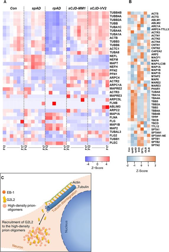
Shafiq et al. Molecular Neurodegeneration   (2021) 16:11                                                      Page 2 of 14

APP, or PRNP genes coding for presenilin isoforms, the       dysregulation of the cytoskeletal machinery [31]. The
amyloid precursor protein, and the cellular prion pro-       cytoskeleton plays a crucial role in the growth and func-
tein, respectively [11, 12]. Patients with rpAD exhibit a    tion of the neurons [32]. Effective transport systems in
distinct profile of CSF biomarker for neurodegeneration.     the neuronal cytoplasmic processes, the intracellular
A study from Abu Rumeileh et al. (2017) reported a           organization of organelles, and a degree of signal trans-
remarkably higher CSF tau level in rpAD cases (me-           duction are primarily orchestrated by the microtubule
dian = 1223 pg/mL, n = 44) compared to that of spAD          system in the neurons [33–37]. This microtubular sys-
(median = 697 pg/mL, n = 45) [13, 14]. However, CSF          tem works with the association of dyneins [38], kinesins
levels of phosphorylated tau (p-tau), Aβ40, Aβ42, α-         [39], spectrins, plakins and spectraplakins [40]. Neurons
synuclein and YKL-40 do not show significant differ-         also rely on a more transient actin system for establish-
ences between rpAD and spAD patients [15, 16].               ing the dendritic spines, extending the neurites, and for
Additionally, in a large scale study comprising over 300     inter-neuronal connections, in cooperation with the as-
patient samples, the CSF p-tau/tau ratio in rpAD pa-         sociated proteins such as integrins, cofilin and formin
tients was found to be lower than that in spAD [17].         [41–44]. Various cytoskeletal anomalies are associated
Other features differentiating rpAD from spAD include        with Alzheimer’s disease as well. Synaptotoxicity in AD
younger age of onset, lower frequency of the APOE4           is attributed to the malfunctioning in the Rho-associated
allele [12], increased serum levels of proinflammatory       protein kinase (ROCK) and cofilin-actin machinery [45,
cytokines in rpAD (G-CSF, TNFα, IL-6 and IL-13) [18],        46], along with altered drebrin / Ca2+ /F-actin-controlled
and a higher incidence of rpAD cases testing positive for    microtubule dynamics [47]. Amyloid-β has also been re-
the 14–3-3 protein in the CSF [12]. Although not dir-        ported to alter the dendritic morphology in hippocampal
ectly comparable to our study due to difference in defin-    neurons [48]. Microtubule / kinesin-mediated axonal
ition of rapid progression (survival in our study,           vesicle transport is also affected in the course of the dis-
cognitive decline in study of Ba et al., 2017); rpAD         ease, caused by detachment of tau from microtubule fila-
patients are also reported to exhibit region-specific        ments after hyperphosphorylation [38, 49]. Drummond
hypometabolism in [18F]fluorodeoxyglucose-positron           et al. (2017) described cytoskeletal proteins differentially
emission tomography by Ba et al., (2017) [17]. More-         associated with the amyloid plaques in rpAD and spAD.
over, a higher abundance of low molecular weight             The expression of the POTE ankyrin domain family
(LMW) amyloid oligomers has been associated uniquely         member E, tubulin polymerization-promoting protein,
with AD cases showing accelerated progression rates,         and tubulin alpha-4A chain was found significantly in-
thus suggesting their possible involvement in the rapid      creased in the cortical amyloid plaques of rpAD patients,
progression [19]. In a nuclear magnetic resonance study,     encouraging further studies of cytoskeletal proteins in
Qiang and coworkers have reported unique Aβ40 fibrils        rpAD brains [50]. PrPC is also reported to interact
prepared by seeded growth from extracts of brain cortex      with cytoskeletal components including cofilin [51],
tissues of rpAD patients. However, no physiological rele-    actin [52], and tubulin resulting in the inhibition of mi-
vance of theses variants has yet been studied further        crotubules assembly [53–55]. In the current study we
[20]. In an earlier study, we described the presence of      aimed to identify cytoskeletal proteins interacting with
high molecular weight oligomers of the cellular prion        the previously described high density PrPC oligomers
protein (PrPC) in the frontal cortex, specifically in pa-    (HDPs) in the brains of rpAD patients, thereby gaining
tients with a rapidly progressive form of AD [8]. Physio-    insight into the pathophysiological relevance of HDPs in
logical involvement of PrPC in the progression of AD         rpAD.
pathology is well described [21]. Previous studies have
emphasized the role of PrPC at the neuronal surface in       Methods
neurotoxic signaling utilizing cAMP/PKA or Erk acti-         Sample collection and processing
vated Fyn kinase pathway [22, 23], whereas more neuro-       Patient material was obtained after the approval of local
protective functions seem to be mediated by                  ethics committees at the University Medical Center,
extracellular fragments of PrPC, which sequester amyloid     Goettingen. Frontal cortex samples from patients with
oligomers and may inhibit their spread and toxicity [24,     spAD (n = 10), rpAD (n = 9), dementia with Lewy bodies
25]. However, the physiological relevance of the prion       (DLB) (n = 3), age-matched non-demented controls
protein oligomers consistently identified in the brains of   (Con) (n = 10) and other rapid dementias including small
rpAD patients has not been explored.                         vessel disease (SVD) (n = 3), rapidly progressive demen-
   In the course of AD, the brain tissue undergoes many      tia with Lewy bodies (rDLB) (n = 2), and dementia with
systemic changes including, more prominently, the de-        frontotemporal lobar degeneration (DFTL) (n = 3) cases
velopment of neurofibrillary tau tangles [2], amyloid-β      were provided by the brain bank of the Institute of
senile plaques [26, 27], synaptic damage [28–30] and         Neuropathology (HUB-ICO-IDIBELL Biobank) and the
Shafiq et al. Molecular Neurodegeneration   (2021) 16:11                                                      Page 3 of 14

biobank of the Hospital Clinic-IDIBAPS, Spain, accord-        30, 35, 40, 45% w/v in PBS/1% N-lauryl sarkosyl, pH
ing to their biomedical study legislation (Ley de la Inves-   7.4). Ultracentrifugation was carried out at 5 °C, 50,000
tigación Biomédica 2013 and Real DecretoBiobancos,            rpm for 73 min in an Optima TL 60 ultracentrifuge
2014). Frontal cortex samples from patients with spor-        equipped with a SW-55ti rotor (Beckman Coulter). From
adic Creutzfeldt-Jakob disease (sCJD) subtypes (MM1:          each sample, twenty density fractions (200 μL each) were
n = 6, MM/MV2: n = 6, VV2: n = 6) were obtained from          collected from top (lighter) to bottom (denser) fractions
the Department of Neurology at the University Medical         separately (Fig. 1a).
Center, Göttingen, Germany. The rpAD patients met the
current selection criteria for rpAD [6–9, 11, 12, 19].        Preparation of protein and peptide pools from high-density
These inclusion criteria are as follows:                      gradient fractions
                                                              For co-immunoprecipitation and subsequent mass
  1. Initial classification as prion diseases based on        spectrometric analysis, equal volumes of corresponding
     clinical features                                        density gradient fractions from the biological replicates
  2. Presence of typical AD pathological features, i.e.,      (n = 6) of each pathological cohort were pooled together
     higher Braak stages                                      (Fig. 2). The pooled high density factions (HDFs) were
  3. Post diagnostic survival time (disease duration)         further subjected for mass spectrometric analysis and
     shorter than four years                                  co-immunoprecipitation assays.
  4. Exclusion of other forms of rapid progressive
     dementias and copathologies e.g. prion diseases,         Antibodies
     extensive Lewy body pathology, vascular damage or        All primary and secondary antibodies used for immuno-
     tumors based on postmortem neuropathological             blotting and co-immunofluorescence in this study are
     examination                                              listed in Suppl. Tables 1 and 2.
  5. Absence of a family history suggestive of familial
     AD                                                       Co-immunoprecipitation of density fractions
                                                              To study unique interactomics signatures of PrPC
   Cortex samples of the non-demented controls ex-            oligomers in the different density fractions, co-
hibited only mild AD pathology (Braak stage I – II).          immunoprecipitation was employed using the
Both, rpAD and spAD samples presented AD patholo-             anti-PrP SAF70 antibody. Phosphatase- and protease-
gies ranging from Braak stage IV to VI. Likewise, the         inhibitor cocktails were added before proceeding to
sCJD subtypes cohort (used as positive controls for           the co-immunoprecipitation assay. Co-IP kits (catch
the PrP-oligomers) presented classical profiles for           and release HT immunoprecipitation kit, Merck) were
prionopathies. The rDLB samples had co-pathology of           used     to    ensure    assay    homogeneity.    Co-
AD, argyrophilic grain disease (AGD), tauopathy, or           immunoprecipitation experiments were carried out in
progressive supranuclear palsy (PSP). Patients from           a well-controlled setting with resin-only controls
the DFTL cohort also showed features of motor                 (HDFs were directly incubated with the antibody-
neuron disease (MND) and TDP-43 pathology. All of             binding resin without adding the antibody). The pro-
the vascular pathology patients (SVD) co-exhibited            teins detected in the resin-only controls were consid-
higher stages of AD pathology. Further details includ-        ered to be due to unspecific binding to the antibody-
ing neuropathological assessment and the processing           binding resin and, when detected, were excluded
of the samples for different analyses in the current          from the HDF-IP eluates.
study are reported as supplementary data (Add-
itional file 2). There were no significant differences in     Mass spectrometric analysis
age distribution and postmortem intervals among the           Protein/peptide sequence identification by data-dependent
disease groups included in this study, as shown in            acquisition
supplementary data (Suppl. Fig. 1).                           The HDFs and the Co-IP eluates were separated on
                                                              4–12% Bis-Tris gradient gels (NuPAGE Novex Bis-
Sucrose density gradients                                     Tris Mini gels, Invitrogen). Following Coomassie
Density gradient fractions were prepared as described         staining, the stained areas were excised, diced, and
previously [8]. Briefly, frontal cortex homogenates (10%      washed in ddH2O. Peptide digestion and identification
w/v in PBS/2% sarkosyl, pH 7.4) were centrifuged at 500       was carried out following protocols described previ-
x g for 5 min. Then 400 μL of the supernatant was             ously [52, 56]. The eluents were analyzed on a Q
layered on a 10–45% sucrose gradient prepared in a            Exactive hybrid quadrupole/orbitrap mass spectrom-
13 × 51 mm Beckman thin-wall polyallomer tube by              eter using Excalibur v2.4 software (Thermo Fisher
layering serially diluted sucrose solution (10, 15, 20, 25,   Scientific) and a top10 method in data dependent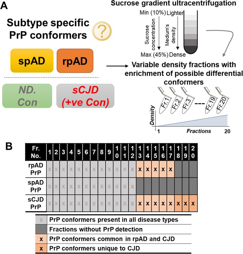
Shafiq et al. Molecular Neurodegeneration    (2021) 16:11                                                                       Page 4 of 14

 Fig. 1 Experimental setup and characterization of disease-specific PrP conformers. A) Scheme of the fractionation of different conformers.
 Density gradient centrifugation with a 10–45% sucrose step gradient was used to separate the density variants. Centrifugation was carried
 out at 50,000 rpm and 5 °C. Twenty density fractions were taken from top to bottom (lighter to dense) and used for downstream
 biochemical assays. B) Profile of high-density PrP (HDP) oligomer occurrence in cortical isolates of rpAD, spAD, sCJD patients (+ve con)
 and non-demented controls (ND. Con). In contrast to spAD, HDP oligomers were detected in density fractions 12 to 17 in rpAD, thus
 overlapping with HDPs isolated from sCJD samples

acquisition mode for analyzing the peptide ions.                        Sequential windowed Acquisition of all Theoretical
Raw2MSM v1.17 software (MPI for Biochemistry,                           Fragment ion Mass Spectra (SWATH)-based proteomics
Martinsried, Germany) extracted tandem mass spectra                     For global proteomic analysis, frontal cortex homoge-
and performed database searching. MS/MS spectra                         nates (50 μg total protein per sample), of various demen-
were evaluated using Mascot (Matrix Science,                            tia groups (spAD: n = 3, rpAD: n = 3, DLB: n = 3, SVD:
London, UK; version 2.4.1) instructed to search the                     n = 3, DFTL: n = 3, and rDLB: n = 2) and controls (n = 3)
Homo sapiens reference proteome (UniProt/SwissProt,                     were utilized. Two independent MS/MS measurements
revision 02–2017, 92,928 entries) with a 5 ppm pre-                     (technical replicates) were made for each sample to im-
cursors mass tolerance and a 0.02 Da mass tolerance                     prove the statistical confidence.
for fragments. Each of the Co-IP eluates and HDFs                          To prepare the peptide library, homogenates from
was analyzed twice for MS/MS (two technical dupli-                      each sample were pooled and separated into eight frac-
cates) to reduce data noise. Only peptides identified                   tions using a reversed phase spin column (Pierce High
with a confidence level greater than 95.0% were ac-                     pH Reversed-Phase Peptide Fractionation Kit, Thermo
cepted, and a minimum peptide score of two was re-                      Fisher Scientific). The separated fractions were then sub-
quired for a peptide identification to be considered as                 jected to tryptic digestion as described previously [52,
valid.                                                                  56]. The protein digests were analyzed on an Eksigent
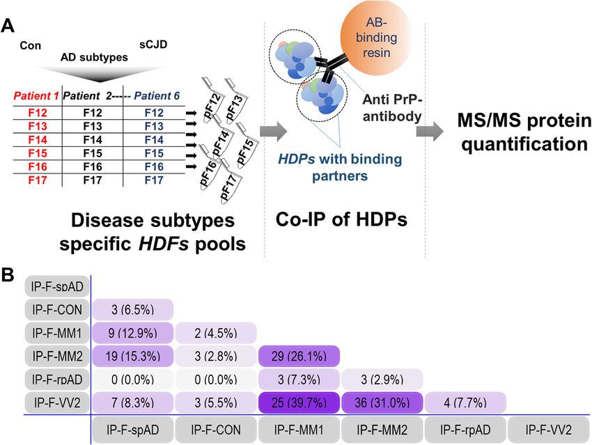
Shafiq et al. Molecular Neurodegeneration     (2021) 16:11                                                                          Page 5 of 14

 Fig. 2 Disease subtype-specific interactors bound to high-density prion protein oligomers. A) The experimental setup for carrying out the co-
 immunoprecipitation of HDPs and their potential interactors. B) Numerical Venn-diagram showing the overlap of subtype-specific HDP interactors.
 pF12-pF17: pools of high-density fractions 12–17 from multiple patient samples. IP-F-Con: high-density PrP (HDP) interactors in control HDFs
 pools from 12 to 17 (collectively), IP-F-spAD: HDP interactors in spAD HDFs pools from 12 to 17, IP-F-rpAD: HDP interactors in rpAD HDFs pools
 from 12 to 17, IP-F-MM1: HDP interactors in sCJD-MM1 HDFs pools from 12 to 17, IP-F-VV2: HDP interactors in sCJD-VV2 HDFs pools from 12 to
 17, IP-F-MM2: HDP interactors in sCJD-MM2 HDFs pools from 12 to 17

nanoLC425 nanoflow chromatography system associated                       using a Top30 data-dependent acquisition method as de-
with a TripleTOF 5600+, hybrid triple quadrupole-TOF                      scribed previously [57].
mass spectrometer equipped with a Nanospray III ion                         For SWATH analysis of the sample homogenates,
source (Ionspray voltage 2400 V, interface heater                         MS/MS data were acquired using 100 variable size
temperature 150 °C, and sheath gas setting 12), con-                      windows across the 400–1200 m/z range. Fragments
trolled by Analyst TF 1.7.1 (AB Sciex). The peptides                      were produced using rolling collision energy settings
were dissolved in loading buffer (2% acetonitrile (ACN)                   for charge state 2+, and fragments acquired over an
and 0.1% formic acid (FA) in ddH2O) to give a final con-                  m/z range of 180–1500 for 40 ms per segment. A
centration of 0.3 μg/μL. For each analysis, 1.5 μg of                     250 ms survey scan resulted in an overall cycle time
digested protein were concentrated on a precolumn                         of 4.3 s. Two replicate injections were acquired for
(0.15 mm ID × 20 mm, self-packed, Reprosil-Pur120                         each biological sample. Protein identification was
C18-AQ 5 μm, Dr. Maisch, Ammerbuch-Entringen,                             achieved using ProteinPilot Software version 5.0 build
Germany) followed by separation on an analytical RP-                      4769 (AB Sciex). A total of 152,341 MS/MS spectra
C18 column (0.075 mm ID × 250 mm, Reprosil-Pur 120                        were searched against the UniProtKB Homo sapiens
C18-AQ, 3 μm, Dr. Maisch) with a 100 min linear gradi-                    reference proteome (revision 02–2017, 92,928 entries).
ent of 5–35% ACN/0.1% FA at a flow rate of 300 nL/                        A total of 1756 proteins were identified with a false
min. Qualitative LC-MS/MS analysis was performed                          discovery rate of 1%.
Shafiq et al. Molecular Neurodegeneration          (2021) 16:11                                                                                 Page 6 of 14

Table 1 HDP-binding interactors in rpAD high-density fractions identified by mass spectrometry assisted co-immunoprecipitation
using anti-PrP antibody
No. IDs        UniProt Protein name                               Occurrence                       Sub-         Disease Relevance
               Acc. No.                                                                            cellular
                                                                                                   location
1    KCC2B Q13554         Calcium/calmodulin-dependent            rpAD-F12,                        C, Ck, Ce,   Alzheimer’s disease [59]
                          protein kinase type II subunit beta     sCJD-MM1-F13, 17, sCJD-          Sr, Sy
                                                                  MM2-F16, 17, sCJD-VV2-F16,
                                                                  17
2    KCC2D Q13557         Calcium/calmodulin-dependent         rpAD-F12,                           Cm, Sl
                          protein kinase type II subunit delta sCJD-MM1-F13, 17, sCJD-
                                                               MM2-F16, 17, sCJD-VV2-F16,
                                                               17
3    EPDR1 Q9UM22 Mammalian ependymin-related                     rpAD-F12,                        S            Unknown
                  protein 1                                       sCJD-MM1-F13–16, sCJD-
                                                                  MM2-F13–16, sCJD-VV2-F12,
                                                                  14, 15
4    DIRA2     Q96HU8     GTP-binding protein Di-Ras2             rpAD-F13, 15,                    Cm           Unknown
                                                                  sCJD-VV2-F17
5    G2L2      Q8NHY3     GAS2-like protein 2                     rpAD-F16                         C, Ck        Unknown
6    1433S     P31947     14–3-3 protein sigma                    rpAD-F17,                        C, Nu, S     Parkinson’s disease [60], Alzheimer’s
                                                                  sCJD-VV2-F17                                  disease, Creutzfeldt Jakob disease [61],
                                                                                                                Epilepsy [62]
F12 to F17: HDF-pool 12 to 17. Ce: centrosome, Sy: synapse, Sr: sarcoplasmic reticulum, C: cytoplasm, Ck: cytoskeleton, Nu: nucleus, S: secreted, Cm: cell
membrane, Sl: sarcolemma, The localization of proteins and accession number are assigned as in the ExPASy protein database and Uniprot data base, respectively.
Disease relevance of the HDP-interacting proteins was identified using Uniprot database search, as well

Co-immunofluorescence analysis                                                    (Fig. 1b) [8]. Due to the unique overlap of properties of
Coronal sections (5 μm thick) for co-immunofluorescence                           the high-density fractions of rpAD and sCJD, the frac-
slides were prepared from formic acid-fixed, paraffin-                            tions 12 to 17 were further used for downstream interac-
embedded cortex samples from AD patients (spAD: n = 5,                            tomics (Fig. 2a).
rpAD: n = 5) and non-demented controls (n = 5) following
a protocol described previously [58]. Confocal microscopy                         Identification of interactors binding to HDPs in brain
was carried out using an SPE laser-scanning microscope                            tissue
(Leica, Germany; 543 and 633 nm helium-neon and 488                               The HDPs with the interacting proteins were isolated
nm argon excitation wavelengths). Ten unbiased micro-                             from the density gradient fractions by immunoprecipi-
graphs were scanned per tissue section from the cortical                          tation and were characterized by MS/MS analysis as
grey matter area. Individual images were separately ana-                          described above. A total of six interactors were identi-
lyzed for co-localization using the ImageJ (WCIF plugin)                          fied, either uniquely present in rpAD-specific HDFs
software. Threshold Mander’s overlap coefficient and                              or commonly shared by rpAD and sCJD subtype-
Pearson’s linear correlation coefficient (rP) values were                         specific HDFs, whereas the HDP interactomes of con-
calculated to quantify fluorescence channel correlations                          trols and spAD subtypes had no common interactors
and to illustrate the strength and direction of the linear re-                    with rpAD. Intriguingly, some of the HDP interactors
lationship between the two fluorescence channels. One-                            from rpAD HDFs were also commonly found as the
way ANOVA followed by Tukey’s post-hoc test was used                              interactors of HDP conformers from sCJD subtypes.
to compare the mean values of Threshold Mander’s over-                            Three     HDP     interactors,   namely     mammalian
lap coefficients.                                                                 ependymin-related protein 1 (EPDR1), Calcium/cal-
                                                                                  modulin-dependent protein kinase type II (CaMKII)
Results                                                                           subunit beta (CAMK2B) and CaMKII subunit delta
High-density prion protein oligomers (HDPs)                                       (CAMK2D), were found in Co-IP eluates of rpAD
Specific occurrence of HDPs has been reported in brain                            HDFs (IP-F-rpAD) and sCJD-MM1, -MM2 and VV2
homogenates of rpAD compared with spAD in our pre-                                subtype fractions, i.e. IP-F-MM1, IP-F-MM2 and IP-
vious work [8]. Density gradient ultracentrifugation was                          F-VV2, respectively. IP-F-VV2 and IP-F-rpAD also
performed using 10–45% step gradients to obtain twenty                            shared another common interactor, namely GTP-
varying density fractions. Oligomeric prion protein was                           binding protein Di-Ras2 (DIRA2) and 14–3-3 protein
detected consistently in the isolated fractions 12 to 17                          sigma (1433S), whereas GAS2-like protein 2 (G2L2)
Shafiq et al. Molecular Neurodegeneration (2021) 16:11 Page 7 of 14
was observed to uniquely interact with rpAD-specific Differential co-localization of G2L2 and HDP affects the
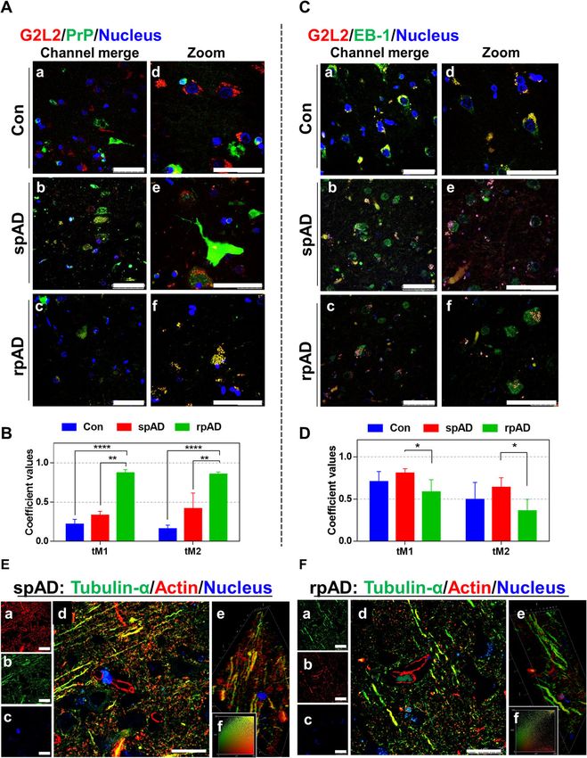
HDPs (Fig. 2, Table 1). Subtype-unique HDP interac- cytoskeletal integrity in the neurons
tors and the interactors commonly occurring in sam- Neuronal co-localization of G2L2 and HDP affects G2L2/EB-
ples from both spAD and sCJD subtypes are 1 binding
discussed in the supplementary data section (Suppl. Immunohistological observations were made in the grey
Tables 3–7). matter areas of the frontal cortex. A certain subtype-
specific trend was observed in G2L2/PrP co-localization.
The highest level of co-localization between PrPC and
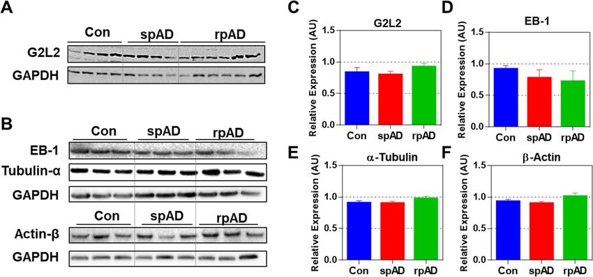
Expression of G2L2 and associated proteins G2L2 was observed in rpAD followed by spAD samples
Among the proteins identified as HDP interactors in (Fig. 4a). Threshold Mander’s overlap coefficients values
rpAD, G2L2 was selected for further functional verifi- have been used to assess the co-localization of red and
cations, given its essential role in cytoskeletal integ- green channels in the microscopic images. Where tM1
rity, i.e. in actin-mediated microtubule growth, and represents the overlaps of pixels from the red channel
the possible relevance of the latter for AD pathology. (G2L2) on those of the green channel (PrP), and tM2-
G2L2 is reported for its role in co-alignment of actin overlaps of pixels from the green channel (PrP) on those
filaments and microtubules, where it acts by cross- of the red channel (G2L2). Mander’s overlap coefficient
linking the two structures. This actin/microtubule values reveal the highest G2L2-PrPC co-localization in
cross-linking is assisted by the end-binding protein-1 rpAD followed by spAD and controls (Fig. 4b). However,
(EB-1) at the plus end of the microtubules [63–65]. a significant decrease in G2L2 and EB-1 co-localization
In order to study G2L2 in the sample cohorts in was observed in rpAD compared with spAD and con-
more detail, proteins functionally associated with the trols (Fig. 4c). The differences in the Mander’s overlap
physiology of G2L2, including EB-1, tubulin and actin, coefficient values (Fig. 4d) also represent the lowest
were also studied. Expression of these proteins in levels of co-localization in rpAD followed by a higher
frontal cortex homogenates was assessed by western overlap in spAD and the highest in the controls.
blot analysis. We found no significant differences in
the levels of G2L2 between the AD subtypes and con- Disturbance in G2L2/EB1 co-localization is associated with
trols. Likewise, no significant differences were de- loss of β-actin/α-tubulin integration
tected between the groups for EB-1, α-tubulin, and β- Actin-tubulin co-alignment was also studied using con-
actin (Fig. 3). focal laser scanning microscopy. Frontal cortex sections
Fig. 3 Expression of G2L2 and associated proteins in the frontal cortex of experimental cohorts. A & B) Immunoblots showing the expression
levels of G2L2, EB-1, α-tubulin, and β-actin. GAPDH served as loading control. C-F) Densitometric quantification for expression of G2L2, EB-1, α-
tubulin, and β-actin in spAD (n = 7), rpAD (n = 7), and control (n = 6) cases, as assessed in three technical replicates. No significant differences
were found for G2L2 and its associated proteins. Statistical significance was calculated with one-way ANOVA followed by Tukey’s post-hoc testShafiq et al. Molecular Neurodegeneration (2021) 16:11 Page 8 of 14 Fig. 4 Neuronal co-localization of G2L2 and HDP effects G2L2 / EB-1 binding and cytoskeletal integrity in the rpAD brain cortex. A) Representative photomicrographs (panels Aa-Af) from frontal cortex sections of rpAD, spAD and controls stained with anti-G2L2 and anti-PrP (SAF70) antibodies. Highest co-localization of G2L2 and PrP was observed in frontal cortex sections from rpAD, followed by spAD and controls. B) Threshold Mander’s coefficient values for the overlap of G2L2 channel pixels to PrP channel pixels (tM1) were significantly higher in rpAD than in spAD. The average tM2 (Mander’s coefficient for the overlap of PrP channel pixels to G2L2 channel pixels) was also significantly higher in rpAD than that of spAD. C) Representative frontal cortex sections of rpAD, spAD and controls stained for G2L2 and EB-1 (panels Ca-Cf) show lowest G2L2/EB-1 co-localization in rpAD frontal cortex sections followed by those of spAD and control, respectively. D) Significantly decreased tM1 (G2L2) and tM2 (EB-1) values were seen in rpAD compared with spAD and Con sections. Statistical significance was calculated with one-way ANOVA followed by Tukey’s post-hoc test. *p < 0.05; **p < 0.005; ***p < 0.001. E&F) Representative micrographs of spAD (E) and rpAD (F) sections stained using anti-α-tubulin and anti-β-actin antibodies are shown. Panels Ea-Ec and Fa-Fc show the single channel images. Panels Ed and Fd show the channel merges. Panels Ee and Fe correspond to the 3D reconstructions from z-stacks of spAD and rpAD, respectively. Insets Ef and Ff show representative IC plots. Relatively stronger actin/tubulin co-localization was observed in spAD compared with rpAD patients. Confocal images were scanned from the frontal cortex sections of spAD (n = 5), rpAD (n = 5) and controls (n = 5). Scale bars in panels A&C = 50 μm; in panels E&F = 25 μm (5 μm thick) were stained for α-tubulin and β-actin. actin-tubulin co-localization was observed in the axonal Confocal z-sections were scanned and later used to con- processes of gray matter neurons of the spAD samples struct three-dimensional images. A more pronounced in comparison with rpAD (Fig. 4e, panel d and Fig. 4f,
Shafiq et al. Molecular Neurodegeneration   (2021) 16:11                                                     Page 9 of 14

panel d). Likewise, correlation plots prepared for the          Diverse sets of prion protein interactors were identi-
actin and tubulin channels revealed a significantly          fied from subtype-specific HDFs. In our study, we identi-
greater overlap between the channel intensities (Fig. 4e,    fied fewer rpAD-specific HDP interactors compared
panel f and Fig. 4f, panel f). Three-dimensional recon-      with sCJD. As reported previously, sCJD tissues exhibit a
structions of the z-sections also showed longer stretches    wider spectrum of PrP/PrPSc oligomers [66, 68, 69] com-
of filaments with actin-tubulin co-localization in spAD      pared with rpAD [8]. A greater diversity of PrP/PrPSc
compared with rpAD, with higher actin and tubulin            oligomers in sCJD pathology can be a potential reason
channel overlap (Fig. 4e, panel e and Fig. 4f, panel e).     for the relatively diverse interactome found for sCJD PrP
                                                             oligomers. There was no overlap of HDP-interactors of
Proteomics of HDFs also indicate higher degree of            rpAD samples with either spAD or control patient tis-
cytoskeletal disintegration in rpAD                          sue, reaffirming the subtype-specific occurrence of cer-
MS/MS analysis of HDF pools was carried out and the          tain PrP species in rpAD. Interactors common for the
observed proteomic variations (unique peptide counts)        rpAD and sCJD datasets point towards the presence of
were compared with the baseline global proteomic data        common PrP oligomers present in both disease entities.
sets obtained by SWATH-MS to recognize variations in         Proteins interacting with the HDPs in rpAD included
cytoskeletal proteins. Levels of tubulin subunits were ob-   CaMKII subunits (KCC2B, KCC2D), EPDR1, DIRA2,
served to be significantly reduced in HDFs from rpAD         G2L2, and 1433S. All of these interactors, except for the
patients compared with HDFs from other groups. A sig-        G2L2 protein, were part of the HDP-interactomes in
nificant decrease was also seen in the levels of certain     both rpAD and sCJD. CaMKII, besides regulating Ca2+
neurofilament subunits, actin and actin-binding proteins     in neurons, is also responsible for reorganizing actin into
in rpAD HDFs. Conversely, significantly higher levels of     bundles in the development of dendritic spines [70].
microtubule-associated proteins (MAP 1A, MAP 1B, and         GTP-binding Di-Ras2 is a Ras GTPase known for its in-
MAP 2), flaming (FLNA), filaggrin (FLG) and plectin          volvement in determining cell morphogenesis. G2L2 has
(PLEC) were observed in rpAD HDFs compared with              been reported to play a role in actin-tubulin communi-
sCJD-specific HDFs. Interestingly, the levels of the         cation and connectivity [71]. Protein 14–3-3 sigma is a
microtubule-associated protein tau (MAPT) were found         protein with many known interactions and is involved in
to be lower in rpAD-specific HDFs compared with              p53, protein kinase C, and AKT/mTOR signaling [72].
spAD-specific HDFs, although higher than the HDFs            Strong evidence had been presented in the literature for
from Con and sCJD subtypes (Fig. 5a, Additional file 4).     the involvement of 14–3-3 isoforms in various neurode-
Among the cytoskeletal proteins from the global prote-       generative disease, particularly in prion dementias and
ome data, the expression of actin G (ACTG) was found         Alzheimer’s disease, where 14–3-3 isoforms are found
to be significantly increased in rpAD compared with          associated with PrPSc and Aβ plaques and serve as bio-
controls, whereas no significant differences were ob-        marker [73, 74]. Physiological outcomes of novel interac-
served between the spAD and rpAD. Expression of              tions of G2L2 with the PrP oligomers are discussed in
ACTG was significantly decreased in SVD and DFTL             the following. Actin and tubulin interlinking and com-
compared with rpAD. Contactin 1 (CNTN1) expression           munication are necessary for the proper functioning of
was also found to be significantly higher in SVD and         cellular transport systems, morphogenesis, repair and
DFTL than in rpAD. Expression of tubulin alpha 1a            many other related functions in the cells. This link is
(TBA1A) was observed to be significantly higher in           mainly controlled by spectraplakins via their ability to
rpAD than in other rapid dementia samples, i.e. DFTL,        connect actin and tubulin filaments [40]. Likewise, the
SVD, and r-DLB. Likewise, expression of TBA4A was            family of growth arrest-specific (GAS) proteins and
differentially higher in rpAD compared with controls,        GAS-like (GASL) proteins plays a role in linking actin
DLB, DFTL, and SVD (Fig. 5b, Additional file 3).             and tubulin filaments [71]. Mutations in the G2L2 pro-
                                                             tein were reported to increase the risk of developing
Discussion                                                   Alzheimer’s disease [75]. Stroud and coworkers [63]
Multiple PrP conformers have previously been described       have provided a comprehensive experimental account of
in association with transmissible spongiform prion dis-      the GAS and GASL and proposed a model in which the
eases in animals and humans [66, 67]. The presence of        GASL proteins G2L1 and G2L2 control microtubule sta-
high-density PrP conformers (HDPs) associated specific-      bility via attachment to end-binding protein-1 (EB-1).
ally with rpAD have recently been identified by our          The interaction of G2L2 and EB-1 with tubulin is also
group [8]. Here we aimed to deepen our understanding         reported to account for microtubular stability [63]. In
of these findings by isolating HDP complexes, identifying    our study, we found a negative correlation between
HDP interactors, and by downstream proteomic                 HDP-G2L2 interaction and G2L2 and EB-1 binding in
analyses.                                                    the rpAD cohort. This disturbance in the G2L2/EB-1
Shafiq et al. Molecular Neurodegeneration (2021) 16:11 Page 10 of 14 Fig. 5 Variations in cytoskeletal proteins in high-density fractions. A) The relative abundance of cytoskeletal proteins in HDFs detected by high- resolution MS/MS analysis is represented as z-score. Significant reduction of cytoskeletal proteins was seen in rpAD-specific HDFs (F12–F17: HDF pool 12 to 17). B) Differences in expression of cytoskeletal and associative proteins as determined by the SWATH-MS. Heatmaps represent the relative protein expression indicated as z-scores for spAD (n = 3), rpAD (n = 3), DLB (n = 3), rDLB (n = 2), D-FTL (n = 3), SVD (n = 3), and controls (n = 3). C) Model showing the recruitment of G2L2 to high-density prion-oligomers. The recruitment of G2L2 towards HDPs results in the loss of its binding to EB-1, affecting the actin-guided microtubule (MT) integrity system may lead to a malfunction of actin-assisted interpreted as a consequence of the disturbances in the microtubule growth in neurons. The shortening of the G2L2/EB-1/tubulin system. Figure 5c illustrates a pos- actin-tubulin co-localization fibers that is specifically ob- sible mechanism of how HDPs interact with the cyto- served in the frontal cortex of rpAD patients can be skeletal system. Proteomic data of HDFs also show more
Shafiq et al. Molecular Neurodegeneration   (2021) 16:11                                                                     Page 11 of 14

extensive cytoskeletal disintegration in rpAD than in         found in the sCJD high-density fractions in our study
spAD and control patients. Decreased levels of tubulin        are correlated to the relatively higher levels of HDPs
in rpAD HDFs are indicative of greater damage to the          with which they interact.
cytoskeletal system, fitting to what has previously been
described [76]. The decrease in tubulin integrity is a sign   Conclusion
of various pathological cascades associated with rpAD         The results suggest an involvement of high-density PrP
that differ from those associated to spAD. PrPC was re-       oligomers in the cytoskeletal damage of the frontal cortex
ported to inhibit microtubule synthesis by its direct         specific to rpAD, as indicated by confocal microscopy and
interaction with tubulin [53, 54]. The PrP oligomers          proteomic profiling. The proposed competitive binding of
uniquely identified in rpAD may be associated with            HDPs to G2L2 resulted in the interruption of G2L2/EB-1/
tubulin-sequestering that results in a greater degree of      Tubulin interaction, which lead to a greater extent of dis-
microtubule damage. Low tubulin HDF levels in rpAD            integration and damage to the cytoskeletal system. These
are accompanied by a decrease in the MAPT levels as           rpAD-specific cytoskeletal alterations can contribute in
well as higher levels of MAP 1 and MAP 2. A previous          the accelerated disease progression of rpAD patients.
report by Ba et al. (2016), with rapid progression defined
on the basis of cognitive decline compared to that of         Supplementary Information
survival time in our study, showed a significant de-          The online version contains supplementary material available at https://doi.
crease in the p-tau/tau ratio in cerebrospinal fluid          org/10.1186/s13024-021-00422-x.
(CSF) of rpAD patients (n = 55) compared with that
                                                               Additional file 1 The document file contains supplementary figures and
in spAD patients (n = 257) [17]. As the p-tau mole-
                                                               tables. Suppl. Fig. 1: Summary of frontal cortex cohorts used in current
cules change their conformation and undergo                    study. Further clinical features and neuropathological details of the
oligomerization, ultimately leading to the formation of        cohort are given in the Additional file 2. Suppl. Fig. 2 Sample cohorts
                                                               used in the study. A) Comparison of ages of the diverse pathological
tau tangles [77], the aforementioned discrepancy in
                                                               cohorts used in the study. B) Graph presents a comparison of post-
CSF p-tau/tau levels in rpAD patients indicates that           mortem intervals to the time of autopsies. Suppl. Table 1: List of primary
the rpAD cohort will show a decrease in MAPT olig-             antibodies and their applications in the current study. Suppl. Table 2: List
                                                               of secondary antibodies and their applications in current study. Suppl.
omers. The cortical expression of MAP 1 and MAP 2
                                                               Table 3: High-density PrP (HDP) interactors commonly found in the HDFs
however does not differ between rpAD and spAD as               of all sCJD subtypes. Suppl. Table 4: High-density PrP (HDP) interactors
seen in the global proteomic data. Elevated HDF                commonly detected between the high-density fractions of sCJD-MM2
                                                               and sCJD-VV2 subtypes.
MAPs levels can result from their self-interaction [78]
                                                               Additional file 2. The spreadsheet contains the details of the sample
or from a stronger interaction with tubulin, actin and
                                                               cohort used in the study including the clinical data, the
other regulatory factors, such as kinases, as previously       neuropathological profiling, and the experiment designing.
reported [78–83]. MAP sequestering also correlates             Additional file 3. The spreadsheet contains the differential expression
with the loss of microtubule integrity in the rpAD             of cytoskeletal and cytoskeletal-associated proteins from the frontal cor-
                                                               tex tissue lysates of Con, spAD, rpAD, SVD, DFTL, rDLB and DLB patients
cortices. Together with the malfunctioning in the
                                                               assessed using the SWATH-MS. Data shown are the normalized SWATH
microtubule system, neurons suffer from a variety of           averages and the corresponding SEM values. The spreadsheet also con-
other degenerative events. Considering the size and            tains the results of the post-hoc intergroup analyses for the differential
                                                               expression SWATH values of these proteins.
complexity of a neuron, a robust intracellular trans-
                                                               Additional file 4. The spreadsheet gives the average peptide counts
port system is obviously necessary to maintain the
                                                               and SEM of the cytoskeletal and cytoskeletal-associated proteins from the
critical connections between the cell body and its dis-        HDF pools from frontal cortex lysates of control brains and those other
tant neural processes for supplying the various types          neurodegenerative samples described in the study. It also contains the
                                                               results of the post-hoc intergroup analyses of these peptide counts.
of neuronal cargo, such as organelles, vesicles, cell
                                                               Additional file 5. The spreadsheet contains the raw global proteomics
signaling molecules, RNA molecules, neurotransmit-
                                                               data (SWATH-MS values) of Con, spAD, rpAD, DLB, rDLB, DFTL, and SVD.
ters, receptors, and adhesion molecules [36, 37]. The          Subsequent normalized data along with the results of pairwise statistical
actin-guided microtubule growth also plays a critical          testing (Welch’s t-test).
role in the structural stability of neurons including          Additional file 6. The spreadsheet contains the raw proteomics data
                                                               (unique peptide counts data) of HDFs, and subsequent normalized data
kinesin-based axon differentiation and polarization
                                                               along with the results of pairwise statistical testing (Welch’s t-test).
[37, 84], MAP-assisted axon growth [85], and finally
                                                               Additional file 7. The additional file contains spreadsheets with
the morphodynamics of dendritic spines [36, 37]. The           averaged normalized mass spectrometric data after removal of
higher levels of the cytoskeletal proteins in sCJD             candidates found in beads only controls (unique peptide counts), for the
                                                               interactors of high-density prion protein oligomers in cerebral cortices
HDFs indicate that a different set of mechanisms is
                                                               from controls and neurodegenerative cases included in the study.
involved in CJD pathology. Interaction between tubu-
lin and PrP has been reported by many previous stud-
                                                              Abbreviations
ies [53, 54]. We argue that the higher levels of              ACN: acetonitrile; AD: Alzheimer’s disease; ADAM: A disintegrin- and
cytoskeletal proteins (tubulin isoforms and MAPs)             metalloproteinase; AgNO3: silver nitrate; AKT/PKB: protein kinase B;
Shafiq et al. Molecular Neurodegeneration (2021) 16:11 Page 12 of 14
APOE: apolipoprotein E; APP: amyloid precursor protein; Aβ: amyloid-β; Competing interests
CaMKII: calcium/calmodulin-dependent protein kinase type II; cAMP: cyclic The authors declare that they have no competing interests.
adenosine monophosphate; Co-IP: co-immunoprecipitation;
CSF: cerebrospinal fluid; ddH2O: double distilled water; DFTL: dementia with Author details
1
fronto-temporal lobar degeneration; DLB: dementia with Lewy bodies; Department of Neurology, University Medicine Goettingen and German
DSG2: desmoglein 2; DTT: dithiothreitol; EDTA: ethylenediaminetetraacetic Center for Neurodegenerative Diseases (DZNE), 37075 Goettingen, Germany.
2
acid; Erk: extracellular signal–regulated kinases; ESI: electrospray ionization; Institute of Neuropathology, University Medical Center Hamburg-Eppendorf
FAD: familial AD; G-CSF: granulocyte-colony stimulating factor; (UKE), 20246 Hamburg, Germany. 3Biomedical Engineering and Sciences
GSK3: glycogen synthase kinase 3-β; HDFs: high density fractions; HDP: high Department, School of Mechanical and Manufacturing Engineering (SMME),
density prions; HMW: high molecular weight; IAA: iodoacetamide; National University of Sciences and Technology (NUST), Islamabad, Pakistan.
4
IgG: immunoglobulin G; IL-13: interleukin 13; IL-6: interleukin 6; JNK: c-jun N- Department of Neurology, Experimental Research in Stroke and
terminal kinases; KCC2: calcium/calmodulin-dependent protein kinase type II; Inflammation (ERSI), University Medical Center Hamburg-Eppendorf, 20246
kDa: kilodalton; LMW: low molecular weight; LTP: long-term potentiation; Hamburg, Germany. 5Institut de Neuropatologica, Servei Anatomia
MAPKs: mitogen-activated protein kinases; MCI: mild Cognitive Impairment; Patològica, IDIBELL-Hospital Universitari de Bellvitge, Universitat de Barcelona,
MCP-1: monocyte chemoattractant protein-1; MMSE: Mini-Mental State Carrer Feixa LLarga sn, 08907, Hospitalet de LLobregat, CIBERNED, Barcelona,
Examination; MS/MS: mass spectrometery; MYLK: myosin light chain kinase; Spain.
Na2S2O3: sodium thiosulfate; NaCl: sodium chloride; NFkB: nuclear factor
kappa-light-chain-enhancer of activated B cells; NFTs: neurofibrillary tangles; Received: 5 April 2020 Accepted: 2 January 2021
NH4HCO3: sodium bicarbonate; NP-40: nonidet P-40; NCAM: neuronal cell
adhesion molecule; p38: mitogen-activated protein kinases P38;
PBS: phosphate buffered saline; PKA: protein kinase A; PKC: protein kinase C;
PLD3: phospholipase D3; PRNP: prion protein coding gene; PrP: prion References
protein; PrPC: cellular prion protein; PrPSc: scrapie form of prion protein; 1. Vos T, Allen C, Arora M, Barber RM, Bhutta ZA, Brown A, et al. Global,
PSEN1: presenilin 1; PSEN2: presenilin 2; p-tau: phosphorylated tau; Q- regional, and national incidence, prevalence, and years lived with disability
TOF: quadrupole time of flight; rDLB: rapidly progressive dementia with Lewy for 310 diseases and injuries, 1990–2015: a systematic analysis for the global
bodies; recPrP: recombinant PrP; RFU: relative fluorescence units; rP: Pearson’s burden of disease study 2015. Lancet. 2016;388:1545–602.
linear correlation coefficient; rpAD: rapidly progressive Alzheimer’s disease; 2. Querfurth HW, LaFerla FM. Alzheimer’s Disease. N Engl J Med. 2010;362:
SAPK: stress-activated phosphokinases; sAPPα: shed-APP; sCJD: sporadic 329–44.
Creutzfeldt-Jakob disease; SDS: sodium dodecyl sulfate; SDS-PAGE: sodium 3. Mann UM, Mohr E, Chase TN. Rapidly progressive Alzheimer’s disease.
dodecyl sulfate- polyacrylamide gel electrophoresis; spAD: sporadic Lancet. 1989;2:799.
Alzheimer’s disease; SRC: proto-oncogene tyrosine-protein kinase; SVD: small 4. Reinwald S, Westner IM, Niedermaier N. Rapidly progressive Alzheimer’s
vessel disease; SWATH-MS: sequential window acquisition of all theoretical disease mimicking Creutzfeldt Jakob disease. J Neurol. 2004;251:1020–2.
mass spectra- mass spectrometry; Tau: tubulin associated unit; TBS: Tris 5. Pillai JA, Appleby BS, Safar J, Leverenz JB. Rapidly progressive Alzheimer’s
buffered saline; TBST: TBS with 0.1% Tween; TEME disease in two distinct autopsy cohorts. J Alzheimers Dis. 2018;64:973–80.
D: tetramethylethylenediamine; Th-T: thioflavin-T; tM: threshold Mander’s 6. Josephs KA, Ahlskog JE, Parisi JE, Boeve BF, Crum BA, Giannini C, et al.
coefficient; TNF: tumor necrosis factor; TREM2: triggering receptor expressed Rapidly progressive neurodegenerative dementias. Arch Neurol. 2009;66:
on myeloid cells 2; Tris: tris (hydroxymethyl)aminomethane; 201–7.
TSEs: transmissible spongiform encephalopathies 7. Schmidt C, Wolff M, Weitz M, Bartlau T, Korth C, Zerr I. Rapidly progressive
Alzheimer disease. Arch Neurol. 2011;68:1124–30.
8. Zafar S, Shafiq M, Younas N, Schmitz M, Ferrer I, Zerr I. Prion protein
Acknowledgements interactome: identifying novel targets in slowly and rapidly progressive
Not applicable. forms of Alzheimer’s disease. J Alzheimers Dis. 2017;59:265–75.
9. Younas N, Zafar S, Shafiq M, Noor A, Siegert A, Arora AS, et al. SFPQ and
Authors’ contributions tau: critical factors contributing to rapid progression of Alzheimer’s disease.
Conceptualization and design of the study: MS, SZ and IZ; Methodology and Acta Neuropathol. 2020;140:317–39.
investigation: MS, SZ, NY, AN, BP, HCA, and MS; Providing important research 10. Grau-Rivera O, Gelpi E, Nos C, Gaig C, Ferrer I, Saiz A, et al.
resources, materials and scientific input: JM, IF, MG, and IZ; Writing the Clinicopathological correlations and concomitant pathologies in rapidly
original manuscript draft: MS, SZ and IZ; Review and Editing: all authors; progressive dementia: a brain Bank series. Neurodegener Dis. 2015;15:
Supervision: SZ and IZ. All authors read and approved the final manuscript. 350–60.
11. Schmidt C, Redyk K, Meissner B, Krack L, von Ahsen N, Roeber S, et al.
Clinical features of rapidly progressive Alzheimer’s disease. Dement Geriatr
Funding Cogn Disord. 2010;29:371–8.
This work was supported by the funding from the Bundesministerium für 12. Schmidt C, Haïk S, Satoh K, Rábano A, Martinez-Martin P, Roeber S, et al.
Bildung und Forschung within the German Network for Degenerative Rapidly progressive Alzheimer’s disease: a multicenter update. J Alzheimers
Dementia (KNDD-2, 2012–2015, determinants for disease progression in AD, Dis. 2012;30:751–6.
grant No.01GI1010C). arroyo572 from DZNE and Helmholtz-Alberta Initiative: 13. Abu Rumeileh S, Lattanzio F, Stanzani Maserati M, Rizzi R, Capellari S, Parchi
HAI SO-083. Helmholtz-Alberta Initiative- Neurodegenerative Disease Re- P. Diagnostic accuracy of a combined analysis of cerebrospinal fluid t-PrP, t-
search (HAI-NDR) grant No. 1.4.2014–30.4.2015. tau, p-tau, and Aβ42 in the differential diagnosis of Creutzfeldt-Jakob
disease from Alzheimer’s disease with emphasis on atypical disease variants.
Availability of data and materials J Alzheimers Dis. 2017;55:1471–80.
All data generated or analyzed during this study are included in this 14. Abu-Rumeileh S, Capellari S, Parchi P. Rapidly progressive Alzheimer’s
published article [and its supplementary information files]. disease: contributions to clinical-pathological definition and diagnosis. J
Alzheimers Dis. 2018;63:887–97.
15. Llorens F, Kruse N, Schmitz M, Gotzmann N, Golanska E, Thüne K, et al.
Ethics approval and consent to participate
Evaluation of α-synuclein as a novel cerebrospinal fluid biomarker in
Patient material was obtained after the approval of local ethics committee of
different forms of prion diseases. Alzheimers Dement. 2017;13:710–9.
the University Medical Center, Göttingen; and according to the legislation of
16. Llorens F, Schmitz M, Karch A, Cramm M, Lange P, Gherib K, et al.
the Spanish authorities (Ley de la Investigación Biomédica 2013 and Real
Comparative analysis of cerebrospinal fluid biomarkers in the differential
DecretoBiobancos, 2014).
diagnosis of neurodegenerative dementia. Alzheimers Dement. 2016;12:
577–89.
Consent for publication 17. Ba M, Li X, Ng KP, Pascoal TA, Mathotaarachchi S, Rosa-Neto P, et al. The
Not applicable. prevalence and biomarkers’ characteristic of rapidly progressive Alzheimer’sShafiq et al. Molecular Neurodegeneration (2021) 16:11 Page 13 of 14
disease from the Alzheimer’s Disease Neuroimaging Initiative database. 45. Rush T, Martinez-Hernandez J, Dollmeyer M, Frandemiche ML, Borel E,
Alzheimer’s Dement Transl Res Clin Interv. 2017;3:107–13. Boisseau S, et al. Synaptotoxicity in alzheimer’s disease involved a
18. Stoeck K, Schmitz M, Ebert E, Schmidt C, Zerr I. Immune responses in rapidly dysregulation of actin cytoskeleton dynamics through cofilin 1
progressive dementia: a comparative study of neuroinflammatory markers phosphorylation. J Neurosci. 2018;38:10349–61.
in Creutzfeldt-Jakob disease. Alzheimer’s disease and multiple sclerosis J 46. Pelucchi S, Stringhi R, Marcello E. Dendritic spines in Alzheimer’s disease:
Neuroinflammation. 2014;11:170. how the actin cytoskeleton contributes to synaptic failure. Int J Mol Sci.
19. Cohen ML, Kim C, Haldiman T, ElHag M, Mehndiratta P, Pichet T, et al. 2020;21:908.
Rapidly progressive Alzheimer’s disease features distinct structures of 47. Merriam EB, Millette M, Lumbard DC, Saengsawang W, Fothergill T, Hu X,
amyloid-β. Brain. 2015;138:1009–22. et al. Synaptic regulation of microtubule dynamics in dendritic spines by
20. Qiang W, Yau WM, Lu JX, Collinge J, Tycko R. Structural variation in amyloid- calcium, F-actin, and Drebrin. J Neurosci. 2013;33:16471–82.
β fibrils from Alzheimer’s disease clinical subtypes. Nature. 2017;541:217–21. 48. Penazzi L, Tackenberg C, Ghori A, Golovyashkina N, Niewidok B, Selle K,
21. Linden R, Martins VR, Prado MAM, Cammarota M, Izquierdo I, Brentani RR. et al. Aβ-mediated spine changes in the hippocampus are microtubule-
Physiology of the prion protein. Physiol Rev. 2008;88:673–728. dependent and can be reversed by a subnanomolar concentration of the
22. Cooper DMF, Crossthwaite AJ. Higher-order organization and regulation of microtubule-stabilizing agent epothilone D. Neuropharmacology. 2016;105:
adenylyl cyclases. Trends Pharmacol Sci. 2006;27:426–31. 84–95.
23. He Q, Meiri KF. Isolation and characterization of detergent-resistant 49. Rodríguez-Martín T, Cuchillo-Ibáñez I, Noble W, Nyenya F, Anderton BH,
microdomains responsive to NCAM-mediated signaling from growth cones. Hanger DP. Tau phosphorylation affects its axonal transport and
Mol Cell Neurosci. 2002;19:18–31. degradation. Neurobiol Aging. 2013;34:2146–57.
24. Linsenmeier L, Altmeppen HC, Wetzel S, Mohammadi B, Saftig P, Glatzel M. 50. Drummond E, Nayak S, Faustin A, Pires G, Hickman RA, Askenazi M, et al.
Diverse functions of the prion protein – does proteolytic processing hold Proteomic differences in amyloid plaques in rapidly progressive and
the key? Biochim Biophys Acta Mol Cell Res. 1864;2017:2128–37. sporadic Alzheimer’s disease. Acta Neuropathol. 2017;133:933–54.
25. Falker C, Hartmann A, Guett I, Dohler F, Altmeppen H, Betzel C, et al. 51. Zafar S, Younas N, Sheikh N, Tahir W, Shafiq M, Schmitz M, et al.
Exosomal cellular prion protein drives fibrillization of amyloid beta and Cytoskeleton-associated risk modifiers involved in early and rapid
counteracts amyloid beta-mediated neurotoxicity. J Neurochem. 2016;137: progression of sporadic Creutzfeldt-Jakob disease. Mol Neurobiol. 2017;55:
88–100. 4009–29.
26. Kayed R, Head E, Thompson JL, McIntire TM, Milton SC, Cotman CW, et al. 52. Zafar S, Asif AR, Ramljak S, Tahir W, Schmitz M, Zerr I. Anchorless 23–230
Common structure of soluble amyloid oligomers implies common PrPC interactomics for elucidation of PrPC protective role. Mol Neurobiol.
mechanism of pathogenesis. Science. 2003;300:486–9. 2014;49:1385–99.
27. Tanzi RE, Bertram L. Twenty years of the Alzheimer’s disease amyloid 53. Nieznanski K, Nieznanska H, Skowronek KJ, Osiecka KM, Stepkowski D. Direct
hypothesis: a genetic perspective. Cell. 2005;120:454–5. interaction between prion protein and tubulin. Biochem Biophys Res
28. Scheff SW, Price DA, Schmitt FA, Dekosky ST, Mufson EJ. Synaptic alterations Commun. 2005;334:403–11.
in CA1 in mild Alzheimer disease and mild cognitive impairment. 54. Nieznanski K, Podlubnaya ZA, Nieznanska H. Prion protein inhibits
Neurology. 2007;68:1501–8. microtubule assembly by inducing tubulin oligomerization. Biochem
29. DeKosky ST, Scheff SW. Synapse loss in frontal cortex biopsies in Alzheimer’s Biophys Res Commun. 2006;349:391–9.
disease: correlation with cognitive severity. Ann Neurol. 1990;27:457–64. 55. Osiecka KM, Nieznanska H, Skowronek KJ, Karolczak J, Schneider G,
30. Masliah E, Mallory M, Alford M, DeTeresa R, Hansen LA, McKeel DW, et al. Nieznanski K. Prion protein region 23-32 interacts with tubulin and inhibits
Altered expression of synaptic proteins occurs early during progression of microtubule assembly. Proteins Struct Funct Bioinforma. 2009;77:279–96.
Alzheimer’s disease. Neurology. 2001;56:127–9. 56. Tahir W, Zafar S, Llorens F, Arora AS, Thüne K, Schmitz M, et al. Molecular
31. Stokin GB. Axonopathy and transport deficits early in the pathogenesis of alterations in the cerebellum of sporadic creutzfeldt–jakob disease subtypes
Alzheimer’s disease. Science. 2005;307:1282–8. with DJ-1 as a key regulator of oxidative stress. Mol Neurobiol. 2018;55:517–37.
32. Eira J, Silva CS, Sousa MM, Liz MA. The cytoskeleton as a novel therapeutic 57. Losensky G, Jung K, Urlaub H, Pfeifer F, Fröls S, Lenz C. Shedding light on
target for old neurodegenerative disorders. Prog Neurobiol. 2016;141:61–82. biofilm formation of Halobacterium salinarum R1 by SWATH-LC/MS/MS
33. Subramanian R, Kapoor TM. Building complexity: insights into self-organized analysis of planktonic and sessile cells. Proteomics. 2017;17:1600111.
assembly of microtubule-based architectures. Dev Cell. 58. Krasemann S, Madore C, Cialic R, Baufeld C, Calcagno N, El Fatimy R, et al.
2012;23:874–85. The TREM2-APOE pathway drives the transcriptional phenotype of
34. Akhmanova A, Steinmetz MO. Tracking the ends: a dynamic protein dysfunctional microglia in neurodegenerative diseases. Immunity. 2017;47:
network controls the fate of microtubule tips. Nat Rev Mol Cell Biol. 2008;9: 566–81.
309–22. 59. Bubber P, Haroutunian V, Fisch G, Blass JP, Gibson GE. Mitochondrial
35. De Forges H, Bouissou A, Perez F. Interplay between microtubule dynamics abnormalities in Alzheimer brain: mechanistic implications. Ann Neurol.
and intracellular organization. Int J Biochem Cell Biol. 2012;44:266–74. 2005;57:695–703.
36. Maday S, Twelvetrees AE, Moughamian AJ, Holzbaur ELF. Axonal 60. Yacoubian TA, Slone SR, Harrington AJ, Hamamichi S, Schieltz JM, Caldwell
transport: cargo-specific mechanisms of motility and regulation. Neuron. KA, et al. Differential neuroprotective effects of 14-3-3 proteins in models of
2014;84:292–309. Parkinson’s disease. Cell Death Dis. 2010;1:e2.
37. Hirokawa N, Niwa S, Tanaka Y. Molecular motors in neurons: transport 61. Llorens F, Schmitz M, Knipper T, Schmidt C, Lange P, Fischer A, et al.
mechanisms and roles in brain function, development, and disease. Neuron. Cerebrospinal fluid biomarkers of Alzheimer’s disease show different but
2010;68:610–38. partially overlapping profile compared to vascular dementia. Front Aging
38. Vicario-Orri E, Opazo CM, Muñoz FJ. The pathophysiology of axonal Neurosci. 2017;9:289.
transport in Alzheimer’s disease. J Alzheimers Dis. 2014;43:1097–113. 62. Schindler CK, Heverin M, Henshall DC. Isoform- and subcellular fraction-
39. Hammond JW, Huang CF, Kaech S, Jacobson C, Banker G, Verhey KJ. specific differences in hippocampal 14-3-3 levels following experimentally
Posttranslational modifications of tubulin and the polarized transport of evoked seizures and in human temporal lobe epilepsy. J Neurochem. 2006;
kinesin-1 in neurons. Mol Biol Cell. 2010;21:572–83. 99:561–9.
40. Suozzi KC, Wu X, Fuchs E. Spectraplakins: master orchestrators of 63. Stroud MJ, Nazgiewicz A, McKenzie EA, Wang Y, Kammerer RA, Ballestrem C.
cytoskeletal dynamics. J Cell Biol. 2012;197:465–75. GAS2-like proteins mediate communication between microtubules and
41. Wiche G. Role of plectin in cytoskeleton organization and dynamics. J Cell actin through interactions with end-binding proteins. J Cell Sci. 2014;127:
Sci. 1998;111:2477–86. 2672–82.
42. Naumanen P, Lappalainen P, Hotulainen P. Mechanisms of actin stress fibre 64. Alberico EO, Zhu ZC, Wu YFO, Gardner MK, Kovar DR, Goodson HV.
assembly. J Microsc. 2008;231:446–54. Interactions between the microtubule binding protein EB1 and F-actin. J
43. Pollard TD, Borisy GG. Cellular motility driven by assembly and disassembly Mol Biol. 2016;428:1304–14.
of actin filaments. Cell. 2003;112:453–65. 65. Bouguenina H, Salaun D, Mangon A, Muller L, Baudelet E, Camoin L, et al.
44. Fletcher DA, Mullins RD. Cell mechanics and the cytoskeleton. Nature. 2010; EB1-binding–myomegalin protein complex promotes centrosomal
463:485–92. microtubules functions. Proc Natl Acad Sci U S A. 2017;114:E10687–96.You can also read