STRUCTURES OF CYANOBACTERIAL BICARBONATE TRANSPORTER SBTA AND ITS COMPLEX WITH PII-LIKE SBTB
←
→
Page content transcription
If your browser does not render page correctly, please read the page content below
Liu et al. Cell Discovery (2021)7:63
https://doi.org/10.1038/s41421-021-00287-w
Cell Discovery
www.nature.com/celldisc
CORRESPONDENCE Open Access
Structures of cyanobacterial bicarbonate
transporter SbtA and its complex with PII-like SbtB
Xiao-Yu Liu1, Wen-Tao Hou1, Liang Wang1, Bo Li1, Yu Chen1, Yuxing Chen1, Yong-Liang Jiang1 and
Cong-Zhao Zhou 1
Dear Editor, conditions, thus functioning as a Ci sensor in
Carbon and nitrogen, the uptake and intracellular cyanobacteria6.
metabolisms of which are tightly coupled1, are the two Here we purified Synechocystis sp. PCC 6803 SbtA and
most fundamental nutrients for all living organisms. As solved its cryo-EM structure at 3.50 Å resolution (Sup-
one of the most ancient autotrophic bacteria, cyano- plementary Fig. S1). SbtA adopts a trimeric structure,
bacteria utilize photosynthesis to convert the inorganic each subunit of which consists of two inverted structural
carbon (Ci) into carbohydrates. Carbon fixation is cata- repeats, namely TM1–5 and TM6–10 (Fig. 1a), which are
lyzed by ribulose-1,5-bisphosphate carboxylase/oxygenase similar to each other in topology but have antiparallel
(RuBisCO), which is a naturally inefficient enzyme2. In orientations within the membrane (Supplementary Fig. S2).
response to gradually decreased CO2 and elevated O2 The two structural repeats possess a root-mean-square
levels in the atmosphere, cyanobacteria have evolved a deviation (RMSD) of 2.63 Å over 135 Cα atoms. At the 3D
unique CO2-concentrating mechanism (CCM), which can level, all 10 TMs of each SbtA subunit are folded into two
substantially accumulate CO2 in the vicinity of RuBisCO domains, a core domain of six TMs (TM3–5 and
1234567890():,;
1234567890():,;
1234567890():,;
1234567890():,;
for improved carboxylation efficiency3. The cyanobacter- TM8–10) and a gate domain of four TMs (TM1–2 and
ial CCM consists of a subcellular self-assembled icosa- TM6–7) (Fig. 1a; Supplementary Fig. S3). The two
hedral microcompartment, termed the carboxysome, and domains have a buried interface of ~2000 Å2, which is
several Ci uptake systems3. To date, five Ci-uptake sys- mainly mediated by hydrophobic interactions between
tems have been identified in cyanobacteria, including residues from TM4–5 to TM9–10 of the core domain and
three bicarbonate transporters BicA, SbtA, and BCT1, in the four TMs of the gate domain (Fig. 1a). Notably, the
addition to two CO2-uptake complexes: NDH-I3 and TM4 and TM9 helices in the core domain are unfolded
NDH-I4. and form a crossover at the middle. In the gate domain,
SbtA, which is ubiquitous in cyanobacteria, is an indu- TM1 and TM6 are divided into two helical moieties,
cible high-affinity sodium-dependent HCO3– symporter forming a kink at the middle (Fig. 1a; Supplementary Fig.
within the TC.2.A.83 family of Na+/solute symporters4,5. S3). In fact, this feature of discontinuous transmembrane
At a low level of intracellular Ci, SbtA of an up-regulated helices is common in previously reported secondary active
level can recruit its partner protein SbtB to the mem- transporters7. Structural analysis showed that SbtA of a
brane6. The sbtB gene from the sbtA–sbtB operon 5 + 5 fold is topologically similar to Neisseria meningitides
encodes a PII-like signaling protein that senses the ASBT8. However, SbtA possesses distinct TM con-
intracellular level of the secondary messenger cAMP that formations from ASBT, especially those at the gate
correlates with high Ci conditions, versus AMP at low Ci domain, with an RMSD of 4.4 Å over 249 Cα atoms.
The tightly packed trimeric structure of SbtA is exclu-
sively stabilized by the gate domains at the center, with a
threefold axis perpendicular to the membrane plane
Correspondence: Yuxing Chen (cyxing@ustc.edu.cn) or Yong- (Fig. 1b). The trefoil-like helical bundle of the gate
Liang Jiang (jyl@ustc.edu.cn) or Cong-Zhao Zhou (zcz@ustc.edu.cn)
1 domains possesses a completely buried interface area of
Hefei National Laboratory for Physical Sciences at the Microscale and School
of Life Sciences, University of Science and Technology of China, Hefei, Anhui, ~3000 Å2, mainly via hydrophobic interactions. As
China
© The Author(s) 2021
Open Access This article is licensed under a Creative Commons Attribution 4.0 International License, which permits use, sharing, adaptation, distribution and reproduction
in any medium or format, as long as you give appropriate credit to the original author(s) and the source, provide a link to the Creative Commons license, and indicate if
changes were made. The images or other third party material in this article are included in the article’s Creative Commons license, unless indicated otherwise in a credit line to the material. If
material is not included in the article’s Creative Commons license and your intended use is not permitted by statutory regulation or exceeds the permitted use, you will need to obtain
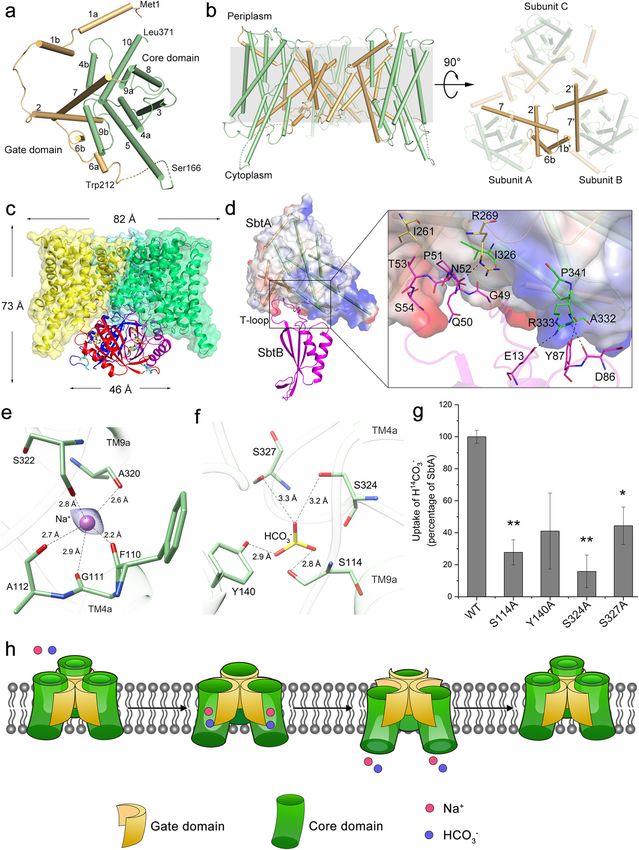
permission directly from the copyright holder. To view a copy of this license, visit http://creativecommons.org/licenses/by/4.0/.Liu et al. Cell Discovery (2021)7:63 Page 2 of 5 Fig. 1 (See legend on next page.)
Liu et al. Cell Discovery (2021)7:63                                                                                                           Page 3 of 5

 (see figure on previous page)
 Fig. 1 Cryo-EM structures of SbtA and its complex with SbtB. a Overall structure of a SbtA subunit viewed from the extracellular side with helices
 shown as cylinders. b Side view of the trimeric structure of SbtA (left) and top view from the intracellular side (right). The gate domains are colored in
 orange, whereas the core domains are colored in green. The missing residues between Ser166 and Trp212 are indicated as a dashed line. c Cartoon
 representation of SbtA–SbtB complex structure. Three subunits of SbtB are colored in magentas, red and blue, respectively. The C-terminal disulfide
 bond between Cys105 and Cys110 is indicated by blue sticks. An AMP molecule in the nucleotide-binding cleft is shown as yellow sticks. d Interfaces
 between a pair of SbtA and SbtB subunits. The interacting residues are shown as sticks, with the polar interactions indicated by dashed lines. e The
 putative Na+-binding site. The Na+ is shown as a violet sphere whereas the Na+-binding residues are shown as sticks. The polar interactions are
 indicated by dashed lines. The cryo-EM density map of Na+ is shown in blue mesh. f The putative HCO3–-binding site modeled by HADDOCK. The
 HCO3– molecule is shown as sticks and colored by atoms. g The HCO3– transport activity assays of the wild-type SbtA and mutants in E. coli
 membrane vesicles. Three independent experiments were performed for each assay. The means and standard deviations were calculated and the
 data are presented as means ± SD. Two-tailed Student’s t-test is used for the comparison of statistical significance. The P values of < 0.05 and < 0.01
 are indicated with * and **, respectively. h A proposed elevator mechanism of Na+-dependent bicarbonate transport of the trimeric SbtA. The Na+
 and HCO3– are shown as red and blue spheres, respectively. SbtA of an outward-open conformation recruits the substrates HCO3– and Na+ from the
 periplasm, accompanied by a rigid-body movement of core domains (green) against the immobile gate domains (yellow). Afterwards, the substrates
 are released into the cytosol followed by the turnover of SbtA into the resting state.

observable from the extracellular side, the N-terminal                          runs along the hydrophobic inter-domain cleft of SbtA,
moieties of three TM7 helices are bent and interact with                        forming extensive hydrophobic interactions (Fig. 1d) via
each other, whereas from the intracellular side, the N-                         relatively conserved residues (Supplementary Fig. S6).
termini of three TM2 helices form pairwise crossovers via                       Similar to the complex structure of AmtB–GlnK, in which
hydrophobic interactions (Fig. 1b). In addition, TM1b of                        the T-loop of PII protein GlnK inserts deeply into the
one subunit is positioned against TM6b of the neigh-                            cytoplasmic pore exit of the ammonia channel AmtB11,
boring subunit via hydrophobic interactions, further sta-                       the SbtA–SbtB structure showed that SbtB T-loop par-
bilizing the trimeric structure at the lateral side (Fig. 1b).                  tially blocks the substrate tunnel exit of SbtA, indicating
  It has been reported that when SbtB and SbtA are co-                          that SbtB might attenuate the transport activity of SbtA
expressed in Escherichia coli (E. coli), they form a com-                       under certain physiological conditions. Consistently, the
plex9. To elucidate the fine interaction mode between                            previous results showed that SbtB inhibited the SbtA-
SbtA and SbtB, we purified the SbtA–SbtB complex in the                          mediated HCO3– uptake in E. coli9. However, a recent
presence of AMP and solved its 3.15 Å cryo-EM structure                         report demonstrated that the light-regulated SbtA func-
(Supplementary Fig. S4). The SbtB trimer is associated                          tion in vivo is independent of SbtB in Synechococcus PCC
with the intracellular face of the SbtA trimer (Fig. 1c),                       794212. Thus the fine in vivo regulatory mechanism of
sharing an overlapped threefold axis. The overall shape of                      SbtA–SbtB remains unsolved, but might be involved in
the complex resembles a cylindrical cone of ~73 Å in                            the coordinated regulation of light–dark transitions, the
height, and ~82 and 46 Å in diameter for the SbtA and                           intracellular homeostasis of adenyl-nucleotides and redox
SbtB trimers, respectively (Fig. 1c). Similar to the pre-                       status.
viously reported PII and PII-like structures10, SbtB also                         At the peripheral side of TM crossover within the center
adopts a trimeric structure, each subunit of which shows a                      of SbtA core domain in the SbtA–SbtB complex, there is a
canonical PII ferredoxin-like fold with a well-structured                       clear cryo-EM density most likely corresponding to a
T-loop (Fig. 1c; Supplementary Fig. S5). The C-terminal                         sodium ion (Fig. 1e), given SbtA is a Na+-dependent
CGPxGC motif, which is conserved in some SbtB                                   transporter. In each subunit, the Na+ forms five coordi-
homologs, also forms a hairpin structure via a disulfide                         nate bonds with the main-chain oxygens of residues
bond between Cys105 and Cys110 (Fig. 1c; Supplemen-                             Phe110, Gly111, and Ala112 from TM4a and Ala320 and
tary Fig. S5). In addition, an AMP molecule binds to the                        Ser322 from TM9a (Fig. 1e). Notably, in the previously
nucleotide-binding cleft between each pair of neighboring                       reported Na+-dependent transporters8,13, Na+ and its co-
SbtB subunits (Fig. 1c; Supplementary Fig. S5).                                 transported substrate are located nearby the crossover of
  In the complex, the core structure of SbtB does not                           TMs. We thus docked HCO3– in the vicinity of the
directly interact with the intracellular surface of SbtA.                       crossover of the complexed SbtA via HADDOCK14. In
Instead, the structured T-loop of SbtB, which extends                           this docking model, the HCO3–-binding site is located at
~20 Å from the core structure of SbtB, is inserted into the                     the interface of the core and gate domains. It binds to a
inter-domain cleft of the corresponding SbtA subunit                            cavity formed by the other side of the crossover and forms
(Fig. 1d), yielding an interface area of ~740 Å2. The inter-                    four hydrogen bonds with Ser114, Tyr140, Ser324, and
domain cleft of SbtA is formed by the intracellular parts of                    Ser327 (Fig. 1f). Sequence analysis revealed that most of
TM2 and TM7 of the gate domain, in addition to TM9                              these putative substrate-binding residues are conserved
and TM10 of the core domain. The extended SbtB T-loop                           among SbtA homologs (Supplementary Fig. S7).
Liu et al. Cell Discovery (2021)7:63                                                                                                                 Page 4 of 5

Furthermore, mutating any of these putative HCO3–-                             on cryo-EM data collection of SbtA–SbtB complex at the Center for Biological
binding residues to Ala led to a dramatic decrease of                          Imaging at the Institute of Biophysics (IBP), Chinese Academy of Sciences. We
                                                                               thank Mr. Jishu Ren (USTC) for the assistance on the transport assays. This
HCO3– transport activity of SbtA in E. coli membrane                           research was supported by the Strategic Priority Research Program of the
vesicles to about 10%–40% of the wild-type level (Fig. 1g).                    Chinese Academy of Sciences (XDB37020301 and XDA24020302), the National
The results demonstrated that these residues are neces-                        Natural Science Foundation of China (31630001 and 31621002), and the
                                                                               Ministry of Science and Technology of China (2016YFA0400900). Y.-L.J. thanks
sary for optimal transport activity of SbtA.                                   the Youth Innovation Promotion Association of Chinese Academy of Sciences
   Structural analysis revealed that the substrate-binding                     for their support (Membership No. 2020452).
sites of free SbtA are inaccessible to the solvent from
                                                                               Author contributions
either intracellular or extracellular side. Thus, the struc-                   C.-Z.Z., Y.-L.J. and Y.C. conceived the project. X.-Y.L. and B.L. performed
ture of free SbtA represents an occluded conformation.                         molecular cloning. X.-Y.L. performed protein purification and cryo-EM sample
Respectively superimposing the two individual domains of                       preparation. Y.-L.J., L.W. and Y.C. conducted cryo-EM data collection, structure
                                                                               determination and model building. W.-T.H. and X.-Y.L. performed transport
each SbtA subunit against those in the SbtB-bound form                         assays. All authors analyzed the data and contributed to the paper preparation.
showed little structural variations (Supplementary Fig.                        Y.-L.J., X.-Y.L. and C.-Z.Z. wrote the paper.
S8a). However, upon binding to SbtB, the two domains of
each SbtA subunit undergo a significant rigid-body                              Data availability
                                                                               The cryo-EM structures of SbtA and SbtA–SbtB have been deposited at PDB
movement against each other. Given the superimposed                            under the codes of 7CYE and 7CYF, respectively. The cryo-EM density maps of
gate domains, each core domain of the complexed SbtA                           SbtA and SbtA–SbtB have been deposited at the Electron Microscopy Data
tilts about 10° against the trefoil-like center, accompanied                   Bank (EMD-30498 for SbtA and EMD-30499 for SbtA–SbtB).

by a 4-Å slide of the core domain toward the intracellular                     Conflict of interest
space (Supplementary Fig. S8b). As a result, the two                           The authors declare no competing interests.
domains of SbtA in SbtB-bound form are relatively
separated from each other, yielding an inward-open
                                                                               Publisher’s note
conformation (Supplementary Fig. S8b). The present                             Springer Nature remains neutral with regard to jurisdictional claims in
structures of SbtA in the free and SbtB-bound forms                            published maps and institutional affiliations.
capture two snapshots in the transport cycle which enable
us to propose a putative model for SbtA-mediated HCO3–                         Supplementary information The online version contains supplementary
                                                                               material available at https://doi.org/10.1038/s41421-021-00287-w.
transport (Fig. 1h). Initially, SbtA was assumed to adopt
an outward-open conformation ready to uptake the sub-                          Received: 20 April 2021 Revised: 20 April 2021 Accepted: 3 June 2021
strates HCO3– and Na+. Substrate binding most likely
promotes SbtA to adopt an occluded conformation, as
seen in our free SbtA structure with a Na+ bound in the
pocket. Afterwards, the core domains of SbtA slide toward                      References
                                                                                1. Zhang, C. C., Zhou, C. Z., Burnap, R. L. & Peng, L. Carbon/Nitrogen Meta-
the cytosolic side against the immobile gate domains,
                                                                                    bolic Balance: lessons from Cyanobacteria. Trends Plant Sci. 23, 1116–1130
resulting in an inward-open conformation of SbtA.                                   (2018).
Accordingly, the substrate tunnel is open toward the                            2. Bracher, A., Whitney, S. M., Hartl, F. U. & Hayer-Hartl, M. Biogenesis and
                                                                                    Metabolic Maintenance of Rubisco. Annu. Rev. Plant Biol. 68, 29–60 (2017).
intracellular space (Supplementary Fig. S8b). Finally, the
                                                                                3. Price, G. D., Badger, M. R., Woodger, F. J. & Long, B. M. Advances in under-
substrates are released to the cytosol, accompanied by the                          standing the cyanobacterial CO2-concentrating-mechanism (CCM): functional
turnover of SbtA to the resting state for the next cycle of                         components, Ci transporters, diversity, genetic regulation and prospects for
                                                                                    engineering into plants. J. Exp. Bot. 59, 1441–1461 (2008).
transport. This transport model of SbtA is reminiscent of
                                                                                4. Shibata, M. et al. Genes essential to sodium-dependent bicarbonate transport
the elevator alternating-access transport mechanism, in                             in cyanobacteria: function and phylogenetic analysis. J. Biol. Chem. 277,
which the substrate-binding domain moves along the                                  18658–18664 (2002).
                                                                                5. Price, G. D., Shelden, M. C. & Howitt, S. M. Membrane topology of the cya-
relatively immobile scaffold domain15.
                                                                                    nobacterial bicarbonate transporter, SbtA, and identification of potential
   In summary, this study provides structural insights into                         regulatory loops. Mol. Membr. Biol. 28, 265–275 (2011).
the cyanobacterial high-affinity HCO3– transporter SbtA,                         6. Selim, K. A., Haase, F., Hartmann, M. D., Hagemann, M. & Forchhammer, K. PII-
which exhibits a new architecture of Na+/solute sym-                                like signaling protein SbtB links cAMP sensing with cyanobacterial inorganic
                                                                                    carbon response. Proc. Natl. Acad. Sci. USA 115, E4861–E4869 (2018).
porters and its fine interaction pattern with a PII-like                         7. Screpanti, E. & Hunte, C. Discontinuous membrane helices in transport pro-
protein SbtB. Structural analysis combined with site-                               teins and their correlation with function. J. Struct. Biol. 159, 261–267 (2007).
                                                                                8. Hu, N. J., Iwata, S., Cameron, A. D. & Drew, D. Crystal structure of a bacterial
directed mutagenesis enabled us to identify the substrate-
                                                                                    homologue of the bile acid sodium symporter ASBT. Nature 478, 408–411
binding pocket and propose an elevator transport                                    (2011).
mechanism of SbtA.                                                              9. Du, J., Forster, B., Rourke, L., Howitt, S. M. & Price, G. D. Characterisation of
                                                                                    cyanobacterial bicarbonate transporters in E. coli shows that SbtA homologs
                                                                                    are functional in this heterologous expression system. PLoS ONE 9, e115905
Acknowledgements
                                                                                    (2014).
We thank Dr. Yong-Xiang Gao for technical support on cryo-EM data collection
                                                                               10. Forchhammer, K. & Luddecke, J. Sensory properties of the PII signalling protein
of SbtA at the Center for Integrative Imaging of University of Science and
                                                                                   family. FEBS J. 283, 425–437 (2016).
Technology of China (USTC). We thank Dr. Li-Hong Chen for technical support
Liu et al. Cell Discovery (2021)7:63 Page 5 of 5
11. Conroy, M. J. et al. The crystal structure of the Escherichia coli AmtB–GlnK 13. Wang, C. et al. Structural mechanism of the active bicarbonate transporter
complex reveals how GlnK regulates the ammonia channel. Proc. Natl. Acad. from cyanobacteria. Nat. Plants 5, 1184–1193 (2019).
Sci. USA 104, 1213–1218 (2007). 14. de Vries, S. J., van Dijk, M. & Bonvin, A. M. The HADDOCK web server for data-
12. Förster, B. et al. Regulatory adenylnucleotide-mediated binding of the PII-like driven biomolecular docking. Nat. Protoc. 5, 883–897 (2010).
protein SbtB to the cyanobacterial bicarbonate transporter SbtA is controlled by 15. Drew, D. & Boudker, O. Shared Molecular Mechanisms of Membrane Trans-
the cellular energy state. bioRxiv https://doi.org/10.1101/2021.02.14.431189 (2021). porters. Annu. Rev. Biochem. 85, 543–572 (2016).You can also read