UBE4B, a microRNA-9 target gene, promotes autophagy-mediated Tau degradation
←
→
Page content transcription
If your browser does not render page correctly, please read the page content below

ARTICLE
                  https://doi.org/10.1038/s41467-021-23597-9                 OPEN

                  UBE4B, a microRNA-9 target gene, promotes
                  autophagy-mediated Tau degradation
                  Manivannan Subramanian1,2,7, Seung Jae Hyeon3,7, Tanuza Das4, Yoon Seok Suh1, Yun Kyung Kim                                                    2,

                  Jeong-Soo Lee 1,2, Eun Joo Song 5 ✉, Hoon Ryu3 ✉ & Kweon Yu 1,2,6 ✉
1234567890():,;

                  The formation of hyperphosphorylated intracellular Tau tangles in the brain is a hallmark of
                  Alzheimer’s disease (AD). Tau hyperphosphorylation destabilizes microtubules, promoting
                  neurodegeneration in AD patients. To identify suppressors of tau-mediated AD, we perform a
                  screen using a microRNA (miR) library in Drosophila and identify the miR-9 family as sup-
                  pressors of human tau overexpression phenotypes. CG11070, a miR-9a target gene, and its
                  mammalian orthologue UBE4B, an E3/E4 ubiquitin ligase, alleviate eye neurodegeneration,
                  synaptic bouton defects, and crawling phenotypes in Drosophila human tau overexpression
                  models. Total and phosphorylated Tau levels also decrease upon CG11070 or UBE4B over-
                  expression. In mammalian neuroblastoma cells, overexpression of UBE4B and STUB1, which
                  encodes the E3 ligase CHIP, increases the ubiquitination and degradation of Tau. In the
                  Tau-BiFC mouse model, UBE4B and STUB1 overexpression also increase oligomeric Tau
                  degradation. Inhibitor assays of the autophagy and proteasome systems reveal that the
                  autophagy-lysosome system is the major pathway for Tau degradation in this context. These
                  results demonstrate that UBE4B, a miR-9 target gene, promotes autophagy-mediated Tau
                  degradation together with STUB1, and is thus an innovative therapeutic approach for AD.

                  1 Metabolism and Neurophysiology Research Group, KRIBB, Daejeon, Korea. 2 Convergence Research Center of Dementia, KIST, Seoul, Korea. 3 Center for

                  Neuroscience, Brain Science Institute, KIST, Seoul, Korea. 4 Biomedical Research Institute, KIST, Seoul, Korea. 5 Graduate School of Pharmaceutical Sciences
                  and College of Pharmacy, Ewha Womans University, Seoul, Korea. 6 Department of Functional Genomics, UST, Daejeon, Korea. 7These authors contributed
                  equally: Manivannan Subramanian, Seung Jae Hyeon. ✉email: esong@ewha.ac.kr; hoonryu@kist.re.kr; kweonyu@kribb.re.kr

                  NATURE COMMUNICATIONS | (2021)12:3291 | https://doi.org/10.1038/s41467-021-23597-9 | www.nature.com/naturecommunications                                  1
ARTICLE                                                                 NATURE COMMUNICATIONS | https://doi.org/10.1038/s41467-021-23597-9

A
         lzheimer’s disease (AD) is one of the most common age-              Results
         related neurodegenerative diseases, and includes the                Identification of miR-9 as a modifier of hTau in Drosophila by
         pathological hallmarks of extracellular amyloid plaques by          genome-wide miRNA screening. The overexpression of human
abnormally folded amyloid-β 42 (Aβ-42) and intracellular neu-                tau (hTau) in Drosophila eyes using the eye-specific GMR-GAL4
rofibrillary tangles (NFTs) by Tau hyperphosphorylation in the                promoter induces eye neurodegeneration, which is grossly char-
brain1,2. Tau is an essential soluble intracellular protein that             acterized by decreased eye size in the rough eye phenotype7. This
associates with and stabilizes axon microtubules3. Hyperpho-                 phenotype was used to screen 131 UAS-miRNA lines covering 144
sphorylation disrupts the physiological function of Tau, resulting           Drosophila miRNAs23. We analyzed the eye morphology of each
in microtubule destabilization and formation of intracellular                line, quantitatively measured eye sizes, and arranged the library in
NFTs, potentiating neurodegeneration and memory impairment                   the order of increasing eye size (Fig. 1a, Supplementary Fig. 1a, b).
in AD patients. Neurodegenerative disorders with Tau inclusions              Eye-specific overexpression of many miRNAs in hTau flies
are referred to as tauopathies4.                                             (GMR > hTau) affected the eye size. Quantification of eye sizes
   Drosophila melanogaster has emerged as an important model                 was plotted in a volcano plot by the ratio of eye size to p-value of
system for investigating the pathology of tauopathies at the cel-            miRNA-overexpressing lines relative to the control GMR > hTau
lular and molecular levels. Drosophila tauopathy AD models                   line (Fig. 1b). The most significant reductions in eye sizes were
exhibit visible phenotypes such as brain vacuole formation, neu-             induced by the overexpression of miR-9a, miR-9b, and miR-9c
romuscular junction defects, larval and adult locomotor defects,             (Fig. 1c, d), which are members of the evolutionarily conserved
impairment of learning and memory, and reduced lifespan5,6.                  miR-9 family (Fig. 1e). The reduced eye size of miR-9a, miR-9b or
Ectopic expression of human tau in Drosophila eyes induces a                 miR-9c in the absence of hTau expression (Fig. 1c, d, and Sup-
rough eye phenotype, which is widely used to screen for modifiers             plementary Fig. 1) may be due to the involvement of these
of Tau pathology7. Similarly, tissue-specific ectopic expression of           miRNAs during eye development where these miRNAs may
human tau or mutant tau in the Drosophila brain or mushroom                  regulate other target genes and affect morphology and develop-
body induces neurodegeneration, manifesting in phenotypes such               ment of eye sizes. However, when these miRNAs were expressed
as locomotor and cognitive impairment8. The pathogenic                       in the presence of hTau, the reduction of eye size was enhanced
mechanisms of Tau are similar in humans and Drosophila9,10.                  drastically. This indicated an important regulatory role for the
   MicroRNAs (miRNAs) are non-coding RNAs comprised of 19                    miR-9 family in Drosophila Tau toxicity. We focused on miR-9a
to 25 nucleotides, which suppress target mRNAs by pairing to                 for subsequent studies, as miR-9a has 100% homology to mam-
complementary sequences in the 3’ UTRs of target genes,                      malian miR-9 (Fig. 1e).
impairing translation and in some cases mRNA stability11,12.
miRNAs are implicated in diverse brain functions including
                                                                             Modulation of hTau by CG11070, a miR-9a target identified by
development, cognition, and synaptic plasticity13. Dysregulation
                                                                             secondary screening. Because miR-9a was identified as a modifier
of miRNAs can be detrimental, and is associated with several
                                                                             of Tau toxicity, we searched miR-9a target genes using three
human diseases ranging from metabolic and inflammatory dis-
                                                                             separate microRNA target prediction programs, TargetScan
ease to neoplasia14–16. miRNA expression-profiling studies have
                                                                             (www.targetscan.org), miRanda (www.microRNA.org), and
identified that multiple miRNAs are dysregulated in the brains of
                                                                             miRbase (www.mirbase.org), detecting 34 miR-9a targets com-
AD patients, but the functional implications of these changes
                                                                             mon to all three platforms (Supplementary Fig. 2a). We then
remain unclear17.
                                                                             screened the 34 putative target genes against the eye phenotype of
   The ubiquitin-proteasome system (UPS) and autophagy-
                                                                             hTau overexpression (GMR > hTau) using stably expressed RNAi
lysosome system (ALS) are the primary protein degradation
                                                                             knockdown in the GMR > hTau background, and arranged the
pathways in eukaryotic cells18. Proteins that undergo proteasomal
                                                                             RNAi lines in order of increasing eye size (Fig. 2a, Supplementary
degradation are polyubiquitinated at Lys48 by ubiquitin ligases
                                                                             Fig. 2b, c). Volcano plot analysis was conducted by the ratio of
and targeted for 26S proteasome complex degradation and pro-
                                                                             eye size to p-value of the 34 RNAi lines relative to the control
teins poly-ubiquitinated at Lys63 are degraded by the ALS.
                                                                             GMR > hTau flies. CG11070-RNAi flies exhibited the most pro-
Interestingly, proteostasis is disrupted in AD brains19. A prior
                                                                             minent reduction in eye size (Fig. 2b, Supplementary Fig. 2c). The
study reported that Tau phosphorylation induces ubiquitination
                                                                             eye size of CG11070-RNAi flies in the GMR > hTau background
and subsequent degradation by the UPS20. STUB1 (STIP1
                                                                             (GMR > hTau + CG11070-RNAi) was significantly reduced rela-
homology and U-Box containing protein1) is an E3 ubiquitin
                                                                             tive to the eye size of control GMR > hTau flies (Fig. 2c, d).
ligase that ubiquitinates phosphorylated Tau for proteasomal
                                                                                Because microRNAs bind to 3′-UTR region of target mRNAs
degradation in vitro21 and has also been implicated in the
                                                                             to suppress translation and/or mRNA stability, we assessed
clearance of truncated Tau by the ALS22.
                                                                             binding of miR-9a to CG11070 using a miRNA–mRNA pull-
   In the present study, we report the results of a Drosophila
                                                                             down assay23. Similar to the known miR-9a/miR-9 targets
miRNA library screening, which identified the evolutionarily
                                                                             senseless24 and sNPFR123, we found that miR-9a bound to and
conserved miR-9 and its target CG11070 as strong modifiers of
                                                                             enriched CG11070 mRNA in Drosophila S2 cells compared to its
the neurodegenerative Drosophila rough eye phenotype induced
                                                                             control scrambled miRNA (Fig. 2e). Consistently, eye-specific
by human tau overexpression. Further, Drosophila CG11070 and
                                                                             overexpression of miR-9a with GMR-GAL4 flies decreased
its mammalian orthologue UBE4B (Ubiquitin conjugation E4 B)
                                                                             CG11070 mRNA levels (Fig. 2f). Taken together, these findings
alleviated the neurodegenerative phenotypes induced by neuronal
                                                                             suggested that CG11070 was a legitimate target of miR-9a.
human tau overexpression in Drosophila. In addition, total and
phosphorylated Tau in aged tau-overexpressing flies was
decreased by CG11070 or mammalian UBE4B overexpression. In                   The overexpression of Drosophila CG11070 and its mammalian
mammalian neuroblastoma cells and a mouse tau overexpression                 orthologue UBE4B alleviated hTau phenotypes in Drosophila.
model, UBE4B, and STUB1 E3 ligases ubiquitinated Tau proteins                CG11070 and UBE4B proteins had 26% amino acid identities and
and induced autophagy-mediated Tau degradation. These find-                   similar functional domains (Supplementary Fig. 3a). Interestingly,
ings suggest that CG11070/UBE4B is a E4 ubiquitin ligase that                the 3′-UTR region of UBE4B mRNA also contained miR-9
regulates Tau degradation via the ALS, and is a putative ther-               binding sequences, similar to the 3′-UTR region of CG11070
apeutic target for the treatment of tauopathies such as AD.                  mRNA (Supplementary Fig. 3b). In addition to miR-9a, UBE4B is

2                NATURE COMMUNICATIONS | (2021)12:3291 | https://doi.org/10.1038/s41467-021-23597-9 | www.nature.com/naturecommunications
NATURE COMMUNICATIONS | https://doi.org/10.1038/s41467-021-23597-9                                                              ARTICLE

also regulated by miR-26, miR-148/miR-152, and miR-15/16/195/                   relative to control GMR > hTau flies, we determined if the over-
424/497. However, CG11070 is regulated by miR-9a only in                        expression of CG11070 and its mammalian orthologue UBE4B
Drosophila. Therefore, CG11070/UBE4B was chosen for further                     could alleviate hTau phenotypes in GMR > hTau flies.
analysis due to similarity in regulation by miR-9. Because the                     In GMR > hTau flies, the overexpression of CG11070 (GMR >
knockdown of CG11070 in GMR > hTau flies decreased eye size                      hTau + GC11070) and UBE4B (GMR > hTau + UBE4B) increased

NATURE COMMUNICATIONS | (2021)12:3291 | https://doi.org/10.1038/s41467-021-23597-9 | www.nature.com/naturecommunications                     3
ARTICLE NATURE COMMUNICATIONS | https://doi.org/10.1038/s41467-021-23597-9
Fig. 1 Genome-wide Drosophila miRNA library screening identified miR-9 family miRNAs as modifiers of hTau in Drosophila eyes. a Screening of the
miRNA library in GMR > hTau Drosophila eyes revealed significant phenotypic enhancement, as indicated by decreased eye size, in flies overexpressing miR-
9 family miRNAs compared with GMR > hTau control flies. Eye sizes were arranged from smallest to largest. b Volcano plot of mean eye sizes in flies
expressing various miRNAs in the GMR > hTau background versus their respective p-values derived from a one-way analysis of variance followed by
pairwise t-tests and a Bonferroni correction for multiple comparisons. All points above the dotted line can be considered significant. This analysis identified
that flies overexpressing miR-9 family miRNAs exhibited a severe Tau toxicity phenotype, as indicated by decreased eye size. N = 3 biologically
independent experiments. c, d The overexpression of miR-9a, miR-9b, or miR-9c in GMR > hTau Drosophila eyes significantly reduced eye sizes relative to
GMR > hTau Drosophila. N = 5 biologically independent experiments. Data are presented as the mean ± s.e.m. Statistical significance was determined with a
two-tailed Student’s t-test. In the box plots the whiskers represent the 5th to 95th percentile range. e Alignment of mature Drosophila miR-9a, miR-9b, and
miR-9c sequences with human and murine miR-9 sequences identified that miR-9a had 100% identity with mammalian miR-9 sequences. Statistical
source data.
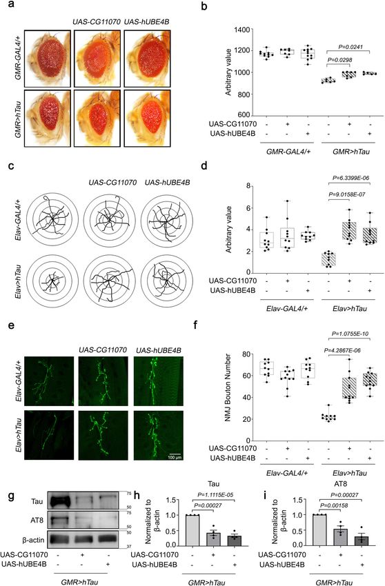
eye size, alleviating the rough eye neurodegenerative phenotype STUB1 co-expression significantly increased Tau ubiquitination
(Fig. 3a, b). Neuron-specific ectopic hTau expression (Elav > (Fig. 4a, lane 5 compared with lane 3 and 4). Subsequently, we
hTau) decreased Drosophila larva locomotion, which was determined that STUB1 activity was important for UBE4B-
alleviated by CG11070 and UBE4B overexpression (Fig. 3c, d). mediated Tau ubiquitination, as co-expression with a dominant-
The bouton number of neuromuscular junctions (NMJ) in negative mutant (STUB1H260Q) failed to enhance Tau ubiquiti-
Drosophila larvae is directly correlated with locomotion, and is nation (Fig. 4a, lane 6). These results suggested that UBE4B and
decreased in Drosophila AD models25,26. Neuronal hTau-expres- STUB1 co-regulated Tau ubiquitination.
sing larvae exhibited significantly reduced bouton numbers Due to the essential regulatory roles of these ubiquitin ligases
relative to the control, which was alleviated by the overexpression in protein degradation, we examined UBE4B and STUB1-
of CG11070 and UBE4B (Fig. 3e, f). mediated Tau degradation. By inhibiting protein synthesis with
The knockdown of miR-9a by miR-9a-sponge (SP) showed no cycloheximide (CHX), we observed that Tau degradation was
change in the eye sizes when compared with GMR > hTau flies increased by STUB1 (Fig. 4b, lanes f–i, Fig. 4c) and UBE4B
(Supplementary Fig. 4a, b). However, the knockdown by miR-9a- overexpression (Fig. 4b, lanes 1–4 and Fig. 4c), and was further
SP in neurons of Elav > hTau rescued the larval locomotion increased by co-expression of STUB1 and UBE4B (Fig. 4b, lanes
phenotype similar to the overexpression of CG11070 and hUBE4B 6–9 relative to lanes 1–4, Fig. 4c). Because UBE4B overexpression
(Supplementary Fig. 4c, d). Similarly, the knockdown by miR-9a- alone degraded Tau (Fig. 4b, c), we knocked down endogenous
SP in neurons also rescued NMJ bouton numbers when STUB1 with siRNA (Supplementary Fig. 5a) and examined
compared with the overexpression of CG11070 and hUBE4B in UBE4B-mediated Tau degradation (Supplementary Fig. 5b, c).
neurons (Supplementary Fig. 4e, f). This data also confirmed that Interestingly, UBE4B overexpression did not affect Tau degrada-
miR-9a and CG11070/UBE4B forms a common axis involved in tion when STUB1 was knocked down (Supplementary Fig. 5b,
regulating Tau toxicity in Drosophila. lanes 6–9 compared with lanes 1–4). Similarly, the knockdown of
Because the overexpression of CG11070 and UBE4B alleviated STUB1 showed no change in Tau degradation when compared
hTau phenotypes, we further examined whether the overexpres- with the siControl (Supplementary Fig. 5c). Collectively, these
sion of these genes affected Tau degradation. We performed results indicated that UBE4B was a critical factor that enhanced
western blots on 30-day-old fly heads with eye-specific hTau the ubiquitination activity of STUB1 to ubiquitinate and degrade
expression (GMR > hTau), and identified that total hTau protein Tau. To evaluate the biochemical interactions of UBE4B with
was significantly decreased by the overexpression of CG11070 STUB1 and Tau, we performed immunoprecipitation analyses.
(GMR > hTau + GC11070) and UBE4B (GMR > hTau + UBE4B) When Tau was co-expressed with UBE4B and STUB1, UBE4B co-
(Fig. 3g, h). In addition, phosphorylated Tau, detected by AT8 (p- precipitated with STUB1 (Fig. 4d). However, UBE4B did not
S202/T205) antibody, was also significantly decreased by the directly interact with STUB1, as UBE4B did not co-precipitate
overexpression of CG11070 and UBE4B (Fig. 3g, i). Similarly, with STUB1 in the absence of Tau (Fig. 4d). Previous studies
other Tau phosphorylated forms, detected by AT180 (p-T231) demonstrated that STUB1 directly interacts with and ubiquiti-
and PHF-1 (p-S396/S404) antibodies, were also reduced in these nates Tau, targeting it for degradation21,27,28. UBE4B also directly
genotypes (Supplementary Fig. 3c–e). Taken together, these interacted with Tau protein (Fig. 4e). These results suggested that
findings suggested that the overexpression of either Drosophila UBE4B did not directly interact with STUB1, but rather that Tau
CG11070 or human UBE4B rescued both larval locomotion/NMJ mediated the interaction between STUB1 and UBE4B.
defects and adult eye phenotypes in hTau-overexpressing
Drosophila by increasing Tau degradation. STUB1 knockdown reduces UBE4B-mediated alleviated hTau
phenotypes in Drosophila. Since the knockdown of STUB1 in
Ubiquitination and degradation of Tau by UBE4B and STUB1 neuroblastoma cell lines showed no change in Tau levels in the
in mammalian neuroblastoma cells. To investigate the presence of UBE4B (Supplementary Fig. 5b, c), we further
mechanism by which UBE4B degraded Tau, we first determined examined the importance of STUB1 in the in vivo Drosophila
if UBE4B affected Tau ubiquitination in SH-SY5Y neuroblastoma model system. Studies from the in vivo model system showed that
cells (Fig. 4a). Previous studies have demonstrated that UBE4B the knockdown of STUB1 gene in the eyes of flies expressing
has E4 ubiquitin ligase activity, and that Tau is ubiquitinated by GMR > hTau + hUBE4B, significantly reduced eye phenotype
STUB1 E3 ligase21,27,28. STUB1 was chosen due to its role in when compared with GMR > hTau + hUBE4B flies (Fig. 5a, b).
ubiquitination of Tau proteins and is not regulated by miR-9. To Similarly, the knockdown of STUB1 in neurons expressing Elav >
confirm the effect of STUB1 on Tau ubiquitination, we evaluated hTau + hUBE4B also significantly reduced larval locomotion
ubiquitination of Tau by STUB1, and found that STUB1 over- phenotype when compared with Elav > hTau + hUBE4B in neu-
expression alone did not affect Tau ubiquitination (Fig. 4a, lane rons (Fig. 5c, d). However, NMJ phenotype showed no change in
3). Similarly, UBE4B overexpression alone did not significantly larvae expressing Elav > hTau + hUBE4B + STUB1-RNAi when
affect Tau ubiquitination (Fig. 4a, lane 4). However, UBE4B and compared with Elav > hTau + hUBE4B larvae (Fig. 5e, f). These
4 NATURE COMMUNICATIONS | (2021)12:3291 | https://doi.org/10.1038/s41467-021-23597-9 | www.nature.com/naturecommunications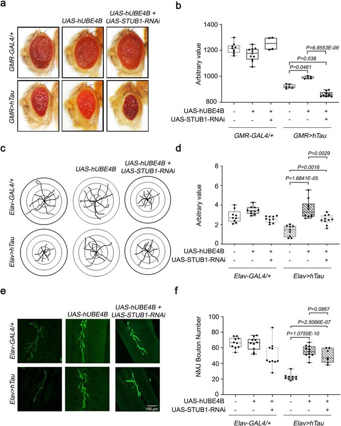
NATURE COMMUNICATIONS | https://doi.org/10.1038/s41467-021-23597-9                                                                 ARTICLE

studies indicate that alleviated hTau phenotypes by hUBE4B                      UBE4B and STUB1 affects Tau degradation, we generated AAV-
overexpressing are dependent on STUB1 function.                                 CMV-UBE4B and AAV-CMV-STUB1 constructs (Fig. 6a). AAVs
                                                                                were delivered to the dentate gyri of Tau-BiFC mice by stereotaxic
Degradation of Tau by UBE4B and STUB1 in the Tau-BiFC                           injection (Fig. 6b, c) and pathological examinations were per-
mouse model. To determine whether in vivo overexpression of                     formed as shown in the work flow (Fig. 6d). In the Tau-BiFC

NATURE COMMUNICATIONS | (2021)12:3291 | https://doi.org/10.1038/s41467-021-23597-9 | www.nature.com/naturecommunications                        5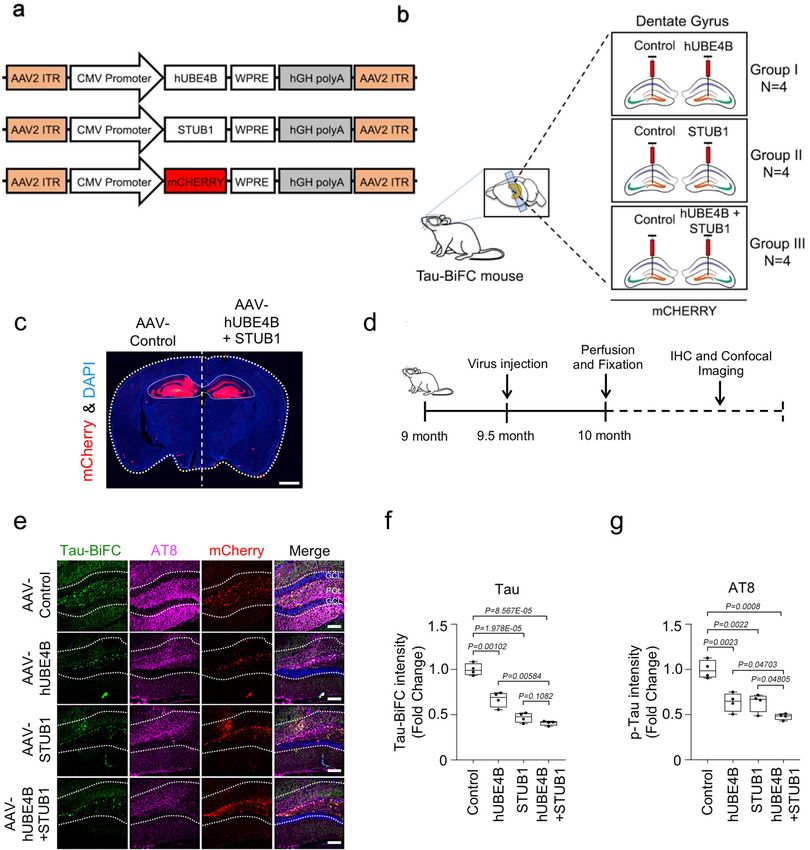
ARTICLE                                                                   NATURE COMMUNICATIONS | https://doi.org/10.1038/s41467-021-23597-9

Fig. 2 CG11070, a miR-9a target identified from secondary screening, strongly modulated hTau in Drosophila eyes. a Screening of flies with RNAi
knockdown of miR-9a targets in GMR > hTau Drosophila eyes identified a significant reduction in eye size in CG11070-RNAi flies relative to the control. b
Volcano plot of mean eye sizes of flies expressing various miR-9a target gene RNAis in GMR > hTau flies versus their respective p-values derived from a
one-way analysis of variance followed by pairwise t-tests and a Bonferroni correction for multiple comparisons. All points above the dotted line can be
considered significant. CG11070-RNAi flies exhibited more severe ocular Tau toxicity, as demonstrated by decreased eye size. N = 3 biologically
independent experiments. c, d The knockdown of CG11070 (CG11070-RNAi) in GMR > hTau flies significantly decreased eye size relative to GMR > hTau
controls. N = 5 biologically independent experiments. In the box plots the whiskers represent the 5th to 95th percentile range. Data are presented as the
mean ± s.e.m. Statistical significance was determined with a two-tailed Student’s t-test. e miRNA–mRNA–RISC pull-down assays in Drosophila S2 cells
revealed that miR-9a bound to CG11070 mRNA. Transfection of miR-9a enriched CG11070 mRNA levels, similar to the known miR-9a targets senseless and
sNPFR1, as demonstrated by qRT-PCR. N = 4 biologically independent experiments. Data are presented as the mean ± s.e.m. Statistical significance was
determined with a two-tailed Student’s t-test. f Expression of miR-9a using GMR-GAL4 significantly decreased CG11070 expression. N = 3 biologically
independent experiments. Data are presented as the mean ± s.e.m. Statistical significance was determined with a two-tailed Student’s t-test. Statistical
source data.

mouse model system, Tau oligomerization can be visualized by                   measured LC3 and p62 levels in our model system. Co-
BiFC fluorescence29. Consistent with the in vitro data, the over-               expression of UBE4B and STUB1 significantly decreased LC3
expression of either UBE4B or STUB1 induced degradation of                     and p62 levels relative to control in the dentate gyri of Tau-BiFC
Tau oligomer, as indicated by decreased Tau fluorescence relative               mice, whereas autophagy inhibitors elevated LC3 and p62 levels
to control (Fig. 6e). Similarly, co-overexpression of UBE4B and                relative to the control in the dentate gyri of UBE4B + STUB1-
STUB1 in Tau-BiFC mice further decreased Tau fluorescence                       expressed Tau-BiFC mice (Fig. 7i, j). Furthermore, autophagy
(Fig. 6e, f). Similar to reduction of oligomeric Tau levels, phos-             inhibitors modulated BECN1 levels compared to the control in
phorylated Tau levels in the dentate gyrus of Tau-BiFC mice, as                the dentate gyri of UBE4B + STUB1-expressed Tau-BiFC mice
detected by the AT8 (p-S202/T205) antibody, were also decreased                (Supplementary Fig. 8). Collectively, these results suggested that
by the overexpression of either UBE4B or STUB1, and were                       monomeric and oligomeric Tau degradations by STUB1 and
further decreased by co-expression of UBE4B and STUB1 (Fig. 6e                 UBE4B are mediated through autophagy pathway rather than the
and g). Additional phosphorylated forms of Tau were detected by                ubiquitin-proteasome system.
AT180 (p-T231) and PHF-1 (p-S396/S404) antibodies. Both p-
T231 and p-S396/S404 Tau levels were also decreased by the
overexpression of either UBE4B or STUB1 alone, and were further                Discussion
decreased by co-expression of UBE4B and STUB1 (Supplemen-                      In the present study, we demonstrated that Drosophila miR-9a
tary Fig. 6a–c). These results demonstrated that UBE4B and                     regulated Tau toxicity and increased Tau phosphorylation in the
STUB1 additively degraded Tau in vivo.                                         eyes. MiR-9a-mediated Tau toxicity arose through disruption of
                                                                               the miR-9a target gene CG11070, the human orthologue of
UBE4B and STUB1-mediated autophagy Tau degradation. The                        UBE4B, which when overexpressed mimicked the miR-9a
two reported major pathways of Tau degradation are the UPS and                 knockdown phenotype. The overexpression of either Drosophila
ALS30,31. To determine the primary pathway of UBE4B and                        CG11070 or human UBE4B alleviated Tau-associated phenotypes,
STUB1-mediated Tau degradation, we co-overexpressed UBE4B                      including increased eye size, improved larval crawling/NMJ
and STUB1 with Tau in neuroblastoma cells, and treated the cells               phenotypes, and decreased total and phosphorylated Tau levels.
with either the UPS inhibitor MG132 or the ALS inhibitors.                     Further, we demonstrated that clearance of Tau proteins by
Interestingly, MG132 treatment did not inhibit Tau degradation                 UBE4B occurred primarily through ubiquitin-dependent ALS
relative to the control cells (Fig. 7a). In contrast, Chloroquine              rather than the proteasome system.
significantly inhibited Tau degradation (Fig. 7b). In addition,                    Previous studies have demonstrated that miRNAs mediate
pepstatin A (PEPA) alone and E64D plus PEPA (E64D + PEPA),                     neurodegeneration, including that of AD33,34. AD-related miR-
autophagy inhibitors, significantly inhibited Tau degradation                   NAs regulate multiple stages of AD pathology and enhance Tau
(Fig. 7c), suggesting that ALS was the major pathway of UBE4B                  toxicity35,36. Studies in AD cadaver samples demonstrated that
and STUB1-mediated Tau degradation in neuroblastoma cells.                     miR-125b is increased in AD, and the overexpression of miR-125b
Our in vitro study showed that the turnover of monomeric Tau                   in mice increases Tau hyperphosphorylation by regulating kinases
molecule by UBE4B/STUB1 is preferentially mediated by                          and phosphatases37. Also, downregulation of miR-132/212 pro-
autophagy-dependent manner rather than the proteasome path-                    motes Tau phosphorylation, which in turn enhances AD
way in SH-SY5Y cells (Fig. 7a–c).                                              phenotypes38. In addition to these miRNAs, miR-138 regulates
   Because autophagy inhibitor treatment blocked autophagic Tau                Tau phosphorylation through GSK3β39 and miR-922 affects Tau
degradation in vitro, we tested this in the in vivo system by                  phosphorylation through UCHL140. Functional analysis of miR-9
injecting autophagy inhibitors into the dentate gyri of Tau-BiFC               has revealed its involvement in the regulation of neuronal pro-
mice with co-expression of UBE4B and STUB1 (Fig. 7d, e). We                    genitor cells and differentiation of neuronal cells during
measured oligomeric Tau and phosphorylated Tau levels in the                   development41. In AD patients, miR-9 expression is elevated42. In
dentate gyrus of Tau-BiFC mice detected with the AT8 antibody                  Drosophila, miR-9a plays an important role in the development of
(S202/T205) (Fig. 7f–h). Inhibition of ALS by Chloroquine and                  sensory organs by suppressing its target gene senseless24 and also
E64D + PEPA significantly increased oligomeric and phosphory-                   regulates body growth through sNPFR signaling23. In the Dro-
lated Tau (S202/T205) (Fig. 7f–h). Similarly, the phosphorylated               sophila Tau AD model, we observed increased Tau toxicity when
forms of Tau p-S396/S404 (PHF-1) and p-T231 (AT180) were                       miR-9a was overexpressed. The Drosophila miR-9a binding
also significantly increased in Chloroquine and E64D + PEPA-                    sequence was 100% identical to that of mammalian miR-9
treated Tau-BiFC mice (Supplementary Fig. 7a–d). Since ALS                     (Fig.1e). Our studies clearly demonstrated that miR-9a regulated
disruption changes LC3 and p62/SQSTM1 levels, which are                        Tau toxicity, exacerbating the rough eye phenotype and sig-
inversely correlated with the efficiency of autophagy32, we                     nificantly decreasing eye size.

6                  NATURE COMMUNICATIONS | (2021)12:3291 | https://doi.org/10.1038/s41467-021-23597-9 | www.nature.com/naturecommunications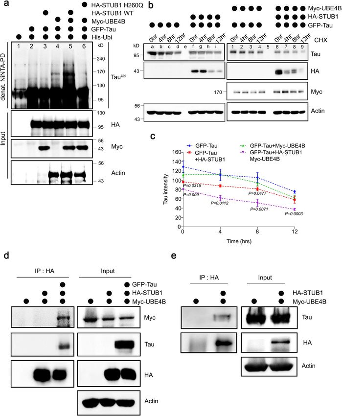
NATURE COMMUNICATIONS | https://doi.org/10.1038/s41467-021-23597-9                                                                ARTICLE

   miR-9a targets a multitude of genes that are involved in diverse             identify a distinct gene, CG11070, as a strong modifier of hTau
cellular processes, and we used in silico analysis to identify 34               (Fig. 2). Mammalian orthologues of Drosophila CG11070 include
putative targets, which were predicted as miR-9a target genes                   UBE4A and UBE4B. UBE4B, but not UBE4A, contained miR-9
using three separate in silico platforms. We then performed a                   binding sequences in its 3′ UTR like those of CG11070 (Supple-
secondary screening of the putative 34 miR-9a target genes to                   mentary Fig. 3b), and is extensively expressed in the brain43. We

NATURE COMMUNICATIONS | (2021)12:3291 | https://doi.org/10.1038/s41467-021-23597-9 | www.nature.com/naturecommunications                       7
ARTICLE                                                                   NATURE COMMUNICATIONS | https://doi.org/10.1038/s41467-021-23597-9

Fig. 3 The overexpression of Drosophila CG11070 and its mammalian orthologue UBE4B alleviated hTau phenotypes in Drosophila. a, b Eye-specific
overexpression of Drosophila CG11070 or its mammalian orthologue UBE4B in GMR > hTau flies increased eye size relative to GMR > hTau controls. N = 5
biologically independent experiments. In the box plots the whiskers represent the 5th to 95th percentile range. c, d Neuronal overexpression of Drosophila
CG11070 or its mammalian orthologue UBE4B using Elav-Gal4 in Elav > hTau flies significantly increased larval crawling. N = 10 biologically independent
experiments. In the box plots the whiskers represent the 5th to 95th percentile range. e, f Neuronal overexpression of Drosophila CG11070 or its mammalian
orthologue UBE4B using Elav-Gal4 in Elav > hTau flies significantly increased synaptic bouton numbers relative to Elav > hTau controls. Scale bar 100 µm.
N = 10 biologically independent experiments. In the box plots the whiskers represent the 5th to 95th percentile range. g–i Western blotting revealed that
ocular overexpression of Drosophila CG11070 or its mammalian orthologue UBE4B in GMR > hTau flies significantly reduced total and phosphorylated Tau
protein levels relative to GMR > hTau controls. N = 4 biologically independent experiments. Data are presented as the mean ± s.e.m. Statistical significance
was determined with a two-tailed Student’s t-test. Statistical source data.

therefore considered UBE4B as the mammalian orthologue of                      autophagy rather than the UPS. In FTDP-17 mutant P301S mice,
Drosophila CG11070 in the context of miR-9a regulation of Tau                  trehalose treatment promotes autophagy with reduced insoluble
toxicity. The overexpression of UBE4B or CG11070 in Drosophila                 and pTau proteins53. Similarly, the knockout of Atg7 (autophagy
not only alleviated hTau eye neurodegenerative phenotypes, larval              marker) in neurons give rise to increased pTau levels and neu-
crawling defects, and synaptic dysfunction in NMJs (Fig. 3a, f),               rodegeneration with aging54. A number of autophagy inhibitors,
but also decreased total and phosphorylated Tau levels (Fig. 3g–i,             including chloroquine, NH4Cl, 3-methyl adenine (3-MA), and
Supplementary Fig. 3c–e). These results indicated that Tau                     cathepsin inhibitors, delay Tau degradation and enhance the
degradation was strongly regulated by UBE4B/CG11070. Since                     formation of high molecular weight Tau aggregates55,56. Con-
Tau is involved in maintaining the stability of microtubules in                trastingly, the autophagy inducer rapamycin facilitates insoluble
neurons, the degradation of Tau proteins affects microtubule                   Tau degradation, alleviating Tau toxicity in Drosophila30. Tre-
stability as seen in tauopathy and Alzheimer’s disease models.                 halose, an mTOR-independent autophagy activator31, improves
However, recent studies have shown that reduction in Tau pro-                  neuronal survival by decreasing Tau aggregation in tauopathy
teins leads to increased accumulation of MAP6 proteins on the                  mouse model57,58. This suggests that basal autophagy activity is
microtubules and enhances the stability of microtubules in                     essential for prevention of neuronal Tau aggregate accumulation,
neurons44. The rescue phenotype seen in UBE4B/CG1170 may be                    which is regulated in part by UBE4B activation. Considering
a result of compromised function of Tau and MAP6 proteins in                   autophagy inhibitors not only modulate neuronal autophagy
the neurons. Reduction in Tau proteins leads to reduced pTau                   pathway but also affect neuroinflammation pathway, it remains to
levels in UBE4B/CG11070 rescue animals, due to increased ubi-                  be determined whether other neuroinflammatory pathways are
quitination of Tau proteins. Studies have shown that the acet-                 involved in Tau pathology beyond the autophagy pathway in
ylation of Tau inhibits degradation of phosphorylated Tau and                  future studies.
contributes to increased tauopathy45. Both ubiquitination and                     Lys63-linked polyubiquitination of Tau facilitates the forma-
acetylation shares common Lys residue Lys28046 which is                        tion of disease-associated Tau inclusions, which are preferentially
involved in stability of Tau proteins. The UBE4B proteins are                  cleared by the ALS59, while Lys48-linked polyubiquitinated Tau is
distinct from E3 ligases in their U-Box domains, which possess                 likely degraded by the UPS60. Lys63 ubiquitinated substrates are
both E3 ligase and E4 ligase activities47. UBE4B ligase ubiquiti-              recognized by autophagy receptors present on autophagosomes,
nates the tumor suppressor genes p53, p63 and p73, and Wal-                    mediating ALS-dependent degradation61–63. Recent studies have
lerian degeneration proteins in conjunction with various E3                    shown that the knockdown of UBE4B affects Lys48 and Lys63
ligases48. In the Drosophila spinocerebellar ataxia type 3 model,              polyubiquitination in Tax binding proteins and Tax mediated
UBE4B ubiquitinates ataxin, targeting it for proteasomal degra-                activation of NF-κB64. Therefore, in the context of the present
dation. However, the dominant-negative UBE4B mutation                          study, UBE4B acted synergistically with STUB1 to facilitate Tau
enhances neurodegeneration49. These previous studies suggest                   degradation by ALS, and could polyubiquitinate Tau proteins by
that UBE4B potentiates the degradation of various proteins                     Lys63 ubiquitin linkage. The specific ubiquitin chain linkage for
through the UPS, unlike our current findings, which suggest                     Tau ubiquitination by STUB1/UBE4B is a potential target for
synergistic regulation of Tau degradation through the ALS in                   lysosomal targeting of Tau. However, the mechanism underlying
conjunction with STUB1.                                                        ALS-mediated Tau degradation remains to be elucidated in future
   Prior reports have demonstrated that Tau is ubiquitinated by                studies.
two E3 ubiquitin ligases, STUB121,27,28 and TRAF650, both of                      Tau oligomers play a central role in tauopathies. To under-
which are colocalized with NFTs in AD brains. In the present                   stand the importance of Tau oligomers in vivo, Bimolecular
study, we found that Tau was ubiquitinated by the E3 ligase                    Fluorescence Complementation (BiFC) technology was applied to
STUB1 in the presence of UBE4B, a further ubiquitin conjugation                visualize Tau oligomerization65. In this system, full-length human
factor (Fig. 4a). In addition, Tau degradation was increased by                Tau protein is fused to the non-fluorescent N- and C-terminal
UBE4B expression, and further increased by STUB1 and UBE4B                     termini of Venus fluorescent protein. In the transgenic
co-expression (Fig. 4b, c). Studies on Tau degradation have                    TauP301L-BiFC mouse model, Venus fluorescence is activated
shown that the proteasomal pathway plays a crucial role. Inhi-                 only when Tau is aggregated29. In our studies using the
bition of proteasome in HEK cells increases the levels of full-                TauP301L-BiFC mouse model, the overexpression of UBE4B,
length Tau proteins50. Similar results were seen in SH-SY5Y cells              STUB1, or both significantly reduced the fluorescence generated
with increased accumulation of both full-length Tau protein and                by Tau oligomers (Fig. 6). Similarly, Tau phosphorylation was
mutant P301L Tau protein levels when proteasome degradation is                 also decreased in the dentate gyri of TauP301L-BiFC mice with
inhibited51,52. However, from our studies, STUB1- and UBE4B-                   the overexpression of UBE4B, STUB1, or both. These results
mediated Tau degradation was inhibited by the autophagy inhi-                  indicate that Tau oligomers, a primary cause of tauopathy, can be
bitors such as Chloroquine, PEPA, and E64D (Fig. 7b, c) but not                degraded by UBE4B and STUB1 in vivo. In TauP301L-BiFC mice,
by the proteasomal inhibitor MG132 (Fig. 7a), suggesting that                  treatment with the autophagy inhibitors (Chloroquine, PEPA,
STUB1/UBE4B-mediated Tau degradation was facilitated by                        and E64D) increased oligomeric and phosphorylated Tau levels

8                  NATURE COMMUNICATIONS | (2021)12:3291 | https://doi.org/10.1038/s41467-021-23597-9 | www.nature.com/naturecommunications
NATURE COMMUNICATIONS | https://doi.org/10.1038/s41467-021-23597-9                                                                    ARTICLE

Fig. 4 Tau was ubiquitinated and degraded by UBE4B and STUB1 in mammalian neuroblastoma cells. a Tau was co-expressed with His-Ubiquitin, UBE4B,
and STUB1 WT or dominant-negative mutant STUB1H260Q in SH-SY5Y neuroblastoma cells. Ubiquitinated Tau was significantly increased by co-expression
of UBE4B and STUB1 (lane 5), but not by expression of UBE4B (lane 4) or STUB1 (lane 3) alone. Tau ubiquitination by co-expression of UBE4B and STUB1
required the ligase activity of STUB1 (lanes 5 and 6). b, c Tau protein degradation was enhanced by co-expression of UBE4B and STUB1 (lane 6–9)
compared with expression of UBE4B (lane 1–4) or STUB1 (lane f–i) alone. Quantification of Tau levels was normalized to the amount of β-actin protein in
each case. Data represent the mean ± s.e.m of three independent experiments (*P < 0.05, **P < 0.005, ***P < 0.001 two-tailed Student’s t-test). d UBE4B
was co-expressed with HA-STUB1 and Tau in SH-SY5Y cells and immunoprecipitated on anti-HA-agarose beads. UBE4B did not directly interact with STUB1
in the absence of Tau, but indirectly interacted with STUB1 in the presence of Tau. e HA-UBE4B was co-expressed with Tau in SH-SY5Y cells and
immunoprecipitated on anti-HA-agarose beads. Co-precipitated Tau was detected by Western blot, revealing that Tau directly interacted with UBE4B. All
western blots were performed more than three times. Statistical source data.

(Fig. 7b, c, f–h, Supplementary Fig. 7). Notably, while the over-               of AAV-STUB1/UBE4B injected TauP301L-BiFC mice (Fig. 7i–l
expression of UBE4B and STUB1 decreased protein levels of LC3,                  and Supplementary Fig. 8). Collectively, these results further
p62, and BECN1, autophagy markers, autophagy inhibitors sig-                    suggested that UBE4B promoted oligomeric Tau clearance via
nificantly elevated LC3, p62, and BECN1 levels in the dentate gyri               ALS through a STUB1-dependent mechanism.

NATURE COMMUNICATIONS | (2021)12:3291 | https://doi.org/10.1038/s41467-021-23597-9 | www.nature.com/naturecommunications                             9
ARTICLE                                                                   NATURE COMMUNICATIONS | https://doi.org/10.1038/s41467-021-23597-9

Fig. 5 STUB1 knockdown reduces UBE4B-mediated alleviated hTau phenotypes in Drosophila. a, b Eye-specific knockdown of Drosophila STUB1
significantly reduced eye size relative to GMR > hTau + hUBE4B flies. N = 5 biologically independent experiments. In the box plots the whiskers represent
the 5th to 95th percentile range. c, d Neuronal knockdown of Drosophila STUB1 using Elav-Gal4 in Elav > hTau + hUBE4B flies significantly reduced larval
crawling. N = 10 biologically independent experiments. In the box plots the whiskers represent the 5th to 95th percentile range. e, f Neuronal knock down
of Drosophila STUB1 using Elav-Gal4 in Elav > hTau + hUBE4B flies significantly reduced synaptic bouton numbers relative to Elav > hTau controls. Scale bar
100 µm. N = 10 biologically independent experiments. In the box plots the whiskers represent the 5th to 95th percentile range. Data are presented as the
mean ± s.e.m. Statistical significance was determined with two-tailed Student’s t-test. Statistical source data.

   In the present study, we identified that Drosophila CG11070                  mice, the overexpression of UBE4B and STUB1 also decreased
and its mammalian orthologue UBE4B, which are targets of                       oligomeric Tau and phosphorylated Tau levels. These Tau
miR-9a/miR-9, rescued human Tau phenotypes in flies by                          degradations occurred primarily via ALS in mammalian
decreasing total and phosphorylated Tau levels. In mamma-                      in vitro and in vivo systems. These results demonstrated that
lian neuroblastoma cells, UBE4B-mediated Tau clearance was                     UBE4B promoted autophagy-mediated Tau degradation
accelerated by co-expression of STUB1, which encodes an                        synergistically with STUB1, providing an innovative ther-
ubiquitin E3 ligase for Tau. In the dentate gyri of Tau BiFC                   apeutic approach for AD.

10                 NATURE COMMUNICATIONS | (2021)12:3291 | https://doi.org/10.1038/s41467-021-23597-9 | www.nature.com/naturecommunications
NATURE COMMUNICATIONS | https://doi.org/10.1038/s41467-021-23597-9                                                                                    ARTICLE

Fig. 6 Tau oligomers were degraded by UBE4B and STUB1 in the Tau-BiFC mouse model. a Schematic representation of AAV-CMV-mCherry and AAV-
UBE4B-mCherry virus constructs. b Schematic illustration of AAV-CMV-mCherry or AAV-UBE4B-mCherry virus delivery into the dentate gyrus of Tau-BiFC
mice. c A fluorescence staining image indicating the foci (red) of AAV-CMV-mCherry or AAV-UBE4B + STUB1-mCherry virus delivery in the dentate gyrus
and hippocampus of Tau-BiFC mice. Scale bar (white), 1 mm. d Schematic illustration of the work flow for virus injection and pathological examination in
Tau-BiFC mice. e AAV-UBE4B and AAV-STUB1 decreased oligomer Tau-BiFC and pTau (S202/T205) levels in the dentate gyrus compared with the control.
GCL, granular cell layer; POL, polymorphic layer. Scale bars (white), 80 μm. These experiments were performed four times. f, g Densitometry analysis
revealed that AAV-UBE4B and STUB1 significantly decreased both Tau-BiFC and pTau (S202/T205) levels in the dentate gyrus relative to the control
(AAV-Cont N = 4; AAV-UBE4B N = 4; AAV-STUB1 N = 4; AAV-UBE4B + AAV-STUB1, N = 4; N = 4 biologically independent animals), respectively. In the
box plots the whiskers represent the 5th to 95th percentile range. Data are presented as means as ± s.e.m. Statistical significance was determined with
two-tailed Student’s t-test. Statistical source data.

Methods                                                                              using Effectene transfection reagent (Qiagen) following the manufacturer’s
Drosophila culture and stocks. Drosophila melanogaster were maintained at 25 oC      instructions. SiRNAs were transfected with lipofectamine 2000 transfection reagent.
on standard cornmeal, yeast, sugar, and agar medium. UAS-hTau, GMR-GAL4, and
ElavGAL4 fly lines were obtained from Bloomington Stock Centre (Bloomington,
USA). miR-9a target RNAi stocks were obtained from Bloomington Stock Centre
                                                                                     Mouse model and virus injection. Male TauP301L-BiFC mice were a kind gift of
(Bloomington, USA) and Vienna Drosophila Research Centre (Vienna, Austria).
                                                                                     Dr. Yunkyung Kim (KIST, KOR)29. Brain specimens of TauP301L-BiFC mice were
pUAS-CG11070 and pUAS-UBE4B flies were generated by the p-element-mediated
                                                                                     prepared as previously described66. AAV-CMV-UBE4B and AAV-CMV-STUB1
germline transformation method with cDNA containing the coding regions of
                                                                                     viruses were injected using a stereotaxic micro-injector (Stoelting Co.). Control
CG11070 or UBE4B.
                                                                                     groups were injected with AAV-CMV. Neuropathological experiments were per-
                                                                                     formed at 2 weeks after injection. Mice were housed on a 12:12 h light-dark cycle
Cell culture and transfections. SH-SY5y neuroblastoma cells were maintained in       and maintained at 18–23 °C with humidity between 40 and 60% in pathogen-free
Dulbecco’s modified Eagle’s medium (DMEM) supplemented with 10% heat-                 facilities at Korea Institute of Science and Technology. All animal experiments were
inactivated fetal bovine serum, penicillin (10 U/mL) and streptomycin (100 μg/mL).   performed in accordance with the National Institutes of Health Guide for the Care
Cells were incubated at 37 °C in 5% CO2 and transfected with desired plasmids        and Use of Laboratory Animals of the Korea Institute of Science and Technology.

NATURE COMMUNICATIONS | (2021)12:3291 | https://doi.org/10.1038/s41467-021-23597-9 | www.nature.com/naturecommunications                                              11
ARTICLE NATURE COMMUNICATIONS | https://doi.org/10.1038/s41467-021-23597-9
All animal experiments were approved by the Korea Institute of Science and Digiretina 16 camera, and eye size was measured using Image J v1.44 software
Technology Animal Care Committee. (National Institutes of Health, Bethesda, USA). Values obtained from these mea-
surements were plotted using a volcano plot in Graphpad Prism 9.1.0.
Quantification of eye phenotypes in Drosophila screens. GMR > hTau flies were
crossed with either UAS-miRNAs23 or miR-9a target UAS-RNAi flies, and the Larval crawling assay. Wandering third instar larvae were briefly washed with
progeny were scored for Tau toxicity in the eyes. Eye images were captured using a PBS to remove residual food. Larvae were dried for a short time on clean filter
12 NATURE COMMUNICATIONS | (2021)12:3291 | https://doi.org/10.1038/s41467-021-23597-9 | www.nature.com/naturecommunicationsNATURE COMMUNICATIONS | https://doi.org/10.1038/s41467-021-23597-9 ARTICLE
Fig. 7 Tau was degraded by UBE4B and STUB1 primarily via autophagy. a Treatment of SH-SY5Y cells with the proteasome inhibitor MG132 did not affect
Tau degradation mediated by UBE4B/STUB1. b Treatment with chloroquine (CQ), an autophagy inhibitor, affected Tau degradation by UBE4B/STUB1. c
Pepstatin A (PEPA) and E64D, autophagy inhibitors, inhibited Tau degradation by UBE4B/STUB1. All western blots were performed three times. d Schematic
illustration of autophagy inhibitor injection to the dentate gyrus of Tau-BiFC mice. e Schematic illustration of the work flow for autophagy inhibitor injection and
pathological examination in Tau-BiFC mice. f CQ and E64D plus PEPA (E64D + PEPA) increased pTau (S202/T205) levels in the dentate gyrus relative to
saline-injected controls. GCL granular cell layer, POL polymorphic layer. g, h Densitometry analysis revealed that chloroquine and ED64 + PEPA significantly
increased both pTau (S202/T205) and Tau-BiFC levels in the dentate gyrus relative to saline-injected controls (AAV-UBE4B + AAV-STUB1 (saline control), N =
4; CQ + AAV-UBE4B + AAV-STUB1, N = 4; E64D/PEPEA + AAV-UBE4B + AAV-STUB1, N = 4; N = 4 biologically independent animals). In the box plots the
whiskers represent the 5th to 95th percentile range. i AAV-UBE4B and AAV-STUB1 decreased LC3 levels in the dentate gyrus relative to control. Scale bars
(white): 40 μm. j AAV-UBE4B and AAV-STUB1 decreased p62 levels in the dentate gyrus relative to control. Scale bars (white): 40 μm. k Densitometry analysis
revealed that autophagy inhibitors significantly increased LC3 levels in the dentate gyrus relative to control (AAV-Control, N = 4; AAV-UBE4B + AAV-STUB1, N
= 4; CQ + AAV-UBE4B + AAV-STUB1, N = 4; E64D/PEPA + AAV-UBE4B + AAV-STUB1, N = 4; N = 4 biologically independent animals). In the box plots the
whiskers represent the 5th to 95th percentile range. l Densitometry analysis revealed that autophagy inhibitors significantly increased p62 levels in the dentate
gyrus relative to control (AAV-Control, N = 4; AAV-UBE4B + AAV-STUB1, N = 4; CQ + AAV-UBE4B + AAV-STUB1, N = 4; E64D/PEPA + AAV-UBE4B + AAV-
STUB1, N = 4; N = 4 biologically independent animals). In the box plots the whiskers represent the 5th to 95th percentile range. Data are presented as means ±
s.e.m. Statistical significance was determined with a two-tailed Student’s t-test. Statistical source data. Each exact p value was listed in Statistical source data.
paper and placed on a 2% agar grape juice-coated petri dish. Each genotype was pH 7.5, 150 mM NaCl, 1.5 mM MgCl2, 5 mM KCl, 0.1% Tween-20, 2 mM DTT,
allowed to crawl freely for 90 s. To quantify crawling distance, lines were drawn to and protease inhibitor cocktail (Roche)). Lysates were centrifuged at 9700 × g for
track the crawling larvae, and total distance was measured using Image J 30 min at 4 °C, and the collected supernatants were incubated with anti-HA
v1.44 software. Approximately 10–20 animals were tested for each genotype25,26. agarose beads (Sigma) at 4 °C for 4 h. The beads were then washed with buffer
containing 50 mM HEPES pH 7.5, 150 mM NaCl, 1.5 mM MgCl2, 5 mM KCl,
0.1% Tween-20, and 2 mM DTT, and the bound proteins were eluted with 2× SDS
Immunohistochemistry of neuromuscular junction. Third instar larvae were
sample buffer. Samples were quantified by Western blot after heating at 95 °C for
dissected in PBS, fixed in 4% formaldehyde in PBS for 15 min, and washed three
10 min.
times in 0.1% Triton X-100 in PBS. FITC-conjugated anti-HRP was used at 1:100
and incubated for 1.5 h at room temperature. Larvae were mounted in Slow Fade
Antifade media. Confocal images were captured using Zeiss confocal microscopes.
Quantification of the NMJ was performed by counting the number of boutons in His-ubiquitin pull-down assay. PCS2-His-ubiquitin was co-transfected with the
each genotype using Image J v1.44 software with cell counter plugin25,26. indicated plasmids and after 24 h of transfection, transfected cells were treated with
10 µM MG132 for an additional 6 h. Cells were lysed in urea lysis buffer (8 M Urea,
Quantitative PCR. The heads of 20 adult Drosophila per group were collected and 0.3 M NaCl, 0.5 M Na2HPO4, 0.05 M Tris, 0.001 M PMSF, 0.01 M imidazole, pH 8.0)
total RNA was isolated with Trizol reagent. After treating the RNA samples with and sonicated for 4 min. Cell lysates were transferred to equilibrated Ni-NTA
RNase-free DNase I, cDNA was synthesized using the SuperScript III First-Strand agarose and incubated for 4 h at room temperature. Beads were then washed five
Synthesis System (TAKARA, Japan). Quantitative reverse transcription–PCR times with urea wash buffer (8 M Urea, 0.3 M NaCl, 0.5 M Na2HPO4, 0.05 M Tris,
(qRT–PCR) analysis was performed using a StepOnePlus Sequence Detection System 0.001 M PMSF, 0.02 M imidazole, pH 6.5) and conjugated proteins were eluted with
(BioRAD, USA) with SYBR Green PCR Core reagents (BioRAD). Each experiment 40 µL 2X Laemmli/Imidazole (200 mM imidazole). Eluted proteins were analyzed by
was performed at least in triplicate. The comparative cycle threshold was utilized to Western blotting after heating the samples at 95 °C for 10 min.
quantify the fold change of each specific mRNA after normalizing to rp49 levels.
miRNA–mRNA pull-down assay. The miRNA–mRNA pull-down assay was Protein stability analysis. SH-SY5y cells were transfected with the indicated
performed23 with minor modifications. Briefly, cells were harvested 24 h after plasmids or siRNAs, and treated with 100 μg/mL cyclohexamide (CHX) or vehicle
transfection and lysed in lysis buffer (Cell Signaling, USA) containing 20× protease after 24 h of transfection. Cells were collected at the specified time points after
inhibitor (Roche) and 60 U RNaseOUT (Invitrogen). Protein A Dynabeads CHX treatment and immunoblotted with antibodies against the specified proteins.
(Invitrogen, USA) and 2 µg AGO-1-specific antibody were used for immunopre- To evaluate autophagic degradation, Tau was co-transfected with STUB1 and
cipitation. The immunoprecipitate was treated with 20 µg/ml proteinase K for 10 UBE4B for 24 h, and cells were treated with 50 µM Chloroquine or vehicle for 8 h.
min at 37 °C. RNA was extracted using the easy-BLUE kit (iNTRON, Korea), and Cells were collected and lysed, and lysates were immunoblotted with antibodies
cDNAs were synthesized with the SuperScript III First-Strand Synthesis System against the specified proteins.
(Invitrogen). To determine if miR-9a directly bound CG11070, primers that
amplified the fragments of its 3′-UTR that included the predicted miR-9a seed
sequence matches were designed. Senseless and sNPFR1 were used as positive
In vivo confocal microscopy and image analyses. Immunofluorescence staining
controls and tubulin was used as a negative control (Supplementary Table 1).
for anti-Tau 5 (1:200, ab3931, Abcam), anti-pTau AT8 (1:200, Cat no. MN1020,
Invitrogen), anti-pTau AT180 (1:200, Cat no. MN1040, Invitrogen), anti-pTau
Western blot. Briefly, 20 fly heads for each genotype were homogenized in RIPA PHF-1 (1:200, Cat no. MN1050, Invitrogen), anti-LC3 (1:200, Cat no. M152-3,
buffer, and lysates were loaded in each lane of 10% SDS gels and transferred to MBL), anti-P62 (1:200, Cat no. PM045, MBL) and anti-Beclin (1:200, Cat no.
nitrocellulose membrane. Membranes were blocked in 5% BSA and incubated with PD017, MBL) was performed in Tau-BiFC mice models29. Fluorescence was
primary antibodies at 4 °C overnight. After washing membranes with TBS-T, observed by confocal microscopy (Nikon A1R, JAPAN). Pre-absorption with
membranes were incubated with the appropriate secondary antibody. Using the excess target protein or omission of primary antibody was used to demonstrate
ECL Western blotting detection reagent, membranes were developed and images antibody specificity and remove background generated by the detection assay. Co-
were captured using FluorChem E image processor. Antibodies used were anti-Tau localization and quantitative assessment of images were conducted using NIH
(1:1000, T46, Cat no. 13-6400, Invitrogen), anti-AT180 (1:1000, Cat no. MN1040, Image J v1.44 software.
Invitrogen), anti-PHF-1 (1:1000, Cat no. MN1050, Invitrogen), anti-AT8 (1:1000,
Cat no. MN1020, Invitrogen) and anti-β-actin (1:1000, Cat no. JLA20, DHSB). β-
actin was used as a loading control. Signal intensity was quantified using ImageJ
(NIH) software. Flies used were 30 days old after eclosion. For Western blot Statistics and reproducibility. All experiments were performed more than three
analysis of SH-SY5y cells, the following antibodies were used: anti-Tau (Cat no. times. In case of animal study, ‘N’ represents the number of biologically inde-
ab64193, Abcam), anti-β-actin (Cat no. LF-PA0207, AB Frontier), anti-Myc (Cat pendent animals. Boxplots were generated using the standard style except that the
no. C3956, Sigma), anti-HA (Cat no. H6908, Sigma), and anti-CHIP (Cat no. sc- whiskers represent minimum to maximum. In bar charts, unless otherwise noted,
133066, Santa Cruz Biotechnology). data are presented as mean ± SEM, and comparisons between groups were con-
ducted using the Student’s t-test considering p < 0.05 to be statistically significant.
For multiple comparisons, we performed a one-way analysis of variance followed
Immunoprecipitation. Cells were transfected with the indicated plasmids. After by pairwise t-tests using the Bonferroni method to adjust the p-value threshold for
24 h of transfection, cells were harvested and lysed with IP buffer (50 mM HEPES significance.
NATURE COMMUNICATIONS | (2021)12:3291 | https://doi.org/10.1038/s41467-021-23597-9 | www.nature.com/naturecommunications 13ARTICLE NATURE COMMUNICATIONS | https://doi.org/10.1038/s41467-021-23597-9
Reporting summary. Further information on research design is available in the Nature Assembly of beta-Amyloid Peptides and Disintegrates Their Aggregates. Sci.
Research Reporting Summary linked to this article. Rep. 7, 7523 (2017).
27. Hatakeyama, S. et al. U-box protein carboxyl terminus of Hsc70-interacting
protein (CHIP) mediates poly-ubiquitylation preferentially on four-repeat Tau
Data availability and is involved in neurodegeneration of tauopathy. J. neurochemistry 91,
The data that support the findings of this study are available from the corresponding
299–307 (2004).
author upon reasonable request. Source data are provided with this paper.
28. Shimura, H., Schwartz, D., Gygi, S. P. & Kosik, K. S. CHIP-Hsc70 complex
ubiquitinates phosphorylated tau and enhances cell survival. J. Biol. Chem.
Received: 3 August 2020; Accepted: 29 April 2021; 279, 4869–4876 (2004).
29. Shin, S. et al. Visualization of soluble tau oligomers in TauP301L-BiFC
transgenic mice demonstrates the progression of tauopathy. Progress in
neurobiology, 101782, https://doi.org/10.1016/j.pneurobio.2020.101782 (2020).
30. Berger, Z. et al. Rapamycin alleviates toxicity of different aggregate-prone
proteins. Hum. Mol. Genet. 15, 433–442 (2006).
31. Sarkar, S., Davies, J. E., Huang, Z., Tunnacliffe, A. & Rubinsztein, D. C.
References Trehalose, a novel mTOR-independent autophagy enhancer, accelerates the
1. Ballatore, C., Lee, V. M. & Trojanowski, J. Q. Tau-mediated
clearance of mutant huntingtin and alpha-synuclein. J. Biol. Chem. 282,
neurodegeneration in Alzheimer’s disease and related disorders. Nat. Rev.
5641–5652 (2007).
Neurosci. 8, 663–672 (2007).
32. Chen, X. et al. Promoting tau secretion and propagation by hyperactive p300/
2. Haass, C. & Selkoe, D. J. Soluble protein oligomers in neurodegeneration:
CBP via autophagy-lysosomal pathway in tauopathy. Mol. neurodegeneration
lessons from the Alzheimer’s amyloid beta-peptide. Nat. Rev. Mol. cell Biol. 8,
15, 2 (2020).
101–112 (2007).
33. Hebert, S. S. & De Strooper, B. Alterations of the microRNA network cause
3. Kadavath, H. et al. Tau stabilizes microtubules by binding at the interface
neurodegenerative disease. Trends Neurosci. 32, 199–206 (2009).
between tubulin heterodimers. Proc. Natl Acad. Sci. USA 112, 7501–7506
34. Wang, M., Qin, L. & Tang, B. MicroRNAs in Alzheimer’s Disease. Front.
(2015).
Genet. 10, 153 (2019).
4. Lee, V. M., Goedert, M. & Trojanowski, J. Q. Neurodegenerative tauopathies.
35. Absalon, S., Kochanek, D. M., Raghavan, V. & Krichevsky, A. M. MiR-26b,
Annu. Rev. Neurosci. 24, 1121–1159 (2001).
upregulated in Alzheimer’s disease, activates cell cycle entry, tau-
5. Sarkar, S. Neurofibrillary tangles mediated human neuronal tauopathies:
phosphorylation, and apoptosis in postmitotic neurons. J. Neurosci.: Off. J. Soc.
insights from fly models. J. Genet. 97, 783–793 (2018).
Neurosci. 33, 14645–14659 (2013).
6. Sivanantharajah, L., Mudher, A. & Shepherd, D. An evaluation of Drosophila
36. Dickson, J. R., Kruse, C., Montagna, D. R., Finsen, B. & Wolfe, M. S.
as a model system for studying tauopathies such as Alzheimer’s disease. J.
Alternative polyadenylation and miR-34 family members regulate tau
Neurosci. Methods 319, 77–88 (2019).
expression. J. neurochemistry 127, 739–749 (2013).
7. Shulman, J. M. & Feany, M. B. Genetic modifiers of tauopathy in Drosophila.
37. Ma, X., Liu, L. & Meng, J. MicroRNA-125b promotes neurons cell apoptosis and
Genetics 165, 1233–1242 (2003).
Tau phosphorylation in Alzheimer’s disease. Neurosci. Lett. 661, 57–62 (2017).
8. Kosmidis, S., Grammenoudi, S., Papanikolopoulou, K. & Skoulakis, E. M.
38. Smith, P. Y. et al. miR-132/212 deficiency impairs tau metabolism and promotes
Differential effects of Tau on the integrity and function of neurons essential
pathological aggregation in vivo. Hum. Mol. Genet. 24, 6721–6735 (2015).
for learning in Drosophila. J. Neurosci. 30, 464–477 (2010).
39. Wang, L. et al. Dihydrotanshinone I induced apoptosis and autophagy
9. Chanu, S. I. & Sarkar, S. Targeted downregulation of dMyc restricts
through caspase dependent pathway in colon cancer. Phytomedicine: Int. J.
neurofibrillary tangles mediated pathogenesis of human neuronal tauopathies
Phytother. phytopharmacology 22, 1079–1087 (2015).
in Drosophila. Biochimi biophys Acta Mol. Basis Dis. 1863, 2111–2119 (2017).
40. Zhao, Z. B. et al. MicroRNA-922 promotes tau phosphorylation by
10. Wittmann, C. W. et al. Tauopathy in Drosophila: neurodegeneration without
downregulating ubiquitin carboxy-terminal hydrolase L1 (UCHL1) expression
neurofibrillary tangles. Science 293, 711–714 (2001).
in the pathogenesis of Alzheimer’s disease. Neuroscience 275, 232–237 (2014).
11. Bartel, D. P. MicroRNAs: target recognition and regulatory functions. Cell
41. Coolen, M., Katz, S. & Bally-Cuif, L. miR-9: a versatile regulator of
136, 215–233 (2009).
neurogenesis. Front. Cell. Neurosci. 7, 220 (2013).
12. Brodersen, P. & Voinnet, O. Revisiting the principles of microRNA target
42. Delay, C., Mandemakers, W. & Hebert, S. S. MicroRNAs in Alzheimer’s
recognition and mode of action. Nat. Rev. Mol. Cell Biol. 10, 141–148 (2009).
disease. Neurobiol. Dis. 46, 285–290 (2012).
13. Edbauer, D. et al. Regulation of synaptic structure and function by FMRP-
43. Kaneko, C. et al. Characterization of the mouse gene for the U-box-type ubiquitin
associated microRNAs miR-125b and miR-132. Neuron 65, 373–384 (2010).
ligase UFD2a. Biochemical biophysical Res. Commun. 300, 297–304 (2003).
14. Krutzfeldt, J. & Stoffel, M. MicroRNAs: a new class of regulatory genes
44. Qiang, L. et al. Tau Does Not Stabilize Axonal Microtubules but Rather
affecting metabolism. Cell Metab. 4, 9–12 (2006).
Enables Them to Have Long Labile Domains. Curr. Biol.: CB 28, 2181–2189
15. Lu, J. et al. MicroRNA expression profiles classify human cancers. Nature 435,
e2184 (2018).
834–838 (2005).
45. Min, S. W. et al. Acetylation of tau inhibits its degradation and contributes to
16. Poy, M. N. et al. A pancreatic islet-specific microRNA regulates insulin
tauopathy. Neuron 67, 953–966 (2010).
secretion. Nature 432, 226–230 (2004).
46. Morris, M. et al. Tau post-translational modifications in wild-type and human
17. Schaefer, A. et al. Cerebellar neurodegeneration in the absence of microRNAs.
amyloid precursor protein transgenic mice. Nat. Neurosci. 18, 1183–1189
J. Exp. Med. 204, 1553–1558 (2007).
(2015).
18. Goldberg, A. L. Protein degradation and protection against misfolded or
47. Wu, H. et al. UBE4B promotes Hdm2-mediated degradation of the tumor
damaged proteins. Nature 426, 895–899 (2003).
suppressor p53. Nat. Med. 17, 347–355 (2011).
19. Keller, J. N., Hanni, K. B. & Markesbery, W. R. Impaired proteasome function
48. Zeinab, R. A., Wu, H., Sergi, C. & Leng, R. UBE4B: a promising regulatory
in Alzheimer’s disease. J. neurochem. 75, 436–439 (2000).
molecule in neuronal death and survival. Int. J. Mol. Sci. 13, 16865–16879
20. Myeku, N. et al. Tau-driven 26S proteasome impairment and cognitive
(2012).
dysfunction can be prevented early in disease by activating cAMP-PKA
49. Matsumoto, M. et al. Molecular clearance of ataxin-3 is regulated by a
signaling. Nat. Med. 22, 46–53 https://doi.org/doi:10.1038/nm.4011 (2016).
mammalian E4. EMBO J. 23, 659–669 (2004).
21. Petrucelli, L. et al. CHIP and Hsp70 regulate tau ubiquitination, degradation
50. Babu, J. R., Geetha, T. & Wooten, M. W. Sequestosome 1/p62 shuttles
and aggregation. Hum. Mol. Genet. 13, 703–714 (2004).
polyubiquitinated tau for proteasomal degradation. J. neurochemistry 94,
22. Dolan, P. J. & Johnson, G. V. A caspase cleaved form of tau is preferentially
192–203 (2005).
degraded through the autophagy pathway. J. Biol. Chem. 285, 21978–21987
51. David, D. C. et al. Proteasomal degradation of tau protein. J. neurochemistry
(2010).
83, 176–185 (2002).
23. Suh, Y. S. et al. Genome-wide microRNA screening reveals that the
52. Wong, E. S. et al. Autophagy-mediated clearance of aggresomes is not a
evolutionary conserved miR-9a regulates body growth by targeting sNPFR1/
universal phenomenon. Hum. Mol. Genet. 17, 2570–2582 (2008).
NPYR. Nat. Commun. 6, 7693 (2015).
53. Schaeffer, V. et al. Stimulation of autophagy reduces neurodegeneration in a
24. Li, Y., Wang, F., Lee, J. A. & Gao, F. B. MicroRNA-9a ensures the precise
mouse model of human tauopathy. Brain: a J. Neurol. 135, 2169–2177 (2012).
specification of sensory organ precursors in Drosophila. Genes Dev. 20,
54. Inoue, K. et al. Macroautophagy deficiency mediates age-dependent
2793–2805 (2006).
neurodegeneration through a phospho-tau pathway. Mol. neurodegeneration
25. Lee, B. I. et al. Photoexcited Porphyrins as a Strong Suppressor of beta-
7, 48 (2012).
Amyloid Aggregation and Synaptic Toxicity. Angew. Chem. 54, 11472–11476
55. Hamano, T. et al. Autophagic-lysosomal perturbation enhances tau
(2015).
aggregation in transfectants with induced wild-type tau expression. Eur. J.
26. Lee, B. I., Suh, Y. S., Chung, Y. J., Yu, K. & Park, C. B. Shedding Light on
Neurosci. 27, 1119–1130 (2008).
Alzheimer’s beta-Amyloidosis: Photosensitized Methylene Blue Inhibits Self-
14 NATURE COMMUNICATIONS | (2021)12:3291 | https://doi.org/10.1038/s41467-021-23597-9 | www.nature.com/naturecommunicationsYou can also read