Unraveling the Mechanism and Kinetics of Binding of an LCI-eGFP-Polymer for Antifouling Coatings - RWTH Publications
←
→
Page content transcription
If your browser does not render page correctly, please read the page content below
RESEARCH ARTICLE
 www.mbs-journal.de

Unraveling the Mechanism and Kinetics of Binding of an
LCI-eGFP-Polymer for Antifouling Coatings
Dominik Söder, Manuela Garay-Sarmiento, Khosrow Rahimi, Fabian Obstals,
Sarah Dedisch, Tamás Haraszti, Mehdi D. Davari, Felix Jakob, Christoph Heß,
Ulrich Schwaneberg, and Cesar Rodriguez-Emmenegger*

 Merrill, Peppas, Andrade, Langer, and oth-
The ability of proteins to adsorb irreversibly onto surfaces opens new ers, there have been enormous advances
possibilities to functionalize biological interfaces. Herein, the mechanism and in biomaterials that improve healthcare,
kinetics of adsorption of protein-polymer macromolecules with the ability to which combine advanced synthetic and
equip surfaces with antifouling properties are investigated. These natural polymers with state-of-the-art fab-
 rication techniques.[1–4] Such systems in-
macromolecules consist of the liquid chromatography peak I peptide from
 clude passive materials aiming to perform
which antifouling polymer brushes are grafted using single electron a given function and active materials that
transfer-living radical polymerization. Surface plasmon resonance have to integrate in the human body to
spectroscopy reveals an adsorption mechanism that follows a Langmuir-type perform, replace, or augment a function,
of binding with a strong binding affinity to gold. X-ray reflectivity supports this such as in tissue engineering or implant
 technology.[5] However, the contact of any
by proving that the binding occurs exclusively by the peptide. However, the
 material with the biological milieu invari-
lateral organization at the surface is directed by the cylindrical eGFP. The ably elicits unspecific interactions, which
antifouling functionality of the unimolecular coatings is confirmed by contact often dictate its outcome.[6] Proteins, small
with blood plasma. All coatings reduce the fouling from blood plasma by molecular components of the biological me-
8894% with only minor effect of the degree of polymerization for the studied dia, rapidly diffuse to the foreign surface
range (DP between 101 and 932). The excellent antifouling properties, and adsorb.[7,8] Subsequently, they act as
 a biological transducer that translates the
combined with the ease of polymerization and the straightforward coating
 presence of the surface into the language
procedure make this a very promising antifouling concept for a multiplicity of of the surrounding tissues. The ensuing
applications. adsorption of albumin, fibrinogen, or fac-
 tor XII initiates uncontrolled coagulation
 at surfaces in contact with blood, e.g., of
 stents, membranes, or vascular grafts.[9–12]
1. Introduction In implants it causes inflammation, which may lead to foreign
 body response, support of bacterial adhesion, or may promote
With an increasingly aging population, the need for advanced the activation of the inflammasome with dreadful consequences
medical technologies and devices, pharmaceuticals, and diag- such as the need of revision surgery.[13–15] In a similar vein,
nostics is steadily increasing. Since the pioneering work from protein adsorption has a deleterious effect on drug delivery

D. Söder, M. Garay-Sarmiento, Dr. K. Rahimi, F. Obstals, S. Dedisch, D. Söder, F. Obstals
Dr. T. Haraszti, Dr. F. Jakob, Prof. U. Schwaneberg, Institute of Technical and Macromolecular Chemistry
Dr. C. Rodriguez-Emmenegger RWTH Aachen University
DWI – Leibniz Institute for Interactive Materials 52074 Aachen, Germany
52074 Aachen, Germany M. Garay-Sarmiento, S. Dedisch, Dr. M. D. Davari, Dr. F. Jakob,
E-mail: rodriguez@dwi.rwth-aachen.de Prof. U. Schwaneberg
 Lehrstuhl für Biotechnologie
 RWTH Aachen University
 52074 Aachen, Germany
 The ORCID identification number(s) for the author(s) of this article
 can be found under https://doi.org/10.1002/mabi.202100158 Prof. C. Heß
 Faculty of Technology and Bionics
© 2021 The Authors. Macromolecular Bioscience published by Rhine-Waal University of Applied Sciences
Wiley-VCH GmbH. This is an open access article under the terms of the 47533 Kleve, Germany
Creative Commons Attribution-NonCommercial License, which permits
use, distribution and reproduction in any medium, provided the original
work is properly cited and is not used for commercial purposes.
DOI: 10.1002/mabi.202100158

Macromol. Biosci. 2021, 2100158 2100158 (1 of 10) © 2021 The Authors. Macromolecular Bioscience published by Wiley-VCH GmbH
www.advancedsciencenews.com www.mbs-journal.de

vehicles for which the formation of a protein corona affects their cialized labs. Their translation to medical devices demands de-
stability, the ability to reach their target, or might signal their veloping strategies to bind initiators to surfaces irreversibly and
opsonization.[16–18] requires facile processes to graft hydrophilic polymer chains di-
 Most synthetic polymers display a rather high interfacial en- rectly. To date, only a limited number of works have demon-
ergy with water, e.g., PDMS-water ≈ 40 mN m−1 .[19] As proteins strated initiator-coupling strategies that address the broad range
adsorb to minimize the interfacial energy, the minimal require- of surface chemistries of different materials, such as the use of
ments for a surface to be antifouling is a low or zero interfacial plasma sputtered polymers,[64] formation of a priming film of
energy with water and electroneutrality to minimize the thermo- polydopamine,[65–67] and the use of C−H insertion reactions.[68,69]
dynamic drive for adsorption. However, proteins still manage to Moreover, the stringent polymerization conditions necessary for
adsorb on hydrophilic surfaces by changing their conformation controlled polymerization are often too complex or expensive
so that they maximize the number and strength of weak interac- to translate into an industrial process. Other approaches to cir-
tions between different amino acid residues and the surface.[20,21] cumvent these difficulties include physisorption of polymers
Several hydrophilic coatings have been designed with the aim of and grafting-onto via end groups or blocks. For the coatings
preventing protein adsorption, which work by creating a strong via physisorption, polymer segments adsorb onto the substrate
hydration layer on self-assembled monolayers,[22–24] or entropic by weak interactions resulting in trains of loops and dangling
repulsion in hydrophilic polymers such as poly(ethylene gly- chains. These coatings provide limited protection against protein
col) (PEG),[25–28] polyglycerols,[29,30] polysaccharides,[31,32] among adsorption.[70–72] The second approach is based on linking a semi-
others.[33–35] However, only polymer brushes enabled complete telechelic polymer to the surface.[27] Yet, stretched conformations
suppression of protein adsorption.[36–40] Polymer brushes are are only attainable if the favorable enthalpic contribution of the
end-tethered polymer chains that have a stretched conforma- binding of a single end group to the substrate overcomes the en-
tion. They provide the best repellency in water by a combi- tropic cost of stretching the whole chain. Thus, this method only
nation of enthalpic and entropic effects.[25,35,41] The most sig- achieves very thin brushes and resistance to merely simple pro-
nificant results include the prevention of protein adsorption tein solutions has been reported.[25,28,39,73,74]
from diverse bodily fluids and cell adhesion.[42,43] Polymer Recently, we introduced a new concept for antifouling coatings
brushes based on oligo(ethylene glycol) methacrylate, hydrox- based on fully hydrophilic macromolecules that do not form ag-
yethyl methacrylate, phosphorylcholine methacrylate, and sulfo- gregates in water while the presence of an interface leads to se-
betaine methacrylate could reduce fouling from blood plasma lective adsorption of one block and the segregation of the sec-
by up to 90%.[38,40] Brushes consisting of either carboxybetaines ond block to generate a brush-like antifouling structure.[75] The
or N-(2-hydroxypropyl) methacrylamide (HPMA) fully prevented key challenge was to design a surface-affine block that adsorbs
blood plasma protein adsorption.[44–47] The excellent antifoul- at interfaces to minimize the interfacial energy, like a macro-
ing properties of the betaines have been ascribed to their abil- molecular amphiphile, but in the absence of the surface, it is
ity to tightly bind and organize water molecules at their outer- fully dissolved and does not form any supramolecular aggregates,
most interface.[48–51] From its earliest use, poly(HPMA) found which would pose as a kinetic barrier to adsorption. Proteins and
its way as an excellent bioinert polymer for drug delivery sys- peptides are particularly well-suited for this as they are molec-
tems (macro and supramolecular) capable of extending the cir- ularly dissolved in water while the presence of an interface al-
culation time.[52–56] Such effect was presumed to be related ters the balance of intramolecular/solvent interactions, enabling
to the generation of interfaces with low protein adsorption.[57] to change conformation and to selectively expose amino acid
Later, brushes from this polymer were prepared by grafting-from residues and maximize the adsorption to virtually any surface.[3]
approaches[40,43,58,59] and as graft copolymers where the back- For this, the surface-affine grafting block was built by the ge-
bone was adsorbed onto the surface and the side poly(HPMA) netic fusion of the antimicrobial amphiphilic liquid chromatog-
chains generated the brushes.[60] Not only did these brushes raphy peak I (LCI) peptide with the enhanced green fluorescent
prevented protein adsorption from the most fouling bodily flu- protein (eGFP). LCI derives from Bacillus subtilis, consists of 47
ids but also created a strong repellency to bacteria. The force amino acids, and comprises four antiparallel -sheets that confer
and work to detach a living Yersinia pseudotuberculosis were 60 high thermal stability.[76] By means of the different amino acids,
pN and 9 aJ which correspond to 0.2% of the work necessary LCI is capable of exerting a multiplicity of weak interactions (H-
to detach it from Teflon.[61] However, the excellent antifouling bonding, hydrophobic effect, ionic interactions) that drive its ph-
properties of poly(HPMA), lacking ionic moieties, must rely ysisorption from aqueous dilute solutions onto highly diverse
on a different mechanism than the betaines. Thermodynamic surfaces including cyclic olefin copolymer, polytetrafluoroethy-
analysis based on the van Oss acid–base approach[62] indicated lene, titanium, hair, teeth, leaves,[75,77,78] as well as polystyrene,
that the Gibbs free energy of interaction between two surfaces polypropylene, stainless steel, gold, and silicon wafer.[79–83] The
coated with poly(HPMA) brushes across water was unfavorable antifouling block was based on a hydrophilic polymer (pHPMA)
(ΔGhydrophobicity = 45.5 mJ m−2 > 0),[63] indicating strong inter- grafted via single electron transfer-living radical polymerization
action with water in spite of lacking ionic groups. Furthermore, (SET-LRP)[84–90] directly from an initiator linked to a cysteine
analysis of the components of the surface energy revealed that the residue in the eGFP.
polar component ( AB ) was double the one of PEG-based brushes But does the adsorption proceed exclusively by LCI or by a com-
and that only a smaller fraction stemmed from the electron ac- bination of mechanisms? And what is the role of eGFP and of
ceptor component ( + ), suggesting that poly(HPMA) brushes the degree of polymerization (DP) in the formation of antifouling
were poor H-bond donor.[63] Nevertheless, despite the success of coatings? To unravel the mechanism of binding, we synthesized
these coatings, their application has not extended outside spe- LCI-eGFP-pHPMA with varying DP by SET-LRP and studied

Macromol. Biosci. 2021, 2100158 2100158 (2 of 10) © 2021 The Authors. Macromolecular Bioscience published by Wiley-VCH GmbH
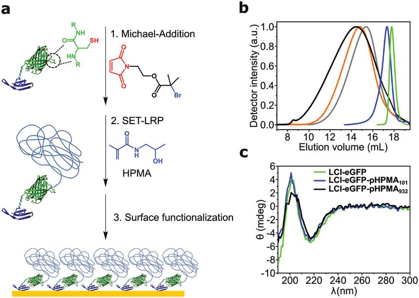
www.advancedsciencenews.com www.mbs-journal.de

Figure 1. a) Linkage of the maleimide initiator to the cysteine residue of eGFP, followed by grafting of poly(HPMA) using SET-LRP and the oriented
adsorption process to surfaces. b) SEC of the bare protein (green) and LCI-eGFP-pHPMA with DPs of 101 (blue), 699 (gray), 843 (orange), and 932
(black). c) CD spectra of LCI-eGFP (green) and LCI-eGFP-pHPMA with a DP of 101 (blue) and 932 (black).

their assembly on gold. By utilizing X-ray reflectivity (XRR) and initiator ratios to have a broad variation in the DP (Table S1,
atomic force microscopy (AFM), we investigated the organization Supporting Information). Hereafter, the samples are referred
of the LCI-eGFP-pHPMA. By a multilayer fitting we analyzed the to as LCI-eGFP-pHPMAX , where X corresponds to number
thickness, roughness, and scattering length density of every com- average DP calculated from Mn obtained from size exclusion
ponent (three layers) and studied the influence of the DP and chromatography (SEC) in PBS. All polymerizations afforded
grafting density. Lastly, surface plasmon resonance (SPR) spec- polymers as confirmed by 1 H NMR (Supporting Information,
troscopy measurements demonstrated the dependency of the DP Figure S1). SEC revealed molecules with monomodal distri-
and the surface grafting density on the antifouling capabilities bution of molecular weight and no presence of free initiator
against the adsorption of blood plasma proteins. (Figure 1b), indicating a quantitative initiation efficiency, well in
 line with previous reports for aqueous SET-LRP.[92,94] The molec-
2. Results and Discussion ular weight distribution increased with the monomer-to-initiator
 ratio and with conversion. The Mn were well beyond LCI-eGFP
2.1. Synthesis of LCI-eGFP-pHPMA Macromolecules ranging from 51,430 to 170,420 g mol−1 , which correspond to
 DPs from 101 to 932 (Figure 1b and Table S1, Supporting Infor-
The macroinitiator was synthesized by conjugating one mation). Some broadening of the molecular weight distribution
maleimide-initiator to the free cysteine of LCI-eGFP (eGFP, posi- was observed for the sample LCI-eGFP-pHPMA932 , however
tion 69)[75,91] via a thiol-maleimide Michael addition (Figure 1a). it remained monomodal. Thus, the polymerization condition
Poly(HPMA) was directly grafted from LCI-eGFP-initiator in allowed the polymerization of very large molecular weights even
phosphate-buffered saline (PBS) buffer at room temperature us- in aqueous medium.
ing SET-LRP. We selected SET-LRP because it accounts for very Circular dichroism (CD) spectroscopy was performed for the
fast kinetics and control even for methacrylamide monomers in LCI-eGFP-pHPMAs to assess whether the secondary structure
aqueous media with minimal bimolecular termination.[89,90,92] of the peptide was affected by grafting high molecular weight
The latter is necessary to prevent dimer formation that would polymers. Figure 1c shows positive (200 nm) and negative bands
obscure the adsorption process, whereas the compatibility with (218 nm) corresponding to four antiparallel -sheets of LCI and
water is necessary to maintain the integrity of the proteins. The 11 -sheets of eGFP which remained unchanged after polymer-
polymerization was performed using a hydrazine-activated cop- ization. Thus, CD confirmed that the secondary structure of LCI-
per wire as catalyst,[93] the LCI-eGFP-initiator, and the following eGFP remained unaltered even by grafting polymers of tenfold
ratios of [Monomer]:[CuBr2 ]:[Me6 TREN]:[LCI-eGFP-initiator] = size. This could also be inferred by the unmodified fluorescence
[X]:[0.1]:[0.6]:[1], where X represents the molar ratio of monomer stemming from the eGFP structure.
ranging from 300 to 800 (Table S1, Supporting Information). An important part of the concept is that the LCI-eGFP-
We performed the polymerization at different monomer to pHPMAs remain molecularly dissolved, as aggregates would

Macromol. Biosci. 2021, 2100158 2100158 (3 of 10) © 2021 The Authors. Macromolecular Bioscience published by Wiley-VCH GmbH
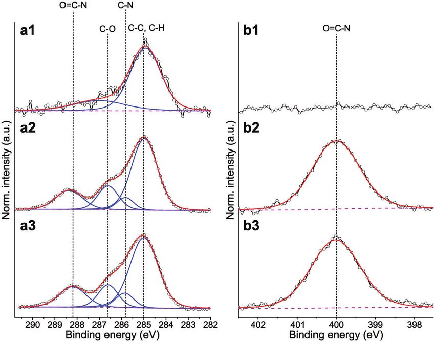
www.advancedsciencenews.com www.mbs-journal.de

Figure 2. High-resolution C1s (left) and N1s (right) XPS spectra of surface modifications on gold; a1/b1 = bare gold, a2/b2 = LCI-eGFP, a3/b3 =
LCI-eGFP-pHPMA491 .

pose kinetic barriers to adsorption. Dynamic light scattering (a1) depicting signals characteristic of C−C, C−H at 285.0 eV and
(DLS) measurements showed that LCI-eGFP displayed a hydro- C−O around 286.6 eV. No nitrogen was observed in the N1s spec-
dynamic diameter of 5.3 nm. Grafting poly(HPMA) from it re- trum. It is of note that the amount of counts in the C1s is 5–10
sulted in a gradual increase of the hydrodynamic radius from 6.2 times lower on gold than on any of the coated surfaces.
to 18.7 nm (Figure S2 and Table S2, Supporting Information). After adsorption of LCI-eGFP and the LCI-eGFP-pHPMA,
No aggregates could be observed in the intensity, volume, and the C1s spectra (a2 and a3) revealed new signals at 285.8
number distributions of hydrodynamic radii. Thus, DLS mea- and 286.6 eV, corresponding to the signals of C−N and C−O,
surements demonstrate that the LCI-eGFP-pHPMAs do not form respectively.[47] The presence of these signals correlates with the
supramolecular aggregates in water. chemical structures of the protein and HPMA. Importantly, the
 peak at 288.2 eV indicates a strong presence of amide bonds,
 which can be ascribed to the peptide bonds in the protein and
2.2. Formation of LCI-eGFP-pHPMA Film on Gold the methacrylamide backbone of poly(HPMA). In contrary to the
 bare gold surface (b1), the high-resolution N1s spectra of the
The coating was assembled on gold by the molecular physisorp- functionalized samples (b2 and b3) clearly show a component at
tion of LCI-eGFP-pHPMA. PBS solutions of the macromolecules 400 eV, stemming from amide bonds.[95]
were contacted with gold-coated silicon wafers for 60 min, fol-
lowed by copious rinsing with PBS and ultrapure water. The re-
sulting films had a thickness of about 5 nm by ellipsometry, sug- 2.3. Unraveling the Mechanism of LCI-eGFP-pHPMA Binding
gesting that the coatings were only one molecule thick. Addition- and Film Formation
ally, AFM of the LCI-eGFP-pHPMA coatings revealed an ultra-
thin film that followed the facets of the polycrystalline gold sub- To elucidate the mechanism of binding of LCI-eGFP-pHPMA
strate (Figure S3, Supporting Information). on gold, we determined the adsorption isotherms and electron
 The chemical composition of the coatings was confirmed us- density profiles. For the studies of binding kinetics, we utilized
ing X-ray photoelectron spectroscopy (XPS). Figure 2 depicts the LCI-eGFP-pHPMA491, as it exhibits a narrow molecular weight
high-resolution C1s (a) and N1s (b ) spectra of a bare gold sur- distribution (homogeneity of sizes), while providing a molecular
face and gold surfaces after functionalization with LCI-eGFP and weight significantly higher than LCI-eGFP. We quantified the
LCI-eGFP-pHPMA491 via physisorption at a concentration of 400 surface adsorption of LCI-eGFP-pHPMA491 at different concen-
µg mL−1 . The presence of adventitious carbon adsorbed from the trations (0.04–1000 µg mL−1 , 0.37 × 10⁻⁹ to 9.33 × 10⁻⁶ m) by
atmosphere could be observed on the C1s spectrum of bare gold SPR. Here, the change in baseline refractive index before and

Macromol. Biosci. 2021, 2100158 2100158 (4 of 10) © 2021 The Authors. Macromolecular Bioscience published by Wiley-VCH GmbH
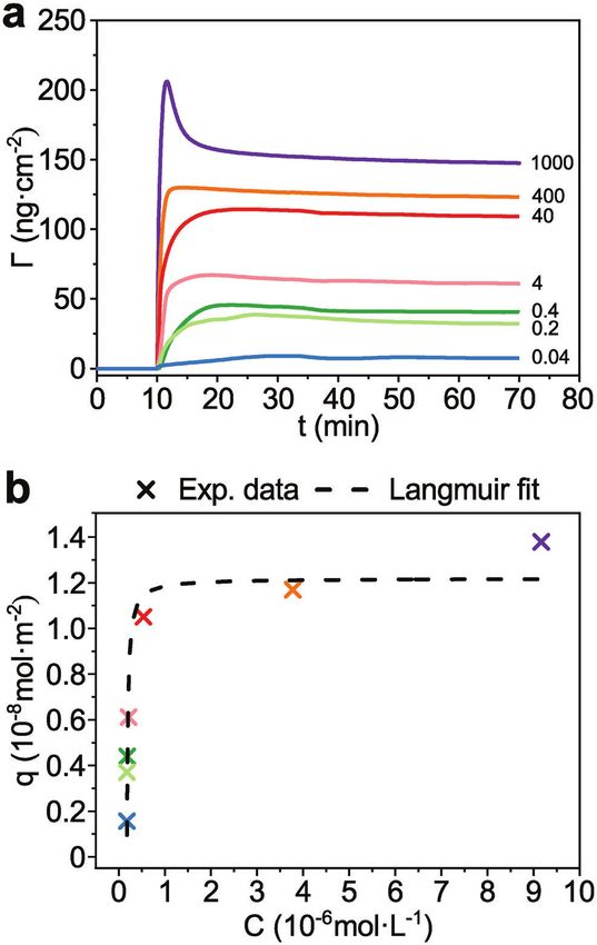
www.advancedsciencenews.com www.mbs-journal.de

 dynamic equilibrium of adsorption and desorption is achieved
 in the course of the experiment.[96,97] Such model could be fitted
 only if one type of adsorbate was present, suggesting that LCI
 preferentially binds the surface compared to poly(HPMA) part.
 However, eGFP may also adsorb. Thus, control experiments
 monitoring the kinetics of eGFP binding to gold were carried
 out demonstrating that the affinity of the eGFP was much lower
 than the one of LCI (Supporting Information, Figure S5). Thus,
 LCI governs the binding process. The values Keq and Q can
 be determined by fitting Equation (1). By fitting Equation (1)
 to an isotherm plot of q (in mol m−2 ) versus C (in mol L−1 )
 (Figure 3b), one can determine Keq , Q, and the Gibbs free
 energy of adsorption Gads . The average Keq is 3.75 × 10⁷ L mol−1 ,
 resulting in an average ∆Gads of −10.22 kcal mol−1 (−42.78 kJ
 mol−1 , see the Supporting Information), a promising adsorption
 strength when compared to the interaction of biotin and avidin,
 one of the strongest noncovalent binding in nature with a
 ∆Gbinding of −20.4 kcal mol−1 .[98] From Q, we derived a maximal
 obtainable surface mass of 128 ng cm−2 (1.19 pmol cm−2 ). It
 is worth noting that a similar amount of protein adsorption is
 observed when bare gold is contacted with a model protein like
 serum albumin Γalbumin = 126 ng cm−2 .[64] The Langmuir-type
 adsorption isotherm indicates a single mode of binding, i.e.,
 the LCI-eGFP-pHPMA must adsorb using the same part of the
 macromolecule, which was assumed to be the LCI. To assess the
 validity of our hypothesis, we studied the grafting density and
 electron density of the films using XRR. This method provides
 information on the thickness, roughness, as well as the electron
 density profile of the thin film along the surface normal. XRR
 was performed on gold-coated silicon wafers for different surface
 coatings consisting of bare LCI-eGFP and LCI-eGFP-pHPMA of
 different molecular weights. After fitting (Figure S7, Supporting
 Information), the experimental data were simulated using the
 parrat formalism.[99] The modeling of the film was performed
 using a multi-slab model with separated SiO2 , TiO2 , Au, LCI,
Figure 3. a) Binding of LCI-eGFP-pHPMA491 at different concentrations eGFP, and poly(HPMA) layers. The scattering length densities
on gold as obtained by SPR. Sensograms were obtained at a flow rate of
10 µL min−1 and are labeled with the sample concentrations in µg mL−1 .
 (SLD), thickness, and roughness for each sample computed from
b) Surface coverage values (crosses) as obtained from (a). The dashed line the fit parameters are shown in Figure 4b,c,e,g and summarized
represents the Langmuir fit of the experimental isotherm. in Table S5 in the Supporting Information.
 First, we analyzed the coating of LCI-eGFP (no polymer)
 which was modeled as a film with two slabs. The first slab (I) was
after injection is proportional to the mass adsorbed to the surface positioned at the interface with Au, while the second (II) at the
(Figure 3a, see Supporting Information for calculation and Table interface with air. The fitting resulted in a thickness of 1.9 nm
S3). Comparison of all data plots (Figure 3b, crosses) illustrates for slab (I) and 4.8 nm for slab (II), close to the height of eGFP
a fast-initial increase in mass adsorbed with higher sample (4.2 nm), with an SLD of 0.6 and 1.4 × 10⁻⁵ Å−2 , respectively.
concentrations, until 400 µg mL−1 (3.73 × 10⁻⁶ m) and thereafter The height of the slab I is in close agreement with the height of
a plateau is observed. The adsorption isotherm could be fitted an LCI peptide fully spread calculated using molecular modeling
with a Langmuir-like profile (Equation (1), see the Supporting with YASARA (Supporting Information, Figure S6 and Table S4).
Information for derivation)[96] Furthermore, the XRR data suggest that the top layer consists of
 eGFP that are packed with their axis preferably orthogonal to the
 QC surface. Therefore, in this model slab (I) should consist of LCI
q= (1)
 C + Keq
 −1
 and its linker, an -helix (17 amino acids).[75] Figure 4d illustrates
 a model for the assembly of LCI-eGFP consistent with the XRR.
where Q is the amount of solute attached at surface saturation Geometrical analysis of eGFP and LCI indicates that their cross-
and q is the amount of solute at the surface for a given concen- sections are similar; 4.5 and 4.0 nm2 , respectively.[80] However,
tration (C) with Keq being the adsorption equilibrium constant. there are striking differences in the SLD of both slabs despite
This model relies on the following assumptions: 1) the sample the peptides mainly consisting of -sheets and -helix (from
saturates as a monolayer; 2) the surface consists of homogenous the linker) of similar density. The dimensions of slab (II) are
adsorption sites available for one type of adsorbate; and 3) a consistent with the packing of eGFP presumably driven by lateral

Macromol. Biosci. 2021, 2100158 2100158 (5 of 10) © 2021 The Authors. Macromolecular Bioscience published by Wiley-VCH GmbH
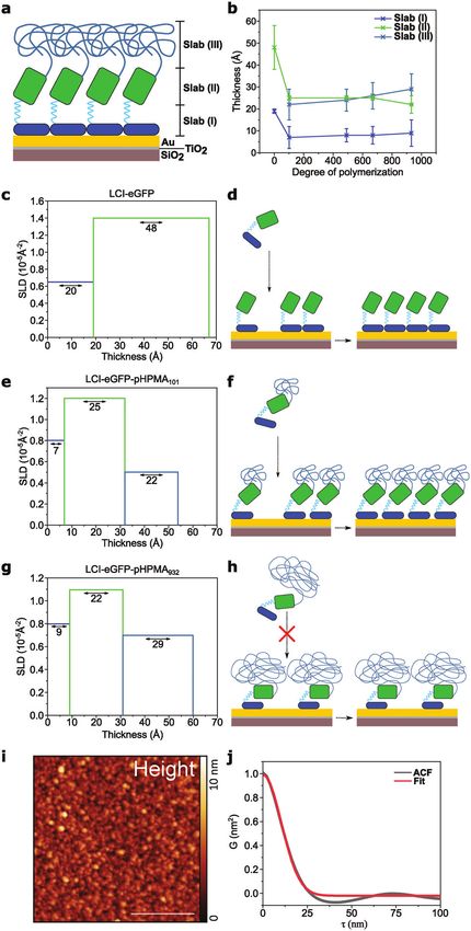
www.advancedsciencenews.com www.mbs-journal.de

Figure 4. a) Sketch of layers of the modeled multi-slab surface film. b) Thickness of slab (I), (II), and (III) as a function of DP. c) SLD and thickness of slab
(I) and (II) of LCI-eGFP. d) Scheme of surface adsorption of LCI-eGFP on gold. e) SLD and thickness of slab (I), (II), and (III) of LCI-eGFP-pHPMA101 .
f) Scheme of surface adsorption of LCI-eGFP-pHPMA with low DP. g) SLD and thickness of slab (I), (II), and (III) of LCI-eGFP-pHPMA932 . h) Scheme
of surface adsorption of LCI-eGFP-pHPMA with high DP. i) AFM height image of LCI-eGFP-pHPMA932 on mica, scale bar is 400 nm. j) 1D ACF of the
surface topography with a Gaussian function fit.

Macromol. Biosci. 2021, 2100158 2100158 (6 of 10) © 2021 The Authors. Macromolecular Bioscience published by Wiley-VCH GmbH
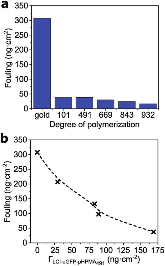
www.advancedsciencenews.com www.mbs-journal.de

associations. However, the thickness of slab (I) is higher than
the molecular thickness of LCI indicating that the resulting SLD
is the combination of the SLD of LCI and peptide linker—which
is orthogonal to the surface—and of air. This model suggests
that the assembly is driven by LCI but once on the surface lateral
interactions between eGFP determine the maximum density.
 Grafting poly(HPMA) from LCI-eGFP had a clear effect in
the assembly. A third slab (III) was necessary to account for
poly(HPMA). The thickness of slab (III) increased with the incre-
ment of the degree of polymerization while the SLD remains con-
stant. However, the bulky size of the polymer caused differences
in the slab beneath. Even the polymer with the smallest degree
of polymerization resulted in a marked decrease in the thickness
from 48 to 25 Å and a reduction of the SLD from 1.4 to 1.2. In-
creasing the degree of polymerization from 101 to 669 did not
result in changes in the thickness of slab (II) nor the SLD. This
suggests that there was no increase in the separation between the
eGFP at that range of degree of polymerization. But how can slab
(II) maintain the same SLD when the steric repulsion between
the poly(HPMA) top groups aim at forcing it apart? We hypoth-
esize that tilting of eGFP can account for the need of a larger
interface area with poly(HPMA) and for the observed decrease
in thickness between the LCI-eGFP and LCI-eGFP-pHPMA (Fig-
ure 4f,h). The tilting of eGFP requires strong tipping of the rigid
peptide linker. This is reflected in the lower thickness and corre-
sponding higher SLD of slab (I) in the presence of poly(HPMA).
 Furthermore, we utilized AFM to assess how LCI-eGFP-
pHPMA arranged when confined to an interface, at the surface
of mica. In these studies, we utilized mica to rule out the effect
of the roughness of the substrate. The topographic images ev-
idenced that the LCI-eGFP-pHPMA coating consisted of gran-
ules packed without pinholes (Figure 4i,j). To estimate the gran-
ule size, we used a 1D autocorrelation function (ACF), which was
calculated along the fast scanning axis and was averaged for all
lines. The ACF was then fitted to a Gaussian function to obtain
the average radius of an equivalent disk that has the same pro- Figure 5. a) Fouling of 10% blood plasma on bare gold and on surfaces
jected area than the granules. The obtained radii for LCI-eGFP- coated with LCI-eGFP-pHPMA with increasing DP. b) The reduction of
pHPMA932 were 14.5 nm ± 0.25 nm (Supporting Information, fouling of 10% blood plasma for LCI-eGFP-pHPMA491 at different surface
Figure S4). Such average radius was larger than the hydrody- densities.
namic radius (9 nm) measured for the same molecule by DLS.
This indicates that upon adsorption the poly(HPMA) part of the
molecule spreads at the interface increasing the interfacial area We analyzed the effect of the DP on the antifouling behav-
while decreasing the height, well in line with the XRR observa- ior at a concentration of 2400 µg mL−1 , and compared the foul-
tion and model (Figure 4h). ing of blood plasma on gold before and after coating with LCI-
 eGFP-pHPMA (Figure 5a). Even the sample having the lowest
 DP already reduced the fouling by 88% (38 ng cm−2 ). Slight im-
2.4. Influence of Degree of Polymerization and Grafting Density provements were observed by increasing DP in the range of 669–
on Antifouling Capabilities 932 to reach a decrease in fouling by 94% (17 ng cm−2 ). The
 values of fouling are almost on par to those of best polymer
We studied the antifouling capabilities utilizing coatings pre- brushes[40,58,100,101] and much lower than for other hydrophilic
pared from LCI-eGFP-pHPMA with varying DP as well as dif- coatings based on grafted linear[39,102] or star-shaped[103] poly-
ferent surface densities and evaluated their resistance to protein mers, hydrogels[104] and even four orders of magnitude lower
fouling from blood plasma with SPR. Protein adsorption from than microgels.[105]
blood plasma (10% in PBS) was measured after surface satura- Additionally, we investigated the antifouling properties in re-
tion with the LCI-eGFP-pHPMA solutions was achieved. The dif- gards of the surface density. We systematically varied the surface
ference in sensor response (detected as Δ RIU) between base- density of LCI-eGFP-pHPMA491 and assessed the fouling from
lines before and after injection is proportional to the mass ad- 10% blood plasma. Figure 5b shows that increasing the grafting
sorbed on the surface (see Supporting Information for calcula- density from 30 to 170 ng cm−2 results in a drastic reduction
tion and Figure S8). of adsorbed proteins, highlighting the importance of the close

Macromol. Biosci. 2021, 2100158 2100158 (7 of 10) © 2021 The Authors. Macromolecular Bioscience published by Wiley-VCH GmbH
www.advancedsciencenews.com www.mbs-journal.de

arrangement of the LCI-eGFP. Interestingly, surface densities Adsorption Kinetics by SPR: Sensor slides (gold coated, Cenibra, Ger-
as low as 89 ng cm−2 (formed from a solution c = 400 µg mL−1 ) many) were pre-cleaned by rinsing with absolute ethanol and ultra-pure
were already sufficient to fully prevent adsorption of albumin water. Adsorption kinetics were monitored using the MP-SPR Navi 210A
 VASA (BioNavis, Finland) by injecting a solution of LCI-eGFP-pHPMA491
and to decrease blood plasma fouling to 98 ng cm−2 which is
 at concentrations of 1000, 400, 40, 20, 4, 0.4, 0.2, and 0.04 µg mL−1 in
equivalent to a reduction of 68% of fouling on gold. On the other PBS at a flow rate of 10 µL min−1 for 25 min, followed by flow of PBS
hand, after achieving complete surface saturation (168 ng cm−2 ), at 10 µL min−1 until a stable baseline was reached. To evaluate the re-
only 20 ng cm−2 of blood plasma could be detected. Such level is sults, SPR-Navi Data Viewer (Version 4.3.5.2) was used. Measurements
in pair with the best antifouling polymer brushes.[39,40,43,44,58] results at 670 nm were used for data comparison and analysis. Adsorption
 isotherms were built from the obtained values of equilibrium adsorption.
 Formation of Monomolecular Coatings of LCI-eGFP-pHPMA on Gold:
 Gold-coated silica wafers were cut into pieces of 2 cm² and rinsed twice
3. Conclusion with pure ethanol and ultrapure water. The coating was carried out by im-
We studied the adsorption and assembly of peptide-polymer mersing the wafers in a solution of LCI-eGFP-pHPMA (400 µg mL−1 ) in
 PBS for 1 h. Subsequently, the solution was diluted by adding and remov-
macromolecules for universal antifouling surface functionaliza-
 ing 5 × 1 mL of PBS and 2 × 1 mL ultrapure water. The samples were
tion based on LCI-eGFP. Adsorption isotherms of LCI-eGFP- stored in ultrapure water and were dried just before use. These samples
pHPMA determined by SPR followed a Langmuir-type behav- were utilized for ellipsometry, XRR, and XPS analysis.
ior, indicating that the adsorption was driven by a single part of Determination of Density of Monomolecular Coatings of LCI-eGFP-
the LCI-eGFP-pHPMA molecule, i.e., oriented immobilization. pHPMA by XRR: Gold-coated silicon wafers were covered with LCI-eGFP
XRR and AFM measurements confirmed that the binding occurs as well as LCI-eGFP-pHPMA with DPs of 101, 491, 669, and 932 as de-
 scribed above. XRR measurements were performed using an Empyrean
solely by the LCI-peptide. However, it also demonstrated that the
 setup from PANalytical with a Cu X-ray tube (line source of 12 × 0.04 mm2 )
packing of the cylindrical eGFPs controls the assembly at the gold providing Cu K radiation with = 0.1542 nm. A divergence slit of 1/32°
surface. The grafting of LCI-eGFP-pHPMA results in eGFP tilted was set to illuminate part of the parabolic graded multilayer system (Gö-
from the normal to the surface. The tilt increases with the DP bel mirror), while the latter was converted 0.8° from the divergent beam
of poly(HPMA). This allows for a larger interface between eGFP into an almost parallel beam (divergence ≤ 55 mdeg). This geometry was
and poly(HPMA) without reducing the attractive interactions be- used to improve the measurement at low angles. On the diffraction side,
tween LCI-surface and the packing of eGFPs. a receiving slit, 0.04 Soller slit, and PANalytical pixel detector (256 × 256
 pixels of 55 µm) were used to collect the scattered signals. The setup was
 The formed coatings displayed antifouling properties on par calibrated using a high-quality Si-wafer, which resulted in 26 mdeg resolu-
with the best polymer brushes grafted from surface. Only minor tion. X’Pert reflectivity software was used for the fitting process, whereas
changes in the repulsion to proteins were observed for the DPs Parratt32 (Version 1.6.0) was used for electron density profiling.
evaluated, however, a pronounced deterioration of the antifouling Protein Adsorption on LCI-eGFP-pHPMA Coatings: Unspecific protein
efficacy was observed when the density of the LCI-eGFP-pHPMA adsorption was measured by flowing blood plasma (10% in PBS) over LCI-
was decreased away from the equilibrium saturation. eGFP-pHPMA coatings at a flow rate of 6 µL min−1 while monitoring the
 resonant angle in SR7500DC SPR (Reichert Technologies, USA). The coat-
 Interestingly, concentrations as low as 400 µg mL−1 (89 ng
 ings were formed in situ by flowing a solution of LCI-eGFP-pHPMA at a
cm−2 ) were already sufficient to fully prevent adsorption of albu- concentration of 2400 µg mL−1 in PBS at a flow rate of 6.0 µL min−1 un-
min and to decrease blood plasma fouling to 98 ng cm−2 which til equilibrium was reached, followed by PBS washing until a stable base-
is equivalent to a reduction of 68% of fouling. Further increase line was acquired. Immediately afterward, a 10% blood plasma solution
of solution concentration to 2400 µg mL−1 (168 ng cm−2 ) allowed was flowed until equilibration, followed by a final PBS washing step until
to obtain surface saturation and excellent antifouling properties, a stable baseline was again discernible. To assess the effect of the grafting
 density in the antifouling properties, LCI-eGFP-pHPMA491 coatings were
paving the way to the translation of this technology to the medical
 formed at different concentrations (2400, 400, 4, and 0.04 µg mL−1 ) and
field. fouling was determined as described above.

4. Experimental Section Supporting Information
 Further details on procedures and instrumentation are given in the Sup- Supporting Information is available from the Wiley Online Library or from
porting Information. the author.
 Chemicals and Materials: All chemicals were analytical purity and were
used as received unless stated otherwise. LCI-eGFP was produced in E. coli
and purified as previously described (details can be found in the Support- Acknowledgements
ing Information).[80]
 Synthesis of LCI-eGFP-pHPMA via SET-LRP: HPMA (see Table S1, Sup- The authors thank the support of the research association of
porting Information), PBS (0.5 mL), CuBr2 (9.8 µg, 29.3 µmol, 0.1 eq.), Forschungskuratorium Textil e.V. supported via AiF (“Arbeitsgemein-
ME6TREN (30 µL, 170 µmol, 0.6 eq.), and protein-initiator solution (1 mL, schaft Industrielle Forschungsvereinigungen Otto von Guericke e.V.”),
0.38 mol, 1 eq.) were added to a 10 mL Schlenk tube. Subsequently, a research project IGF-No. 19893 N within the promotion program of
hydrazine-activated copper wire (4.5 cm x 0.1 cm) wrapped around a mag- “Industrielle Gemeinschaftsforschung” (IGF) of the Federal Ministry for
netic stir bar was added to the Schlenk flask and kept out of the liquid with Economic Affairs and Energy on the basic of a decision by the German
a magnet. The solution was degassed by at least five freeze-pump-thaw cy- Bundestag. This work was also partially performed at the Center for
cles. The reaction was initiated by dropping the Cu-wire into the solution Chemical Polymer Technology CPT, which was supported by the EU and
at 28 °C and allowed to proceed for 18–20 h. The samples were purified the federal state of North Rhine-Westphalia (grant EFRE 30 00 883 02).
by using centrifugal filter units (Amicon Ultra-15, 10 kDa cut-off, Merck Table of content figure was partially created with BioRender.com
Millipore Ltd.) and two washing steps. Open access funding enabled and organized by Projekt DEAL.

Macromol. Biosci. 2021, 2100158 2100158 (8 of 10) © 2021 The Authors. Macromolecular Bioscience published by Wiley-VCH GmbH
www.advancedsciencenews.com www.mbs-journal.de

Conflict of Interest [30] M. Wyszogrodzka, R. Haag, Biomacromolecules 2009, 10, 1043.
 [31] M. Morra, C. Cassineli, J. Biomater. Sci., Polym. Ed. 1999, 10, 1107.
The authors declare no conflict of interest. [32] S. L. McArthur, K. M. McLean, P. Kingshott, H. A. W. St John, R. C.
 Chatelier, H. J. Griesser, Colloids Surf., B 2000, 17, 37.
 [33] D. Xiao, H. Zhang, M. Wirth, Langmuir 2002, 18, 9971.
Data Availability Statement [34] Y. Mei, T. Wu, C. Xu, K. J. Langenbach, J. T. Elliott, B. D. Vogt, K. L.
 Beers, E. J. Amis, N. R. Washburn, Langmuir 2005, 21, 12309.
Data available on request from the authors. [35] R. Barbey, L. Lavanant, D. Paripovic, N. Schuwer, C. Sugnaux, S.
 Tugulu, H. A. Klok, Chem. Rev. 2009, 109, 5437.
 [36] Y. Chang, S. Chen, Z. Zhang, S. Jiang, Langmuir 2006, 22, 2222.
Keywords [37] S. Chen, L. Liu, S. Jiang, Langmuir 2006, 22, 2418.
 [38] Z. Zhang, T. Chao, S. Chen, S. Jiang, Langmuir 2006, 22, 10072.
adhesion peptides, antifouling, aqueous SET-LRP, biomimetic coating, [39] C. Rodriguez Emmenegger, E. Brynda, T. Riedel, Z. Sedlakova, M.
surface functionalization Houska, A. B. Alles, Langmuir 2009, 25, 6328.
 [40] C. Rodriguez-Emmenegger, E. Brynda, T. Riedel, M. Houska, V. Subr,
 Received: April 16, 2021 A. B. Alles, E. Hasan, J. E. Gautrot, W. T. Huck, Macromol. Rapid
 Revised: May 15, 2021 Commun. 2011, 32, 952.
 Published online: [41] L. Li, S. Chen, J. Zheng, B. D. Ratner, S. Jiang, J. Phys. Chem. B 2005,
 109, 2934.
 [42] M.-C. Sin, S.-H. Chen, Y. Chang, Polym. J. 2014, 46, 436.
 [43] C. Rodriguez-Emmenegger, M. Houska, A. B. Alles, E. Brynda,
 [1] N. A. Peppas, E. W. Merrill, J. Biomed. Mater. Res. 1977, 11, 423. Macromol. Biosci. 2012, 12, 1413.
 [2] N. A. Peppas, R. Langer, Science 1994, 263, 1715. [44] A. de los Santos Pereira, S. Sheikh, C. Blaszykowski, O. Pop-
 [3] J. D. Andrade, Surface and Interfacial Aspects of Biomedical Polymers, Georgievski, K. Fedorov, M. Thompson, C. Rodriguez-Emmenegger,
 Springer, Boston, MA 1985. Biomacromolecules 2016, 17, 1179.
 [4] R. Langer, D. A. Tirrell, Nature 2004, 428, 487. [45] Z. Wu, H. Chen, X. Liu, Y. Zhang, D. Li, H. Huang, Langmuir 2009,
 [5] F. M. Chen, X. Liu, Prog. Polym. Sci. 2016, 53, 86. 25, 2900.
 [6] D. F. Williams, Biomaterials 2009, 30, 5897. [46] G. Cheng, G. Li, H. Xue, S. Chen, J. D. Bryers, S. Jiang, Biomaterials
 [7] L. Vroman, A. L. Adams, J. Biomed. Mater. Res. 1969, 3, 43. 2009, 30, 5234.
 [8] L. Vroman, A. L. Adams, in Proteins at Interfaces, Vol. 343 (Eds: T. A. [47] F. Obstals, M. Vorobii, T. Riedel, A. de Los Santos Pereira, M. Bruns,
 Horbett, J. Brash), ACS Publications, Washington 1987, Ch. 10. S. Singh, C. Rodriguez-Emmenegger, Macromol. Biosci. 2018, 18,
 [9] S. de Maat, C. Maas, J. Thromb. Haemostasis 2016, 14, 1498. 1700359.
 [10] B. Sivaraman, R. A. Latour, Biomaterials 2010, 31, 1036. [48] C. Leng, X. Han, Q. Shao, Y. Zhu, Y. Li, S. Jiang, Z. Chen, J. Phys.
 [11] B. Sivaraman, R. A. Latour, Biomaterials 2010, 31, 832. Chem. C 2014, 118, 15840.
 [12] B. D. Ratner, A. S. Hoffman, F. J. Schoen, J. E. Lemons, Biomaterials [49] Q. Shao, S. Jiang, Adv. Mater. 2015, 27, 15.
 Science: An Introduction to Materials in Medicine, Elsevier, New York [50] C. Leng, S. Sun, K. Zhang, S. Jiang, Z. Chen, Acta Biomater. 2016,
 2004. 40, 6.
 [13] J. M. Anderson, Annu. Rev. Mater. Res. 2001, 31, 81. [51] C. A. Del Grosso, C. Leng, K. Zhang, H.-C. Hung, S. Jiang, Z. Chen,
 [14] D. Pavithra, M. Doble, Biomed. Mater. 2008, 3, 034003. J. J. Wilker, Chem. Sci. 2020, 11, 10367.
 [15] R. Maitra, C. C. Clement, B. Scharf, G. M. Crisi, S. Chitta, D. Paget, [52] J. Kopeček, H. Baẑilová, Eur. Polym. J. 1973, 9, 7.
 P. E. Purdue, N. Cobelli, L. Santambrogio, Mol. Immunol. 2009, 47, [53] K. R. Whiteman, V. Subr, K. Ulbrich, V. P. Torchilin, J. Liposome Res.
 175. 2001, 11, 153.
 [16] S. Behzadi, V. Serpooshan, R. Sakhtianchi, B. Muller, K. Landfester, [54] S. G. Krimmer, H. Pan, J. Liu, J. Yang, J. Kopecek, Macromol. Biosci.
 D. Crespy, M. Mahmoudi, Colloids Surf., B 2014, 123, 143. 2011, 11, 1041.
 [17] S. Ritz, S. Schottler, N. Kotman, G. Baier, A. Musyanovych, J. [55] A. Kelsch, S. Tomcin, K. Rausch, M. Barz, V. Mailänder, M. Schmidt,
 Kuharev, K. Landfester, H. Schild, O. Jahn, S. Tenzer, V. Mailander, K. Landfester, R. Zentel, Biomacromolecules 2012, 13, 4179.
 Biomacromolecules 2015, 16, 1311. [56] L. Nuhn, M. Barz, R. Zentel, Macromol. Biosci. 2014, 14, 607.
 [18] G. Settanni, J. Zhou, T. Suo, S. Schottler, K. Landfester, F. Schmid, [57] F. A. de Oliveira, L. J. C. Albuquerque, K. A. Riske, E. Jager, F. C.
 V. Mailander, Nanoscale 2017, 9, 2138. Giacomelli, J. Colloid Interface Sci. 2020, 574, 260.
 [19] H. W. Fox, P. W. Taylor, W. A. Zisman, Ind. Eng. Chem. 1947, 39, 1401. [58] M. Vorobii, A. de los Santos Pereira, O. Pop-Georgievski, N. Y.
 [20] T. McPherson, A. Kidane, I. Szleifer, K. Park, Langmuir 1998, 14, 176. Kostina, C. Rodriguez-Emmenegger, V. Percec, Polym. Chem. 2015,
 [21] A. Halperin, Langmuir 1999, 15, 2525. 6, 4210.
 [22] Y. Y. Luk, M. Kato, M. Mrksich, Langmuir 2000, 16, 9604. [59] A. R. Kuzmyn, A. T. Nguyen, L. W. Teunissen, H. Zuilhof, J. Bagger-
 [23] R. L. C. Wang, H. J. Kreuzer, M. Grunze, J. Phys. Chem. B 1997, 101, man, Langmuir 2020, 36, 4439.
 9767. [60] E. Roeven, A. R. Kuzmyn, L. Scheres, J. Baggerman, M. M. J. Smul-
 [24] R. E. Holmlin, X. X. Chen, R. G. Chapman, S. Takayama, G. M. White- ders, H. Zuilhof, Langmuir 2020, 36, 10187.
 sides, Langmuir 2001, 17, 2841. [61] C. Rodriguez-Emmenegger, S. Janel, A. de los Santos Pereira, M.
 [25] S. I. Jeon, J. H. Lee, J. D. Andrade, P. G. De Gennes, J. Colloid Interface Bruns, F. Lafont, Polym. Chem. 2015, 6, 5740.
 Sci. 1991, 142, 149. [62] C. J. Van Oss, M. K. Chaudhury, R. J. Good, Chem. Rev. 1988, 88,
 [26] L. D. Unsworth, H. Sheardown, J. L. Brash, Biomaterials 2005, 26, 927.
 5927. [63] B. Lopez-Mila, P. Alves, T. Riedel, B. Dittrich, F. Mergulhao, C.
 [27] L. D. Unsworth, H. Sheardown, J. L. Brash, Langmuir 2005, 21, 1036. Rodriguez-Emmenegger, Bioinspir. Biomim. 2018, 13, 065001.
 [28] L. D. Unsworth, H. Sheardown, J. L. Brash, Langmuir 2008, 24, 1924. [64] C. Rodriguez-Emmenegger, O. Kylian, M. Houska, E. Brynda, A.
 [29] R. K. Kainthan, M. Gnanamani, M. Ganguli, T. Ghosh, D. E. Brooks, Artemenko, J. Kousal, A. B. Alles, H. Biederman, Biomacromolecules
 S. Maiti, J. N. Kizhakkedathu, Biomaterials 2006, 27, 5377. 2011, 12, 1058.

Macromol. Biosci. 2021, 2100158 2100158 (9 of 10) © 2021 The Authors. Macromolecular Bioscience published by Wiley-VCH GmbH
www.advancedsciencenews.com www.mbs-journal.de

 [65] C. Rodriguez-Emmenegger, C. M. Preuss, B. Yameen, O. Pop- [84] V. Percec, T. Guliashvili, J. S. Ladislaw, A. Wistrand, A. Stjerndahl,
 Georgievski, M. Bachmann, J. O. Mueller, M. Bruns, A. S. Gold- M. J. Sienkowska, M. J. Monteiro, S. Sahoo, J. Am. Chem. Soc. 2006,
 mann, M. Bastmeyer, C. Barner-Kowollik, Adv. Mater. 2013, 25, 6123. 128, 14156.
 [66] O. Pop-Georgievski, C. Rodriguez-Emmenegger, A. L. S. Pereira, V. [85] B. M. Rosen, V. Percec, Chem. Rev. 2009, 109, 5069.
 Proks, E. Brynda, F. Rypacek, J. Mater. Chem. B 2013, 1, 2859. [86] N. Zhang, S. R. Samanta, B. M. Rosen, V. Percec, Chem. Rev. 2014,
 [67] X. Fan, L. Lin, J. L. Dalsin, P. B. Messersmith, J. Am. Chem. Soc. 2005, 114, 5848.
 127, 15843. [87] G. Lligadas, S. Grama, V. Percec, Biomacromolecules 2017, 18,
 [68] M. Dahm, B. J. Chang, O. Prucker, M. Pierkes, T. Alt, E. Mayer, J. 1039.
 Ruhe, H. Oelert, Ann. Thorac. Surg. 2001, 71, S437. [88] G. Lligadas, S. Grama, V. Percec, Biomacromolecules 2017, 18,
 [69] O. Sterner, A. Serrano, S. Mieszkin, S. Zurcher, S. Tosatti, M. E. Cal- 2981.
 low, J. A. Callow, N. D. Spencer, Langmuir 2013, 29, 13031. [89] X. Feng, D. S. Maurya, N. Bensabeh, A. Moreno, T. Oh, Y. Luo, J.
 [70] D. Knoll, J. Hermans, J. Biol. Chem. 1983, 258, 5710. N. Lejnieks, M. Galià, Y. Miura, M. J. Monteiro, Biomacromolecules
 [71] K. Holmberg, F. Tiberg, M. Malmsten, C. Brink, Colloids Surf., A 1997, 2020, 21, 250.
 123–124, 297. [90] D. S. Maurya, A. Malik, X. Feng, N. Bensabeh, G. Lligadas, V. Percec,
 [72] E. P. Currie, W. Norde, M. A. Cohen Stuart, Adv. Colloid Interface Sci. Biomacromolecules 2020, 21, 1902.
 2003, 100–102, 205. [91] A. Royant, M. Noirclerc-Savoye, J. Struct. Biol. 2011, 174, 385.
 [73] P. Uhlmann, H. Merlitz, J. U. Sommer, M. Stamm, Macromol. Rapid [92] N. H. Nguyen, C. Rodriguez-Emmenegger, E. Brynda, Z. Sedlakova,
 Commun. 2009, 30, 732. V. Percec, Polym. Chem. 2013, 4, 2424.
 [74] S. Zurcher, D. Wackerlin, Y. Bethuel, B. Malisova, M. Textor, S. [93] N. H. Nguyen, V. Percec, J. Polym. Sci., Part A: Polym. Chem. 2010,
 Tosatti, K. Gademann, J. Am. Chem. Soc. 2006, 128, 1064. 48, 5109.
 [75] S. Dedisch, F. Obstals, A. los Santos Pereira, M. Bruns, F. Jakob, [94] N. H. Nguyen, J. Kulis, H.-J. Sun, Z. Jia, B. van Beusekom, M. E.
 U. Schwaneberg, C. Rodriguez-Emmenegger, Adv. Mater. Interfaces Levere, D. A. Wilson, M. J. Monteiro, V. Percec, Polym. Chem. 2013,
 2019, 6, 1900847. 4, 144.
 [76] W. Gong, J. Wang, Z. Chen, B. Xia, G. Lu, Biochemistry 2011, 50, 3621. [95] M. Kaba, N. Raklaoui, M. F. Guimon, A. Mas, J. Appl. Polym. Sci.
 [77] R. A. Meurer, S. Kemper, S. Knopp, T. Eichert, F. Jakob, H. E. Gold- 2005, 97, 2088.
 bach, U. Schwaneberg, A. Pich, Angew. Chem., Int. Ed. Engl. 2017, [96] R. A. Latour, J. Biomed. Mater. Res., Part A 2015, 103, 949.
 56, 7380. [97] A. Savara, C. M. Schmidt, F. M. Geiger, E. Weitz, J. Phys. Chem. C
 [78] P. Schwinges, S. Pariyar, F. Jakob, M. Rahimi, L. Apitius, M. Hunsche, 2009, 113, 2806.
 L. Schmitt, G. Noga, C. Langenbach, U. Schwaneberg, U. Conrath, [98] N. M. Green, Avidin, Academic Press, New York 1975.
 Green Chem. 2019, 21, 2316. [99] L. G. Parratt, Phys. Rev. 1954, 95, 359.
 [79] S. Dedisch, A. Wiens, M. D. Davari, D. Söder, C. Rodriguez- [100] H. Ma, M. Wells, T. P. Beebe, A. Chilkoti, Adv. Funct. Mater. 2006, 16,
 Emmenegger, F. Jakob, U. Schwaneberg, Biotechnol. Bioeng. 2020, 640.
 117, 49. [101] E. van Andel, S. C. Lange, S. P. Pujari, E. J. Tijhaar, M. M. J. Smulders,
 [80] K. Rübsam, B. Stomps, A. Boker, F. Jakob, U. Schwaneberg, Polymer H. F. J. Savelkoul, H. Zuilhof, Langmuir 2019, 35, 1181.
 2017, 116, 124. [102] J. G. Archambault, J. L. Brash, Colloids Surf., B 2004, 39, 9.
 [81] K. Rübsam, L. Weber, F. Jakob, U. Schwaneberg, Biotechnol. Bioeng. [103] J. Groll, Z. Ademovic, T. Ameringer, D. Klee, M. Moeller, Biomacro-
 2018, 115, 321. molecules 2005, 6, 956.
 [82] K. Rubsam, M. D. Davari, F. Jakob, U. Schwaneberg, Polymers 2018, [104] A. Wörz, B. Berchtold, K. Moosmann, O. Prucker, J. Rühe, J. Mater.
 10, 423. Chem. 2012, 22, 19547.
 [83] M. Noth, Z. Zou, I. El-Awaad, L. C. de Lencastre Novaes, G. Dilarri, [105] P. Saha, M. Santi, M. Emondts, H. Roth, K. Rahimi, J. Grosskurth,
 M. D. Davari, H. Ferreira, F. Jakob, U. Schwaneberg, Biotechnol. Bio- R. Ganguly, M. Wessling, N. K. Singha, A. Pich, ACS Appl. Mater.
 eng. 2021, 118, 1520. Interfaces 2020, 12, 58223.

Macromol. Biosci. 2021, 2100158 2100158 (10 of 10) © 2021 The Authors. Macromolecular Bioscience published by Wiley-VCH GmbH
You can also read