ZAP70 Activation Compensates for Loss of Class IA PI3K Isoforms Through Activation of the JAK-STAT3 Pathway
←
→
Page content transcription
If your browser does not render page correctly, please read the page content below
CANCER DIAGNOSIS & PROGNOSIS
2: 391-404 (2022)                                                                                                                       doi: 10.21873/cdp.10122

ZAP70 Activation Compensates for Loss of
Class IA PI3K Isoforms Through Activation
of the JAK–STAT3 Pathway
MELIKE DEMIR and ONUR CIZMECIOGLU

Department of Molecular Biology and Genetics, Bilkent University, Ankara, Turkey

Abstract. Background/Aim: Tyrosine kinases have crucial                                             transduction pathways, which are crucial for many biological
functions in cell signaling and proliferation. The                                                  processes (1). The most well-characterized protein kinases in
phosphatidylinositol 3-kinase (PI3K) pathway is frequently                                          the human genome are protein serine/threonine kinases and
deregulated in human cancer and is an essential regulator                                           protein tyrosine kinases (2). High levels of tyrosine
of cellular proliferation. We aimed to determine which                                              phosphorylation correspond to enhanced proliferative state (3).
tyrosine kinases contribute to resistance elicited by PI3K                                             The phosphatidylinositol 3-kinase (PI3K) pathway is one
silencing and inhibition. Materials and Methods: To mimic                                           of the most activated signaling pathways in human cancer.
catalytic inactivation of p110α/β, specific p110α (BYL719)                                          Upon activation of receptor tyrosine kinases (RTKs),
and p110β (KIN193) inhibitors were used in addition to                                              recruitment of PI3K to the cell membrane occurs via
genetic knock-out in in vitro assays. Cell viability was                                            coupling the Src homology-2 (SH2) domains of p85 with
assessed using crystal violet staining, whereas cellular                                            tyrosine phosphorylated residues of activated receptors (4).
transformation ability was analyzed by soft-agar growth                                             Oncogenic RAS and other tyrosine kinases can cause PI3K
assays. Results: Activated zeta chain of T-cell receptor-                                           activation. This pathway is also a crucial regulator of cell
associated protein kinase 70 (ZAP70) generated resistance                                           proliferation, survival and motility (5). Although many
to PI3K inhibition. This resistance was via activation of the                                       findings support the idea that targeting the PI3K pathway is
Janus kinase/signal transducer and activator of transcription                                       one of the most promising approaches in cancer therapy (4,
3 (JAK/STAT3) axis. We demonstrated that activated ZAP70                                            6), inhibition of class I PI3K activity by using
has a high transforming capability associated with the                                              pharmacological inhibitors leads to retardation of cell
formation of malignant phenotype in untransformed cells                                             proliferation rather than induction of apoptosis, bringing
and has the potential to be a tumor-initiating factor in                                            about a problem with generation of drug resistance in the
cancer cells. Conclusion: ZAP70 may be a potent driver of                                           longer run (7).
proliferation and transformation in untransformed cells and                                            Previous studies have identified the contribution of several
is implicated in resistance to PI3K inhibitors in cancer cells.                                     activated tyrosine or serine/threonine kinases to growth
Protein phosphorylation causes activation of signal                                                 compensation upon PI3K inhibition (8-10). In addition to
                                                                                                    this, pharmacological inhibition of the PI3K pathway in
                                                                                                    cancer cells leads to up-regulation of receptor tyrosine
                                                                                                    kinases through feedback loops that circumvent baseline
Correspondence to: Onur Cizmecioglu, Department of Molecular                                        level of PI3K inactivation. These compensatory mechanisms
Biology and Genetics, Bilkent University, Ankara, Turkey. E-mail:                                   include forkhead box O (FOXO) transcription factors along
onur.cizmecioglu@bilkent.edu.tr                                                                     with mitogen-activated protein kinase (MAPK)-dependent
                                                                                                    pathways (11, 12). Elucidation of the primary mechanisms
Key Words: PI3K, cell signaling, tyrosine kinases, growth
                                                                                                    of PI3K resistance might be a significant therapeutic
resistance, ZAP70.
                                                                                                    approach for tumorigenesis (6). As an example, Pim-1
©2022 International Institute of Anticancer Research                                                serine/threonine kinase (PIM1) regulates cell proliferation
www.iiar-anticancer.org                                                                             and survival that might lead to reduction of therapeutic drug
                                                                                                    efficacy by promoting the Janus kinases/signal transducer
                                                                                                    and activator of transcription proteins (JAK/STAT) signaling
                 This article is an open access article distributed under the terms and
                                                                                                    pathway in MYC proto-oncogene (MYC)-driven cancer (13-
                                                                                                    16), particularly in breast cancer.
                 conditions of the Creative Commons Attribution (CC BY-NC-ND) 4.0
                 international license (https://creativecommons.org/licenses/by-nc-nd/4.0).

                                                                                              391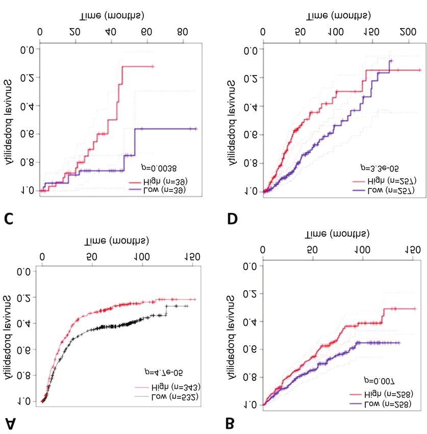
CANCER DIAGNOSIS & PROGNOSIS 2: 391-404 (2022)

Table I. Activated tyrosine kinase library. The list of activated tyrosine kinase pools is presented. Pool 9, which was investigated in further detail
in this study, containing zeta chain of T-cell receptor-associated protein kinase 70 (ZAP70) tyrosine kinase, is shown in bold.

Pool #                            Gene symbol                            Full name

1                                    ERBB3                               erb-b2 receptor tyrosine kinase 3
                                     ERBB4                               erb-b2 receptor tyrosine kinase 4
                                       FES                               FES proto-oncogene, tyrosine kinase
                                      FGR                                FGR proto-oncogene, Src family tyrosine kinase
2                                      FRK                               Fyn-related Src family tyrosine kinase
                                      HCK                                HCK proto-oncogene, Src family tyrosine kinase
                                     IGF1R                               Insulin-like growth factor 1 receptor
                                       ITK                               Interleukin 2 inducible T-cell kinase
3                                    MATK                                Megakaryocyte-associated tyrosine kinase
                                     NTRK2                               Neurotrophic receptor tyrosine kinase 2
                                    PKMYT1                               Protein kinase, membrane associated tyrosine/threonine 1
                                       TEC                               Tec protein tyrosine kinase
                                      PTK7                               Protein tyrosine kinase 7 (inactive)
4                                      TXK                               TXK tyrosine kinase
                                     ZAP70                               Zeta chain of T-cell receptor-associated protein kinase 70
                                     ERBB2                               erb-b2 receptor tyrosine kinase 2
                                      MET                                MET proto-oncogene, receptor tyrosine kinase
5                                     LCK                                LCK proto-oncogene, Src family tyrosine kinase
                                     NTRK1                               Neurotrophic receptor tyrosine kinase 1
                                    PDGFRA                               Platelet-derived growth factor receptor alpha
                                    PDGFRB                               Platelet-derived growth factor receptor beta
6                                     TNK1                               Tyrosine kinase non receptor 1
                                      FLT3                               Fms-related receptor tyrosine kinase 3
                                     EPHB4                               EPH receptor B4
                                      DDR2                               Discoidin domain receptor tyrosine kinase 2
                                     MST1R                               Macrophage-stimulating 1 receptor
7                                      RET                               Ret proto-oncogene
                                       SYK                               Spleen-associated tyrosine kinase
                                      TYK2                               Tyrosine kinase 2
                                      ABL2                               ABL proto-oncogene 2, non-receptor tyrosine kinase
8                                     PTK2                               Protein tyrosine kinase 2
                                     STYK1                               Serine/threonine/tyrosine kinase 1
                                     FGFR2                               Fibroblast growth factor receptor 2
                                       LYN                               LYN proto-oncogene, Src family tyrosine kinase
9                                    PTK2B                               Protein tyrosine kinase 2 beta
                                      PTK6                               Protein tyrosine kinase 6
                                     ZAP70                               Zeta chain of T-cell receptor-associated protein kinase 70
                                     NTRK3                               Neurotrophic receptor tyrosine kinase 3
10                                    DDR1                               Discoidin domain receptor tyrosine kinase 1
                                     EPHA3                               EPH receptor A3
                                      FLT1                               Fms-related receptor tyrosine kinase 1
                                     MUSK                                Muscle-associated receptor tyrosine kinase
                                       AXL                               AXL receptor tyrosine kinase
11                                   LMTK2                               Lemur tyrosine kinase 2
                                      JAK2                               Janus kinase 2
                                     TYRO3                               TYRO3 protein tyrosine kinase
                                       ALK                               ALK receptor tyrosine kinase
12                                     BTK                               Bruton tyrosine kinase
                                     EPHA4                               EPH receptor A4
                                     EPHA6                               EPH receptor A6
                                       FER                               FER tyrosine kinase
13                                   FGFR1                               Fibroblast growth factor receptor 1
                                      FLT4                               Fms-related receptor tyrosine kinase 4
                                     INSRR                               Insulin receptor related receptor
                                      KDR                                Kinase insert domain receptor
14                                   EPHA2                               EPH receptor A2
                                      ROR2                               Receptor tyrosine kinase-like orphan receptor 2

                                                                                                                                  Table I. Continued

                                                                        392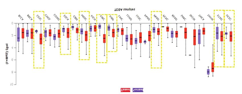
Demir and Cizmecioglu: Investigation of PI3K Functional Compensation via Activated Tyrosine Kinases

Table I. Continued

Pool #                        Gene symbol                           Full name

                                 CSF1R                              Colony-stimulating factor 1 receptor
                                 EPHA1                              EPH receptor A1
15                               EPHB1                              EPH receptor B1
                                 EPHB6                              EPH receptor B6
                                  JAK1                              Janus kinase 1
                                 FGFR3                              Fibroblast growth factor receptor 3
                                  JAK3                              Janus kinase 3
16                                 SRC                              SRC proto-oncogene, non-receptor tyrosine kinase
                                 EGFR                               Epidermal growth factor receptor
                                  BMX                               BMX non-receptor tyrosine kinase
                                  FYN                               FYN proto-oncogene, Src family tyrosine kinase
17                                YES1                              YES proto-oncogene 1, Src family tyrosine kinase
                                  ABL1                              ABL proto-oncogene 1, non-receptor tyrosine kinase
                                  BLK                               BLK proto-oncogene, Src family tyrosine kinase
                                  CSK                               C-Terminal Src kinase
                                  TIE1                              Tyrosine kinase with immunoglobulin like and interleukin-like domains 1

   Zeta chain of T-cell receptor associated protein kinase 70             with 4.5 g/l D-glucose and 1 mM L-glutamine (Sigma–Aldrich)
(ZAP70) is a cytoplasmic tyrosine phosphoprotein. It belongs              supplemented with 8% fetal bovine serum (FBS, Biowest, Nuaillé,
to spleen tyrosine kinase (SYK) family of nonreceptor tyrosine            France), and 100 IU/ml penicillin and 100 μg/ml streptomycin.
                                                                          Human telomerase reverse transcriptase-immortalized retinal
kinases (17). ZAP70 has a crucial role in T-cell receptor
                                                                          pigment epithelial-1 (RPE1-hTERT) cells were grown in DMEM/F-
signaling activation and T-cell development (18). Based on                12, supplemented with 8% FBS, 1 mM L-glutamine and 100 IU/ml
structural studies, T-cell activation by antigens results in              penicillin and 100 μg/ml streptomycin (Thermo Fisher Scientific,
phosphorylation of LCK proto-oncogene, Src family tyrosine                Waltham, MA, USA). The non-tumorigenic human epithelial breast
kinase (LCK) and binding of SH2 domains of ZAP70 to                       cells MCF10A and human mammary epithelial cells (HMECs) were
tyrosine phosphorylated immunoreceptor tyrosine-based                     cultured in DMEM/F-12, supplemented with 4% FBS, penicillin
activation motifs which leads to its recruitment to the                   (100 IU/ml), streptomycin (100 μg/ml), L-glutamine, 10 ng/ml
                                                                          epidermal growth factor (Sigma–Aldrich), 10 μg/ml insulin (Sigma–
membrane (19). Existing literature has also described non-
                                                                          Aldrich) and 0.5 μg/ml hydrocortisone (Sigma–Aldrich). Jurkat cells
immunological functions of ZAP70. ZAP70 was shown to                      were maintained in RPMI 1640 medium supplemented with 8%
promote migration and invasion of prostate cancer cell lines              FBS, 1 mM L-glutamine, and 100 IU/ml penicillin/streptomycin. All
(20). Moreover, in mammalian oocytes and embryonic stem                   cell lines were incubated at 37˚C with 5% CO2. The Jurkat and
cells, ZAP70 has a role in maintaining stemness and                       hTERT-RPE1 cells were purchased from the American Type Culture
differentiation by regulating the JAK–STAT3–MYC signaling                 Collection (Manassas, VA, USA). All the other cell lines have
axis (21). Additionally, ZAP70 was identified as a prognostic             previously been validated and characterized (23).
                                                                             To identify a tyrosine kinase that potentially contributes to
marker in colorectal cancer for radiation response (22). In this
                                                                          generating resistance to PI3K abrogation, we screened an activated
study, we wanted to identify potential tyrosine kinases that              tyrosine kinase library in MEFs which are genetically engineered
contribute to generating resistance to PI3K inhibition. ZAP70             with LoxP sites, inserted into the first exons of PIK3CA and
was identified as a mediator of resistance to PI3K ablation in            PIK3CB (23, 24). The LoxP sequences in these cells can be targeted
our genetic screen. We then aimed to understand the potential             by Cre recombinase or LacZ-expressing adenoviruses (Ad/Cre,
role of activated ZAP70 in tumor initiation, carcinogenesis               Ad/LacZ; The University of Iowa, Viral Vector Core, Iowa City, IA,
and resistance to PI3K inhibition.                                        USA) for excision of the targeted exons of PIK3CA and PIK3CB to
                                                                          generate p110α/β double knock-out cells. The activated tyrosine
                                                                          kinase library has 73 open-reading frames (Table I). This pooled
Materials and Methods                                                     tyrosine kinase library consists of modified receptor and non-
                                                                          receptor tyrosine kinases. The activated tyrosine kinase library
Generation of cell lines expressing activated tyrosine kinase and         constructs have C-terminal dimerization tag; ETS variant
cell culture. p110αflox/flox; p110βflox/flox Mouse embryonic              transcription factor 6 (ETS6) dimerization domain (TEL) and are
fibroblasts (MEFs) and SV40 large T-antigen-expressing human              tagged with FLAG epitope (25). The tyrosine kinase pools were
embryonic kidney 293 (HEK293T) cells, T47D and MCF7 luminal               stably expressed in MEFs and screened using crystal violet growth
A type breast cancer cells were cultured in Dulbecco’s modified           assay. The TEL-RTK library was stably expressed in cells using the
Eagle’s medium (DMEM; Sigma–Aldrich, St Louis, MO, USA)                   transfection protocol of Lipofectamine 2000 Reagent (Invitrogen,

                                                                    393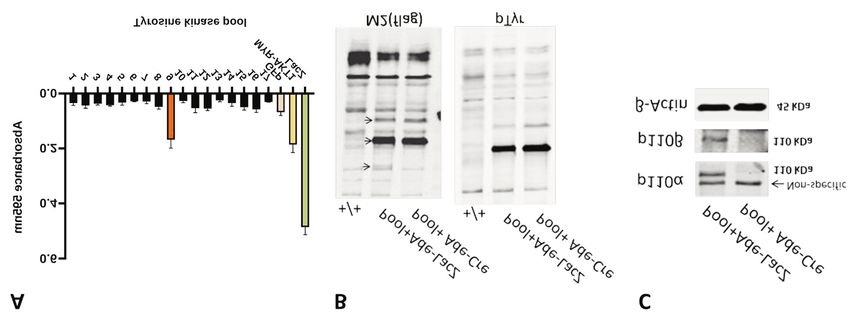
CANCER DIAGNOSIS & PROGNOSIS 2: 391-404 (2022)

Carlsbad, CA, USA). To generate HEK293T cells transiently                      Cell Signaling, Danvers, MA, USA), PI3K p110β (C-8; Santa Cruz
expressing the activated tyrosine kinase library, Lipofectamine 3000           Biotechnology, Dallas, TX, USA), phospho-STAT3 (Tyr705) (Cell
Reagent (Invitrogen) was used. The pWzl green fluorescent protein-             Signaling), ZAP70 (1E7.2) (Santa Cruz Biotechnology), phospho-
expressing (#12269; Addgene, Watertown, MA, USA) and                           ZAP70 (Tyr493/Syk (Tyr526) (Cell Signaling), STAT3 (Cell
myristoylated AKT serine/threonine kinase 1 (MYR-AKT1)-                        Signaling), phospho- AKT (Ser473) (Cell Signaling), phospho-AKT
expressing (#15294; Addgene) MEFs were generated by retroviral                 (Thr308) (D25E6) (Cell Signaling), phospho-p44/42 extracellular
transduction. The H-Ras G12V construct (#9051; Addgene) was                    signal-regulated protein kinase 1/2 (ERK1/2) (Thr202/Tyr204) (Cell
stably expressed in MEFs and RPE1-hTERT cells using the protocol               Signaling), phospho-p70 S6 kinase (Thr389) (Cell Signaling), S6
of Lipofectamine 2000 reagent (Invitrogen).                                    ribosomal protein (Cell Signaling), glyceraldehyde-3-phosphate
                                                                               dehydrogenase (GAPDH) (Cell Signaling), β-actin (clone AC74;
Cell viability assays and inhibitor treatment. The cells were seeded           Sigma–Aldrich) phospho-tyrosine 4G10 (Sigma–Aldrich).
into 12-well or 6-well plates at a confluency of 30-40% and then
treated with increasing concentrations of p110α-specific inhibitor             In-silico analysis. Gene Expression Profiling Interactive Analysis
BYL719 (0.3, 0.5 or 1 μM, Alpelisib; Selleck Chem, Houston, TX,                (GEPIA; http://gepia2.cancer-pku.cn) is an interactive platform for
USA) or p110β-specific inhibitor KIN193 (1 μM, AZD6482;                        analyzing RNA sequencing expression data of 9,736 tumors and 8,587
Selleck Chem) or with both inhibitors combined (0.3, 0.5 or 1 μM)              normal samples from The Cancer Genome Atlas (TCGA) and the
for approximately 7-10 days. The inhibitors were supplemented in               Genotype Tissue Expression (GTEx) projects. Analysis of survival was
reduced serum medium (4% FBS) to maintain physiologically                      conducted by selecting disease-free survival as the endpoint with a
relevant levels of growth factors. When control cells reached about            median cutoff of 50% for each gene of interest to generate Kaplan–
80% confluency, cells were fixed in 10% acetic acid and 10%                    Meier survival curves. In addition, survival curves were generated using
ethanol for at least 24 hours at room temperature. Cells were then             KM plotter by restricting the analysis of progression-free survival by
washed with 1× non-sterile phosphate-buffered saline and stained               grade and chemotherapy treatment (26). University of Alabama Cancer
with 0.4% crystal violet and 20% ethanol for 1 hour at RT. Cells               web portal (UALCAN) database was used to depict the comparison of
were washed with distilled water and air-dried. DNA intercalating              mRNA expression levels between tumor and normal TCGA datasets
dye was removed with 1 ml 10% acetic acid. Plates were incubated               (27). The cBio Cancer Genomics Portal was used to indicate the
in destaining solution for 1 hour at room temperature. The optical             alteration frequency of ZAP70 in various type of cancer using US
density of the solution was measured at 595 nm.                                National Cancer Institute (NCI)-60 cancer cell line studies (28).

Adenovirus/Cre recombinase transduction of MEFs. MEF cell lines                Statistical analysis. Two-way analysis of variance and paired t-test
were seeded in 6-well plates in duplicates at a density of 1×10⁵ cells         were used for differential comparison between variables. All statistical
per well. The day after seeding, cells were treated with Ad/Cre at a           analyses were performed in GraphPad Prism 8.0 (San Diego, CA,
multiplicity of infection of 75 in medium containing 2% FBS and                USA). p-Values less than 0.05 were considered significant.
incubated for 6-8 h. Then the medium was removed and growth
medium was added into cells. The infection was repeated three                  Results
times to facilitate near-complete excision of the first exons of
endogenous PIK3CA and PIK3CB. The adenovirus/Cre-treated cells                 Library screen for identifying factors that generate
were seeded into 12-well plates at a density of 2×10³ cells per well           resistance to PI3K inhibition. Our activated tyrosine kinase
and cultured for 6 days for cellular viability assay.
                                                                               library comprises 73 open-reading frames of receptor and
Soft-agar assay. The soft-agar assay was performed in 6-well plates
                                                                               non-receptor tyrosine kinases which are activated via C-
as triplicates. To form the bottom layer, 2.5 ml DMEM containing               terminal dimerization domain of the TEL transcription factor
2.25% low-melting agarose (Sigma–Aldrich) mixture was poured                   (Table I) (25). We stably expressed activated tyrosine kinases
into each well and were solidified at room temperature for at least            in MEFs and generated p110α/β double knock-out cells. As
30 minutes. Then 2×10⁵ cells were resuspended in 9 ml DMEM                     a result of our screening, Pool 9, which included protein
solution and mixed with 3.2 ml 2.25% low-melting agarose. Two                  tyrosine kinase 2 beta (PTK2B), protein tyrosine kinase 6
millimeters of this mixture was poured into each well and allowed
                                                                               (PTK6), ZAP70 and neurotrophic receptor tyrosine kinase 3
to solidify. A total of 2-3 ml complete growth medium was added
to each well and refreshed every 4-5 days. After 4 weeks, the
                                                                               (NTRK3), partially restored growth capability upon dual loss
colonies formed were stained with 0.005% crystal violet and 10%                of p110α/β (Figure 1A). In our screen, green fluorescent
ethanol solution and counted under a microscope.                               protein-expressing MEFs were used as a negative control,
                                                                               whereas MYR-AKT1-expressing MEFs served as a positive
Antibodies and western blotting. The protein lysates were prepared             control as MYR-AKT1 localizes to the cell membrane and
from MEFs, RPE1-hTERT, MCF10A and HEK293T cell pellets.                        becomes constitutively activated (29). Then, we extracted
Then 20-30 μg of protein was resolved by sodium dodecyl                        proteins from MEFs that expressed FLAG-tagged activated
sulphate–polyacrylamide gel electrophoresis and transferred to
                                                                               tyrosine kinases in Pool 9 before and after Ad/Cre treatment
nitrocellulose membranes (Bio-Rad, Hercules, CA, USA). The
membranes were blocked with 5% bovine serum albumin (Serva,                    and performed western blot analysis. Immunoblotting
Heidelberg, Germany) in TBS and then incubated with primary                    experiments confirmed the presence of immunoreactive
antibodies in TBS with 5% bovine serum albumin and 0.1% Tween.                 bands compatible with the expected sizes of NTRK3, ZAP70
The following primary antibodies were used: PI3K p110α (C73F8;                 and PTK6, whereas PTK2B expression was not detected

                                                                         394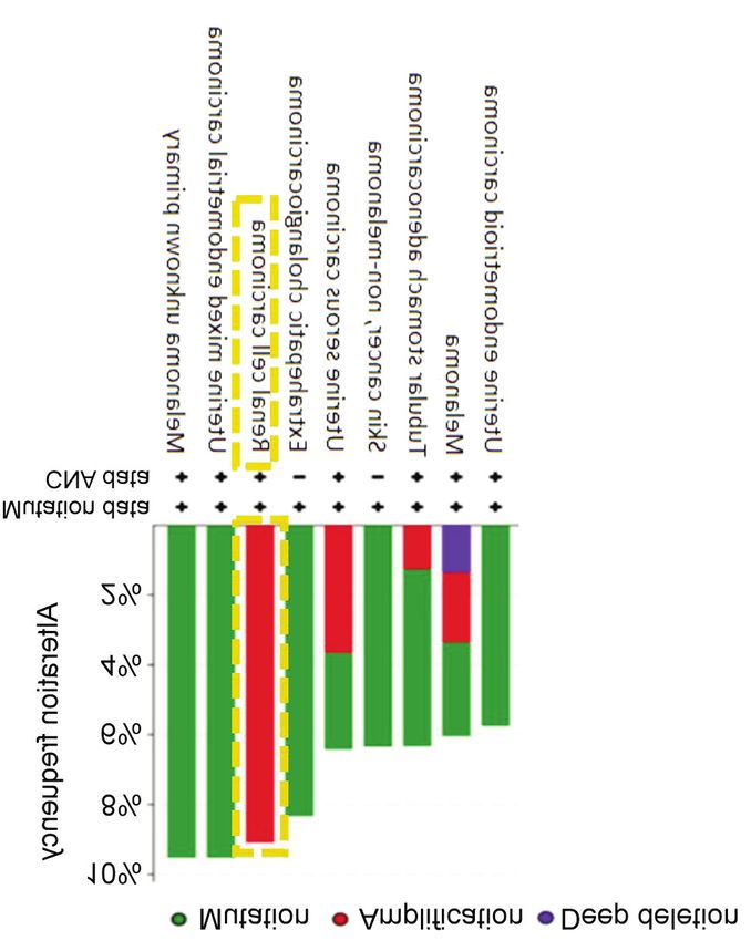
Demir and Cizmecioglu: Investigation of PI3K Functional Compensation via Activated Tyrosine Kinases
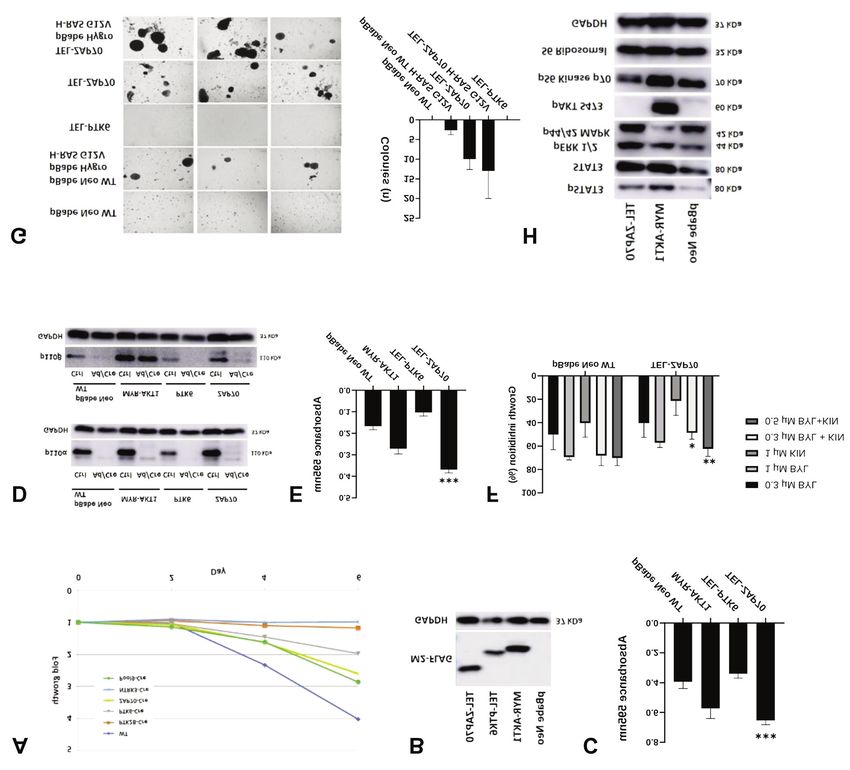
Figure 1. Screening of ETS variant transcription factor 6 dimerization domain (TEL)-tyrosine kinase library. A: The TEL-tagged, activated tyrosine
kinases were expressed in mouse embryonic fibroblasts (MEFs). Cells were then treated with Cre-expressing adenoviruses (Ade-Cre). Crystal violet
assays were performed and relative cell growth for each pool was calculated. MEFs were transfected with green fluorescent protein (GFP) as a
negative control and myristoylated AKT serine/threonine kinase 1 (MYR-AKT1) acted as a positive control. LacZ-expressing MEFs were used as a
control for adenoviral infection. The data are the mean±standard deviation from experimental triplicates. B: Immunoblot analysis of tyrosine kinase
pool 9 including neurotrophic receptor tyrosine kinase 3 (NTRK3), zeta chain of T-cell receptor-associated protein kinase 70 (ZAP70), protein
tyrosine kinase 6 (PTK6) and protein tyrosine kinase 2 beta (PTK2B) expression in MEFs by using M2-Flag and phospho-tyrosine primary antibodies
(4G10 clone). C: p110α/β Knockout upon Ade-Cre treatment in pool 9-expressing MEFs. Western blot analysis showed the knockout efficiency in
these cells using primary antibodies for p110α and p110β.
(Figure 1B). We also checked for the efficiency of PIK3CA demonstrated efficient p110α/β ablation in Ad/Cre-treated
and PIK3CB knock-out in the cells and found that p110α and MEFs in comparison to controls (Figure 2D). The knockout
p110β protein expression was abolished, confirming Ad/Cre of p110α/β caused growth retardation in control MEFs.
targeting (Figure 1C). Nevertheless, TEL-ZAP70 expression in MEFs partially
restored the impairment of cellular growth imposed by
TEL-ZAP70 promoted cell proliferation. Cellular growth assays p110α/β knockout, as did MYR-AKT1 expression in the
using individual Pool 9 components demonstrated that positive control (Figure 2E). Of note, the compensation was
standalone expression of activated ZAP70 enabled the MEFs to more pronounced in TEL-ZAP70-expressing MEFs in
grow efficiently upon Ad/Cre treatment in comparison to those comparison to MYR-AKT1-expressing MEFs.
bearing other tyrosine kinases in the pool and to controls For pharmacological inhibition, we used Food and Drug
(Figure 2A). To understand the role of activated tyrosine kinases Administration-approved small-molecule inhibitor of PI3K-
in MEFs, we stably transfected these cells with TEL-ZAP70 or p110α, Alpelisib (BYL719), and a specific PI3K-p110β
TEL-PTK6, using MEFs transfected with MYR-AKT1 as a inhibitor, KIN193 (AZD6482) to selectively inhibit kinase
positive control and pBabe Neo (empty) as a negative control activities of p110α and p110β respectively. In general, TEL-
(Figure 2B). We conducted crystal violet cell viability assays. ZAP70 MEFs were growth-inhibited to a lesser degree than
Our results showed that TEL-ZAP70 expression increased the control MEFs in crystal violet growth assays. We particularly
rate of cellular growth to a level similar to MYR-AKT1- observed a statistically significant reduction of growth
expressing MEFs. However, TEL-PTK6 expression in MEFs inhibition in response to the combination of BYL719 and
had no significant effect on cellular growth (Figure 2C). KIN193 when the cells expressed activated ZAP70 (Figure
2F). These results suggest that activation of ZAP70-mediated
Investigation of the growth-compensatory potential of signaling induces growth resistance to genetic or
activated ZAP70 upon PI3K knockout. In order to confirm pharmacological inhibition of p110α/β.
the tyrosine kinase library screening, we followed two
complementary approaches to inhibit PI3K signaling in TEL-ZAP70 promoted anchorage-independent growth in
MEFs. FirstIy, we took advantage of molecular genetics and MEFs. MEFs are untransformed cells which are not capable
induced genetic silencing of PIK3CA as well as PIK3CB, of growing in an anchorage-independent manner or initiating
and secondly, we employed pharmacological inhibitors tumors (30, 31). To determine whether ZAP70 has the ability
specific for p110α and -β isoforms. The immunoblot analysis to transform cells, we performed soft-agar assays with our
395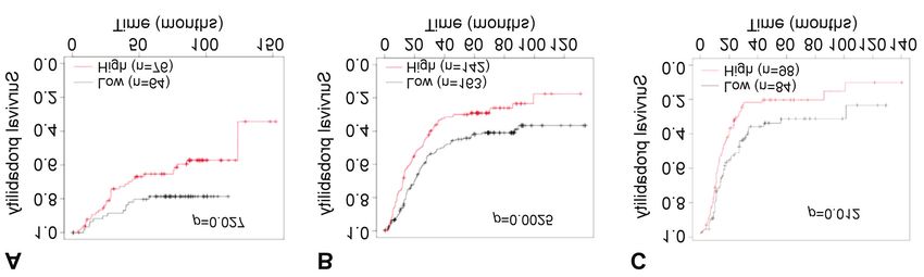
CANCER DIAGNOSIS & PROGNOSIS 2: 391-404 (2022)

Figure 2. Impact of activated zeta chain of T-cell receptor-associated protein kinase 70 (ZAP70) on proliferation of mouse embryonic fibroblasts
(MEFs). A: Crystal violet growth assay was conducted for MEFs that express ZAP70, neurotrophic receptor tyrosine kinase 3 (NTRK3), protein
tyrosine kinase 6 (PTK6), protein tyrosine kinase 2 beta (PTK2B) and pool 9. Cells were fixed and stained 2, 4 and 6 days after initial seeding.
WT: Wild-type. B: Western blot analysis was conducted to confirm the protein expression of individual activated tyrosine kinases in MEFs
using M2-FLAG primary antibody. Glyceraldehyde-3-phosphate dehydrogenase (GAPDH) was used as a loading control. C: Crystal violet
growth assays showing growth rate of MEFs that expressed activated ZAP70, PTK6, and myristoylated serine/threonine kinase 1 (MYR-AKT1)
as positive control and pBabe Neo WT MEFs as negative control. Data shown are the mean±standard deviation (SD), of three independent
experiments analyzed by using two-way analysis of variance. ***Significantly different from pBabe Neo WT at p
Demir and Cizmecioglu: Investigation of PI3K Functional Compensation via Activated Tyrosine Kinases

Figure 3. Detection of zeta chain of T-cell receptor-associated protein kinase 70 (ZAP70) expression in different cell lines. A: Western blot analysis
showing an endogenous ZAP70 expression. The size difference between positive controls was caused by the ETS variant transcription factor 6
(ETV6/TEL) tag for ZAP70 expression in T47D cells. B: ZAP70 phosphorylation in different cell lines. Asterisks indicate the position of endogenous
ZAP70. Glyceraldehyde-3-phosphate dehydrogenase (GAPDH) was used as a loading control.

Figure 4. Investigation of the function of zeta chain of T-cell receptor-associated protein kinase 70 (ZAP70) expression in non-transformed epithelial
cell lines. A: The immunoblots showing ETS variant transcription factor 6 (TEL)-ZAP70 expression in MCF10A and RPE1-hTERT cells using M2-
FLAG antibody. B: Western blot analysis was performed to detect activated signaling pathways with antibodies against phospho-specific signal
transducer and activator of transcription 3 (pSTAT3-Tyr705), total STAT3, p-S6K p70 (Thr389), p-S6 (Ser235/236), phospho-specific mitogen-
activated protein kinase 3/1 (pERK1/2-Thr202/Tyr204), phospho-AKT serine/threonine kinase 1 (p-AKT-Ser473) and MYC. Glyceraldehyde-3-
phosphate dehydrogenase (GAPDH) was used as a loading control. C: Crystal violet growth assay analysis showing growth inhibition of depicted
RPE1-hTERT lines upon treatment with p110α- (BYL719) and p110β (KIN193)-specific inhibitors. Data shown are the mean±standard deviation,
of triplicate independent experiments and analysed using two-way analysis of variance. **Significantly different from pBabe Neo WT at p
CANCER DIAGNOSIS & PROGNOSIS 2: 391-404 (2022)

                                                                             AKT1-expressing MEFs, which can be explained by the
                                                                             essential role of STAT3 in PI3K-induced oncogenic
                                                                             transformation (36).
                                                                                Additionally, a slight increase in ERK1/2 phosphorylation
                                                                             was observed (Figure 2H). However, the level of pS6K did
                                                                             not change. The lack of a change in the level of pS6K for
                                                                             ZAP70-expressing MEFs indicates a PI3K-independent
                                                                             mode of growth compensation. Taken together, these
                                                                             observations indicate that growth compensation mediated by
                                                                             activated ZAP70 might occur via phosphorylation/activation
                                                                             of JAK/STAT or MAPK signaling pathways.
                                                                                To understand if endogenous ZAP70 expression is present
                                                                             in adherent cell lines, we conducted western blot analysis.
                                                                             To this end, Jurkat CD4+ T-cells and TEL-ZAP70-expressing
                                                                             T47D cells were used as a positive control. The immunoblot
                                                                             showed that all cell lines, except for p110α/β+/+ wild-type
                                                                             MEFs, expressed endogenous ZAP70 to various extents
                                                                             (Figure 3A). LCK tyrosine kinase was found to be an
                                                                             important mediator of phosphorylation-dependent activation
                                                                             of ZAP70 (37). To further illustrate if endogenously
                                                                             expressed ZAP70 is phosphorylated and therefore activated,
                                                                             we examined phosphorylation levels of ZAP70. We observed
                                                                             elevated levels of phospho-ZAP70 in TEL-ZAP70-
                                                                             expressing T47D cells, along with HEK293T and MCF7
Figure 5. Zeta chain of T-cell receptor-associated protein kinase 70         cells (Figure 3B). Taken together, we detected expression of
(ZAP70) alterations found in cBioPortal for the US National Cancer           endogenous and possibly activated ZAP70 expression in
Institute (NCI)-60 cancer cell line studies. Based on these studies,         several adherent cell lines, including HEK293T, MCF7 and
ZAP70 amplification occurs highly in renal cell carcinomas. CNS:             T47D. Several studies showed LCK expression in several
Copy-number alteration.
                                                                             solid cancer types [reviewed in (37)], therefore it seems
                                                                             plausible that LCK phosphorylates and activates ZAP70 in a
                                                                             context-dependent manner.
Neo empty vector-expressing MEFs were not able to form
colonies, ZAP70 overexpression triggered colony formation                    Functional analysis of TEL-ZAP70 expression in alternative,
on soft agar (Figure 2G, right panel). Moreover, co-                         non-transformed epithelial cell culture models. The
expression of TEL-ZAP70 and H-Ras G12V had an                                morphological characteristics of MEFs are different from
additive effect on colony formation, implicating a                           epithelial cells as they represent cells of mesenchymal origin.
cooperation of ZAP70-mediated signaling with the MAPK                        Since most carcinomas are derived from epithelial cells, we
pathway (Figure 2G).                                                         used non-transformed RPE1-hTERT cells and non-
                                                                             tumorigenic epithelial human breast cells (MCF10A) to
ZAP70 expression leads to activation of the JAK/STAT3                        understand the impact of activated ZAP70 on initiation of
pathway. Receptor and non-receptor tyrosine kinases might                    carcinogenesis. We generated cell lines with stable expression
activate alternative signaling pathways besides PI3K via                     of activated ZAP70 along with positive and negative controls
phosphorylation of downstream targets (34, 35). We                           in MCF10A and RPE1-hTERT (Figure 4A). Next, we wanted
hypothesized that the JAK/STAT and MAPK pathways,                            to analyze the activity of ZAP70-mediated signaling
which have been implicated in functional compensation of                     pathways in these cells. Our results showed that the phospho-
PI3K (10, 16), might be activated upon ZAP70-induced                         STAT3 level in both cell lines expressing activated ZAP70
partial resistance. To assess the molecular mechanisms that                  was elevated (Figure 4B). Since activated ZAP70 had little
promote compensation of growth upon PI3K knockout in                         effect on pERK and pS6K levels in our study, these results
activated ZAP70-expressing MEFs, we conducted western                        imply that the mechanistic target of rapamycin kinase
blots to analyze activation of relevant pathways. According                  (mTOR) and MAPK pathways might not be universally
to our results, the level of STAT3 phosphorylation was                       affected by ZAP70 activation. However, JAK/STAT signaling
elevated in TEL-ZAP70-expressing MEFs (Figure 2H). In                        pathway appears to be consistently up-regulated by activated
addition, STAT3 phosphorylation was also elevated in MYR-                    ZAP70 in a broad range of cellular models.

                                                                       398
Demir and Cizmecioglu: Investigation of PI3K Functional Compensation via Activated Tyrosine Kinases
Figure 6. Zeta chain of T-cell receptor-associated protein kinase 70 (ZAP70) mRNA expression in tumor and normal tissue in different cancer types
from The Cancer Genome Atlas (TCGA). The data were obtained from the University of Alabama Cancer Database (UALCAN). The yellow boxes
indicate significantly elevated ZAP70 mRNA expression in solid tumor tissues compared to matched normal tissues. Error bars represent standard
deviation. BLCA: Bladder urothelial carcinoma; BRCA: breast invasive carcinoma; COAD: colon adenocarcinoma; ESCA: esophageal carcinoma;
CESC: cervical squamous cell carcinoma and endocervical adenocarcinoma; HNSC: head and neck squamous carcinoma; KICH: kidney
chromophobe; KIRC: kidney renal clear -cell carcinoma; KIRP: kidney renal papillary cell carcinoma; LIHC: liver hepatocellular carcinoma;
LUAD: lung adenocarcinoma; LUSC: lung squamous cell carcinoma; PAAD: pancreatic adenocarcinoma; PCPG: pheochromocytoma and
paraganglioma; PRAD: prostate adenocarcinoma; READ: rectal adenocarcinoma; SARC: sarcoma; SKCM: skin cutaneous melanoma; STAD:
stomach adenocarcinoma; UCEC: uterine corpus endometrial carcinoma; THCA: thyroid carcinoma; THYM: thymoma.
We next assessed whether activated ZAP70 had a positive Moreover, we investigated ZAP70 mRNA expression
effect on cell viability upon PI3K inhibition in RPE1-hTERT changes in tumor vs normal tissue for solid tumors. For this
cells. We used TEL-ZAP70-expressing RPE1 cells along purpose, we used the University of Alabama cancer web portal
with controls (pBabe Neo and MYR-AKT1-expressing RPE1 in UALCAN and found that in many cancer types, such as renal
cells). Crystal violet growth assays demonstrated that TEL- clear-cell carcinoma, renal papillary-cell carcinoma, cervical
ZAP70 expression in RPE1-hTERT cells partially squamous cell carcinoma, endocervical adenocarcinoma,
compensated growth upon BYL719-mediated inhibition of esophageal carcinoma, prostate adenocarcinoma, stomach
p110α (Figure 4C). adenocarcinoma, uterine corpus endometrial carcinoma, as well
After determining the importance of activated ZAP70 for as head and neck squamous carcinoma, ZAP70 mRNA levels
cellular proliferation in RPE1-hTERT cells, we wanted to see were found to be higher in tumor tissues in comparison to
if TEL-ZAP70 had the ability to form colonies in anchorage- matched controls (Figure 6).
independent growth assays. We performed soft-agar growth We wished to expand our observations to other types of
assays and found that TEL-ZAP70-expressing RPE1-hTERT cancer. To this end, we performed overall survival analysis
cells were able to form colonies (Figure 4D), although in various cancer types. The Kaplan–Meier plots showed that
constitutively activated AKT was unable to induce colony low expression of ZAP70 in gastric cancer correlated with
formation. Taken together, expression of activated ZAP70 in better survival (Figure 7A). Additionally, the Kaplan–Meier
non-transformed epithelial cells can increase their curves generated using the GEPIA2 platform demonstrated
proliferative potential as well as their ability to transform that low ZAP70 expression correlated with better survival in
into more malignant phenotypes. renal clear-cell carcinoma, low-grade glioma, and uveal
melanoma (Figure 7B-D). Next, to delineate whether ZAP70
has a differential effect on distinct stages of tumorigenesis,
Analyzing the involvement of ZAP70 in solid tumors in silico. we plotted stage-specific Kaplan–Meier survival curves. Our
ZAP70 activation has been associated with leukemia (38). results showed that low expression of ZAP70 in stage 2 and
We performed in silico analyses to determine if it might also stage 3 gastric cancer correlated with better survival (Figure
be involved in initiation or progression of solid tumors. 8A and B). These in silico analyses support our findings
Analyses of the US National Cancer Institute’s NCI-60 cell implying a causative role for ZAP70 in various types of solid
lines studied using the cBioPortal platform revealed that, tumor. To further investigate involvement of ZAP70 in solid
ZAP70 is moderately altered in renal cell carcinomas (9%), tumors, we plotted Kaplan–Meier survival curves in ovarian
and these alterations mostly occur in the form of gene cancer using KM Plotter database. In survival analysis of
amplification (Figure 5). patients with grade 1-2 ovarian cancer treated with
399CANCER DIAGNOSIS & PROGNOSIS 2: 391-404 (2022)
Figure 7. Kaplan–Meier graphs depicting overall survival according to expression of zeta chain of T-cell receptor-associated protein kinase 70
(ZAP70) in patients with gastric cancer using KM Plotter database for mRNA gene chip data (2104032_at) (A) and with renal clear-cell carcinoma
(B), uveal melanoma (C) and low-grade glioma (D) via GEPIA2.
Figure 8. Kaplan–Meier survival graphs depicting survival according to expression of zeta chain of T-cell receptor-associated protein kinase 70 (ZAP70)
in patients with gastric and ovarian cancer using KM Plot database for mRNA gene chip data (2104032_at). Overall survival of patients with stage 2
(A) and stage 3 (B) gastric cancer. Progression-free survival of patients with grade 1+2 ovarian cancer treated with paclitaxel/cisplatin (C).
400Demir and Cizmecioglu: Investigation of PI3K Functional Compensation via Activated Tyrosine Kinases
paclitaxel/cisplatin chemotherapy, high ZAP70 expression
levels significantly correlated with reduced progression-free
survival (Figure 8C). In conclusion, ZAP70 might be a
crucial prognostic marker for ovarian carcinogenesis and low
ZAP70 expression correlated with better survival in patients
treated with chemotherapy for early-stage ovarian cancer.
The role of ZAP70 overexpression in kidney cell lines.
According to our in-silico analysis, renal cell carcinomas had
the highest frequency of ZAP70 amplifications (Figure 5). We
wanted to investigate the impact of ZAP70 overexpression in
kidney-derived cell lines to consolidate our findings in
previous cellular models. To that end, we used embryonic
kidney-derived HEK293T cells and transiently expressed
activated ZAP70 along with MYR-AKT1 as a positive control
and the empty vector (pBabe Neo). Expression of TEL-ZAP70
dramatically enhanced STAT3 phosphorylation in HEK293T
cells. However, levels of phospho-S6 kinase and phospho-
ERK along with c-MYC did not change (Figure 9). These
results imply that ZAP70 activity triggers activation of the
JAK/STAT signaling pathway in epithelial as well as in
mesenchymal cellular models of tumorigenesis. In conclusion,
activated ZAP70 functions as a novel transforming factor in
Figure 9. Zeta chain of T-cell receptor-associated protein kinase 70
solid tumors and induces partial resistance to PI3K inhibition (ZAP70) overexpression in HEK293T cells. Western blot analysis was
via phosphorylation and activation of STAT3 in the JAK/STAT performed to detect ETS variant transcription factor 6 (TEL)-ZAP70-
signaling axis. mediated activation of downstream signaling components with
antibodies against phospho-specific signal transducer and activator of
Discussion transcription 3 (pSTAT3-Tyr705), total STAT3, p-S6K p70 (Thr389), p-
S6 (Ser235/236), phospho-specific mitogen-activated protein kinase 3/1
(p-ERK1/2-Thr202/Tyr204), phospho-AKT serine/threonine kinase 1 (p-
In this study, we screened an activated tyrosine kinase library AKT-Thr308) and MYC proto-oncogene (MYC). Glyceraldehyde-3-
and identified ZAP70 as mediator of resistance upon phosphate dehydrogenase (GAPDH) was used as a loading control.
simultaneous loss of p110α / p110β in MEFs (Figure 1). MYR-AKT1 was used as positive control for phosphatidylinositol 3-
kinase pathway activation.
ZAP70 plays critical roles in T-cell/B-cell development and
T-cell receptor signaling as well as being involved in
hematological malignancies (38). Our results describe a
potentially novel role for ZAP70 in untransformed cell lines
besides its immunological functions. Our cell viability assays of PI3K, we used clinically relevant p110α_specific
showed that TEL-ZAP70 expression in MEFs significantly Alpelisib and p110β-specific AZD6482 pharmacological
enhanced cellular proliferation in comparison to negative inhibitors. Our results show that active ZAP70 expression
control pBABE NEO and positive control MYR-AKT1 can induce partial resistance to PI3K inhibition in various
(Figure 2). These analyses indicate that active ZAP70 can cellular models comprising both epithelial as well as
promote cellular proliferation in adherent cells. mesenchymal cells (Figure 2 and Figure 4).
Existing literature supports the notion that PI3K is a As proposed by Siveen et al., STAT3 can be activated by
crucial regulator of cellular proliferation, and its impairment Src family kinases (39). Furthermore, Warmuth et al.
has been linked to severe growth defects in various cancer suggested that the STAT3 signaling cascade is crucial in
models (3, 7). In order to abrogate PI3K signaling, we carcinogenesis (40). We performed western blot analysis to
knocked out endogenous p110α/p110β in MEFs. For this, we elucidate the mechanism of TEL-ZAP70-mediated activation
utilized genetically engineered MEFs, where p110α and of downstream pathway signaling upon PI3K ablation. Our
p110β can be effectively knocked out by expression of Ad- results indicate that up-regulation of STAT3 phosphorylation
Cre. Although, active PTK6 expression was not able to on tyrosine 705 residue is a ubiquitous event in active
restore growth in the p110α/p110β knockout system (Figure ZAP70-expressing cell lines including RPE1-hTERT, MEFs,
2), TEL-ZAP70 expressing MEFs largely recovered growth non-tumorigenic epithelial breast (MCF10A) and human
upon p110α/p110β ablation. To abolish the catalytic activity embryonic kidney cells (HEK293T) cell lines (Figure 2,
401CANCER DIAGNOSIS & PROGNOSIS 2: 391-404 (2022)
Figure 4 and Figure 9). Remarkably, p-ERK1/2 and p-S6 cisplatin, low ZAP70 expression correlated with better survival
kinase p70 expression levels did not change in TEL-ZAP70 (Figure 8). Consequently, high ZAP70 expression might have
RPE1 cells. Additionally, dual-tyrosine phosphorylation of a tumorigenic/oncogenic function during the establishment of
ERK in TEL-ZAP70 MEFs (Figure 2) and phosphorylation the disease and correlate with a worse prognosis.
of S6K p70 levels in TEL-ZAP70 HEK293T (Figure 9) only In conclusion, our results implicate ZAP70 in tumor
slightly increased. Cha et al. suggested that the initiation as well as in resistance to PI3K inhibition.
JAK/STAT3/c-MYC signaling axis is negatively regulated to Although ZAP70 expression has been implicated in poor
modulate differentiation capability by ZAP70 in mouse prognosis in hematological malignancies such as chronic
embryonic stem cells (21). In contrast to their findings, our lymphocytic leukemia (44), our results suggest that ZAP70
results suggested that STAT3 activation was positively tyrosine kinase activation can promote cell proliferation and
correlated with ZAP70 expression (21). In addition, we contribute to generation of resistance to PI3K inhibition. The
found out that the c-MYC levels were not altered upon induction of resistance to PI3K inhibition might be explained
expression of active ZAP70 in TEL-ZAP70 expressing cells through activation of JAK/STAT; in particular, the
(Figure 4 and Figure 9). These contrasting observations JAK/STAT3 signaling axis. We believe that these findings
might be explained by different genetic contexts in are of strong clinical relevance as several PI3K inhibitors
pluripotent stem cells versus differentiated or transformed have already passed clinical trials and are being used in the
mesenchymal/epithelial cell lines. clinic (45, 46).
To further explore the function of ZAP70 in
carcinogenesis, we investigated involvement of ZAP70 in Conflicts of Interest
anchorage-independent growth on soft agar in untransformed
cell lines. The morphological characteristics of MEFs are The Authors declare no competing conflicts of interest.
different from epithelial cells as they represent cells of
mesenchymal origin, whereas most carcinomas are of Authors’ Contributions
epithelial origin. Thus, we used non-transformed epithelial
cells, MCF10A and RPE1-hTERT, to understand the impact Onur Cizmecioglu: Conception and design, analysis and
of activated ZAP70 on initiation of carcinogenesis. We found interpretation of data, and revision of the article. Melike Demir:
that ZAP70-expressing MEF and RPE1 cells were capable of Acquisition of data, analysis and interpretation of data, and drafting
of the article.
forming colonies on soft agar (Figure 2 and Figure 4). We
also showed that ZAP70 had an additive effect in anchorage-
independent growth along with H-Ras G12V (Figure 2). Acknowledgements
These findings imply that ZAP70 might have a function in
This study was funded by The Scientific and Technological Research
tumor initiation. Although our research was mainly conducted Council of Turkey (TÜBİTAK) Directorate of Science Fellowships
on untransformed cell lines, it would be important to decipher and Grant Programs (BİDEB), project number 117C040.
the effects of ZAP70 in transformed cancer cells. Based on
the literature, ZAP70 promotes cell migration and invasion of References
prostate cancer cell lines (20). Moreover, ZAP70 was
identified as a prognostic marker in cervical squamous cell 1 Kondapalli L, Soltani K and Lacouture ME: The promise of
carcinoma (41), prostate adenocarcinoma (42) and colorectal molecular targeted therapies: protein kinase inhibitors in the
cancer in response to radiation (22). Furthermore, Sun et al. treatment of cutaneous malignancies. J Am Acad Dermatol 53(2):
indicated that ZAP70 may be a crucial regulator of metastasis 291-302, 2005. PMID: 16021125. DOI: 10.1016/j.jaad.2005.02.011
in prostate cancer (42). Of note, Sadras et al. found that while 2 Hunter T: Protein kinase classification. Methods Enzymol 200: 3-
37, 1991. PMID: 1835513. DOI: 10.1016/0076-6879(91)00125-g
ZAP70 has an oncogenic role, SYK, a member belonging to
3 Krause DS and Van Etten RA: Tyrosine kinases as targets for
the same family, functions as a tumor suppressor in cancer therapy. N Engl J Med 353(2): 172-187, 2005. PMID:
autoimmune diseases and B-cell malignancies (43). 16014887. DOI: 10.1056/NEJMra044389
According to our in-silico analysis, ZAP70 mRNA 4 Liu P, Cheng H, Roberts TM and Zhao JJ: Targeting the
expression levels were found to be elevated in many solid phosphoinositide 3-kinase pathway in cancer. Nat Rev Drug Discov
tumor tissues in comparison to matched controls (Figure 6). 8(8): 627-644, 2009. PMID: 19644473. DOI: 10.1038/nrd2926
Additionally, the Kaplan–Meier plots show that low ZAP70 5 Vivanco I and Sawyers CL: The phosphatidylinositol 3-Kinase
AKT pathway in human cancer. Nat Rev Cancer 2(7): 489-501,
expression in several cancer types, including gastric cancer,
2002. PMID: 12094235. DOI: 10.1038/nrc839
uveal melanoma, kidney clear cell carcinoma and low-grade 6 Yang J, Nie J, Ma X, Wei Y, Peng Y and Wei X: Targeting PI3K
glioma, was associated with better survival (Figure 7). in cancer: mechanisms and advances in clinical trials. Mol
Moreover, our survival analysis depicted that in patients with Cancer 18(1): 26, 2019. PMID: 30782187. DOI: 10.1186/
low-grade ovarian cancer who were treated with paclitaxel and s12943-019-0954-x
402Demir and Cizmecioglu: Investigation of PI3K Functional Compensation via Activated Tyrosine Kinases
7 Cheng CK, Fan QW and Weiss WA: PI3K signaling in glioma— Biochem Biophys Res Commun 469(3): 345-351, 2016. PMID:
animal models and therapeutic challenges. Brain Pathol 19(1): 26620225. DOI: 10.1016/j.bbrc.2015.11.093
112-120, 2009. PMID: 19076776. DOI: 10.1111/j.1750- 21 Cha Y, Moon BH, Lee MO, Ahn HJ, Lee HJ, Lee KA, Fornace
3639.2008.00233.x AJ Jr, Kim KS, Cha HJ and Park KS: Zap70 functions to
8 Michmerhuizen NL, Leonard E, Kulkarni A and Brenner JC: maintain stemness of mouse embryonic stem cells by negatively
Differential compensation mechanisms define resistance to PI3K regulating Jak1/Stat3/c-Myc signaling. Stem Cells 28(9): 1476-
inhibitors in PIK3CA amplified HNSCC. Otorhinolaryngol Head 1486, 2010. PMID: 20641039. DOI: 10.1002/stem.470
Neck Surg 1(2): 44-50, 2016. PMID: 28004037. DOI: 22 Huang MY, Wang JY, Chang HJ, Kuo CW, Tok TS and Lin SR:
10.15761/ohns.1000111 CDC25A, VAV1, TP73, BRCA1 and ZAP70 gene
9 Fruman DA, Chiu H, Hopkins BD, Bagrodia S, Cantley LC overexpression correlates with radiation response in colorectal
and Abraham RT: The PI3K pathway in human disease. Cell cancer. Oncol Rep 25(5): 1297-1306, 2011. PMID: 21344162.
170(4): 605-635, 2017. PMID: 28802037. DOI: DOI: 10.3892/or.2011.1193
10.1016/j.cell.2017.07.029 23 Cizmecioglu O, Ni J, Xie S, Zhao JJ and Roberts TM: Rac1-
10 Serra V, Scaltriti M, Prudkin L, Eichhorn PJ, Ibrahim YH, mediated membrane raft localization of PI3K/p110β is required
Chandarlapaty S, Markman B, Rodriguez O, Guzman M, for its activation by GPCRs or PTEN loss. Elife 5: e17635,
Rodriguez S, Gili M, Russillo M, Parra JL, Singh S, Arribas J, 2016. PMID: 27700986. DOI: 10.7554/eLife.17635
Rosen N and Baselga J: PI3K inhibition results in enhanced 24 Yuzugullu H, Von T, Thorpe LM, Walker SR, Roberts TM, Frank
HER signaling and acquired ERK dependency in HER2- DA and Zhao JJ: NTRK2 activation cooperates with PTEN
overexpressing breast cancer. Oncogene 30(22): 2547-2557, deficiency in T-ALL through activation of both the PI3K-AKT
2011. PMID: 21278786. DOI: 10.1038/onc.2010.626 and JAK-STAT3 pathways. Cell Discov 2: 16030, 2016. PMID:
11 Hanker AB, Kaklamani V and Arteaga CL: Challenges for the 27672444. DOI: 10.1038/celldisc.2016.30
clinical development of PI3K inhibitors: strategies to improve 25 Kuno Y, Abe A, Emi N, Iida M, Yokozawa T, Towatari M,
their impact in solid tumors. Cancer Discov 9(4): 482-491, 2019. Tanimoto M and Saito H: Constitutive kinase activation of the
PMID: 30867161. DOI: 10.1158/2159-8290.CD-18-1175 TEL-Syk fusion gene in myelodysplastic syndrome with
12 Muranen T, Selfors LM, Worster DT, Iwanicki MP, Song L, t(9;12)(q22;p12). Blood 97(4): 1050-1055, 2001. PMID:
Morales FC, Gao S, Mills GB and Brugge JS: Inhibition of 11159536. DOI: 10.1182/blood.v97.4.1050
PI3K/mTOR leads to adaptive resistance in matrix-attached 26 Győrffy B: Survival analysis across the entire transcriptome
cancer cells. Cancer Cell 21(2): 227-239, 2012. PMID: identifies biomarkers with the highest prognostic power in breast
22340595. DOI: 10.1016/j.ccr.2011.12.024 cancer. Comput Struct Biotechnol J 19: 4101-4109, 2021. PMID:
13 Nawijn MC, Alendar A and Berns A: For better or for worse: the 34527184. DOI: 10.1016/j.csbj.2021.07.014
role of Pim oncogenes in tumorigenesis. Nat Rev Cancer 11(1): 27 Chandrashekar DS, Bashel B, Balasubramanya SAH, Creighton
23-34, 2011. PMID: 21150935. DOI: 10.1038/nrc2986 CJ, Ponce-Rodriguez I, Chakravarthi BVSK and Varambally S:
14 Song JH, Singh N, Luevano LA, Padi SKR, Okumura K, Olive V, UALCAN: a portal for facilitating tumor subgroup gene
Black SM, Warfel NA, Goodrich DW and Kraft AS: Mechanisms expression and survival analyses. Neoplasia 19(8): 649-658,
behind resistance to PI3K inhibitor treatment induced by the PIM 2017. PMID: 28732212. DOI: 10.1016/j.neo.2017.05.002
kinase. Mol Cancer Ther 17(12): 2710-2721, 2018. PMID: 28 Cerami E, Gao J, Dogrusoz U, Gross BE, Sumer SO, Aksoy BA,
30190422. DOI: 10.1158/1535-7163.MCT-18-0374 Jacobsen A, Byrne CJ, Heuer ML, Larsson E, Antipin Y, Reva B,
15 Le X, Antony R, Razavi P, Treacy DJ, Luo F, Ghandi M, Castel Goldberg AP, Sander C and Schultz N: The cBio cancer genomics
P, Scaltriti M, Baselga J and Garraway LA: systematic functional portal: an open platform for exploring multidimensional cancer
characterization of resistance to PI3K inhibition in breast cancer. genomics data. Cancer Discov 2(5): 401-404, 2012. PMID:
Cancer Discov 6(10): 1134-1147, 2016. PMID: 27604488. DOI: 22588877. DOI: 10.1158/2159-8290.CD-12-0095
10.1158/2159-8290.CD-16-0305 29 Zhao JJ, Gjoerup OV, Subramanian RR, Cheng Y, Chen W,
16 Chang M, Kanwar N, Feng E, Siu A, Liu X, Ma D and Jongstra Roberts TM and Hahn WC: Human mammary epithelial cell
J: PIM kinase inhibitors downregulate STAT3(Tyr705) transformation through the activation of phosphatidylinositol 3-
phosphorylation. Mol Cancer Ther 9(9): 2478-2487, 2010. kinase. Cancer Cell 3(5): 483-495, 2003. PMID: 12781366.
PMID: 20667852. DOI: 10.1158/1535-7163.MCT-10-0321 DOI: 10.1016/s1535-6108(03)00088-6
17 Chan AC, Irving BA, Fraser JD and Weiss A: The zeta chain is 30 Mori S, Chang JT, Andrechek ER, Matsumura N, Baba T, Yao
associated with a tyrosine kinase and upon T-cell antigen G, Kim JW, Gatza M, Murphy S and Nevins JR: Anchorage-
receptor stimulation associates with ZAP-70, a 70-kDa tyrosine independent cell growth signature identifies tumors with
phosphoprotein. Proc Natl Acad Sci U S A 88(20): 9166-9170, metastatic potential. Oncogene 28(31): 2796-2805, 2009. PMID:
1991. PMID: 1717999. DOI: 10.1073/pnas.88.20.9166 19483725. DOI: 10.1038/onc.2009.139
18 Wange RL and Samelson LE: Complex complexes: signaling at 31 Suzuki J, Sukezane T, Akagi T, Georgescu MM, Ohtani M, Inoue
the TCR. Immunity 5(3): 197-205, 1996. PMID: 8808675. DOI: H, Jat PS, Goff SP, Hanafusa H and Shishido T: Loss of c-abl
10.1016/s1074-7613(00)80315-5 facilitates anchorage-independent growth of p53- and RB- deficient
19 Kaur M, Singh M and Silakari O: Insight into the therapeutic primary mouse embryonic fibroblasts. Oncogene 23(52): 8527-
aspects of ‘Zeta-Chain Associated Protein Kinase 70 kDa’ 8534, 2004. PMID: 15378021. DOI: 10.1038/sj.onc.1207894
inhibitors: a review. Cell Signal 26(11): 2481-2492, 2014. 32 Seeburg PH, Colby WW, Capon DJ, Goeddel DV and Levinson
PMID: 25049080. DOI: 10.1016/j.cellsig.2014.06.017 AD: Biological properties of human c-Ha-ras1 genes mutated at
20 Fu D, Liu B, Zang LE and Jiang H: MiR-631/ZAP70: A novel codon 12. Nature 312(5989): 71-75, 1984. PMID: 6092966.
axis in the migration and invasion of prostate cancer cells. DOI: 10.1038/312071a0
403CANCER DIAGNOSIS & PROGNOSIS 2: 391-404 (2022)
33 Colby WW, Hayflick JS, Clark SG and Levinson AD: 42 Sun X, Wang L, Li H, Jin C, Yu Y, Hou L, Liu X, Yu Y, Yan R
Biochemical characterization of polypeptides encoded by and Xue F: Identification of microenvironment related potential
mutated human Ha-ras1 genes. Mol Cell Biol 6(2): 730-734, biomarkers of biochemical recurrence at 3 years after
1986. PMID: 3537694. DOI: 10.1128/mcb.6.2.730-734.1986 prostatectomy in prostate adenocarcinoma. Aging (Albany NY)
34 Bennasroune A, Gardin A, Aunis D, Crémel G and Hubert P: 13(12): 16024-16042, 2021. PMID: 34133324. DOI: 10.18632/
Tyrosine kinase receptors as attractive targets of cancer therapy. aging.203121
Crit Rev Oncol Hematol 50(1): 23-38, 2004. PMID: 15094157. 43 Sadras T, Martin M, Kume K, Robinson ME, Saravanakumar S,
DOI: 10.1016/j.critrevonc.2003.08.004 Lenz G, Chen Z, Song JY, Siddiqi T, Oksa L, Knapp AM, Cutler J,
35 Gocek E, Moulas AN and Studzinski GP: Non-receptor protein Cosgun KN, Klemm L, Ecker V, Winchester J, Ghergus D, Soulas-
tyrosine kinases signaling pathways in normal and cancer cells. Sprauel P, Kiefer F, Heisterkamp N, Pandey A, Ngo V, Wang L,
Crit Rev Clin Lab Sci 51(3): 125-137, 2014. PMID: 24446827. Jumaa H, Buchner M, Ruland J, Chan WC, Meffre E, Martin T and
DOI: 10.3109/10408363.2013.874403 Müschen M: Developmental partitioning of SYK and ZAP70
36 Hart JR, Liao L, Yates JR 3rd and Vogt PK: Essential role of prevents autoimmunity and cancer. Mol Cell 81(10): 2094-2111.e9,
Stat3 in PI3K-induced oncogenic transformation. Proc Natl Acad 2021. PMID: 33878293. DOI: 10.1016/j.molcel.2021.03.043
Sci U S A 108(32): 13247-13252, 2011. PMID: 21788516. DOI: 44 Shvidel L, Bairey O, Tadmor T, Braester A, Ruchlemer R, Fineman
10.1073/pnas.1110486108 R, Joffe E, Berrebi A, Polliack A and Israeli CLL Study Group:
37 Bommhardt U, Schraven B and Simeoni L: Beyond TCR Absolute lymphocyte count with extreme hyperleukocytosis does
signaling: emerging functions of lck in cancer and not have a prognostic impact in chronic lymphocytic leukemia.
immunotherapy. Int J Mol Sci 20(14): 3500, 2019. PMID: Anticancer Res 35(5): 2861-2866, 2015. PMID: 25964568.
31315298. DOI: 10.3390/ijms20143500 45 Juric D, Rodon J, Tabernero J, Janku F, Burris HA, Schellens
38 Au-Yeung BB, Shah NH, Shen L and Weiss A: ZAP-70 in JHM, Middleton MR, Berlin J, Schuler M, Gil-Martin M, Rugo
signaling, biology, and disease. Annu Rev Immunol 36: 127-156, HS, Seggewiss-Bernhardt R, Huang A, Bootle D, Demanse D,
2018. PMID: 29237129. DOI: 10.1146/annurev-immunol- Blumenstein L, Coughlin C, Quadt C and Baselga J:
042617-053335 Phosphatidylinositol 3-kinase α-selective inhibition with
39 Siveen KS, Prabhu KS, Achkar IW, Kuttikrishnan S, Shyam S, alpelisib (BYL719) in PIK3CA-altered solid tumors: Results
Khan AQ, Merhi M, Dermime S and Uddin S: Role of non from the first-in-human study. J Clin Oncol 36(13): 1291-1299,
receptor tyrosine kinases in hematological malignances and its 2018. PMID: 29401002. DOI: 10.1200/JCO.2017.72.7107
targeting by natural products. Mol Cancer 17(1): 31, 2018. 46 Engelman JA: Targeting PI3K signalling in cancer:
PMID: 29455667. DOI: 10.1186/s12943-018-0788-y opportunities, challenges and limitations. Nat Rev Cancer 9(8):
40 Warmuth M, Damoiseaux R, Liu Y, Fabbro D and Gray N: SRC 550-562, 2009. PMID: 19629070. DOI: 10.1038/nrc2664
family kinases: potential targets for the treatment of human
cancer and leukemia. Curr Pharm Des 9(25): 2043-2059, 2003.
PMID: 14529415. DOI: 10.2174/1381612033454126
41 Qin R, Cao L, Ye C, Wang J and Sun Z: A novel prognostic
prediction model based on seven immune-related RNAs for
predicting overall survival of patients in early cervical squamous Received December 20, 2021
cell carcinoma. BMC Med Genomics 14(1): 49, 2021. PMID: Revised February 28, 2022
33588862. DOI: 10.1186/s12920-021-00885-3 Accepted March 2, 2022
404You can also read


