Modified Sijunzi Decoction Inhibits Epithelial-Mesenchymal Transition of Non-Small Cell Lung Cancer by Attenuating AKT/GSK3β Pathway in vitro and ...
←
→
Page content transcription
If your browser does not render page correctly, please read the page content below
ORIGINAL RESEARCH
                                                                                                                                               published: 17 January 2022
                                                                                                                                          doi: 10.3389/fphar.2021.821567

                                           Modified Sijunzi Decoction Inhibits
                                           Epithelial-Mesenchymal Transition of
                                           Non-Small Cell Lung Cancer by
                                           Attenuating AKT/GSK3β Pathway
                                           in vitro and in vivo
                                           Niu Shao 1, Yao Xiao 1, Jiaxin Zhang 1, Yuying Zhu 1, Shenglong Wang 2 and Suzhen Bao 1*
                                           1
                                            College of Basic Medical Science, Zhejiang Chinese Medical University, Hangzhou, China, 2The First College of Clinical Medicine,
                                           Zhejiang Chinese Medical University, Hangzhou, China

                                           Modified Sijunzi Decoction (MSJZD) is an empirical prescription of Traditional Chinese
                                           Medicine (TCM) and has been corroborated to be effective in multiple human diseases, but
                                           its role in non-small cell lung cancer (NSCLC) is enigmatic. Here we mainly analyze the
                                           function and mechanism of MSJZD in NSCLC. In this study, we used a method that
                            Edited by:
                                           coupled ultra-performance liquid chromatography to quadrupole time-of-flight mass
                      Jiang-Jiang Qin,     spectrometry (UPLC-Q-TOF-MS) to investigate the major constituents in MSJZD with
Institute of Cancer and Basic Medicine
                                           positive and negative ion modes. Additionally, in in vitro experiments, the effects of serum-
                          (CAS), China
                                           containing MSJZD on the biological behavior of NSCLC cells induced by TGF-β1 were
                    Reviewed by:
                        Wen Zhou,          assessed by cell function experiments. Then, the influences of serum-containing MSJZD
    Guangzhou University of Chinese        on epithelial-mesenchymal transition (EMT)-related markers were examined by
                   Medicine, China
                           Qi Zeng,
                                           immunofluorescence and western blot assays. Also, the AKT/GSK3β pathway and
            Xidian University, China       apoptosis-related markers were estimated by western blotting. Tumor xenografts were
                  *Correspondence:         generated by subcutaneously injecting A549 cells into BALB/c nude mice to determine the
                         Suzhen Bao
                                           effects of MSJZD in vivo. We first analyzed the composition of MSJZD. In positive ion
              suzhenbao93@126.com
                                           mode, 47 kinds of components were identified. In negative ion mode, 45 kinds of
                  Specialty section:       components were identified. We also found that TGF-β1 contributed to inducing cell
       This article was submitted to       morphological changes and EMT progression. In vitro, surprisingly, cell proliferation,
 Pharmacology of Anti-Cancer Drugs,
             a section of the journal      migration as well as invasion in NSCLC cells induced by TGF-β1, could be weakened
          Frontiers in Pharmacology        by serum-containing MSJZD, and apoptosis was intensified. Moreover, serum-containing
      Received: 24 November 2021           MSJZD weakened EMT passage and AKT/GSK3β pathway activation and induced
      Accepted: 20 December 2021
                                           apoptosis-related markers in NSCLC cells triggered by TGF-β1. In vivo, we discovered
       Published: 17 January 2022
                                           that MSJZD attenuated the tumor growth, promoted histopathological damage, and
                             Citation:
     Shao N, Xiao Y, Zhang J, Zhu Y,       induced apoptosis in A549 tumor-bearing mice. Importantly, MSJZD has also restrained
 Wang S and Bao S (2022) Modified           the development of EMT, AKT/GSK3β pathway, and TGF-β1 expression levels in nude
  Sijunzi Decoction Inhibits Epithelial-
Mesenchymal Transition of Non-Small        mice. These findings demonstrated that MSJZD significantly weakened NSCLC
Cell Lung Cancer by Attenuating AKT/       progression by modulating EMT and AKT/GSK3β pathway.
 GSK3β Pathway in vitro and in vivo.
        Front. Pharmacol. 12:821567.       Keywords: non-small cell lung cancer, modified sijunzi decoction, apoptosis, epithelial-mesenchymal transition,
      doi: 10.3389/fphar.2021.821567       Akt/GSK3β pathway

Frontiers in Pharmacology | www.frontiersin.org                                     1                                          January 2022 | Volume 12 | Article 821567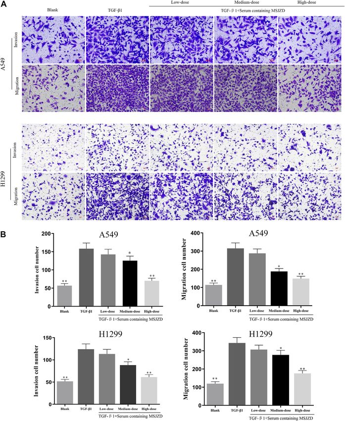
Shao et al.                                                                                                         MSJZD Inhibits EMT in NSCLC

INTRODUCTION                                                              Fritillariae Thunbergii Bulbus, Herba Hedyotis, and Glycyrrhizae
                                                                          Praeparata cum Melle Radix et Rhizoma. The most basic theory of
Lung cancer is the most common malignant tumor in the world               TCM in the treatment of NSCLC is regulating the deficiency of
and its morbidity and mortality rate remain high (Adjei, 2019;            body energy to inhibit tumor cell growth, proliferation, invasion,
Sharma et al., 2019). According to the 2020 Global Cancer                 and migration (Li et al., 2021). MSJZD is a TCM prescription as
Statistics report released by the International Agency for                adjuvant therapy in the standard treatment of NSCLC, which is
Research on Cancer (IARC), there were about 1.8 million                   commonly used clinically for patients with lung- and spleen-Qi
deaths and 2.2 million new cases of lung cancer worldwide in              deficiency. Additionally, modern studies have found that
2020, accounting for 18% of the deaths and 11.4% of the new               deficiency of spleen-Qi is also closely associated with the
cases of malignant tumors, ranking first and second among all              body’s anti-cancer immunity (Li, 2007; Li et al., 2021).
malignant tumors (Sung et al., 2021). Non-small cell lung cancer          Accumulated studies have demonstrated the nourish spleen-Qi
(NSCLC) accounts for about 80% of all lung cancer cases, about            effect of SJZD in chemotherapy-induced immunotoxicity and on
75% of patients are already in the advanced stage when                    immune function in post-operative patients (Guan et al., 2018;
discovered, and the 5-years survival rate is only 15% (Wakelee            Chen et al., 2019). According to TCM’s theory, the spleen
et al., 2014). Currently, the treatment of NSCLC includes surgery,        contributes to the production of sputum, as well as lung stores
radiotherapy, chemotherapy, molecular targeted therapy, and               the sputum; Thus, phlegmatic hygrosis also affects the metastasis
immunotherapy, etc (Ruiz et al., 2014; Hirsch et al., 2016;               of lung cancer in patients with lung- and spleen-Qi deficiency.
Gadgeel, 2017; Allen et al., 2018; Yang and Mu, 2018).                    Because of this, the MSJZD used mentioned above used Sijunzi
Although the above treatments have certain effects, distant               decoction as a major formula and combined it with the Pinelliae
metastasis in most patients is still the cause of the high                rhizoma, Fritillariae thunbergii Bulbus, Herba hedyotis, and
mortality rate of NSCLC (Du et al., 2016). Therefore, it is of            Astragali Radix, that possess synergistic or additive activity to
great significance to research the invasion and metastasis of              promote the invigorating spleen and replenishing qi of the Sijunzi
tumors for the treatment of NSCLC.                                        decoction. Besides, Many components of MSJZD have good
    Epithelial-mesenchymal transition (EMT) is a key event in the         clinical effects on mitigating cancer-related symptoms. For
process of early tumor metastasis and progression. A variety of           example, Quercetin, Liquiritigenin, Peimine, and Liquiritin all
tumor cells can lose the epithelial cell-like phenotypic                  manifested good anti-tumor proliferation and metastasis effects
characteristics such as cell-to-cell contact and cell polarity            (Wei et al., 2017; Meng and Lin, 2019; Reyes-Farias and Carrasco-
through EMT, thereby gaining invasion and migration                       Pozo, 2019; Tan et al., 2020). Although the clinical efficacy of
capabilities (Aiello et al., 2017; Ye et al., 2017). Transforming         MSJZD in the treatment of lung cancer metastasis has been
growth factor-β1 (TGF-β1) is a multifunctional cell regulator,            confirmed, however, its therapeutic mechanism is still
which plays a bidirectional role in the development of cancer.            ambiguous.
TGF-β1 was overexpressed in NSCLC tissues, the prognosis of                   In this study, we start with MSJZD as a whole and further
patients with overexpression of TGF-β1 was poor, and its high             evaluate the anti-tumor effect of MSJZD on EMT in lung cancer
expression may indicate the progression or metastasis of NSCLC            by preparing serum-containing MSJZD and establishing an A549
(Huang et al., 2014). Meanwhile, as one of the earliest                   cell tumor-bearing model in mice, to provide a scientific basis for
multifunctional cytokines that can induce EMT, TGF-β1 can                 guiding clinical treatment.
participate in a variety of biological processes by inducing EMT,
such as fibrotic diseases, embryonic development, and cancer
(Yang and Weinberg, 2008). Consequently, it is of major                   MATERIALS AND METHODS
significance to study the mechanism of NSCLC metastasis and
investigate effective drugs against NSCLC metastasis.                     Ethics Statement
    Traditional Chinese medicine (TCM) is rich in bioactive               The animal programs complied with the guidelines of the China
components, which play a role by targeting multiple molecular             Animal Care and Use Committee. Approval was acquired from
networks associated with the disease. Whereas, TCM is a                   the Committee of Laboratory Animals of Zhejiang Chinese
potential candidate drug that can be developed for the                    Medical University Laboratory Animal Research Center
prevention and treatment of cancer. In recent years, TCM has              (License number: SYXK (Zhe) 2018-0012). All efforts were
shown unique advantages and curative effects in many aspects of           made to alleviate the suffering of animals.
lung cancer treatment, such as reducing the toxic and side effects
of radiotherapy and chemotherapy, adjusting the immune                    Preparation Aqueous Extract of MSJZD
function of the body, stabilizing tumor foci, etc. (Jiang et al.,         Codonopsis Radix 15 g, Astragali Radix 30 g, Rhizoma
2019; Xu et al., 2019). Scientific research proved that single TCM         Atractylodis Macrocephalae 20 g, Poria 15 g, Pinelliae Rhizoma
extracts, such as Astragali Radix, and Poria, have certain effects        12 g, Fritillariae Thunbergii Bulbus 12 g, Herba Hedyotis 30 g, and
on inducing lung cancer cell apoptosis, inhibiting cell invasion,         Glycyrrhizae Praeparata cum Melle Radix et Rhizoma 10 g were
metastasis, and angiogenesis (Cheng et al., 2014; Lee et al., 2018;       actually weighed and soaked in 1500 ml of distilled water for
Xu et al., 2018; Lin et al., 2020). Modified Sijunzi Decoction             60 min (min), then boiled at 100°C for 45 min. After harvesting
(MSJZD) was composed of Astragali Radix, Poria, Codonopsis                the decoction solution, another 1200 ml of distilled water was
Radix, Rhizoma Atractylodis Macrocephalae, Pinelliae Rhizoma,             added for the second extraction at 100°C for 30 min. After

Frontiers in Pharmacology | www.frontiersin.org                       2                                  January 2022 | Volume 12 | Article 821567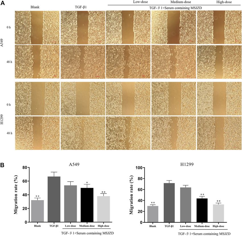
Shao et al.                                                                                                       MSJZD Inhibits EMT in NSCLC

combining the filtrates, the decoction solution was concentrated          30 g/kg (2 times clinical equivalent dose), respectively for
to 100 ml with a final concentration of 2.8 g/ ml. The 2.8 g/ ml          5 days. The control group was fed with normal saline at 1 ml
MSJZD solution was diluted with ultrapure water to 0.288 g/ ml           per 100 g body weight for five consecutive days. The drug clinical
aqueous extract of MSJZD. Finally, 0.5 ml MSJZD solution                 equivalent dose conversion formula was as follows: (human dose
(0.288 g/ ml) was diluted to 0.144 g/ ml test solution with              of crude herbs in clinic /60 kg)×6.3. They were administered
0.5 ml methanol for UPLC-Q-TOF-MS analysis.                              MSJZD twice a day for 5 days with an interval of 10 h. After
                                                                         the first MSJZD intragastric administration of rats on the 5th day
UPLC-Q-TOF-MS Analysis                                                   for 1 h, we utilized pentobarbital sodium (40 mg/ kg
The aqueous extract of the MSJZD sample was analyzed on a                intraperitoneally, bm-007, Merck, Germany) to anesthetize rats
Waters ACQUITY UPLC I-Class PLUS system (Waters                          (Mao et al., 2015). Blood samples were harvested through the
Corporation, Milford, MA, United States) equipped with a                 abdominal aorta and left at room temperature for 0.5 h and then
Waters UPLC BEH C18 column (100 mm × 2.1 mm, 1.7 µm                      centrifuged at 3500 r/min for 20 min. The upper serum was the
particle size) at a column temperature of 40°C. The mobile phase         serum-containing MSJZD which was then inactivated at a 56°C
consisted of acetonitrile (A) and water (B), each containing 0.1%        water bath for 30 min. Then, filter the serum using a syringe filter
formic acid. The elution procedure was as follows: 99–99% B at           with a 0.22-µm pore size hydrophilic polyethersulfone
0–1 min; 99%~50% B at 1–15 min; 50%~40% B at 15–17 min;                  membrane. After filtration, the filtrate was harvested and
40%~1% B at 17–18 min; 1% B at 18–21 min. The flow rate was               stored at -20°C for subsequent cell experiments.
0.3 ml/ min, and the injection volume was 2 μL.
   The mass spectrometric data were collected using a time-of-           Cell Lines and Cell Culture
flight analyzer with TurboIonSpray ion source in both positive            Human NSCLC cell lines, A549, and H1299 were obtained from
and negative ion modes. The specific conditions were as follows:          the American type culture collection (United States), and
nebulizing gas (N2): 55 psi; drying gas (N2): 45 psi; curtain gas        cultured in DMEM medium (SH30243.01, Hyclone,
(CUR): 35 psi; source temperature: 600°C; ions apart voltage             United States) with 10% fetal bovine serum (FBS) (11011-
floating (ISVF): 5500 V/-4500 V; TOF MS scan m/z range:                   8615, Tianhang, China) and 1% penicillin-streptomycin
50–1500 Da; TOF-MS/MS scan m/z range: 25–1000 Da; TOF                    (HyClone, Logan, Utah, United States). Also, the A549 and
MS scan accumulation time: 0.25 s/spectra; product ion scan              H1299 cells were grown as a monolayer in a cell incubator
accumulation        time:   0.035 s/spectra.   Secondary     mass        (BB150, ThermoFisher, United States) with standard culture
spectrometry was obtained by information Dependent                       conditions (5% CO2, 37°C, 95% air atmosphere).
Acquisition (IDA) and high sensitivity mode. Declustering
potential (DP) was ±60 V (two modes of positive and negative             Experimental Design
ions); collision energy was 35 ± 15 eV; IDA setup was as follows:        We first utilized 5 ng/ ml TGF-β1 (RP00161, ABclonal,
Exclude isotopes within 4 Da; candidate ions to monitor per cycle        United States) to treat cells for 0, 24, 48, and 72 h and observed
was 12. The data were processed using SCIEX OS software with             the alterations of cellular morphology and EMT-related markers at
multiple confidence criteria, including quality accuracy, retention       72 h. Then, to check the roles of serum-containing MSJZD on cells,
time, isotopes, and matching use of compound libraries. In the           the cells were assigned to the blank group (cells were exposed to
current study, the TCM MS/MS Library in the SCIEX OS                     10% control rat serum), TGF-β1 group (cells were subjected to
software was employed to identify the major constituents in              5 ng/ ml TGF-β1 and 10% control serum), low-dose group (cells
MSJZD according to the first-order accurate mass number,                  were subjected to 5 ng/ ml TGF-β1 and low-dose serum-containing
isotope distribution ratio, and MS/MS of the constituents.               10% MSJZD), medium-dose group (cells were stimulated with
                                                                         5 ng/ ml TGF-β1 and medium-dose serum-containing 10%
Preparation of Serum-Containing MSJZD                                    MSJZD), and high-dose group (cells were exposed to 5 ng/ ml
A total of 40 male Sprague-Dawley (SD) rats (SPF grade,                  TGF-β1 and high-dose serum-containing 10% MSJZD).
weighting 200 ± 10 g) were purchased from Shanghai SLAC
Laboratory Animal Co., Ltd [(SCXK (Hu) 2017-0005,                        Cell Viability
Shanghai, China)]. All rats were raised in the Animal Center             Cells (2×10⁴/well) were grown as a monolayer on 96-well plates
of Zhejiang Chinese Medical University with a light- and                 for 24 h. Then, cells were processed according to the mentioned
temperature-controlled room (room temperature, 20–25°C;                  experimental design. After that, cells were exposed to 10 μL CCK-
relative humidity, 40–60%, 12/12 h light/dark cycle), and                8 solution (HY-K0301, MCE, United States) at 24, 48, and 72 h
received ad libitum access to food and tap water. Before the             for another 1 h. In the end, the optical density of each well was
experiment, the rats were adapted to the laboratory housing              measured by a spectrophotometer at a wavelength of 450 nm
conditions for 1 week.                                                   (EPOCH2, Biotek, United States).
   All animals were randomly divided into four groups with 10
rats in each group, namely the control group, MSJZD low-dose             Apoptosis Assay
group, MSJZD medium-dose group, and MSJZD high-dose                      For the evaluation of apoptosis, the Annexin V-FITC kit (556547,
group. The rats in the MSJZD administration group were                   BD Pharmingen, United States) was used in this study. After
gavaged MSJZD at the dose of 7.5 g/ kg (0.5 times clinical               serum-containing MSJZD treatment, the centrifuged A549 and
equivalent dose), 15 g/ kg (clinical equivalent dose), and               H1299 cells (1.5 × 10⁶/well) were re-suspended in 1 × binding

Frontiers in Pharmacology | www.frontiersin.org                      3                                 January 2022 | Volume 12 | Article 821567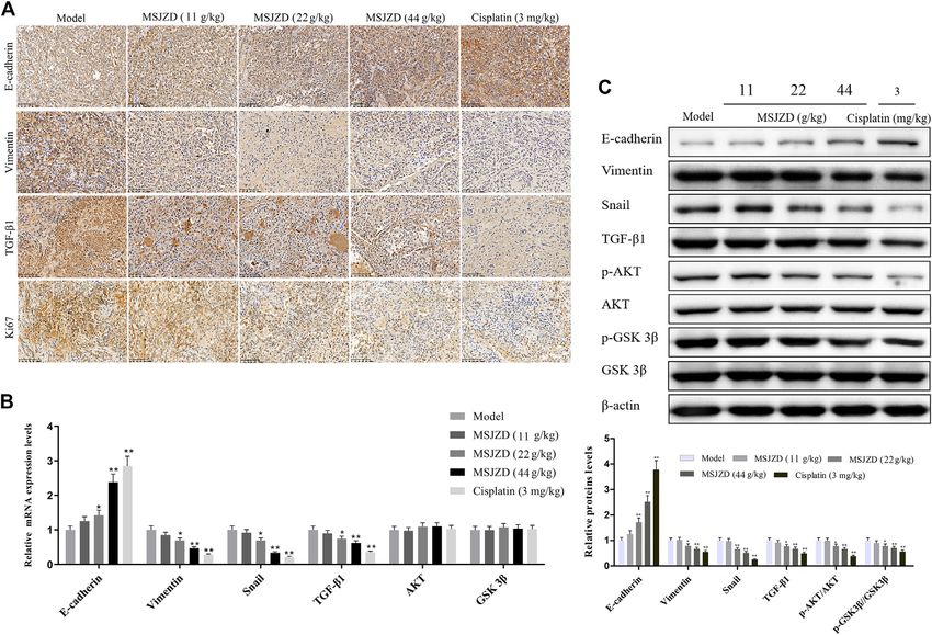
Shao et al. MSJZD Inhibits EMT in NSCLC
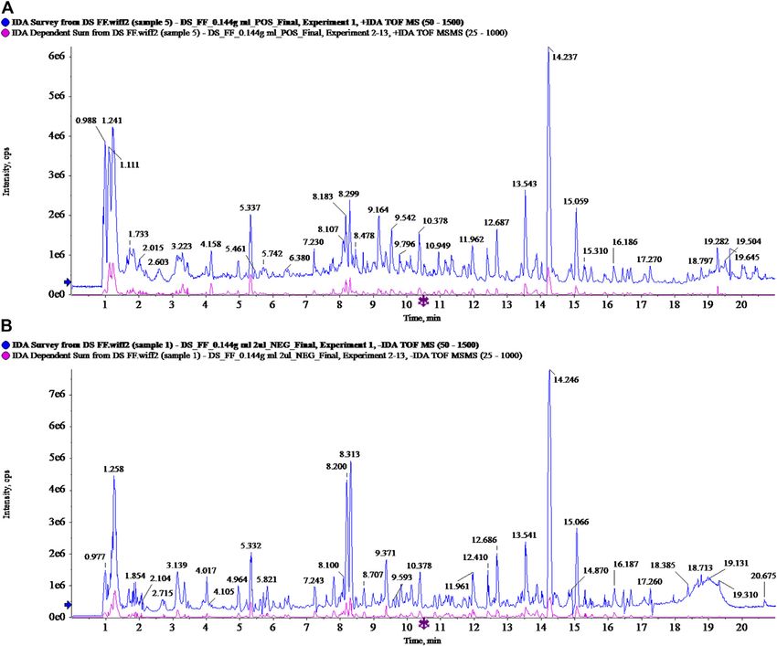
FIGURE 1 | Total ion chromatogram of Modified Sijunzi Decoction (MSJZD) obtained by UPLC/Q-TOF MS analysis in (A) positive ion mode and (B) negative
ion mode.
buffer until the cell concentration was 1.2 × 10⁶/well. After 15 min before staining with 0.1% crystal violet (548-62-9,
centrifugation, the precipitated cells were incubated with 5 μL Qiangshun, China) for 0.5 h. At last, the images were gained
Annexin V-FITC at 37°C away from light for 10 min followed by of the bottom side of the membrane by an optical microscope (AE
reaction with 5 μL PI at 37°C away from light for 5 min. After 2000, Motic, Germany). The number of migrated and invaded
adding 1 × binding buffer, we utilized a flow cytometer cells were quantified by ImageJ 1.52a software.
(CytoFLEX, Beckman Coulter, United States) to determine the
apoptosis rate of A549 and H1299 cells. Wound Healing Analysis
In brief, cells at a concentration of 5 × 10⁵ per well were plated
Transwell Assay into the 6-well plates overnight at 37°C with 5% CO2.
Cell migration was measured using inserts (3422, Corning, Subsequently, we utilized a 0.2 ml yellow sterile pipet tip to
United States) without matrigel matrix, while cell invasion was draw a gap. After reacting for 48 h at 37°C with 5% CO2, the
carried out using inserts with matrigel matrix (356234, BD results were observed and pictures were gained in the optical
Biosciences, United States). The treated cells (1 × 10⁴/well) in microscope at two-time points (0 and 48 h).
100 μL non-FBS medium were plated into the inserts and the
lower compartment was filled with medium augmented with 10% Immunofluorescence
FBS. After reacting for 24 h, the cells on top of the chambers were Cells at a concentration of 1 × 10 5 per well were injected into
cleaned with Q-tips and cells on the lower compartment were the 6-well plates containing cover slips. After being subjected
fixed in 4% paraformaldehyde (DF0135, Leagene, China) for to the indicated treatments, each well was fixed with methanol
Frontiers in Pharmacology | www.frontiersin.org 4 January 2022 | Volume 12 | Article 821567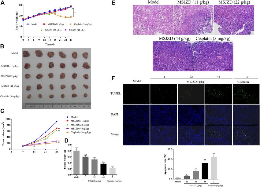
Shao et al.                                                                                                                 MSJZD Inhibits EMT in NSCLC

TABLE 1 | Putative identification of MSJZD in positive ion mode.

NO.                  Component                      Area          Retention    Formula       Precursor    Found     Mass        Library     Isotope
                       name                                         time                       Mass      at Mass     error       score        ratio
                                                                                                                    (ppm)                  difference

1        L (+)-Arginine                           6674000           1.11      C6H14N4O2       175.119    175.1184    −3.3        89.1          0.8
2        Glutamic acid                             299100           1.16      C5H9NO4          148.06    148.0602    −1.4        97.1          1.5
3        Betaine                                   296500           1.17      C5H11NO2        118.086    118.0861    −1.1        100           0.8
4        Trigonelline                             1389000           1.21      C7H7NO2         138.055    138.0545    −3.5        96.9          1.8
5        Proline                                  5169000           1.23      C5H9NO2         116.071    116.0703    −2.4        98.8          1.3
6        Cytidine                                   61590           1.28      C9H13N3O5       244.093    244.0925    −1.3        100           3.7
7        Pipecolinic acid                          792900           1.32      C6H11NO2        130.086    130.0862    −0.7        77.4          0.6
8        Adenine                                   800400           1.66      C5H5N5          136.062    136.0615    −2.1        97.6          2.4
9        Nicotinic acid                            172900           1.74      C6H5NO2         124.039    124.0391    −1.5        99.8          1.2
10       Nicotinamide                              231600           1.86      C6H6N2O         123.055    123.0551    −1.6        98.6           1
11       6-Hydroxypurine                            79120           1.93      C5H4N4O         137.046    137.0457    −0.7        79.4          1.6
12       Adenosine                                1537000           3.31      C10H13N5O4      268.104    268.1036    −1.8        100           4.1
13       10-Deacetylasperulosidic acid              58800           3.35       C16H22O11       408.15    408.1503    0.6         95.8          2.6
14       Cordycepin                                 86000           3.39      C10H13N5O3      252.109    252.1089    −0.7        100           4.6
15       Guanosine                                 433700           3.45      C10H13N5O5      284.099    284.0988    −0.6        99.1          4.9
16       Phenylalanine                             659900           4.16      C9H11NO2        166.086    166.0859    −2.1        99.8          1.7
17       Deacetyl asperulosidic acid methyl       217100            4.96      C17H24O11       422.166    422.1656    −0.3        99.4          8.4
         ester
18       Complanatoside                               7232           5.52     C28H32O16       625.176    625.1759    −0.6        100          12.1
19       Asperuloside                               30430            5.71     C18H22O11       415.123    415.1232    −0.7        87.4          1.9
20       Syringin                                    5659              6      C17H24O9        390.176    390.1753    −1.5        93.9         11.2
21       Chlorogenic acid                          127500              6      C16H18O9        355.102    355.1021    −0.7        99.6          6.2
22       Vitamin B2                                 56030            6.82     C17H20N4O6      377.146    377.1454    −0.3        93.9          5.8
23       Isoquercitrin                              84680            7.23     C21H20O12       465.103    465.1028    0.1         98.7          3.5
24       Quercetin                                 331500            7.23     C15H10O7         303.05    303.0499    −0.2        97.2           5
25       Hyperin                                    84680            7.23     C21H20O12       465.103    465.1028    0.1         99.1          3.5
26       Schaftoside                               319500            7.29     C26H28O14       565.155    565.1555    0.6         87.3          9.6
27       Daidzin                                    29060            7.66     C21H20O9        417.118    417.1182    0.4         100           6.2
28       Rutin                                     358200            7.98     C27H30O16       611.161    611.1604    −0.4        97.2         10.9
29       Calycosin-7-O-glucoside                  2881000             8.1     C22H22O10       447.129    447.1283    −0.6        100          9.4
30       Liquiritigenin                           7249000            8.28     C15H12O4        257.081    257.0806     −1         93.8         4.8
31       Scopoletin                                166200            8.29     C10H8O4          193.05    193.0494    −0.7        94.2         0.5
32       Peimisine                                 798300            8.83     C27H41NO3       428.316    428.3155    −1.1        89.1          9.8
33       Peimine                                   5136000           9.17     C27H45NO3       432.347    432.3468    −0.9        100          11.3
34       Pratensein-7-O-glucoside                    56730            9.3     C22H22O11       463.123    463.1237     0.4        95.2          6.5
35       Naringenin                                 174900           9.42     C15H12O5        273.076    273.0756    −0.7        99.4          5.8
36       Peiminine                                 3702000           9.54     C27H43NO3       430.332    430.3312     −1         100           8.5
37       Ononin                                    2887000          10.38     C22H22O9        431.134    431.1333    −0.7        98.6          8.4
38       Berberine                                  118700          10.66     C20H17NO4       336.123    336.1231    0.2          97           8.8
39       Farrerol                                   747800          10.96     C17H16O5        301.107    301.1069    −0.6        81.1          6.5
40       Calycosin                                 1548000          11.17     C16H12O5        285.076    285.0756    −0.5        95.4          6.2
41       Isomucronulatol                             89900          11.24     C17H18O5        303.123    303.1226    −0.2        91.7          4.5
42       Isomucronulatol-7-O-glucoside               25400          11.24     C23H28O10       465.176    465.1756     0.2        78.6          4.9
43       Astragaloside Ⅳ                            25030           13.85     C41H68O14       785.468    785.4668    −1.7        92.5          7.5
44       Glycyrrhizic acid                        11430000          14.23     C42H62O16       823.411    823.4098    −1.5        98.6          15
45       Parthenolide                                89170           16.6     C15H20O3        249.149    249.1483    −0.8        71.6          4.9
46       Glabridin                                   30850           18.1     C20H20O4        325.143    325.1431    −1.1        96.2          3.5
47       Alantolactone                              324600          18.55     C15H20O2        233.154    233.1532    −1.8        94.4          5.2

“jianguoyun” https://www.jianguoyun.com/p/DUoWHt0Q8-mCChiI5poE

followed by permeabilization with 0.5% Triton X-100. After                         DAPI, which was then captured by fluorescence microscopy
sealing with 1% BSA for 30 min, each well was subjected to                         (Ts2-FC, Nikon, Japan).
anti-E-cadherin antibody (1:500, ab40772, Abcam,
United Kingdom), anti-Vimentin antibody (1:1000,
ab92547, Abcam, United Kingdom), and anti-Fibronectin                              Establishment of A549 Cell Tumor-Bearing
antibody (1:50, ab268020, Abcam, United Kingdom) at 4 °C                           Model in Mice
overnight. Following reaction with anti-rabbit secondary                           Thirty male BALB/c nude mice (SPF grade, weighing 16 ± 2 g,
antibody for 1 h at 37 °C, the nuclei were stained with                            and 3–5 weeks) were obtained from Shanghai SLAC Laboratory
                                                                                   Animal Co., Ltd (Shanghai, China), and raised in the Animal

Frontiers in Pharmacology | www.frontiersin.org                                5                               January 2022 | Volume 12 | Article 821567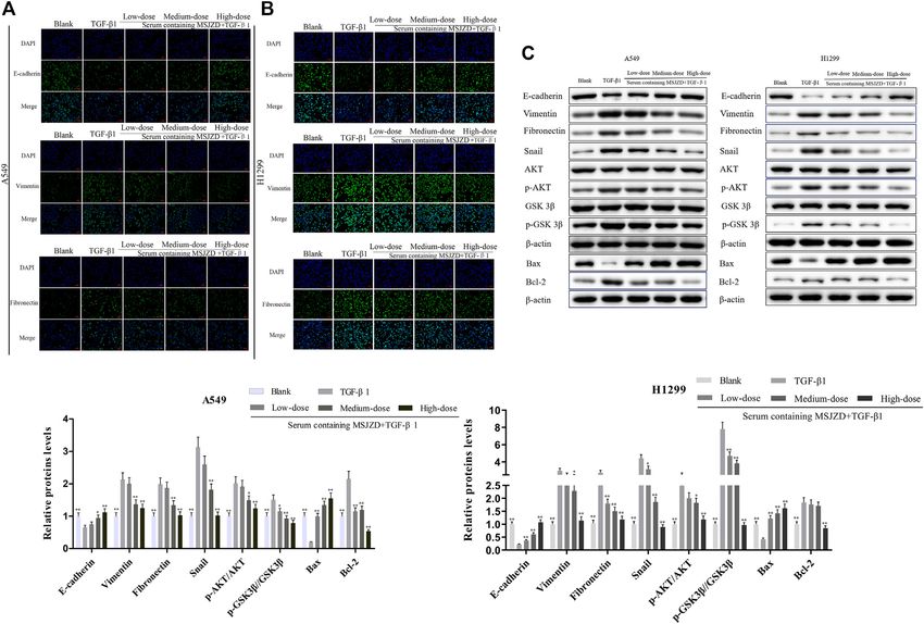
Shao et al.                                                                                                               MSJZD Inhibits EMT in NSCLC

TABLE 2 | Putative identification of MSJZD in negative ion mode.

NO.                 Component                       Area      Retention    Formula       Precursor     Found      Mass        Library     Isotope
                      name                                      time                       Mass       at Mass      error       score        ratio
                                                                                                                  (ppm)                  difference

1        Histidine                                   46050         1.08   C6H9N3O2        154.062     154.0623      0.4        96.3          1.6
2        L (+)-Arginine                             217900          1.1   C6H14N4O2       173.104     173.1042     −0.9        90.5          2.9
3        Glutamic acid                               46660         1.14   C5H9NO4         146.046     146.0459      0.1        93.7          1.1
4        Quinic acid                                694400         1.23   C7H12O6         191.056     191.0559     −1.4        93.8          2.9
5        Maleic acid                                469200         1.34   C4H4O4          115.004     115.0036     −0.4        99.6          0.5
6        Citric acid                               1466000         1.81   C6H8O7           191.02     191.0194     −1.5        98.6          1.6
7        Amber Acid                                 125800         2.65   C4H6O4          117.019     117.0192     −0.8        93.5          1.1
8        Isoleucine                                  64480         3.04   C6H13NO2        130.087     130.0873     −0.5        100           0.3
9        Adenosine                                   21060         3.31   C10H13N5O4      266.089     266.0894     −0.2        96.5          4.2
10       Guanosine                                  636700         3.46   C10H13N5O5      282.084     282.0837     −2.3        98.7          6.3
11       10-Deacetylasperulosidic acid             3209000         4.02   C16H22O11       389.109     389.1084     −1.3        97.9          7.6
12       Phenprobamate                              184000         4.18   C9H11NO2        164.072     164.0715     −1.3        95.6          3.5
13       L-Tryptophan                               127100         5.39   C11H12N2O2      203.083     203.0825     −0.5        91.5          2.4
14       Chlorogenic acid                           559600         6.01   C16H18O9        353.088     353.0874     −1.3        100           7.6
15       Asperuloside                                25180         6.47   C18H22O11       413.109     413.1086     −0.8        94.7          2.9
16       Fraxin                                       4046         6.48   C16H18O10       369.083     369.0825     −0.7         83          17.9
17       Caffeic acid                               223500         6.59   C9H8O4          179.035     179.0348     −1.1        82.1          4.1
18       Vitamin B2                                  14220         6.82   C17H20N4O6      375.131     375.1307     −0.8        99.2          2.6
19       Sibiricose A5                               24920         6.88   C22H30O14       517.156     517.1558     −0.8        94.6          8.2
20       Pratensein-7-O-glucoside                    14210         7.21   C22H22O11       461.109     461.1086     −0.6        98.1          3.7
21       Schaftoside                                278000         7.29   C26H28O14       563.141     563.1402     −0.7        97.9          11
22       Daidzin                                     27360         7.67   C21H20O9        415.103     415.1029     −1.3        92.3          5.3
23       p-Coumaric acid                           1381000         7.81   C9H8O3           163.04     163.0398     −1.6        99.2          4.3
24       Rutin                                      813500         7.98   C27H30O16       609.146     609.1454     −1.2        95.5         11.6
25       Calycosin-7-o-glucoside                   1831000          8.1   C22H22O10       491.119     491.1184     −2.2        99.2          7.1
26       Isoquercitrin                               83380         8.28   C21H20O12       463.088     463.0878     −0.8        89.7          1.4
27       Liquiritin                               14010000         8.31   C21H22O9        417.119     417.1183      −2         97.5          9.1
28       Ferulic Acid                                76560         8.38   C10H10O4        193.051     193.0505     −0.7        78.8          1.4
29       Isochlorogenic acid A                       47060         9.33   C25H24O12       515.119     515.1187     −1.6        94.3          7.5
30       4-Hydroxybenzoic acid                      230900        10.08   C7H6O3          137.024     137.0242     −1.6        94.3          1.3
31       Rhein                                        2069        10.18   C15H8O6         283.025     283.0257     −3.1         70           6.7
32       Ononin                                    2710000        10.39   C22H22O9        475.125     475.1235     −2.4        99.3         10.9
33       Liquiritigenin                            1471000        10.83   C15H12O4        255.066     255.0655     −2.9        91.2          6.8
34       Quercetin                                  420300         11.1   C15H10O7        301.035     301.0345     −2.9        96.2          6.9
35       Calycosin                                 1436000        11.17   C16H12O5        283.061     283.0604     −2.8        96.1          7.9
36       Isomucronulatol-7-O-glucoside              504300        11.24   C23H28O10       463.161     463.1599     −2.3        95.7          9.4
37       3-Hydroxy-9,10-                            120600        11.74   C17H16O5        299.092     299.0919     −1.9        83.1          5.6
         Dimethoxypterocarpan
38       Naringenin                                 67970         12.29   C15H12O5        271.061     271.0608     −1.5        97.2          5.4
39       Dihydroartemisinin                          3205          12.8   C15H24O5        283.155     283.1549     −0.7        86.3          5.2
40       Astragaloside Ⅳ                           152700         13.86   C41H68O14       829.459     829.4573     −2.1        98.8          17
41       Glycyrrhizic acid                        22010000        14.25   C42H62O16       821.397     821.3945     −2.4        95.5         14.7
42       Astragaloside Ⅱ                           852900         14.85   C43H70O15        871.47     871.4677     −2.3        96.9          17
43       Asiatic acid                               29610         16.57   C30H48O5        487.343     487.3423     −1.2        100           3.2
44       Astragaloside I                           966200         16.68   C45H72O16        913.48     913.4781     −2.4        99.7         17.3
45       Glabridin                                  97900         18.11   C20H20O4        323.129     323.1285     −1.1         87           9.4

Center of Zhejiang Chinese Medical University in a specific                     nude mice in the MSJZD group were gavaged with 11 g/kg,
pathogen-free (SPF) facility. These mice have free access to                   22 g/kg, or 44 g/ kg MSJZD, respectively, every day until the
diet and water for 7 days to acclimate to the environment.                     28th day. The nude mice in the cisplatin group were injected
A549 cell suspension (5 × 10⁶ in 100 μL sterilized PBS) was                    intraperitoneally with 3 mg/ kg cisplatin once every 3 days. The
injected subcutaneously into the left armpit of the nude mice.                 nude mice in the model group were given an equal volume of
When the tumor size reached nearly 50 mm3, they were randomly                  normal saline once a day for 28 days.
assigned to five groups, six mice in each group, namely model
group, 11 g/ kg MSJZD group (0.5 times clinical equivalent dose),              Detection of the Tumor Volume and Weight
22 g/ kg MSJZD group (clinical equivalent dose), 44 g/ kg MSJZD                The tumor volume of mice from all groups was measured and
group (2 times clinical equivalent dose), and 3 mg/ kg Cisplatin               calculated every 7 days until the 28th day, and the body
group. The drug clinical equivalent dose conversion formula was                weights of the mice were measured every 3 days during the
as follows: (human dose of crude herbs in clinic /60 kg) × 9.1. The            experiment. The tumor volume (V) was calculated using the

Frontiers in Pharmacology | www.frontiersin.org                            6                                 January 2022 | Volume 12 | Article 821567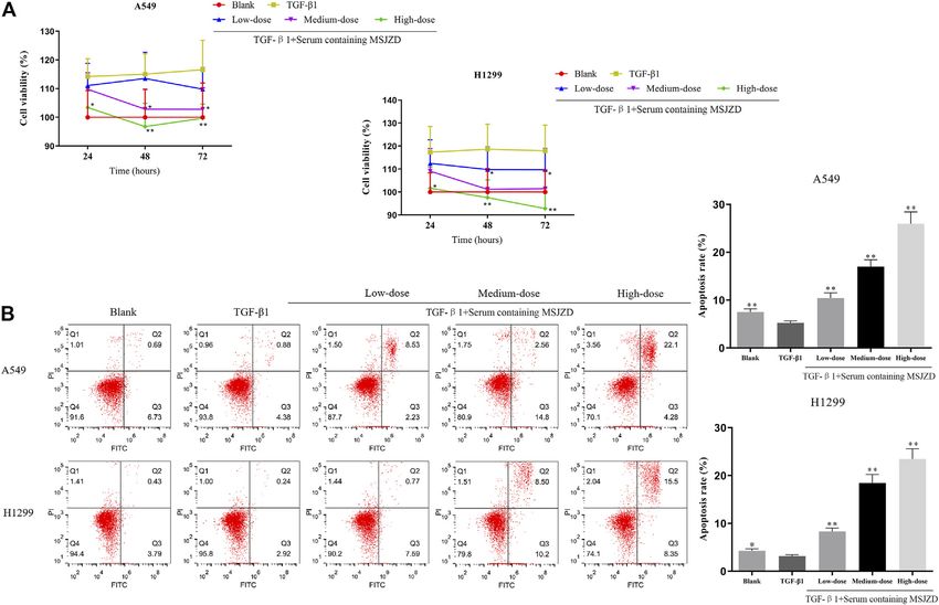
Shao et al.                                                                                                                                MSJZD Inhibits EMT in NSCLC

  FIGURE 2 | Effects of TGF-β1 on cell morphological changes and epithelial-mesenchymal transition of A549 and H1299 cells. (A) Microscopic photo of the lung
  cancer cells A549 and H1299 that treated with transforming growth factor-β1 (TGF-β1, 5 ng/ ml) for 0 h, 24, 48, and 72 h. (B) The levels of E-cadherin, Vimentin, and
  Fibronectin in A549 and H1299 cells treated with TGF-β1 (5 ng/ ml) for 72 h, as determined by western blot. *p < 0.05, **p < 0.01. versus the Blank group.

Frontiers in Pharmacology | www.frontiersin.org                                     7                                          January 2022 | Volume 12 | Article 821567
Shao et al. MSJZD Inhibits EMT in NSCLC
FIGURE 3 | Serum-containing MSJZD weakened the cell viability and augmented apoptosis in NSCLC cells induced by TGF-β1. (A) Cell counting kit-8 (CCK-8)
assay was performed in TGF-β1-induced A549 and H1299 cells treated with serum-containing MSJZD. (B) The A549 and H1299 cells were treated with blank control or
serum-containing MSJZD and analyzed by flow cytometer to indicate cell apoptosis. *p < 0.05, **p < 0.01 vs. the TGF-β1.
following formula: V 0.5 × W2 × L (W: the width of the the prepared TUNEL detection solution at 37°C away from light for
tumor; L: the length of the tumor). After the 28 th day, they 1 h. After washing, the sections were blocked with Antifade Mounting
were sacrificed using 35 mg/kg pentobarbital sodium, and Medium with DAPI (P0131, Beyotime, China) and then observed in
simultaneously, the tumor tissues were aseptically stripped, the fluorescence microscopy with its excitation and emission
weighed, and taken pictures. A portion of the tissues was fixed wavelength at 500 and 565 nm, respectively.
in 4% paraformaldehyde for H&E staining, TUNEL, and
immunohistochemistry (IHC) assay. Other tissues were IHC Assay
utilized for qRT-PCR and western blot assays. The paraffin sections were dewaxed, rehydrated, and then rinsed
with PBS. After antigen repair, the sections were subjected to 3%
H&E Staining hydrogen peroxide solution in the dark at 37°C for 25 min. After
The fixed tissues were dehydrated, which were then embedded in blocking, they were subjected to anti-E-cadherin antibody (1:500),
paraffin. After cutting into 5 μm sections, the sections were dewaxed anti-Vimentin antibody (1:500), anti-TGF-β1 antibody (1:500,
and hydrated followed by coloration with hematoxylin staining ab215715, Abcam, United Kingdom), and anti-Ki67 antibody (1:
solution (G1004, Servicebio, China). After the sections were 200, ab16667, Abcam, United Kingdom) at 37°C overnight. The next
differentiated and washed, they were placed in 1% eosin solution day, the sections were exposed to a secondary antibody at 37°C for
(C0109, Beyotime, China) for 5 min followed by dehydration and 1 h, which were then developed using DAB (abs9210, absin, China).
transparency. In the end, images were observed and taken using the After dehydration, we utilized a mounting medium to seal the
optical microscope after sealing the sections. sections. Lastly, images were observed and obtained under the
optical microscope.
TUNEL Staining
TUNEL Apoptosis Assay Kit (C1086, provided by Beyotime, China) Quantitative Real-Time PCR
was utilized in this research. The paraffin sections were subjected to RNA from tissues was got using Trizol reagent (abs60031, absin,
treatment with dewaxing and rehydration followed by a reaction with China). Afterward, the qRT-PCR reaction was conducted by
DNase-free proteinase K (ST532, Beyotime, China) at 37°C for TaqMan One-Step RT-qPCR Kit (T2210, Solarbio, China) in
20 min. After washing thoroughly, the sections were exposed to an EDC-810 PCR system (Eastwin Life Sciences, Inc.) under the
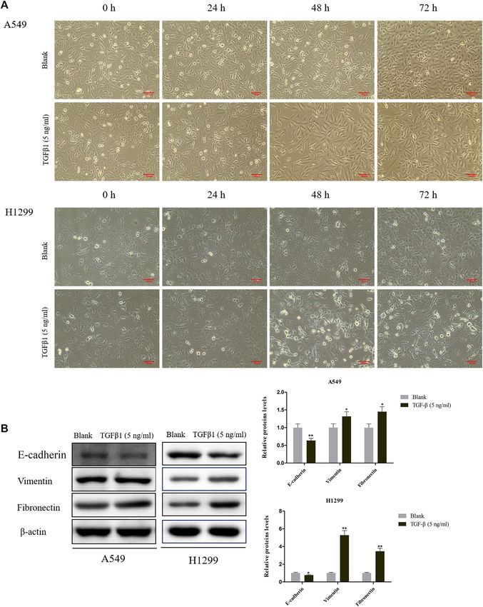
Frontiers in Pharmacology | www.frontiersin.org 8 January 2022 | Volume 12 | Article 821567Shao et al. MSJZD Inhibits EMT in NSCLC FIGURE 4 | Serum-containing MSJZD mitigated the migration and invasion in NSCLC cells triggered by TGF-β1. (A) Transwell assays were conducted to examine the effects of serum-containing MSJZD on A549 and H1299 cell migration and invasion. (B) Statistical analysis of invasion and migration cell number in A549 and H1299 cells. *p < 0.05, **p < 0.01. versus the TGF-β1 group. manufacturer’s instructions. β-actin was taken as the CCAGATTA -3’; Snail forward: 5′-ACTGCAACAAGGAAT normalization control. The 2−ΔΔCT was taken as count the ACCTCAG-3′, Snail reverse: 5′-GCACTGGTACTTCTTGAC relative expressions of the gene (Sun et al., 2020). The primers ATCTG-3’; TGF-β1 forward: 5′-CTAATGGTGGAAACCCAC were as follows: E-cadherin forward: 5′-AAAGGCCCATTTCCT AACG-3′, TGF-β1 reverse: 5′-TATCGCCAGGAATTGTTG AAAAACCT-3′, E-cadherin reverse: 5′-TGCGTTCTCTATCCA CTG-3’; AKT forward: 5′-AGCGACGTGGCTATTGTGAAG- GAGGCT-3’; Vimentin forward: 5′-TGCCGTTGAAGCTGC 3′, AKT reverse: 5′-GCCATCATTCTTGAGGAGGAAGT-3’; TAACTA-3′, Vimentin reverse: 5′-CCAGAGGGAGTGAAT GSK3β forward: 5′-GACTAAGGTCTTCCGACCCC--3′, Frontiers in Pharmacology | www.frontiersin.org 9 January 2022 | Volume 12 | Article 821567
Shao et al.                                                                                                                              MSJZD Inhibits EMT in NSCLC

  FIGURE 5 | Effects of serum-containing MSJZD on the migration of NSCLC cells in vitro. Wound healing (A) and quantitative assay (B) were performed to evaluate
  the migration ability of NSCLC cells. *p < 0.05, **p < 0.01. versus the TGF-β1 group.

GSK3β reverse: 5′-TTAGCATCTGAGCTCTGCTGT-3’; β-actin                                     Fibronectin antibody (1:1000), anti-Snail antibody (1:1000,
forward: 5′-GGAGCGAGATCCCTCCAAAAT-3′, β-actin                                           ab216347), anti-AKT antibody (1:500, ab8805), anti-p-AKT
reverse: 5′-GGCTGTTGTCATACTTCTCATGG-3’.                                                 antibody (1:1000, ab38449), anti-GSK3β antibody (1:8000,
                                                                                        ab32391), anti-p-GSK3β antibody (1:10000, ab75814), anti-Bax
Western Blot                                                                            antibody (1:2000, ab182733), anti-Bcl-2 antibody (1:1000,
After treatment, cells and tissues were treated with RIPA buffer                        ab32124), anti-β-actin antibody (1:200, ab115777) were obtained
(P0013D, Beyotime, China) containing PMSF (ST506, Beyotime,                             from Abcam (United Kingdom).
China) to acquire total proteins followed by quantification with BCA
kit (pc0020, Solarbio, China). After denaturation and electrophoresis,                  Statistical Analysis
the protein was transferred to a nitrocellulose membrane. After                         Each experiment was repeated independently at least three times.
blocking, the nitrocellulose membranes were exposed to specific                          Data were analyzed using Graphpad Prism 8.0 (GraphPad
primary antibodies overnight at 4°C. The next day, the bound                            Software Inc., United States). All data were revealed as
antibodies were subjected to an anti-rabbit secondary antibody for                      mean ± standard deviation. One-way ANOVA followed by
90 min at 37°C. In the end, signals were examined by an ECL reagent                     SNK’s multiple comparison test was utilized when data
(abs920, absin, China) with ChemiScope 3300 mini equipment                              followed a normal distribution. Kruskal-Wallis H test was
(Clinx, China). The primary antibodies of anti-E-cadherin                               utilized when data did not follow a normal distribution. A
antibody (1:40000), anti-Vimentin antibody (1:5000), anti-                              statistically significant difference was exhibited as p < 0.05.

Frontiers in Pharmacology | www.frontiersin.org                                    10                                        January 2022 | Volume 12 | Article 821567
Shao et al. MSJZD Inhibits EMT in NSCLC
FIGURE 6 | Serum-containing MSJZD weakened EMT, AKT/GSK3β pathway and induced apoptosis-related markers in NSCLC cells triggered by TGF-β1. The
EMT protein levels of A549 (A) and H1299 (B) were detected by immunofluorescence. (C) The levels of E-cadherin, Vimentin, Fibronectin, Snail, p-AKT, AKT, p-GSK3β,
GSK3β, and apoptotic proteins were detected by western blot after treatment with serum-containing MSJZD in A549 and H1299 cells. *p < 0.05, **p < 0.01. versus the
TGF-β1 group.
RESULTS changed most obviously after 72 h, which showed that the
epithelial cells dominated by cubes changed to the spindle and
fusiform mesenchymal morphology (Figure 2A). To a certain
Total Ion Chromatogram of Modified Sijunzi extent, TGF-β1 led to the boost of Vimentin and Fibronectin
Decoction (MSJZD) Obtained by UPLC/ levels, as well as the attenuation of E-cadherin in A549 and H1299
Q-TOF MS Analysis cells that were treated for 72 h (Figure 2B).
The MSJZD test solution was firstly analyzed by the UPLC-Q/
TOF-MS system. Figure 1 manifested the total ion
chromatogram of MSJZD in (A) positive ion mode and (B)
Serum Containing MSJZD Weakened the
negative ion mode. In positive ion mode, 47 kinds of Cell Viability and Augmented Apoptosis in
components were identified (Table 1). In negative ion mode, NSCLC Cells Induced by TGF-β1
45 kinds of components were identified (Table 2). We found that In this work, to probe the role of serum-containing MSJZD in
amino acids, polysaccharides, aromatic acids, flavones, NSCLC cells triggered by TGF-β1, we conducted CCK-8 and flow
monoterpene glycosides, and others widely existed in MSJZD. cytometer assays. Cells were subjected to 5 ng/ ml TGF-β1 and 10%
serum-containing MSJZD from different groups. Functionally, the
results unveiled that TGF-β1 led to the boost of cell viability and
TGF-β1 Induced Cell Morphological the inhibition of apoptosis. Interestingly, serum-containing
Changes and EMT MSJZD evidently attenuated cell viability and induced apoptosis
Next, we utilized TGF-β1 (5 ng/ ml) to treat A549 and H1299 in TGF-β1-mediated NSCLC cells and the effect of high-dose was
cells for 24, 48, and 72 h. We discovered that the cell morphology higher than medium-dose and low-dose (Figures 3A,B). These
changed to different degrees at different times after induction by findings revealed that serum-containing MSJZD has an inhibitory
TGF-β1, especially the morphology of A549 and H1299 cell effect in TGF-β1-mediated NSCLC cells.
Frontiers in Pharmacology | www.frontiersin.org 11 January 2022 | Volume 12 | Article 821567Shao et al. MSJZD Inhibits EMT in NSCLC
FIGURE 7 | MSJZD attenuated the tumor growth, promoted histopathological damage, and induced apoptosis in A549 tumor-bearing nude mice. (A) Body weight
of mice after MSJZD treatment was determined every 3 days. (B) Photograph of subcutaneous xenografts in A549 tumor-bearing nude mice after 28 days of treatment
of MSJZD. The volume (C) and weight (D) of xenografts in MSJZD and Cisplatin groups. (E) Microscopic images of xenografts were observed by H&E staining. (F)
TUNEL assay was performed to assess the apoptosis rate of xenografts in MSJZD and Cisplatin groups. *p < 0.05, **p < 0.01. versus the model group.
Serum Containing MSJZD Mitigated the were enhanced and the positive expression of E-cadherin was
Migration and Invasion in NSCLC Cells blunted by TGF-β1, whereas serum-containing MSJZD restrained
the positive expressions of Vimentin and Fibronectin and
Triggered by TGF-β1 augmented the positive expression of E-cadherin in TGF-β1-
To check the function of serum-containing MSJZD on the migration
mediated NSCLC cells, and the influence of high-dose was higher
and invasion in NSCLC cells triggered by TGF-β1, we utilized 5 ng/
than medium-dose and low-dose (Figures 6A,B). The results of
ml TGF-β1 and different doses of serum-containing MSJZD to treat
western blot clarified that TGF-β1 caused the inhibition of
cells. As displayed in Figures 4, 5, we further proved that the
E-cadherin and Bax and the boost of Vimentin, Fibronectin,
migration and invasion of A549 and H1299 cells were extremely
Snail, p-AKT, p-GSK3β, and Bcl-2, while the above effects were
strengthened by TGF-β1 (p < 0.01). More importantly, we
overturned by serum-containing MSJZD (Figure 6C).
discovered that the enhanced effects were partially offset by
serum-containing MSJZD in a dose-dependent way.
MSJZD Attenuated the Tumor Growth,
Promoted Histopathological Damage, and
Serum Containing MSJZD Weakened EMT, Induced Apoptosis in A549 Tumor-Bearing
AKT/GSK3β Pathway and Induced Nude Mice
Apoptosis-Related Markers in NSCLC Cells To further confirm the accuracy of in vitro experiments, we
Triggered by TGF-β1 established an A549 cell tumor-bearing model in mice. MSJZD
Then, to identify the mechanism of MSJZD on NSCLC cells, we failed to affect mice’s body weight, while cisplatin treatment
measured EMT-related factors by immunofluorescence and western significantly reduced the mice’s body weight (Figure 7A). MSJZD
blot assays. The positive expressions of Vimentin and Fibronectin restrained the tumor volume and weight in a dose-dependent way
Frontiers in Pharmacology | www.frontiersin.org 12 January 2022 | Volume 12 | Article 821567Shao et al. MSJZD Inhibits EMT in NSCLC
FIGURE 8 | MSJZD restrained EMT, AKT/GSK3β pathway, and TGF-β1 expression in A549 tumor-bearing nude mice. (A) Immunohistochemical (IHC) staining of
xenograft tumors. (B) The mRNA expression levels of E-cadherin, Vimentin, Snail, TGF-β1, AKT, and GSK3β in the experimental groups were determined by qRT-PCR.
(C) The expression levels of E-cadherin, Vimentin, Snail, TGF-β1, p-AKT, AKT, p-GSK 3β and GSK 3β in the experimental groups were determined by western blot. *p <
0.05, **p < 0.01. versus the model group.
and the inhibitory role of cisplatin was the most obvious (Figures MSJZD and cisplatin augmented the E-cadherin level and weakened
7B–D). The H&E staining results illustrated that in the model group, the Vimentin, Snail, TGF-β1 levels in A549 tumor-bearing nude
the tumor tissue structure and cell morphology were complete mice (Figure 8B). Similarly, western blot revealed that E-cadherin
(Figure 7E). In the high-dose MSJZD group and cisplatin group, expression in the MSJZD and cisplatin groups was significantly
the cell structure of tumor tissue was destroyed and the nucleus was increased compared with the models. Additionally, western blot
seriously condensed. Moreover, TUNEL staining exhibited that showed that the MSJZD and cisplatin markedly abolished the
MSJZD and cisplatin-induced apoptosis of A549 tumor-bearing expression of Vimentin, Snail, TGF-β1, p-AKT, p-GSK 3β in
nude mice and the promotion of 44 g/kg MSJZD on apoptosis A549 tumor-bearing nude mice, but MSJZD had no effect on the
was higher relative to the 11 and 22 g/kg MSJZD (Figure 7F). total protein expression levels of AKT and GSK 3β (Figure 8C). The
regulation of 44 g/kg MSJZD on these genes was the most evident
than 11 and 22 g/kg MSJZD.
MSJZD Restrained EMT, AKT/GSK3β
Pathway, and TGF-β1 Expression in
A549 Tumor-Bearing Nude Mice DISCUSSION
To verify the EMT-related protein (E-cadherin, Vimentin, Snail),
TGF-β1, Ki67, and AKT/GSK-3β pathway associated protein Tumor metastasis is caused by the decrease of intercellular
expressions in vivo, IHC assay, qRT-PCR and western blot assays adhesion and the enhancement of tumor cell motility and
were performed in tumor tissues of mice. IHC assay proved that the invasiveness; the invasive characteristics enable cancer cells to
positive expressions of Vimentin, TGF-β1, and Ki67 were inhibited separate from primary tumors and invade surrounding tissues
by MSJZD in the dose-dependent way and cisplatin, while the through collective or individual cell migration (Otsuki et al.,
positive expression of E-cadherin was enhanced by MSJZD and 2018). Modern medical research illustrated that EMT could give
cisplatin (Figure 8A). Next, the qRT-PCR results illuminated that tumor cells the phenotype and cellular plasticity required for
Frontiers in Pharmacology | www.frontiersin.org 13 January 2022 | Volume 12 | Article 821567Shao et al. MSJZD Inhibits EMT in NSCLC
metastasis to acquire mesenchymal characteristics, thereby augmented apoptosis in NSCLC cells induced by TGF-β1,
possessing high migration and invasion characteristics, and exhibiting that MSJZD has a certain anti-NSCLC effect.
ultimately promote tumor cells to spread and metastasize Invasion and metastasis are not only the important causes of death
(Lamouille et al., 2014; Marcucci et al., 2016). Many factors in NSCLC patients but also are the most intractable problems in
can induce the EMT process, including TGF-β1 (Eger et al., NSCLC treatment. EMT plays a pivotal role in the metastasis of
2004). TGF-β1 is transforming growth factor β, which has NSCLC (Mittal, 2016). EMT mainly involves multiple biological
multiple functions of modulating cell growth, apoptosis, processes, such as the loss of cell-cell adhesion, the destruction of
differentiation, and migration, involving multiple signal tumor basement membrane and extracellular matrix, and the
pathways (Moustakas and Heldin, 2012). A clinical study in reconstruction of the cytoskeleton, which plays a critical role in
NSCLC exhibited that the positive expression rate of TGF-β1 the metastasis of lung cancer (Chen et al., 2017). Tumor cells that
in adenocarcinoma in situ (AIS) was 27.3%, and in minimally occur in EMT usually undergo morphological and genetic changes.
invasive adenocarcinoma (MIA) was 65.2%, demonstrating that Morphological changes mainly include the evolution of cytokeratin
TGF-β1 overexpression makes the tumor more invasive (Imai structure from cubic epithelial cells to spindle-fusiform fibrocytes
et al., 2013). TGF-β1 induced EMT in lung cancer cells, resulting (Odero-Marah et al., 2018); gene changes include down-regulation of
in loss of cell polarity, decreased expression of epithelial marker E-cadherin expression and up-regulation of Vimentin and
E-cadherin, and increased expression of mesenchymal marker Fibronectin expression (Li B et al., 2019). The above changes lead
Vimentin (Massagué, 2008). Additionally, TGF-β 1-induced to the loss of cell-cell interaction, the loss of cell polarity, and the
tumor cell development EMT is a classic pathological model acquisition of interstitial cell “characteristics”, finally resulting in the
used in the experimental study of cancer cell metastasis. In vitro stronger invasion of tumor cells (Konrad et al., 2020).
studies, TGF-β1 induced EMT in NSCLC A549 cells, resulting in Changes in the expression of EMT-related markers are regulated
morphological changes, phosphorylation of Smad2 and Smad3, by transcription factors such as Twist, Snail, and Zeb, which are
down-regulation of E-cadherin and up-regulation of Vimentin, activated in the early stage of EMT to coordinate the suppression of
N-cadherin, Snail, Slug, and MMP2 (Zhao et al., 2015; Feng et al., epithelial genes and the activation of the mesenchymal gene (Bai et al.,
2017; Lim et al., 2017). In this study, A549 and H1299 cells of 2017; Przygodzka et al., 2019). Therefore, blocking the occurrence of
NSCLC were used and induced by TGF-β1 for 72 h. Significant EMT is an effective treatment to limit the spread of tumor cells. In
morphological changes occurred in the cells, including long addition, the AKT/GSK3β pathway plays a major role in the EMT
spindle shape, discrete, and disappearance of intercellular process (Xu et al., 2015). GSK3β is a downstream gene of PI3K/AKT
adhesion. At the same time, the expression of cell epithelial signaling, which can phosphorylate Snail transcription factor to
protein E-cadherin was down-regulated, and the expression of regulate EMT (Qiu et al., 2019). Snail is a kind of DNA binding
interstitial proteins Vimentin and Fibronectin increased notably, protein containing zinc finger structure, which can recognize the
proving that the EMT process occurred in NSCLC cells. E-box region upstream of the E-cadherin promoter, weaken the gene
TCM has a unique advantage in anti-tumor metastasis and has expression of E-cadherin, and promote the occurrence of EMT (Liu
been valued and affirmed by the medical community. The results et al., 2014). It was demonstrated that in lung cancer, OLA1
have clarified that TCM compounds, such as Jiedu Sangen modulated EMT through GSK3β/Snail/E-cadherin, thereby
Decoction, Jianpi Yangzheng Xiaozheng Decoction, and Bu-Fei modulating the invasion and metastasis of lung cancer (Bai et al.,
decoction, could weaken EMT to play the pharmacodynamic role 2016). Similarly, PI3K/AKT/GSK3β signaling has also been found to
of anti-tumor metastasis (He et al., 2017; Wu et al., 2019; Shan et al., regulate EMT in breast and gastric cancer Zhang et al., 2017, Dai et al.,
2020). In the current study, the constituents of MSJZD were analyzed 2016). Moreover, AKT/GSK3β signaling generated a critical role in
using the UPLC-Q-TOF-MS method. Amino acids, polysaccharides, the modulation of apoptosis in lung cancer cells (Li, Y et al., 2019).
aromatic acids, flavones, and monoterpene glycosides may aid in the The boost of pro-apoptotic protein Bax and the reduction of anti-
anti-cancer effects of MSJZD. Among them, Engelen et al. found that apoptotic protein Bcl-2 are the key factors to induce apoptosis (Liao
an impaired endogenous arginine synthesis was related to the et al., 2014). In vitro research, we discovered that TGF-β1 caused the
reduced systemic arginine availability and NO synthesis in inhibition of E-cadherin and Bax and the boost of Vimentin,
advanced NSCLC, and a dietary amino acid mixture is able to Fibronectin, Snail, p-AKT, p-GSK3β, and Bcl-2, while the above
restore systemic arginine availability in cancer (Engelen et al., 2016). effects were overturned by serum-containing MSJZD, exhibiting that
Also, chlorogenic acid, an ester with various pharmacological effects, MSJZD restrained EMT and induced apoptosis through the AKT/
is important in cancer therapy, including NSCLC (Hongtao et al., GSK3β pathway, thereby repressing the migration, invasion, and
2018). Moreover, Liao et al. reported that the combination of promoting apoptosis of NSCLC cells.
cordycepin and apatinib has a synergistically anticancer effect on To further verify the accuracy of in vitro results, we established an
NSCLC cells by down-regulating VEGF/PI3K/Akt signaling pathway A549 cell tumor-bearing model in mice. Our analysis manifested that
(Liao et al., 2020). This result indicated that the constituents of MSJZD attenuated the tumor growth, promoted histopathological
MSJZD could be a promising drug against NSCLC. damage, and induced apoptosis in A549 tumor-bearing nude mice.
Meanwhile, we evaluated the effects of serum-containing More importantly, MSJZD augmented the E-cadherin level and
MSJZD in NSCLC A549 and H1299 cells-induced EMT by weakened the Vimentin, Snail, TGF-β1, p-AKT, and p-GSK3β
TGF-β1 for the first time. The results of cell function levels in A549 tumor-bearing nude mice, which was consistent
experiments illustrated that serum-containing MSJZD with in vitro experiments. Although this study unveiled the anti-
weakened the cell viability, migration, invasion, and tumor mechanism of MSJZD to a certain extent, TCM prescription
Frontiers in Pharmacology | www.frontiersin.org 14 January 2022 | Volume 12 | Article 821567Shao et al. MSJZD Inhibits EMT in NSCLC
exerted its curative effect through multi-targets and multi-pathways. ETHICS STATEMENT
It should be noted that whether MSJZD may regulate EMT through
other pathways, which needs more experimental verification. The animal programs complied with the guidelines of the China
Meanwhile, the composition of MSJZD is extremely complex, and Animal Care and Use Committee. Approval was acquired from
the active substance of its curative effect needs further studied and the Committee of Laboratory Animals of Zhejiang Chinese
confirmed. In addition, Further establish animal models of Medical University Laboratory Animal Research Center
spontaneous metastasis of lung cancer are needed to more (license number: SYXK (Zhe) 2018-0012). All efforts were
intuitively evaluate the pharmacodynamics of MSJZD in made to alleviate the suffering of animals.
modulating the EMT process of lung cancer and weakening
tumor metastasis in vivo.
AUTHOR CONTRIBUTIONS
CONCLUSION SB and NS: Conception and design of the research; YX, JZ, YZ,
SW: Acquisition of data; NS, JZ, YX: Analysis and interpretation
In short, our research is the first one to demonstrate that MSJZD of data; NS and YX: Statistical analysis; NS: Drafting the
restrains EMT through the AKT/GSK3β pathway, thereby manuscript; SB and NS: Revision of manuscript for important
repressing the migration and invasion of NSCLC cells, which intellectual content. All authors reviewed the results and
is expected to become a new therapeutic target for NSCLC. approved the final version of the manuscript.
DATA AVAILABILITY STATEMENT FUNDING
The raw data supporting the conclusions of this article will be This work was financially supported by the Zhejiang TCM
made available by the authors, without undue reservation. Science and Technology Program (2017ZB025).
Eger, A., Stockinger, A., Park, J., Langkopf, E., Mikula, M., Gotzmann, J., et al.
REFERENCES (2004). Beta-Catenin and TGFbeta Signalling Cooperate to Maintain a
Mesenchymal Phenotype after FosER-Induced Epithelial to Mesenchymal
Adjei, A. A. (2019). Lung Cancer Worldwide. J. Thorac. Oncol. 14 (6), 956. Transition. Oncogene 23 (15), 2672–2680. doi:10.1038/sj.onc.1207416
doi:10.1016/j.jtho.2019.04.001 Engelen, M. P., Safar, A. M., Bartter, T., Koeman, F., and Deutz, N. E. (2016).
Aiello, N. M., Brabletz, T., Kang, Y., Nieto, M. A., Weinberg, R. A., and Stanger, B. Reduced Arginine Availability and Nitric Oxide Synthesis in Cancer Is Related
Z. (2017). Upholding a Role for EMT in Pancreatic Cancer Metastasis. Nature to Impaired Endogenous Arginine Synthesis. Clin. Sci. (Lond) 130 (14),
547 (7661), E7–E8. doi:10.1038/nature22963 1185–1195. doi:10.1042/CS20160233
Allen, A. M., Shochat, T., Flex, D., Kramer, M. R., Zer, A., Peled, N., et al. (2018). Feng, H. T., Zhao, W. W., Lu, J. J., Wang, Y. T., and Chen, X. P. (2017).
High-Dose Radiotherapy as Neoadjuvant Treatment in Non-small-cell Lung Hypaconitine Inhibits TGF-B1-Induced Epithelial-Mesenchymal Transition
Cancer. Oncology 95 (1), 13–19. doi:10.1159/000487928 and Suppresses Adhesion, Migration, and Invasion of Lung Cancer A549
Bai, J. W., Zhang, Y. Q., Li, Y. C., and Zhang, G. J. (2017). Analysis of Epithelial- Cells. Chin. J. Nat. Med. 15 (6), 427–435. doi:10.1016/S1875-5364(17)30064-X
Mesenchymal Transition Induced by Overexpression of Twist. Methods Mol. Gadgeel, S. M. (2017). Role of Chemotherapy and Targeted Therapy in Early-Stage
Biol. 1652, 259–274. doi:10.1007/978-1-4939-7219-7_17 Non-small Cell Lung Cancer. Am. Soc. Clin. Oncol. Educ. Book 37, 630–639.
Bai, L., Yu, Z., Zhang, J., Yuan, S., Liao, C., Jeyabal, P. V., et al. (2016). OLA1 doi:10.1200/EDBK_175188
Contributes to Epithelial-Mesenchymal Transition in Lung Cancer by Guan, Z., Wu, J., Wang, C., Zhang, F., Wang, Y., Wang, M., et al. (2018).
Modulating the GSK3β/snail/E-Cadherin Signaling. Oncotarget 7 (9), Investigation of the Preventive Effect of Sijunzi Decoction on Mitomycin
10402–10413. doi:10.18632/oncotarget.7224 C-Induced Immunotoxicity in Rats by 1H NMR and MS-based Untargeted
Chen, J. M., Yang, T. T., Cheng, T. S., Hsiao, T. F., Chang, P. M., Leu, J. Y., et al. Metabolomic Analysis. J. Ethnopharmacol 210, 179–191. doi:10.1016/
(2019). Modified Sijunzi Decoction Can Alleviate Cisplatin-Induced j.jep.2017.08.021
Toxicity and Prolong the Survival Time of Cachectic Mice by He, X. R., Han, S. Y., Li, X. H., Zheng, W. X., Pang, L. N., Jiang, S. T., et al. (2017).
Recovering Muscle Atrophy. J. Ethnopharmacol 233, 47–55. doi:10.1016/ Chinese Medicine Bu-Fei Decoction Attenuates Epithelial-Mesenchymal
j.jep.2018.12.035 Transition of Non-small Cell Lung Cancer via Inhibition of Transforming
Chen, T., You, Y., Jiang, H., and Wang, Z. Z. (2017). Epithelial-mesenchymal Growth Factor β1 Signaling Pathway In Vitro and In Vivo. J. Ethnopharmacol
Transition (EMT): A Biological Process in the Development, Stem Cell 204, 45–57. doi:10.1016/j.jep.2017.04.008
Differentiation, and Tumorigenesis. J. Cel Physiol 232 (12), 3261–3272. Hirsch, F. R., Suda, K., Wiens, J., and Bunn, P. A., Jr (2016). New and Emerging
doi:10.1002/jcp.25797 Targeted Treatments in Advanced Non-small-cell Lung Cancer. Lancet 388
Cheng, X., Gu, J., Zhang, M., Yuan, J., Zhao, B., Jiang, J., et al. (2014). Astragaloside (10048), 1012–1024. doi:10.1016/S0140-6736(16)31473-8
IV Inhibits Migration and Invasion in Human Lung Cancer A549 Cells via Hongtao, L., Xiaoqi, G., Junni, L., Feng, X., Guodong, B., and Liang, Y. (2018).
Regulating PKC-α-Erk1/2-NF-Κb Pathway. Int. Immunopharmacol 23 (1), Chlorogenic-induced Inhibition of Non-small Cancer Cells Occurs through
304–313. doi:10.1016/j.intimp.2014.08.027 Regulation of Histone Deacetylase 6. Cel Mol Biol (Noisy-le-grand) 64 (10),
Dai, J., Qian, C., Su, M., Chen, M., and Chen, J. (2016). Gastrokine-2 Suppresses 134–139. doi:10.14715/cmb/2018.64.10.22
Epithelial Mesenchymal Transition through PI3K/AKT/GSK3β Signaling in Huang, A. L., Liu, S. G., Qi, W. J., Zhao, Y. F., Li, Y. M., Lei, B., et al. (2014). TGF-β1
Gastric Cancer. Tumour Biol. 37 (9), 12403–12410. doi:10.1007/s13277-016- Protein Expression in Non-small Cell Lung Cancers Is Correlated with Prognosis.
5107-x Asian Pac. J. Cancer Prev. 15 (19), 8143–8147. doi:10.7314/apjcp.2014.15.19.8143
Du, L., Waqar, S. N., and Morgensztern, D. (2016). Multimodality Therapy for Imai, K., Minamiya, Y., Goto, A., Nanjo, H., Saito, H., Motoyama, S., et al. (2013).
NSCLC. Cancer Treat. Res. 170, 151–163. doi:10.1007/978-3-319-40389-2_7 Bronchioloalveolar Invasion in Non-small Cell Lung Cancer Is Associated with
Frontiers in Pharmacology | www.frontiersin.org 15 January 2022 | Volume 12 | Article 821567You can also read