A locomotor neural circuit persists and functions similarly in larvae and adult Drosophila
←
→
Page content transcription
If your browser does not render page correctly, please read the page content below
RESEARCH ADVANCE

                                   A locomotor neural circuit persists and
                                   functions similarly in larvae and adult
                                   Drosophila
                                   Kristen Lee, Chris Q Doe*

                                   Institute of Neuroscience, Howard Hughes Medical Institute, University of Oregon,
                                   Eugene, United States

                                   Abstract Individual neurons can undergo drastic structural changes, known as neuronal
                                   remodeling or structural plasticity. One example of this is in response to hormones, such as during
                                   puberty in mammals or metamorphosis in insects. However, in each of these examples, it remains
                                   unclear whether the remodeled neuron resumes prior patterns of connectivity, and if so, whether
                                   the persistent circuits drive similar behaviors. Here, we utilize a well-characterized neural circuit in
                                   the Drosophila larva: the moonwalker descending neuron (MDN) circuit. We previously showed that
                                   larval MDN induces backward crawling, and synapses onto the Pair1 interneuron to inhibit forward
                                   crawling (Carreira-Rosario et al., 2018). MDN is remodeled during metamorphosis and regulates
                                   backward walking in the adult fly. We investigated whether Pair1 is remodeled during
                                   metamorphosis and functions within the MDN circuit during adulthood. We assayed morphology
                                   and molecular markers to demonstrate that Pair1 is remodeled during metamorphosis and persists
                                   in the adult fly. MDN-Pair1 connectivity is lost during early pupal stages, when both neurons are
                                   severely pruned back, but connectivity is re-established at mid-pupal stages and persist into the
                                   adult. In the adult, optogenetic activation of Pair1 resulted in arrest of forward locomotion, similar
                                   to what is observed in larvae. Thus, the MDN-Pair1 neurons are an interneuronal circuit – a pair of
                                   synaptically connected interneurons – that is re-established during metamorphosis, yet generates
                                   similar locomotor behavior at both larval and adult stages.

*For correspondence:
cdoe@uoregon.edu

Competing interest: See            Introduction
page 13
                                   Large-scale changes in neuronal morphology and function occur during mammalian puberty
Funding: See page 13               (Barendse et al., 2018; Mills et al., 2016; Sisk and Zehr, 2005), as well as several neurobiological
Preprinted: 27 April 2021          disorders including depression (Patel et al., 2019), or chronic pain (Kuner and Flor, 2017). Similarly,
Received: 27 April 2021            major changes in neuronal numbers and type occur as a result of insect metamorphosis
Accepted: 13 July 2021             (Kanamori et al., 2015; Truman and Reiss, 1976; Yaniv and Schuldiner, 2016). Despite these
Published: 14 July 2021            changes, there are documented cases of individual insect neurons persisting from larval to adult
                                   stages. In Drosophila, individual motor and sensory neurons have been shown to persist throughout
Reviewing editor: Ronald L
Calabrese, Emory University,
                                   metamorphosis and undergo dramatic remodeling (Consoulas et al., 2002; Consoulas et al., 2000;
United States                      Yaniv and Schuldiner, 2016; Yu and Schuldiner, 2014). Similar findings have been reported for the
                                   insect mushroom body, where Kenyon cells partners (projection neurons, DANs) exist at both larval
   Copyright Lee and Doe. This
                                   and adult stages (Li et al., 2020; Marin et al., 2005). Yet, it remains unclear whether the remodeled
article is distributed under the
                                   neurons re-establish connectivity with the identical neurons in the larva and adult.
terms of the Creative Commons
Attribution License, which             During Drosophila metamorphosis, the animal changes from a crawling limbless larva to a walking
permits unrestricted use and       six-legged adult (Riddiford, 1980; Riddiford et al., 2003). Despite the obvious differences, some
redistribution provided that the   behaviors are similar: both larvae and adults undergo forward locomotion in search of food, back-
original author and source are     ward locomotion in response to noxious stimuli, and pausing in between antagonistic behaviors (Car-
credited.                          reira-Rosario et al., 2018). We and others identified a neuron that, when activated, can trigger

Lee and Doe. eLife 2021;10:e69767. DOI: https://doi.org/10.7554/eLife.69767                                                         1 of 15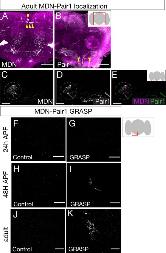
Research advance                                                                                                     Neuroscience

                                   backward locomotion in both larvae and adults (Bidaye et al., 2014; Carreira-Rosario et al., 2018;
                                   Sen et al., 2017), despite the obvious differences in limbless and six-legged locomotion. This neu-
                                   ron, named mooncrawler/moonwalker descending neuron (MDN), is present as a bilateral neuronal
                                   pair in each brain lobe, with all four MDNs having similar synaptic partners, and all four MDNs capa-
                                   ble of eliciting backward larval locomotion in larvae (Carreira-Rosario et al., 2018). Larval MDNs
                                   function within a neural circuit that induces backward locomotion and coordinately arrests forward
                                   locomotion. Halting forward locomotion is achieved via activation of the Pair1 descending interneu-
                                   ron, which inhibits the A27h premotor neuron. Given that the A27h interneuron is required for for-
                                   ward locomotion, its inhibition via MDN-induced Pair1 activation prevents forward locomotion
                                   (Carreira-Rosario et al., 2018). Activating backward locomotion is likely to be due, in part, to MDN
                                   activation of the A18b premotor neuron, which is specifically active during backward locomotion
                                   (Carreira-Rosario et al., 2018). Thus, MDN-Pair1 are synaptically coupled members of a locomotor
                                   circuit in the Drosophila larva.
                                       Here, we follow our previous work showing that MDN is remodeled during metamorphosis and
                                   persists into the adult (Carreira-Rosario et al., 2018) by asking: Is the MDN partner neuron Pair1
                                   also maintained in the adult? Does the adult Pair1 induce an inhibition in forward locomotion, similar
                                   to its role in larvae? And, are the adult Pair1 and MDN synaptically coupled? We find that all of these
                                   questions are answered in the affirmative, showing that the core MDN-Pair1 interneuron circuit (a
                                   pair of synaptically connected interneurons) is re-established during metamorphosis despite pro-
                                   found neurite remodeling, and that this circuit coordinates forward/backward locomotion in both lar-
                                   vae and adults.

                                   Results
                                   The Pair1 neuron persists from larval to adult stages
                                   To determine if Pair1 neurons were present in the adult, we mapped expression of a Pair1-Gal4 line
                                   (R75C02-Gal4) from early larval to adult stages. We identified the larval Pair1 neurons based on their
                                   characteristic cell body position in the medial subesophageal zone (SEZ), dense local ipsilateral den-
                                   dritic arborizations (defined as dendritic based on enrichment for post-synapses in the TEM recon-
                                   struction of the larval Pair1 neuron; Figure 1—figure supplement 1), and contralateral axons
                                   descending into the ventral nerve cord (VNC) in an extremely lateral axon tract (Carreira-
                                   Rosario et al., 2018). Using the Pair1-Gal4 line, we could identify Pair1 neurons with this morphol-
                                   ogy at 28 and 96 hr after larval hatching (ALH; Figure 1A,B). The Pair1 neuron cell bodies and proxi-
                                   mal neurites could still be observed at 24 hr after pupal formation (APF), but virtually all of the
                                   dendridic processes and descending axonal process are pruned (Figure 1C, only one neuron
                                   labeled). This is expected, given that many or all neurons undergo axon/dendrite remodeling during
                                   metamorphosis (Kanamori et al., 2015; Truman and Reiss, 1976; Yaniv and Schuldiner, 2016). At
                                   48 hr APF, Pair1 neurons exhibited dendritic branching in the SEZ and a descending axon into the
                                   VNC, regaining morphological features similar to that of larval Pair1 neurons (Figure 1D). The axon
                                   innervated the T1 (prothoracic) neuropil and descended further down the VNC. These morphological
                                   features were maintained into the adult fly, where we could trace the Pair1 axon to primarily inner-
                                   vate the T1 neuropil (Figure 1E), with less extensive innervation of the mesothoracic (T2) and meta-
                                   thoracic (T3) neuropils.
                                       Although we can use the Pair1-Gal4 line to track neurons with Pair1 morphological features from
                                   larva to adult, it remains possible that the Gal4 line switches off in Pair1 and switches on in a similar
                                   descending neuron at a stage in between those we assayed. To conclusively demonstrate that the
                                   larval Pair1 neuron survives into adulthood, we used a genetic technique to permanently label or
                                   ‘immortalize’ the larval Pair1 neurons and assay for their presence in the adult brain. Briefly, the
                                   method achieves spatial specificity by using Pair1-Gal4 to drive UAS-FLP which removes a stop cas-
                                   sette from nSyb-FRT-stop-FRT-LexA resulting in permanent LexA expression in Pair1-Gal4 neurons;
                                   it achieves temporal specificity (e.g. labeling only larval Pair1-Gal4+ neurons) by using a heat induc-
                                   ible KD recombinase to ‘open’ the lexAop-KDRTstopKDRT-HA reporter (see Materials and methods
                                   for additional details). Thus, a heat shock will permanently label all Pair1-Gal4+ neurons at the time

Lee and Doe. eLife 2021;10:e69767. DOI: https://doi.org/10.7554/eLife.69767                                                          2 of 15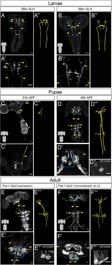
Research advance                                                                                                                Neuroscience

                                   Figure 1. The Pair1 neuron persists from larval to adult stages. (A–B) Pair1 neurons (cell body: yellow asterisk;
                                   neurites: yellow arrowhead) in the larval CNS (gray outline) at 28 hr after larval hatching (ALH) (A) and 96 hr ALH
                                   (B). Here and in subsequent panels are maximum intensity projections of confocal sections containing the Pair1
                                   neurons; anterior, up; dorsal view. Significant ‘off-target’ expression marked with white arrowheads. Scale bar, 50
                                   Figure 1 continued on next page

Lee and Doe. eLife 2021;10:e69767. DOI: https://doi.org/10.7554/eLife.69767                                                                     3 of 15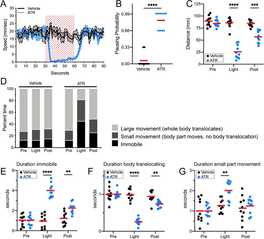
Research advance Neuroscience
Figure 1 continued
mm. (A’–B’) Enlargement of the brain regions boxed in A,B. Scale bar, 20 mm. (A”–B”) Tracing to show Pair1
neuron morphology. Genotype: +; UAS-myr::GFP; R75C02-Gal4. (C–D) Pair1 neurons (cell body: yellow asterisk;
neurites: yellow arrowhead) in the pupal CNS (gray outline) at 24 hr after pupal formation (APF) (C) and 96 hr APF
(D). Significant ‘off-target’ expression marked with white arrowheads. Scale bar, 50 mm. (C’–D’) Enlargement of the
brain regions boxed in C, D; cell body: yellow asterisk, neurites: yellow arrowhead. Scale bar, 10 mm. (C”) Tracing
to show Pair1 neuron morphology. (D”) Focal plane showing Pair1 cell bodies (region boxed in D’, cell body
marked with yellow asterisks). Scale bar, 10 mm. (D”’) Tracing to show Pair1 neuron morphology. Note that Pair1
can be followed to T1 in the 3D confocal stack but is difficult to represent here due to fasciculation of Pair1 with
off-target neurons. Genotype: +; UAS-myr::GFP; R75C02-Gal4. (E) Pair1 neurons (cell body: yellow asterisk;
neurites: yellow arrowhead) in the 4-day adult CNS (gray outline) Significant ‘off-target’ expression marked with
white arrowheads. Scale bar, 50 mm. (E’) Enlargement of the brain region boxed in E. Scale bar, 10 mm. (E”) Focal
plane showing Pair1 cell bodies (region boxed in E’, cell body marked with yellow asterisks). Scale bar, 10 mm. (E”’)
Tracing to show Pair1 neuron morphology. Genotype: +; UAS-myr::GFP; R75C02-Gal4. (F) Pair1 neurons (cell body:
yellow asterisk; neurites: yellow arrowhead) permanently labeled at 96 hr ALH and visualized in the 4-day old adult.
See Materials and methods for details. Significant ‘off-target’ expression marked with white arrowheads. Scale bar,
50 mm. (F’) Enlargement of the brain region boxed in F; Pair1 cell body: yellow asterisk; Pair1 neurites: yellow
arrowhead. Scale bar, 10 mm. (F”) Focal plane showing Pair1 cell bodies (region boxed in F’, cell body marked with
yellow asterisks). Scale bar, 10 mm. (F”’) Tracing to show Pair1 neuron morphology. Genotype: Hs-KD,3xUAS-FLP;
13xLexAop(KDRT.Stop)myr:smGdP-Flag/+; 13xLexAop(KDRT.Stop)myr:smGdP-V5, 13xLexAop(KDRT.Stop)myr:
smGdP-HA, nSyb(FRT.Stop)LexA::p65.
The online version of this article includes the following figure supplement(s) for figure 1:
Figure supplement 1. Moonwalker descending neuron (MDN) axon and Pair1 dendrite target the same neuropil
in the larval brain.
of heat shock. We immortalized Pair1 neurons in the larva, and assayed expression in the adult, and
observed the two bilateral Pair1 neurons, based on characteristic medial SEZ cell body position, local
ipsilateral arbors, and contralateral descending axons that preferentially innervate the prothoracic
neuropil (Figure 1F). Pair1 innervation is clearer in neurons immortalized during larval stages, which
reduces the off-target neuron expression in the adult VNC, and reveals an greatly enriched level of
innervation in the T1 neuropil (Figure 1F).
The Pair1-Gal4 line is expressed in several off-target neurons in addition to Pair1. One of these, a
sensory neuron from the proboscis can be removed from the adult Pair1 pattern by cutting off the
proboscis a day prior to analysis (see Materials and methods) but is present at the 48 hr APF time-
point (Figure 1D,E). In addition, there are off-target neurons that innervate all three thoracic neuro-
pils (T1-T3), obscuring Pair1 innervation (Figure 1E). We took advantage of the sparse labeling of
the immortalization genetics and found brains that maintained preferential targeting of Pair1 to the
prothoracic neuropil but lacked T1-T3 off-target innervation, confirming that they are indeed off-tar-
get neurons (Figure 1F–F’’’).
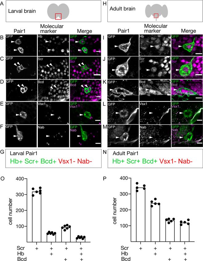
Pair1 neurons maintain the same molecular profile from larval to adult
stages
If Pair1 neurons persist from larva to adult, they may express the same transcription factor (TF) pro-
file at both stages. We screened a small collection of TF markers for expression in the larval and
adult Pair1 neurons, and in all cases we found identical expression (Figure 2A–N). Larval and adult
Pair1 neurons expressed Hunchback (Hb), Sex combs reduced (Scr), and Bicoid (Bcd); but did not
express Visual system homeobox (Vsx1) or Nab (Figure 2A–N). However, there were many other
Scr/Bcd/Hb triple-positive neurons in the SEZ (Figure 2O,P), showing that additional factors would
be necessary to uniquely specify Pair1 identity. These results support the conclusion that Pair1 per-
sists from larva to adult, maintaining both molecular and morphological features, and raises the
interesting possibility that the three TFs (Hb, Bcd, Scr) may be part of a molecular code that directs
both larval and adult Pair1 morphology and/or connectivity.
Pair1 activation arrests forward locomotion in adults
We previously showed that larval MDN persists in adults and can induce backward locomotion at
both stages despite the obvious difference in motor output – limbless crawling versus six-legged
Lee and Doe. eLife 2021;10:e69767. DOI: https://doi.org/10.7554/eLife.69767 4 of 15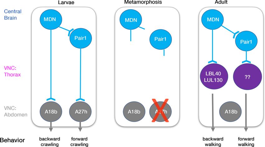
Research advance                                                                                                                Neuroscience

Figure 2. The Pair1 neuron expresses the same molecular markers at larval and adult stages. (A) Schematic of the larval brain showing region of Pair1
neurons (red box) enlarged in panels below. Anterior up, dorsal view. (B–G) Larval Pair1 neurons (left column), indicated markers (middle column), and
merge (right column) at 28 hr after larval hatching (ALH). In some cases the second Pair1 neuron is out of the focal plane, but both Pair1 neurons always
have the same gene expression profile. Markers detect the following transcription factors: Hb, Hunchback; Scr, Sex combs reduced; Bcd, Bicoid; Vsx1,
Figure 2 continued on next page

Lee and Doe. eLife 2021;10:e69767. DOI: https://doi.org/10.7554/eLife.69767                                                                       5 of 15
Research advance                                                                                                               Neuroscience

Figure 2 continued
Visual system homeobox 1; and Nab. Scale bar, 5 mm. (G) Summary: marker expression matches that in adults. Genotype: +; UAS-myr::GFP; R75C02-
Gal4. (H) Schematic of the adult brain showing region of Pair1 neurons (red box) enlarged in panels below. Anterior up, dorsal view. (I–N) Adult Pair1
neurons (left column), indicated markers (middle column), and merge (right column) in 4-day old adult. Scale bar, 5 mm. (N) Summary: marker
expression matches that in larvae. Genotype: +; UAS-myr::GFP; R75C02-Gal4. (O–P) The number of cells expressing Scr (first column), Scr/Hb (second
column), Scr/Bcd (third column), and Scr/Hb/Bcd (fourth column) in larvae (O) and adults (P). n = 5–6 whole brains.
The online version of this article includes the following source data for figure 2:
Source data 1. Raw cell counts - Figure 2.

                                     walking (Carreira-Rosario et al., 2018). This raised the question of whether the adult Pair1 neuron
                                     also maintains its larval function, that is, to pause forward locomotion. To test this hypothesis, we
                                     used Pair1-Gal4 to express the red light-gated cation channel CsChrimson (Chrimson) to activate
                                     Pair1 neurons in the adult. Experimental flies were fed all-trans retinal (ATR; required for Chrimson
                                     function) whereas control flies were fed vehicle only.
                                         Control flies exposed to red light did not pause or arrest forward locomotion, did not show an
                                     increased probability of pausing, and did not have a decrease in distance traveled during the stimu-
                                     lus interval. In contrast, experimental flies expressing Chrimson in Pair1 neurons showed a near-com-
                                     plete arrest of forward locomotion, an increased probability of pausing, and a reduced distance
                                     traveled during the stimulus interval (Figure 3A–C; Figure 3—figure supplement 1). These effects
                                     were reversed after turning off the red light, with the exception of a slightly reduced distance trav-
                                     eled, likely due to a lingering physiological effect of the 30 s Pair1 activation (Figure 3A,C). Pair1
                                     activation resulted in an increase in immobile flies (Figure 3E) and a corresponding decrease in
                                     whole body translocation (Figure 3F, defined as ‘large movements’). Importantly, Pair1 activation
                                     did not prevent small body part movements such as those involved in grooming (Figure 3G, defined
                                     as ‘small movements’). Note that Pair1-Gal4 off-target expression is common but variable from fly to
                                     fly, whereas its expression in Pair1 neurons is fully penetrant; because the Chrimson-induced behav-
                                     ior is also fully penetrant, we conclude that the arrest in forward locomotion is due to Chrimson acti-
                                     vation of the Pair1 neurons. We conclude that Pair1 activation prevents a single behavior – forward
                                     locomotion – but does not produce general paralysis or interfere with non-translocating limb
                                     movements.

                                     MDN and Pair1 are synaptic partners during adulthood
                                     Given that MDN and Pair1 are synaptic partners in the larvae (Figure 1—figure supplement 1),
                                     MDN and Pair1 persist into adulthood (Figures 1 and 2), and MDN and Pair1 both regulate the
                                     same behavior in larvae and adults (Figure 3; Carreira-Rosario et al., 2018), we hypothesized that
                                     MDN and Pair1 may also be synaptic partners during adulthood. To test this hypothesis, we used
                                     the MDN-LexA and Pair1-Gal4 to label MDN and Pair1 neurons individually in the same animal
                                     (Figure 4A,B). We observed MDN and Pair1 neurites in close proximity to each other (Figure 4C–E).
                                        To determine if MDN and Pair1 are synaptic partners in this region of neuropil, we utilized
                                     t-GRASP (targeted GFP reconstitution across synaptic partners), an activity-independent method to
                                     label synaptic contact sites (Shearin et al., 2018). MDN-Pair1 connectivity was absent at 24 hr APF
                                     (Figure 4F,G) when neurite pruning was maximal (Figure 1C), but was re-established at 48 hr APF
                                     (Figure 4H,I) when neurite regrowth was occurring (Figure 1D) and maintained into the adult
                                     (Figure 4J,K).
                                        Across each timepoint, control flies only expressing pre-t-GRASP in MDN did not have detectable
                                     t-GRASP signal (Figure 4F,H and J). Thus, MDN-Pair1 larval connectivity is established twice during
                                     development: initially established in the late embryo, and then re-established in the pupae.

                                     Discussion
                                     Together with our earlier work (Carreira-Rosario et al., 2018), our results here show that a core
                                     interneuron circuit is preserved from larval stages into the adult. This neuronal circuit contains MDN
                                     and its monosynaptically coupled Pair1 neuron, allowing the fly to switch between antagonistic
                                     behaviors: forward versus backward locomotion. Our work raises several interesting questions: Do
                                     many other larval neural circuits persist and have similar function in adults? Do these results

Lee and Doe. eLife 2021;10:e69767. DOI: https://doi.org/10.7554/eLife.69767                                                                      6 of 15
Research advance Neuroscience Figure 3. Pair1 activation for 30 s arrests forward locomotion but does not cause paralysis in adults. (A) Speed (mm/s) of adult flies expressing Chrimson in Pair1 neurons following neuronal activation (+ATR [all-trans retinal] , blue) or no activation (vehicle control, black) in a closed loop arena. Speed was recorded for the 30 s prior to activation, the 30 s light-induced activation (red stipple), and 30 s after activation. Mean ± SEM, n = 10. Genotype for this and all subsequent panels: UAS-CsChrimson::mVenus; +; R75C02-Gal4. (B) Probability of forward locomotion pausing upon light- induced Pair1 activation (ATR treatment, blue) compared to vehicle control (black). Statistics: t-test, p < 0.001; n = 10. (C) Total distance traveled pre- light stimulus (‘pre’), during the light stimulus (‘light’) and post-light stimulus (‘post’) (terminology used here and in subsequent panels) of flies fed ATR (Pair1 activation, blue) compared to controls (fed vehicle, no Pair1 activation, black). Statistics: two-way ANOVA: drug treatment, F(1, 18) = 111.3, p < 0.0001; time, F(1.867, 33.61) = 47.03, p < 0.0001; interaction F(2, 26) = 38.24, p < 0.001; Bonferroni’s multiple comparisons between drug treatments within each timepoint: pre, p > 0.9999; light, p < 0.0001; post, p = 0.0001; n = 10. (D) Percent time doing large movements (whole body translocation, light gray), small movements (body part movement but no translocation, dark gray) or no movements (immobile, black) of flies fed vehicle (left side) or ATR (right side) during each time phase (pre, light, post). (E) Normalized duration of time spent immobile during each timepoint (pre, light, post) for flies fed ATR (Pair1 activation, blue) compared to controls fed vehicle (black). Statistics: two-way ANOVA: drug treatment, F(1, 18) = 112.8, p < 0.0001; time, F(1.930, 34.74) = 25.55, p < 0.0001; interaction, F(2, 36) = 27.81, p < 0.0001; Bonferroni’s multiple comparisons between drug treatments within each timepoint: pre, p > 0.9999; light, p < 0.0001; post, p = 0.0022; n = 10. (F) Normalized duration of time spent doing small movements during each timepoint (pre, light, post) for flies fed ATR (Pair1 activation, blue) compared to controls fed vehicle (black). Statistics: two-way ANOVA: drug treatment, F(1, 18) = 5.111, p = 0.036; time, F(1.923, 34.62) = 10.82, p = 0.0003; interaction, F(2, 36) = 4.225, p = 0.0225; Bonferroni’s multiple comparisons between drug treatments within each timepoint: pre, p > 0.9999; light, p = 0.0022; post, p > 0.9999; n = 10. (G) Normalized duration of time spent doing large movements during each time phase (pre, light, post) for flies fed ATR (Pair1 activation, blue) compared to controls fed vehicle (black). Statistics: two-way ANOVA: drug treatment, F(1, 18) = 53.56, p < 0.0001; time, F(1.869, 33.64) = 53.44, p < 0.0001; interaction, F(2, 36) = 52.20, p < 0.0001; Bonferroni’s multiple comparisons between drug treatments within each timepoint: pre, p > 0.9999; light, p < 0.0001; post, p = 0.0074; n = 10. The online version of this article includes the following source data and figure supplement(s) for figure 3: Source data 1. Raw behavior data - Figure 3. Figure supplement 1. Pair1 activation for 4 s arrests forward locomotion but does not cause paralysis in adults in an open field arena. Lee and Doe. eLife 2021;10:e69767. DOI: https://doi.org/10.7554/eLife.69767 7 of 15
Research advance                                                                                                               Neuroscience

Figure 4. Moonwalker descending neurons (MDN) and Pair1 are synaptic partners in adults. (A–E) MDN and Pair1 show close membrane
apposition. (A, B) MDN neurons (A) and Pair1 neurons (B) in the adult central brain. Neurons are in white, nc82 counterstain in magenta for whole brain
orientation; cell bodies marked by yellow arrowheads. Here and in subsequent panels shows maximum intensity projection of volume; anterior, up;
dorsal view. Scale bar, 50 mm. Genotype: UAS-mCD8::RFP, LexAop-mCD8::GFP; VT044845-LexA; R75C02-Gal4. (C–E) MDN neurites (C), Pair1 neurites
Figure 4 continued on next page

Lee and Doe. eLife 2021;10:e69767. DOI: https://doi.org/10.7554/eLife.69767                                                                     8 of 15
Research advance                                                                                                         Neuroscience

Figure 4 continued
(D), and merge (E) in the left subesophageal ganglion (red box in schematic). Scale bar, 10 mm. Genotype: UAS-mCD8::RFP, LexAop-mCD8::GFP;
VT044845-LexA; R75C02-Gal4. (F–K) t-GRASP (targeted GFP reconstitution across synaptic partners) between MDN and Pair1. In all panels: Scale bar,
10 mm. Genotype:;; LexAop-pre-t-GRASP, UAS-post-t-GRASP/R75C02-Gal4. (F–G) Pupal t-GRASP at 24 hr after pupal formation (APF). (F) No detectable
t-GRASP signal was observed in the subesophageal ganglion without expression of the pre-t-GRASP fragment in MDN. (G) t-GRASP signals between
MDN and Pair1 were lacking in the subesophageal ganglion. (H–I) Pupal t-GRASP at 48 hr APF. (H) No detectable t-GRASP signal was observed in the
subesophageal ganglion without expression of the pre-t-GRASP fragment in MDN. (I) t-GRASP signals between MDN and Pair1 were observed in the
subesophageal ganglion. (J–K) Adult t-GRASP. (J) No detectable t-GRASP signal was observed in the subesophageal ganglion without expression of
the pre-t-GRASP fragment in MDN. (K) t-GRASP signals between MDN and Pair1 were observed in the subesophageal ganglion.

                                   represent a rare occurrence or a common one? Are the cues that establish MDN-Pair1 connectivity
                                   in the larvae also used to re-establish MDN-Pair1 connectivity in the adult?
                                       How much of the larval MDN-Pair1 circuit is maintained into the adult? The larval circuit contains
                                   the MDN partners Pair1, ThDN, and A18b, and the Pair1 partner A27h (Figure 5; Carreira-
                                   Rosario et al., 2018). In addition to MDN, we show here that Pair1 is maintained. There are no Gal4
                                   lines or markers for the ThDN neuron. Although two independent A18b Gal4 lines have extensive
                                   off-target expression, permanently labeling the Gal4 expression during larval stages consistently
                                   labels an adult neuron in the abdominal ganglion (data not shown); since MDN axons do not extend
                                   to the abdominal ganglion, it is unlikely that MDN and A18b are synaptic partners during adulthood.
                                   Additionally, the A27h interneuron, which regulates forward crawling in the larvae, undergoes apo-
                                   ptosis during pupal stages (data not shown) and thus cannot regulate forward walking in the adult.
                                   This is not surprising as the A27h neurons are located in the abdominal segments, which do not
                                   have a role in adult walking.

Figure 5. Model describing the moonwalker descending neuron (MDN)-Pair1 circuit in larval and adult stages. In the larvae, MDN and Pair1 neurons
are located in the central brain. MDN and Pair1 neurons are synaptic partners. MDN neurons also extend axons into the abdominal region of the
ventral nerve cord (VNC) and synapse onto A18b. A18b subsequently regulates backward crawling by synapsing onto motor neurons. Pair1 synapses
onto and inhibits the pre-motor neuron A27h, which generates forward locomotion when activated. During metamorphosis, MDN and Pair1 neurons
remain in the central brain, drastically prune their neurites, and survive. The A18b neuron and processes remain in the abdomen of the VNC and
survives in the adult. The A27h neuron undergoes apoptosis. In the adult, synapses between MDN and Pair1 are re-established. MDN neurons also
extend axons into the thoracic region of the VNC and synapse onto LBL40 and LUL130. Activation of LBL40 and LUL130 generates backward walking.
MDN does not extend axons into the abdominal region of the VNC and is no longer synaptic partners with A18b. Pair1 neurons extend axons into the
thoracic region of the VNC and synapse onto unknown neurons. Activation of Pair1 generates a pausing behavior, likely through the inhibition of
neurons generating forward locomotion. In both larvae and adult, MDN and Pair1 neurons (blue) persist and function as a core circuit to regulate
locomotion.

Lee and Doe. eLife 2021;10:e69767. DOI: https://doi.org/10.7554/eLife.69767                                                               9 of 15
Research advance Neuroscience
How much of the adult MDN-Pair1 circuit is present in the larvae? Recent work mapping the adult
MDN circuit has identified over 30 VNC neurons downstream of MDN, including the LBL40 and
LUL130 neurons required for hindleg backward stepping (Figure 5; Feng et al., 2020). Recent work
has also identified adult neurons important for forward walking (Bidaye et al., 2020), but their rela-
tionship to adult MDN is unknown. In the future, it will be interesting to see if any of these adult neu-
rons are present in the larvae, particularly those regulating forward and backward walking, and
determine if they are also MDN or Pair1 target neurons.
Elegant recent work has shown that initiation of forward walking requires the forelegs, innervated
by motor neurons in the prothoracic segment, whereas initiation of backward walking requires the
hindlegs, innervated by motor neuron in the metathoracic segment (Feng et al., 2020). Similarly,
adult MDN synaptic partners primarily innervate the metathoracic (T3) neuromere (Feng et al.,
2020), a good location for inducing hindleg stepping and initiation of backward walking. A similar
spatial segregation is likely to occur in the larva, where forward crawling is induced by A27h in pos-
terior segments, and backward crawling is induced in anterior segments (Fushiki et al., 2016;
Tastekin et al., 2018).
Is maintenance of neuronal circuits from larval to adult stages rare or common? Individual neurons
that have similar functions in larva and adults have been identified, including select motor neurons,
sensory neurons (Consoulas et al., 2002; Consoulas et al., 2000; Levine, 1984; Truman, 1992;
Weeks, 2003), and Kenyon cells of the mushroom body (Eichler et al., 2017; Li et al., 2020). How-
ever, it remains unknown whether any of their individual synaptic partners also persist and retain the
same pattern of connectivity. To date, the MDN-Pair1 circuit is the first pair of interneurons whose
connectivity is established in the larva and then re-established after remodeling during metamorpho-
sis, all while maintaining a similar function. It is currently unclear how common it is to maintain inter-
neuron connectivity from larva to adult. If maintenance is rare, it raises the question of what is
special about the MDN-Pair1 circuit? If a common occurrence, the MDN-Pair1 circuit provides an
excellent model system to study a widely used mechanism.
Our work is the first, to our knowledge, to show that a pair of synaptically connected interneurons
can persist from larva to adult and perform similar functions at both stages. Remarkably, both MDN
and Pair1 undergo dramatic pruning and regeneration events during metamorphosis, only to re-
form synapses with each other following neuronal remodeling. This suggests that synapse specificity
cues are maintained from the late embryo, where MDN-Pair1 connectivity is first established, into
pupal stages, where MDN-Pair1 connectivity is re-established. The importance of the MDN-Pair1
interneuron circuit is highlighted by its persistence from embryo to adult, despite adapting to differ-
ent sensory input and motor output at each stage. Perhaps other descending or ascending inter-
neurons will also persist into adults, switching inputs and outputs as needed. Indeed, the idea that
an interneuron circuit that is stable across developmental stages is supported by recent elegant
TEM reconstruction of neural circuits at different stages of Caenorhabditis elegans development
(Witvliet et al., 2020). Here, the authors conclude that ‘Across maturation, the decision-making
(interneuron) circuitry is maintained whereas sensory and motor pathways are substantially remod-
eled.’ These results, together with ours, raise the possibility that preservation of interneuron circuit
motifs may be functional modules that can be used adaptively with different sensorimotor inputs
and outputs. The presence of this circuit motif in both flies and worms suggests that it may be an
ancient evolutionary mechanism for assembling sensorimotor circuits.
Materials and methods
Key resources table
Reagent type
(species) or Source or Additional
resource Designation reference Identifiers information
Genetic reagent R75C02-Gal4 BDSC RRID:BDSC_39886 Short genotype:
(Drosophila Pair1-Gal4
melanogaster)
Continued on next page
Lee and Doe. eLife 2021;10:e69767. DOI: https://doi.org/10.7554/eLife.69767 10 of 15Research advance Neuroscience
Continued
Reagent type
(species) or Source or Additional
resource Designation reference Identifiers information
Genetic reagent VT044845-lexA Gift from Short genotype:
(Drosophila B Dickson, JRC MDN-LexA
melanogaster)
Genetic reagent UAS-myr::GFP BDSC RRID:BDSC_32198 Gal4 reporter
(Drosophila
melanogaster)
Genetic reagent UAS- Gift from Vivek Was used to
(Drosophila mChrimson::mVenus Jayaraman, JRC excite/depolarize
melanogaster) neurons of interest
Genetic reagent UAS-mCD8::RFP, BDSC RRID:BDSC_32229 Gal4 and LexA
(Drosophila LexAop-mCD8::GFP reporters
melanogaster)
Genetic reagent LexAop-pre-t- BDSC RRID:BDSC_79039 t-GRASP
(Drosophila GRASP, UAS- (Shearin et al., 2018)
melanogaster) post-t-GRASP
Genetic reagent Hs-KD,3xUAS-FLP; This work Used to permanently
(Drosophila 13xLexAop(KDRT.Stop) label Gal4 pattern
melanogaster) myr:smGdP-Flag/
CyO-YFP; 13xLexAop
(KDRT.Stop)myr:smGdP-V5, 13xLexAop(KDRT.Stop)
myr:smGdP-HA,
nSyb-(FRT.Stop)-
LexA::p65/R75C02-Gal4
Antibody, Rabbit polyclonal Thermo Fisher Scientific, RRID:AB_221569 (1:500)
polyclonal anti-GFP A-11122 Waltham, MA
Antibody, Chicken polyclonal Abcam, Eugene, OR RRID:BDSC_13970 (1:1500)
polyclonal anti-GFP
Antibody, Rabbit polyclonal Thermo Fisher Scientific, RRID:AB_2536526 (1:300); used for t-GRASP
monoclonal anti-GFP (G10362) Waltham, MA
Antibody, Rat monoclonal Sigma, St. Louis, MO SKU: 11867423001 (1:100)
monoclonal anti-HA (3F10)
Antibody, Mouse monoclonal DSHB (Iowa City, IA) RRID:AB_528462 (1:10)
monoclonal anti-Scr
Antibody Rat polyclonal Gift from John (1:100)
anti-Bcd Reinitz, University
of Chicago, IL
Antibody Guinea pig Gift from Claude (1:500)
polyclonal anti-Vsx1 Desplan, NYU,
New York, NY
Antibody Guinea pig Gift from Stefan Thor, (1:500)
polyclonal anti-Nab University of
Queensland, Brisbane,
Australia
Antibody Secondary Jackson (1:400); all Donkey
antibodies ImmunoResearch,
West Grove, PA
Fly husbandry
All flies were reared in a 25˚C room at 50% relative humidity with a 12 hr light/dark cycle. All com-
parisons between groups were based on studies with flies grown, handled, and tested together.
Fly stocks
1. R75C02-Gal4 (Pair1 line; BDSC #39886).
2. UAS-myr::GFP (BDSC #32198).
3. UAS-CsChrimson::mVenus (Vivek Jayaraman, Janelia Research Campus).
4. VT044845-lexA (adult MDN line; a gift from B Dickson, Janelia Research Campus).
Lee and Doe. eLife 2021;10:e69767. DOI: https://doi.org/10.7554/eLife.69767 11 of 15Research advance Neuroscience
5. UAS-mCD8::RFP, LexAop-mCD8::GFP;; (BDSC #32229).
6. LexAop-pre-t-GRASP, UAS-post-t-GRASP (BDSC #79039).
7. Hs-KD,3xUAS-FLP; 13xLexAop(KDRT.Stop)myr:smGdP-Flag/ CyO-YFP; 13xLexAop(KDRT.
Stop)myr:smGdP-V5, 13xLexAop(KDRT.Stop)myr:smGdP-HA, nSyb-(FRT.Stop)-LexA::p65/
R75C02-Gal4 (line to permanently label Pair1; Doe Lab; modified from Ren et al., 2016).
Gal4 driver ‘immortalization’
Immortalization flies (see genotype #7, above) were allowed to lay eggs for 4 hr. Newly hatched lar-
vae were placed in a food vial, and at 96 hr ALH the food vial was partially submerged in a 37˚C
water bath for 5 min, allowing the hs-KD to act as a recombinase to remove the KDRT Stop cassette,
resulting in nSyb-LexA driving HA expression permanently in the neurons expressing Pair1-Gal4 at
the time of heat shock (96 hr ALH). After the heat shock, larvae in the food vial recovered at 18˚C for
5 min, and then grown to adulthood at 25˚C.
Immunostaining and imaging
Standard confocal microscopy and immunocytochemistry methods were performed as previously
described (Carreira-Rosario et al., 2018). Primary antibodies used recognize: GFP (rabbit, 1:500,
Thermo Fisher Scientific, Waltham, MA; chicken, 1:1500, Abcam12970, Eugene, OR), HA (rat, 1:100,
Sigma, St. Louis, MO), Hb (mouse, 1:400, AbcamF18-1G10.2), Scr (mouse, 1:10, Developmental
Studies Hybridoma Bank, Iowa City, IA), Bicoid (rat, 1:100, John Reinitz, University of Chicago, IL),
Vsx1 (guinea pig, 1:500, Claude Desplan, NYU, New York, NY), Nab (guinea pig, 1:500, Stefan Thor,
University of Queensland, Brisbane, Australia), and t-GRASP signal (rabbit GFP G10362, 1:300, Invi-
trogen). Secondary antibodies were from Jackson ImmunoResearch (Donkey, 1:400, West Grove,
PA). Confocal image stacks were acquired on a Zeiss 800 microscope. All images were processed in
Fiji (https://imagej.new/Fiji) and Adobe Illustrator (Adobe, San Jose, CA). Images were processed as
described previously (Carreira-Rosario et al., 2018). The primary neurites of Pair1 were traced using
the Simple Neurite Tracer in Fiji.
Cell counts
Cell counting was done manually using the ‘Cell Counter’ plugin in Fiji (https://imagej.new/Fiji). Only
cells expressing Scr were counted.
Adult behavioral experiment
Adult behavior was assayed using two arenas, a closed loop arena (Figure 3) and an open field
arena (Figure 3—figure supplement 1). For the closed loop arena, adult female flies 1 day after
eclosion were transferred to standard cornmeal fly food supplemented with 100 mL 0.5 mM ATR or
100% ethanol for 4 days (changed every 2 days). Animals, with intact wings, were starved for 4 hr
and then placed in arenas and their behavior was recorded as described previously (Carreira-
Rosario et al., 2018). Flies were exposed to low transmitted light, red light, and low transmitted
light again for 30 s each. This was done three times for each animal. To calculate different parame-
ters, the recorded videos were tracked and analyzed using the CalTech Fly Tracker (Fontaine et al.,
2009) and JABA (Kabra et al., 2013). The speed, distance, and behavior reported were specific to
the first trial. The reported speeds are the average speed of each second. The pausing probability
was calculated as previously described (Carreira-Rosario et al., 2018). ‘Pre’ defines the 30 s prior to
red light exposure, ‘light’ defines the 30 s of red light exposure, and ‘post’ defines the 30 s after red
light exposure. Immobile movements were defined as the fly not translocating and not moving other
body parts. Small movements were defined as the fly not translocating but moving body parts (i.e.
grooming, moving wings). Large movements were defined as the fly translocating its body. All
behavior measures were normalized by dividing them by the group average ‘pre’ values.
For the open field arena, adult flies were fed ATR and vehicle as described above. Three animals
were placed in a circular arena with a diameter of 14.5 cm and height of 0.5 cm. After 5 min for envi-
ronmental acclimation, animal behavior was recorded at 25 FPS using a Basler acA2040-25gm GigE
camera under infrared light for 4 s followed by 4 s under red light and another 4 s under infrared
light, as described previously (Risse et al., 2013). The was repeated three times, and tracked and
analyzed as described above.
Lee and Doe. eLife 2021;10:e69767. DOI: https://doi.org/10.7554/eLife.69767 12 of 15Research advance Neuroscience
t-GRASP
Synapse establishment was investigated via t-GRASP. Flies with MDN-LexA and Pair1-Gal4 driving
expression of t-GRASP (see genotype #6 above) were reared at 25˚C and dissected at 24 hr APF, 48
hr APF, and 4 days post-eclosion. Control flies lacked MDN-LexA.
Statistics
All statistical analysis (t-test, one-way and two-way ANOVA with Bonferroni’s multiple comparison
tests) were performed with Prism 9 (GraphPad Software, San Diego, CA). Numerical data in graphs
show individual measurements (animals), means (represented by red bars) or means ± SEM (dashed
lined), when appropriate. The number of replicates (n) is indicated for each data set in the corre-
sponding legend.
Acknowledgements
We thank John Reinitz, Claude Desplan, and Stefan Thor for antibodies; Barry Dickson, Matthieu
Loius, and Vivek Jayaraman for fly stocks. Transgenic lines were generated by BestGene (Chino Hills,
CA) or Genetivision (Houston, TX). Stocks obtained from the Bloomington Drosophila Stock Center
(NIH P40OD018537) were used in this study. We thank Dr Sen-Lin Lai for the immortalization fly
stock, and Sen-Lin Lai, Emily Heckman, and Arnaldo Carreira-Rosario for comments on the manu-
script. Funding was provided by HHMI (CQD, KML).
Additional information
Competing interests
Chris Q Doe: Reviewing editor, eLife. The other author declares that no competing interests exist.
Funding
Funder Grant reference number Author
Howard Hughes Medical Insti- Chris Q Doe
tute
National Institutes of Health HD27056 Kristen Lee
NIH P40OD018537 Chris Q Doe
The funders had no role in study design, data collection and interpretation, or the
decision to submit the work for publication.
Author contributions
Kristen Lee, Conceptualization, Resources, Data curation, Software, Formal analysis, Validation,
Investigation, Visualization, Methodology, Writing - original draft, Writing - review and editing; Chris
Q Doe, Conceptualization, Data curation, Supervision, Funding acquisition, Writing - original draft,
Project administration, Writing - review and editing
Author ORCIDs
Kristen Lee https://orcid.org/0000-0003-4527-6468
Chris Q Doe https://orcid.org/0000-0001-5980-8029
Decision letter and Author response
Decision letter https://doi.org/10.7554/eLife.69767.sa1
Author response https://doi.org/10.7554/eLife.69767.sa2
Lee and Doe. eLife 2021;10:e69767. DOI: https://doi.org/10.7554/eLife.69767 13 of 15Research advance Neuroscience
Additional files
Supplementary files
. Transparent reporting form
Data availability
All data generated or analysed during this study are included in the manuscript and supporting files.
References
Barendse MEA, Simmons JG, Byrne ML, Seal ML, Patton G, Mundy L, Wood SJ, Olsson CA, Allen NB, Whittle S.
2018. Brain structural connectivity during adrenarche: associations between hormone levels and white matter
microstructure. Psychoneuroendocrinology 88:70–77. DOI: https://doi.org/10.1016/j.psyneuen.2017.11.009,
PMID: 29175736
Bidaye SS, Machacek C, Wu Y, Dickson BJ. 2014. Neuronal control of Drosophila walking direction. Science 344:
97–101. DOI: https://doi.org/10.1126/science.1249964, PMID: 24700860
Bidaye SS, Laturney M, Chang AK, Liu Y, Bockemühl T, Büschges A, Scott K. 2020. Two brain pathways initiate
distinct forward walking programs in Drosophila. Neuron 108:469–485. DOI: https://doi.org/10.1016/j.neuron.
2020.07.032, PMID: 32822613
Carreira-Rosario A, Zarin AA, Clark MQ, Manning L, Fetter RD, Cardona A, Doe CQ. 2018. MDN brain
descending neurons coordinately activate backward and inhibit forward locomotion. eLife 7:e38554.
DOI: https://doi.org/10.7554/eLife.38554, PMID: 30070205
Consoulas C, Duch C, Bayline RJ, Levine RB. 2000. Behavioral transformations during metamorphosis:
remodeling of neural and motor systems. Brain Research Bulletin 53:571–583. DOI: https://doi.org/10.1016/
S0361-9230(00)00391-9, PMID: 11165793
Consoulas C, Restifo LL, Levine RB. 2002. Dendritic remodeling and growth of motoneurons during
metamorphosis of Drosophila Melanogaster. The Journal of Neuroscience 22:4906–4917. DOI: https://doi.org/
10.1523/JNEUROSCI.22-12-04906.2002, PMID: 12077188
Eichler K, Li F, Litwin-Kumar A, Park Y, Andrade I, Schneider-Mizell CM, Saumweber T, Huser A, Eschbach C,
Gerber B, Fetter RD, Truman JW, Priebe CE, Abbott LF, Thum AS, Zlatic M, Cardona A. 2017. The complete
connectome of a learning and memory centre in an insect brain. Nature 548:175–182. DOI: https://doi.org/10.
1038/nature23455
Feng K, Sen R, Minegishi R, Dübbert M, Bockemühl T, Büschges A, Dickson BJ. 2020. Distributed control of
motor circuits for backward walking in Drosophila. Nature Communications 11:6166. DOI: https://doi.org/10.
1038/s41467-020-19936-x
Fontaine EI, Zabala F, Dickinson MH, Burdick JW. 2009. Wing and body motion during flight initiation in
Drosophila revealed by automated visual tracking. Journal of Experimental Biology 212:1307–1323.
DOI: https://doi.org/10.1242/jeb.025379
Fushiki A, Zwart MF, Kohsaka H, Fetter RD, Cardona A, Nose A. 2016. A circuit mechanism for the propagation
of waves of muscle contraction in Drosophila. eLife 5:e13253. DOI: https://doi.org/10.7554/eLife.13253,
PMID: 26880545
Kabra M, Robie AA, Rivera-Alba M, Branson S, Branson K. 2013. JAABA: interactive machine learning for
automatic annotation of animal behavior. Nature Methods 10:64–67. DOI: https://doi.org/10.1038/nmeth.2281
Kanamori T, Togashi K, Koizumi H, Emoto K. 2015. Chapter One - Dendritic Remodeling: Lessons from
Invertebrate Model Systems. In: Jeon K. W (Ed). International Review of Cell and Molecular Biology. Academic
Press. p. 1–25.
Kuner R, Flor H. 2017. Erratum: Structural plasticity and reorganisation in chronic pain. Nature Reviews
Neuroscience 18:113. DOI: https://doi.org/10.1038/nrn.2017.5
Levine RB. 1984. Changes in neuronal circuits during insect metamorphosis. Journal of Experimental Biology
112:27–44. DOI: https://doi.org/10.1242/jeb.112.1.27
Li F, Lindsey JW, Marin EC, Otto N, Dreher M, Dempsey G, Stark I, Bates AS, Pleijzier MW, Schlegel P, Nern A,
Takemura SY, Eckstein N, Yang T, Francis A, Braun A, Parekh R, Costa M, Scheffer LK, Aso Y, et al. 2020. The
connectome of the adult Drosophila mushroom body provides insights into function. eLife 9:e62576.
DOI: https://doi.org/10.7554/eLife.62576, PMID: 33315010
Marin EC, Watts RJ, Tanaka NK, Ito K, Luo L. 2005. Developmentally programmed remodeling of the Drosophila
olfactory circuit. Development 132:725–737. DOI: https://doi.org/10.1242/dev.01614
Mills KL, Goddings A-L, Herting MM, Meuwese R, Blakemore S-J, Crone EA, Dahl RE, Güroğlu B, Raznahan A,
Sowell ER, Tamnes CK. 2016. Structural brain development between childhood and adulthood: Convergence
across four longitudinal samples. NeuroImage 141:273–281. DOI: https://doi.org/10.1016/j.neuroimage.2016.
07.044
Patel D, Kas MJ, Chattarji S, Buwalda B. 2019. Rodent models of social stress and neuronal plasticity: Relevance
to depressive-like disorders. Behavioural Brain Research 369:111900. DOI: https://doi.org/10.1016/j.bbr.2019.
111900
Lee and Doe. eLife 2021;10:e69767. DOI: https://doi.org/10.7554/eLife.69767 14 of 15Research advance Neuroscience
Ren Q, Awasaki T, Huang YF, Liu Z, Lee T. 2016. Cell Class-Lineage analysis reveals sexually dimorphic lineage
compositions in the Drosophila Brain. Current Biology 26:2583–2593. DOI: https://doi.org/10.1016/j.cub.2016.
07.086, PMID: 27618265
Riddiford LM. 1980. Insect endocrinology: action of hormones at the cellular level. Annual Review of Physiology
42:511–528. DOI: https://doi.org/10.1146/annurev.ph.42.030180.002455, PMID: 6996595
Riddiford LM, Hiruma K, Zhou X, Nelson CA. 2003. Insights into the molecular basis of the hormonal control of
molting and metamorphosis from Manduca sexta and Drosophila melanogaster. Insect Biochemistry and
Molecular Biology 33:1327–1338. DOI: https://doi.org/10.1016/j.ibmb.2003.06.001
Risse B, Thomas S, Otto N, Löpmeier T, Valkov D, Jiang X, Klämbt C. 2013. FIM, a novel FTIR-based imaging
method for high throughput locomotion analysis. PLOS ONE 8:e53963. DOI: https://doi.org/10.1371/journal.
pone.0053963, PMID: 23349775
Sen R, Wu M, Branson K, Robie A, Rubin GM, Dickson BJ. 2017. Moonwalker descending neurons mediate
visually evoked retreat in Drosophila. Current Biology 27:766–771. DOI: https://doi.org/10.1016/j.cub.2017.02.
008, PMID: 28238656
Shearin HK, Quinn CD, Mackin RD, Macdonald IS, Stowers RS. 2018. t-GRASP, a targeted GRASP for assessing
neuronal connectivity. Journal of Neuroscience Methods 306:94–102. DOI: https://doi.org/10.1016/j.jneumeth.
2018.05.014
Sisk CL, Zehr JL. 2005. Pubertal hormones organize the adolescent brain and behavior. Frontiers in
Neuroendocrinology 26:163–174. DOI: https://doi.org/10.1016/j.yfrne.2005.10.003
Tastekin I, Khandelwal A, Tadres D, Fessner ND, Truman JW, Zlatic M, Cardona A, Louis M. 2018. Sensorimotor
pathway controlling stopping behavior during chemotaxis in the Drosophila melanogaster larva. eLife 7:
e38740. DOI: https://doi.org/10.7554/eLife.38740, PMID: 30465650
Truman JW. 1992. Developmental neuroethology of insect metamorphosis. Journal of Neurobiology 23:1404–
1422. DOI: https://doi.org/10.1002/neu.480231005
Truman J, Reiss S. 1976. Dendritic reorganization of an identified motoneuron during metamorphosis of the
tobacco hornworm moth. Science 192:477–479. DOI: https://doi.org/10.1126/science.1257782
Weeks JC. 2003. Thinking globally, acting locally: steroid hormone regulation of the dendritic architecture,
synaptic connectivity and death of an individual neuron. Progress in Neurobiology 70:421–442. DOI: https://
doi.org/10.1016/S0301-0082(03)00102-3
Witvliet D, Mulcahy B, Mitchell JK, Meirovitch Y, Berger DR, Wu Y, Liu Y, Koh WX, Parvathala R, Holmyard D,
Schalek RL, Shavit N, Chisholm AD, Lichtman JW, Samuel ADT, Zhen M. 2020. Connectomes across
development reveal principles of brain maturation in C. elegans. bioRxiv. DOI: https://doi.org/10.1101/2020.
04.30.066209
Yaniv SP, Schuldiner O. 2016. A fly’s view of neuronal remodeling. Wiley Interdisciplinary Reviews:
Developmental Biology 5:618–635. DOI: https://doi.org/10.1002/wdev.241
Yu F, Schuldiner O. 2014. Axon and dendrite pruning in Drosophila. Current Opinion in Neurobiology 27:192–
198. DOI: https://doi.org/10.1016/j.conb.2014.04.005, PMID: 24793180
Lee and Doe. eLife 2021;10:e69767. DOI: https://doi.org/10.7554/eLife.69767 15 of 15You can also read