At the Intersection of Cardiology and Oncology: TGFβ as a Clinically Translatable Therapy for TNBC Treatment and as a Major Regulator of ...
←
→
Page content transcription
If your browser does not render page correctly, please read the page content below
Preprints (www.preprints.org) | NOT PEER-REVIEWED | Posted: 7 February 2022                               doi:10.20944/preprints202202.0078.v1

          Review

          At the Intersection of Cardiology and Oncology: TGFβ as a
          Clinically Translatable Therapy for TNBC Treatment and as a
          Major Regulator of Post-Chemotherapy Cardiomyopathy
          Andrew Sulaiman1¥, Jason Chambers2, Sai Charan Chilumula 1, Vishak Vinod1, Rohith Kandunuri 1, Sarah
          McGarry3 and Sung Kim1

                                        1    Department of Basic Science, Kansas City University, 1750 Independence Ave, Kansas City, MO 64106, USA
                                        2    Western University, Schulich School of Medicine, London, Canada
                                        3    Children’s Mercy Hospital Kansas City, 2401 Gillham Rd, Kansas City, MO, 64108, USA
                                        ¥
                                            Correspondence Andrew Sulaiman (asula097@uottawa.ca) Tel: 816-726-2293

                                        Simple Summary: Specific/targeted therapies have been shown to be effective in the treatment of
                                        certain cancers. Unfortunately, there currently is no specific therapy for the treatment of triple neg-
                                        ative breast cancer (TNBC) which is attributed to why this subtype of breast cancer is associated
                                        with reduced patient prognosis. While there is an immense focus on development on new therapies
                                        the issue of cardiotoxicity following chemotherapeutic treatment is commonly overlooked despite
                                        its role as a leading cause of mortality in cancer survivors. This review aims to discuss the connec-
                                        tion of TGF-β signaling, and its role in modulating cardiac fibrosis and remodeling as well as its role
                                        in TNBC tumor progression, cancer stem cell enrichment, chemoresistance and relapse. Together
                                        we highlight the modulation of TGF-β as a method to target two of the greatest causes of morbidity
                                        and mortality in breast cancer patients.

                                         Abstract: Triple-negative breast cancer (TNBC) is a subtype of breast cancer that disproportionally
                                 accounts for the majority of breast cancer-related deaths due to the lack of specific targets for effective
                                 treatments. While there is immense focus on the development of novel therapies for TNBC treatment, a
                                 persistent and critical issue is the rate of heart failure and cardiomyopathy which is a leading cause of
                                 mortality and morbidity amongst cancer survivors. In this review, we highlight mechanisms of cardiotox-
                                 icity post-chemotherapeutic exposure, assess how this is assessed clinically and highlight the transforming
                                 growth factor-beta family (TGF-β) pathway and discuss its role as a mediator of cardiomyopathy. We high-
                                 light recent findings demonstrating TGF-β inhibition as a potent method to prevent cardiac remodeling,
                                 fibrosis and cardiomyopathy. We describe how dysregulation of the TGF-β pathway is associated with
                                 negative patient outcomes across 32 types of cancer including TNBC. We then highlight how TGF-β mod-
                                 ulation may be a potent method to target mesenchymal (CD44+/CD24-) and epithelial (ALDHhigh) cancer
                                 stem cell (CSC) populations in TNBC models. CSCs are associated with tumorigenesis, metastasis, relapse,
                                 resistance, and diminished patient prognosis; however, due to plasticity and differential regulation these
                                 populations remain difficult to target and persist as a major barrier barring successful therapy. TGF-β in-
                                 hibition represents an intersection of two fields: cardiology and oncology. Through inhibiting cardiomyo-
                                 pathy, cardiac damage and heart failure may be prevented and through CSC targeting, patient prognosis
                                 may be improved. Together, both approaches, if successfully implemented would target the two greatest
                                 causes of cancer-related morbidity in patients and potentially lead to a breakthrough therapy.

                                        Keywords: TNBC TGF-β; Cardiology; Oncology; CSC

            © 2022 by the author(s). Distributed under a Creative Commons CC BY license.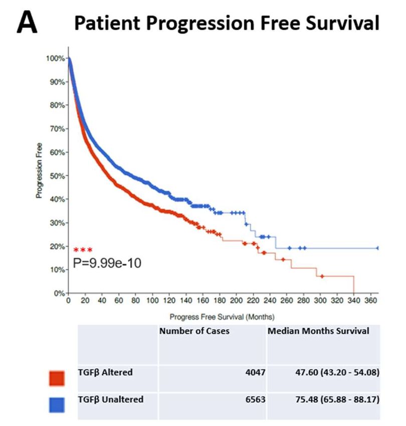
Preprints (www.preprints.org) | NOT PEER-REVIEWED | Posted: 7 February 2022                                doi:10.20944/preprints202202.0078.v1

                                                                                                                                        2 of 29

                                                   1.0 Introduction

                                                   Breast cancer is the most frequent cancer affecting women and accounted for over 2 mil-
                                                   lion breast cancer diagnoses and approximately 600,000 related mortalities in 2018[1].
                                                   TNBC only accounts for the minority of breast cancer incidences (15-20%); however,
                                                   TNBC is disproportionally associated with reduced patient prognosis compared to the
                                                   other breast cancer subtypes[2,3]. TNBC in contrast with other breast cancer subtypes
                                                   lacks expression of the estrogen receptor, progesterone receptor and HER-2. The presence
                                                   of these receptors are associated with the usage of specific therapies; thus non-specific
                                                   chemotherapies and radiotherapies are mainstays for the treatment of TNBC which over-
                                                   all is associated with reduced patient prognosis.

                                                   As such, there is immense focus on the development of targeted therapies to target TNBC.
                                                   However, a critical issue garnering increased attention in preclinical research is the high
                                                   incidence of cardiotoxicity following therapy leading to increased rates of heart failure
                                                   and cardiomyopathy[4]. CVD and its related complications is a leading cause of morbidity
                                                   and mortality in cancer survivors[5]. In a observational study, Patnaik et al demonstrated
                                                   in 63,566 breast cancer patients, that while there were increased adjusted relative hazards
                                                   of comorbidities such as cardiovascular disease, COPD and diabetes; it was found that
                                                   cardiovascular disease was the primary cause of death amongst the patients (15.9%) ex-
                                                   ceeding mortality due to breast cancer (15.1%)[6].

                                                   Moreover, Bardia et al demonstrated in a clinical trial using a 10-year recurrence risk pre-
                                                   diction model in breast cancer patients with early stage breast cancer (stage I-III with
                                                   67.5% having stage I) which calculated CVD and breast cancer recurrence risk [7]. It was
                                                   found that the risk of a CVD event exceeded the risk of breast cancer relapse in 37% of the
                                                   patients and 43% possessed a risk equal to that of breast cancer recurrence[7]. These stud-
                                                   ies highlight that not only is the development of therapeutics for primary tumor manage-
                                                   ment important for patient prognosis, but that the cardiovascular health of the patient
                                                   must be protected due to sensitivity following chemotherapy treatment.

                                                   To highlight this point, a recent study by Sturgeon et al, 3,234,256 US cancer survivors
          Citation: Lastname, F.; Lastname, F.;
                                                   from the year 1973-2012 were assessed and mortality ratios stemming from CVD (consist-
          Last-name, F. Title. Cancers 2021, 13,
                                                   ing of a grouping of heart disease, hypertension, atherosclerosis, cerebrovascular disease,
          x. https://doi.org/10.3390/xxxxx
                                                   aortic aneurysm or aortic dissection) and cancer related causes were assessed[8]. The pa-
          Academic Editor: Firstname Lastname      tients were separated by cancer type and CVD mortality was found to highly elevated in
          Received: date                           patients diagnosed with breast, prostate or bladder cancer (together accounting for 61%
          Accepted: date                           of all CVD mortality) and also in patients diagnosed at an earlier age (
Preprints (www.preprints.org) | NOT PEER-REVIEWED | Posted: 7 February 2022 doi:10.20944/preprints202202.0078.v1
3 of 29
2.0 Post-Chemotherapeutic Cardiomyopathy
Due to the aforementioned lack of specific cellular targeting in TNBC treatment, there is
a strong reliance on standard cytotoxic chemotherapeutic agents in clinical practice.[9]
These regimens often involve the use of anthracycline or taxane class chemotherapeutic
agents.[10] Unfortunately, chemotherapy often induces very severe side effects, with car-
diotoxicity at the forefront of dose limiting toxicity.[11]
Cardiotoxicity is a broad term which includes both early and late onset forms as well as
effects ranging from subclinical impairment of cardiac function to cardiac death.[12] Early
onset, also called “acute”/”subacute” cardiotoxicity develops immediately after chemo-
therapeutic infusion or up to 2-4 weeks after completion and is typically characterized by
reversible arrhythmias, abnormalities in ventricular repolarization, prolongation of the
QT interval, acute coronary syndrome, pericarditis/myocarditis-like syndromes or altered
myocardial function.[13] Late onset cardiotoxicity can be divided into either early-chronic
or late-chronic subtypes. Early chronic cardiotoxicity occurs within 1 year after termina-
tion of chemotherapy while late-chronic cardiotoxicity occurs more than 1 year after ter-
mination.[14] Late-onset cardiotoxicity can result in systolic/diastolic left ventricular dys-
function that leads to congestive cardiomyopathy which can transition towards cardiac
death.[14] Additionally, cardiomyopathy can be classified into two subtypes: type I
(caused by cardiomyocyte death and is irreversible) and type II (caused by cardiomyocyte
impairment of cardiac function and is reversible).[15] This concept was originally pro-
posed by Ewer et al and these subtypes can differentiate the effects of various chemother-
apeutic agents; for example, doxorubicin (an anthracycline class chemotherapeutic) in-
duces type I cardiotoxicity while the biologic trastuzumab (an anti HER-2 chemothera-
peutic agent) induces type II cardiotoxicity.[15]
3.0 Anthracycline and Taxane Mechanisms of Cardiotoxicity
Doxorubicin, an anthracycline, is one of the most frequently prescribed chemothera-
peutic agent for the treatment of TNBC. The toxicity of doxorubicin on cardiac tissue is
mediated through multifactorial mechanisms which remain convoluted. One commonly
proposed mechanism is that anthracycline agents such as doxorubicin are prone to the
generation of reactive oxygen species (ROS) during their metabolism.[11] Specifically, the
univalent reduction of the anthracycline class quinone moiety by mitochondrial complex
I in the electron transport chain (ETC) results in the formation of semiquinone radicals
which rapidly undergo auto-oxidation to form superoxide anions (O2-), thereby also re-
generating the quinone moiety. [16,17] This cycle can then continue under aerobic condi-
tions, producing additional ROS. This process may shed light on the correlation between
anthracycline class chemotherapeutics and the induction of cardiotoxicity as the cardio-
myocytes experience large demand for ATP produced by ETC, and therefore have a
greater density of mitochondria (and subsequently complex I) than other cell types[18].
The high rate of ROS production in the mitochondrion of the cardiomyocyte can then in-
terfere with iron reduction, damaging the cells via ROS mediated reactions which result
in the formation of reactive nitrogen species leading to mitochondrial/cardiomyocyte dys-
function which ultimately promotes apoptosis [19-21].
Another proposed mechanism for the cardiotoxicity of anthracyclines is via its intended
anti-tumor mechanism of DNA-topioisomerase2 (Top2) intercalation, wherein the an-
thracycline forms a Top2-doxorubicin-DNA ternary complex. In humans Top2 is ex-
pressed as the isoenzymes Top2α and Top2β, with the former expressed in proliferative
cells (including cancer cells) and the latter in quiescent cells[22]. Top2 β positive malignant
cells demonstrate ternary complex formation upon anthracycline therapy promoting the
inhibition of DNA replication leading to G1/G2 arrest and apoptosis in cancerous cells.Preprints (www.preprints.org) | NOT PEER-REVIEWED | Posted: 7 February 2022 doi:10.20944/preprints202202.0078.v1
4 of 29
Unfortunately, Top2β is also the primary form expressed in adult cardiac tissue promot-
ing anthracycline binding and cardiotoxicity resulting in mitochondrial and cellular dys-
function.[23],[24]
Paclitaxel (a taxane) is another commonly used chemotherapeutic agent in the treatment
of breast cancer, especially in anthracycline-resistant breast cancer.[25] Although it was
thought that taxanes have negligible cardiotoxicity when compared to anthracyclines,
phase I and II clinical trials revealed acute cardiac reactions upon paclitaxel infusion such
as: cardiac rhythm disturbances, atrioventricular conduction abnormalities, sinus brady-
cardia and ventricular tachycardia [26],[27]. Importantly, the majority of cardiac disturb-
ances were not associated with clinical symptoms and were found incidentally during
cardiac monitoring. Moreover, these cardiac issues were common in taxane treated pa-
tients with 29% of patients having asymptomatic bradycardia when administered the
maximal tolerable dose of taxane therapy(110-250 mg/m2)[28]. One proposed mechanism
for taxane cardiotoxicity is the mediated not by the taxane agent itself but rather through
its formulation vehicle, Cremophor EL (a vehicle to enhance the solubility). It has been
proposed that Cremophor EL induces massive histamine release causing acute cardiovas-
cular reactions.[29] Interestingly, taxanes such as paclitaxel are often used in combination
with anthracyclines; however, it was found in clinical trials that the combination pro-
duced unacceptably high rates of heart failure (18% of patients)[30]. This is thought to be
due to pharmacokinetic interference, where paclitaxel interferes with the clearance of dox-
orubicin possibly through competition for biliary clearance promoting cardiotoxicity.[31]
4.0 Clinical assessment of cardiotoxicity
The severity of cardiomyopathy is important not only for determining therapeutic course
but also for the manifestations of CVD later in life, especially in regards to pediatric pop-
ulations who were administered chemotherapeutic agents. The gold standard for an-
thracycline class cardiotoxicity determination is a cardiac biopsy; however, due to the im-
practicality of this as a clinical assessment it is not typically considered. Rather, cardiac
imaging can be used to monitor cardiac deterioration, where the left ventricle ejection
fraction (LVEF) is used to track progression. LVEF can be determined via TC-99 Multiple
Gated Acquisition Scan (MUGA), also called radionuclide ventriculography[32,33]. Cur-
rent guidelines place cardiotoxicity as one or more of the following: 1) Reduction of LVEF,
either globally or more within the septum. 2) The onset of symptoms associated with heart
failure. 3) A reduction of >5% to < 55% in regards to the ejection fraction (EF) alongside
symptoms of heart failure, or a >10% toPreprints (www.preprints.org) | NOT PEER-REVIEWED | Posted: 7 February 2022 doi:10.20944/preprints202202.0078.v1
5 of 29
ventriculography [37]. Additionally, echocardiography can evaluate for adverse struc-
tural effects, such as valvular disease or pericardial constriction[38,39].
Cardiovascular magnetic resonance imaging (CMR) is another imaging technique for the
evaluation of cardiomyopathies induced by cardiotoxic therapies which has the ad-
vantage of being radiation free[40]. CMR has the ability to detect subclinical cardiac dys-
function prior to detectable LVEF changes in addition to the ability to detect myocardial
edema (a marker of myocardial injury). The high cost and low availability of CMR in con-
trast to echocardiography make it less widely utilized as a screening tool.[38]
The utilization of electrocardiogram (ECG) for cardiac monitoring circumvents the above
problems associated with imaging and has the added benefit of being inexpensive and
readily available. Horacek et al found statistically significant correlation between de-
creased QRS voltage, corrected QT interval (QTc) prolongation and left ventricular dys-
function as visualized by echocardiography[41]. ECG also has the added benefit of being
able to correlate with malignant ventricular arrhythmias via QTc, an important indicator
of acute cardiotoxicity[41]. Additionally, Fukumi et al found that signal-averaged ECG
was able to detect acute and chronic cardiotoxicity from anthracycline chemotherapeutics
at lower cumulative doses than echocardiography based imaging. Such a finding could
allow for earlier insight into cardiac dysfunction[42].
Many well established biomarkers are used to investigate the cardiomyocyte damage. Not
only can troponins serve as an indicator of damage, but their levels correlate with the
clinical severity of the damage that occurs from the insult[43]. This allows for risk stratifi-
cation during an infarct or other cardiac insults[44]. A study by Cardinale et al found that
elevation in Troponin I in patients undergoing high dose chemotherapy (anthracyclines)
preceded and was able to accurately predict the development of future cardiac dysfunc-
tion (via lowered LVEF)[45]. As the elevation of cardiac troponin I is a very specific and
sensitive marker for cardiac damage and is one that many hospitals utilize in their practice
its adoption into chemotherapy related cardiac monitoring remains a popular proposi-
tion[46]. Other markers of interest include natriuretic peptides such as Brain Natriuretic
Peptide (BNP), its preprohormone and cleavage product (NT-proBNP) and Atrial Natri-
uretic Peptide (ANP). These substances serve to regulate blood pressure and circulating
blood volume and are released from cardiomyocytes in response to atrial stretching/vol-
ume overload[47]. Similar to troponins, BNP may allow for the early detection of cardio-
toxicity, although they may have the added advantage of being detectable for longer pe-
riods of time. While troponin was detectable within 4-15 hours until 10-14 days, natriuretic
peptides were detectable within 24 hours and for as long as 2 years[48-52]. Routine assess-
ment of patients exposed to chemotherapy with the detection methods described above
would serve to identify early signs of CVD post-treatment allowing for increased oppor-
tunity to mitigate negative effects and increase prognosis in patients post therapy.
5.0 TGF-β Overview
Extensive studies have shown that transforming growth factor beta (TGF-β) is a major
mediator which modulates multiple cellular steps that promote cardiovascular disease,
cardiac hypertrophy, arrhythmia, fibrosis and cardiac failure [53]. In brief, various pro-
teins/conditions have been found to activate TGF-β secretion [54]. Initially, TGF-β ligand
is bound by the TGF-β binding protein which is activated via binding of αv integrin to the
prodomain of TGF-β1/2 mediated by a myriad of conditions including myofibroblast in-
duced contraction [55-57]. Activated TGF-β signaling is primarily mediated via two dis-
tinctive downstream effectors: the SMAD pathway and the non-canonical pathway.
SMAD signaling is mediated through activated TGF-β interaction with type I (TβRI) and
type II receptor (TβRII) via trans-phosphorylation of multiple serine/threonine residuesPreprints (www.preprints.org) | NOT PEER-REVIEWED | Posted: 7 February 2022 doi:10.20944/preprints202202.0078.v1
6 of 29
of TβRI GS domain [58]. The activated TGF-β type I receptor then activates SMAD2 and
SMAD3 via phosphorylation. Following SMAD2/3 activation, the complex trimerizes with
SMAD4 forming the activated SMAD complex which translocate into the nucleus to reg-
ulate transcription for a variety of downstream effectors including the COL1A1/COL3A1
genes that facilitate production/deposition of collagens, plasminogen activator inhibitor-
1 that builds matrix, and connective tissue growth factor that upregulates the expression
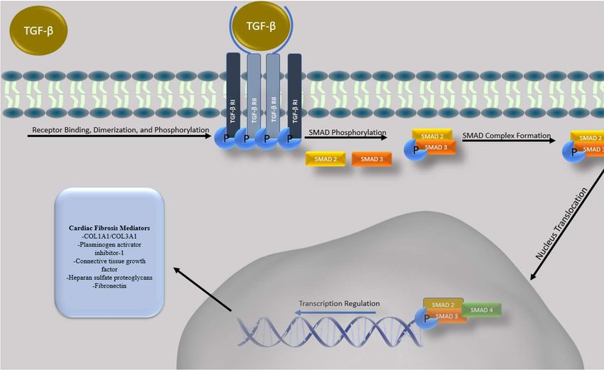
of fibronectin or heparan sulfate proteoglycans (HSPG) (Figure1) [59,61].
Figure 1: Overview of Conventional TGF-β Signaling. A schematic overview of conven-
tional (SMAD mediated) TGF-β signaling occurring after TGF-β ligand binding which
leads to the activation of TGF-β type II and TGF-β type II receptor heteromeric complex
which can induce the phosphorylation of SMAD2 and 3, promoting complex formation
with co-SMAD (SMAD4). This trimeric complex can translocate into the nucleus and in-
duce transcription of numerous genes including those involved in cardiac remodeling and
fibrosis as well as cellular differentiation, survival, invasion and apoptosis.
SMAD-independent pathways are broadly referenced as non-canonical pathways and can
mediate TGF-β signaling independently or work in conjunction with SMAD-dependent
pathways to facilitate/repress the TGF-β pathway [62,63]. Amongst the various non-ca-
nonical mediated intercellular signals, mitogen activated protein (MAP) kinase is one of
the mechanistic pathways which showed growing evidence of its roles in mediating TGF-
β induced cardiac fibrosis [64]. Activated TGF-β receptors can interact with TNF Receptor
Associated Factor 6 (TRAF-6) to induce ubiquitination [62]. Subsequently, ubiquitinated
TRAF-6 recruits TGF-β activated kinase (TAK-1). TAK-1 activation consists of the kinase
domain of TAK-1 forming a complex with TAK1-binding protein (TAB1). The active
TAK1-TAB1 heterometric complex can then upregulate non-canonical mediating effectors
such as MKK4/7 and MKK3/6 via phosphorylation [65]. Phosphorylated MMK4/7 upreg-
ulates the expression of JNK which in turn recruits transcriptional factor c-jun. Similarly,
phosphorylated MMK3/6 can upregulate the expression of p38 which in turn increases the
expression of ATF-2 [62,63]. These non-canonical pathways induce c-jun and ATF-2 co-
transcription factors which can regulate the expression of SMAD dependent fibrosis via
phosphorylation, signifying the intricate cellular interplays between the SMAD depend-
ent and non-canonical induced fibrosis in cardiac models [62,63,66].Preprints (www.preprints.org) | NOT PEER-REVIEWED | Posted: 7 February 2022 doi:10.20944/preprints202202.0078.v1
7 of 29
6.0 TGF-β as a Mediator of Cardiac Fibrosis and Remodeling
Cardiac fibrosis is a hallmark response to injuries of the heart and its onset has been asso-
ciated with myocardial infarction, ventricular remodeling, arrhythmia, dilated cardiomy-
opathy and heart failure [67-69]. Cardiac fibrosis is characterized by the differentiation of
cardiac fibroblasts into myofibroblasts [70,71]. TGF- β is a crucial mediator in the differ-
entiation of myofibroblasts and resistance to apoptosis via activation of the Smad3 path-
way which promotes α-SMA (alpha-smooth muscle actin) transcription in fibroblasts and
induces extracellular matrix protein deposition and myofibroblast differentiation[72-75].
Dobaczewski et al demonstrated via a closed-chest model of coronary occlusion/reperfu-
sion to induce reperfused myocardial infarction in Smad3 null mice that ablation of
SMAD-mediated signaling was associated with a reduction of α-SMA transcription in fi-
broblasts. Furthermore, upon TGFβ1 stimulation, while wild-type mice demonstrated in-
creased α-SMA and fibrosis, Smad3 null mice did not, highlighting the association be-
tween TGFβ-SMAD signaling and the cardiac fibrosis[73]. In another similar study, a
closed-chest model of reperfused myocardial infarction in Smad3 null mice demonstrated
that TGF-β1 stimulation was associated with upregulation of procollagen III but not in
Smad3-null mice, which indicates that TGF-β mediated Smad3 signaling plays an im-
portant role in the extracellular matrix protein synthesis[76]. Using mice subjected to car-
diac pressure overload stimulation via transverse aortic constriction surgery, Khalil et al
showed that TGF-β treated Smad3 and Smad2/3-deleted fibroblasts had a significant re-
duction in fibroblast marker genes (POSTN, COL1A1, and COL3A1) in primary cardiac
fibroblasts, indicating that deletion of Smad3 from newly activated fibroblasts may signif-
icantly attenuate the cardiac fibrosis response[77].
Additionally, angiotensin II of the Renin-Angiotensin-Aldosterone System (RAAS) has
been associated with the onset of cardiac fibrosis. Research has demonstrated the correla-
tion between angiotensin II expression and TGF-β expression in cardiac fibroblasts[78-80].
Wang et al stimulated mouse primary aorta vascular smooth muscle cells (VSMCs) with
angiotensin II in vitro and demonstrated that angiotensin II can mediate Smad2/3 signal-
ing pathway in a TGF-β dependent manner[81]. Furthermore, Zhang et al demonstrated
that chronic angiotensin II infusion upregulates human c-reactive protein (CRP) in CRP
transgenic mice, leading to a 5-fold increase in serum CRP, a biomarker associated with
cardiovascular diseases and events. As angiotensin II-induced cardiac TGF- β1 expression
and activation of the Smad signaling were enhanced in CRP transgenic mice as well this
highlights that angiotensin II mediated activation of TGF-β plays a pathogenic role in car-
diac remodeling[82].
TGF-β can also mediate non-canonical signaling to promote pathological cardiac remod-
eling via activation of TGF-β Activated Kinase 1 (TAK1) as a delayed response to mechan-
ical stress. Transgenic mice that expressed TAK1DN (a constitutive active form of TAK1)
under the control of cardiac specific aMHC promoter (aMHC-TAK1DN) had a 46% in-
crease in cardiac mass at 9-11 days after aortic banding and selective activation of p38 in
myocardium at 9 days (up to 400%). Hearts of mice 9-10 days old showed hypertrophied
myocytes with hyperchromatic nuclei, interstitial fibrosis, and other signs seen in load-
induced hypertrophy and heart failure[83]. Constitutive overexpression of the human tu-
mor suppressor A20 suppressed TAK-1 induced collagen synthesis and TAK-1 dependent
Smad 2/3/4 activation in murine hearts, protecting against cardiac hypertrophy and fibro-
sis[84]. Thus, TGF-β mediated TAK-1 activity plays an important role in myocardial hy-
pertrophy and heart failure.
Thus, TGF- β through SMAD dependent and independent signaling is associated with the
onset of adverse cardiac pathologies and negative clinical outcomes making preclinicalPreprints (www.preprints.org) | NOT PEER-REVIEWED | Posted: 7 February 2022 doi:10.20944/preprints202202.0078.v1
8 of 29
research into TGF-β modulation for the treatment of cardiac disease as potential target for
further clinical consideration. This is highlighted in a study by Laviades et al which
demonstrated that hypertension and microalbuminuria in patients was associated with
left ventricular hypertrophy and higher levels of serum TGF- β1 compared to normoten-
sive participants. In the same hypertensive patient group, treatment with Losartan (a clin-
ically approved angiotensin II receptor antagonist with TGF-β inhibitory activity) de-
creased TGF-β1 levels in patients which correlated with a reduction of microalbuminuria
and left ventricular hypertrophy [85]. To further highlight the importance of TGF-β in
cardiac function, using sequence specific oligonucleotide probing (SSOP), Holweg et al
studied genomic DNA samples from heart transplant recipients and found that Leu>Pro
(codon 10) polymorphism in the TGFB1 gene is associated with end-stage heart failure
caused by dilated cardiomyopathy[69]. Thus, TGF-β through SMAD dependent and in-
dependent signaling is associated with the onset of adverse cardiac pathologies and neg-
ative clinical outcomes making preclinical research into this pathway for the treatment of
cardiac disease an unmet medical need.
7.0 TGF-β Inhibition to Prevent Cardiomyopathy
It has been demonstrated that TGF-β exerts physiologic effects on embryonic develop-
ment, cardiac development and cellular growth and it has been further highlighted that
dysregulated TGF-β signaling is associated with a host of unwanted pathologic conditions
such as fibrosis, cardiac hypertrophy and inflammation [86-89]. Thus, modulation of TGF-
β through pharmacologic agents may be of therapeutically benefit patients with post-
chemotherapy fibrosis, heart failure and cardiomyopathy.
Oliveira et al, demonstrated that GW788388 (a TGF-β inhibitor specific for TβRI/ALK5)
can reduce cardiac fibrosis [90]. This was demonstrated through injecting Swiss mice with
Trypanosoma Cruzi parasites to induce Chagas disease and cardiac fibrosis which was
measured via fibronectin and collagen type I deposition [90]. It was found that this model
induced substantial indications of cardiac fibrosis; however, upon treatment with
GW788388, deposition of fibronectin and collagen type I was reduced in cardiomyocytes
and cardiac electrical conduction was improved [90]. In a separate study by Ferreira et al,
these results were repeated in a chronic Chagas in vivo mouse model consisting of C57BL/6
mice injected with Trypanosoma Cruzi and treated with GW788388 [91]. Mice receiving
treatment demonstrated reduced fibrosis of cardiac tissue indicated by reduced levels of
collagen type I and fibronectin deposition in cardiac tissue. Moreover, GW788388 inhib-
ited TGF-β/pSmad2/3 expression and activity which was correlated with reduced CD3+
inflammatory lymphocyte cell migration into cardiac tissue [91]. Interestingly, these ef-
fects were correlated with increased stem cell antigen-1 (Sac-1+) cardiac cells following
treatment. As Sca-1+ is a marker for cardiac stem cells it was suggested that TGF-β inhibi-
tion can not only inhibit fibrosis but also promote the enrichment of cardiac stem cells
which could promote cardiac recovery [91].
TGF-β has also demonstrated translatability in the treatment of myocardial infarction
(MI). Myocardial infarctions lead to cardiomyocyte death through ischemia, fibrosis and
eventual heart failure. During a MI, there is a well-documented upregulation of TGF-β
isoforms which facilitate healing and repair [87,92,93]. This process however also leads to
fibroblastic extracellular matrix protein deposition and an upregulation of TIMPs (Tissue
inhibitors of metalloproteinases) which inhibits matrix degeneration and ultimately stim-
ulates fibrosis [94]. Khalil et al highlights the importance of TGF-β signaling in the fibrotic
response via deletion of TGF-β receptors TGFβR1/2 and Smad3 in cardiac fibroblasts
which reduced TGF-β–induced gel contraction indicating a disruption in myofibroblast
differentiation. Moreover, a novel in vivo mouse model was used with Periostin-GFP re-
porter tracking of myofibroblasts of the heart in combination with TGFβR1/2, Smad2,Preprints (www.preprints.org) | NOT PEER-REVIEWED | Posted: 7 February 2022 doi:10.20944/preprints202202.0078.v1
9 of 29
Smad3 and Smad2/3 knockouts [95]. This model then induced cardiac pressure overload
via aortic constriction (an in vivo methodology to induce cardiac hypertrophy and heart
failure) which found that deletion of Smad3, Smad2/3, or TGFβR1/2 was able to inhibit
cardiac fibrosis following aortic constriction [95]. Moreover, 12 weeks after aortic con-
striction TGFβR1/2 knockout mice demonstrated reduced ventricular fractional shorten-
ing, preserved diastolic function and reduced cardiac hypertrophy highlighting the tar-
geting of the TGF-β pathway as a viable strategy to reduce cardiac fibrosis [95]. Im-
portantly it was also found that the inhibition of Smad2/3 led to reduced fibroblast prolif-
eration, differentiation and activity which correlated with a reduction of cardiac fibrosis
although it did not lead to altered hypertrophy [95]. Thus, this study demonstrated dif-
ferential effects upon targeting different parts of TGF-β pathway and suggests that inhi-
bition of Smad2/3 can inhibit fibrosis while TGFβR1/2 inhibition can affect fibrosis but
also hypertrophy and other aspects of cardiac signaling.
TGF-β1 has also been shown to induce cardiomyocyte hypertrophy and post-MI remod-
eling through the activation of TGF-β 1/TAK-p38MAPK signaling within non-infarcted
myocardium after acute MI [96]. Thus, inhibition of the TGF-β signaling cascade is an
attractive target for the prevention of cardiac remodeling and cardiomyopathy post-MI.
In this regard, Ellmers et al demonstrated using SD-208 (a TGF- β receptor kinase 1 inhib-
itor) that deleterious cardiac remodeling post-infarction could be inhibited [97]. MI was
induced in mice via left coronary artery ligation and were treated with SD-208 for 30 days.
While there was no difference recorded in ventricular TGFβ gene expression, there was
increased TAK-1 (a downstream effector of TGFβ) in the control which was inhibited upon
treatment with SD-208. The blockade of TGF-β signaling after MI resulted in reduced ven-
tricular expression of TGF-β -activated kinase-1, decreased collagen 1 and decreased car-
diac mass highlighting TGF-β inhibition as a potent method to reduce cardiac remodeling
post-MI [97].
Additionally, as diabetic mortality is primarily due to cardiovascular complications recent
studies have sought to investigate whether TGF-β inhibition can affect diabetic cardiomy-
opathy [98]. A study by Zhang et al demonstrated in Sprague-Dawley rats which were
induced to become diabetic through the injection of streptozotocin that Matrine (an inhib-
itor of the TGF-β/Smad pathway) administration to rats could prevent diabetic cardiomy-
opathy as indicated through reduced fibrosis, recovery of LV function and heart compli-
ance [99].
Together these reports demonstrate that inhibition of TGF-β signaling via pharmacologic
modulation may reduce cardiac fibrosis, improve heart function, and decrease cardiomy-
opathy in a wide variety of preclinical models. The ultimate goal is to translate these find-
ings to the clinic and improve patient prognosis; however, much work remains to be done
to identify effective TGF-β inhibitors which can be translated for effective patient therapy.
As such, we have identified potential TGF-β inhibitors for this purpose which are cur-
rently in active and interventional clinical trials for the treatment of cardiotoxicity or heart
disease (including heart failure, cardiovascular disease, ischemic heart disease, coronary
heart disease and arrhythmia) from the Clinicaltrials.gov database are summarized in Ta-
ble 1. Identified potential TGF-β inhibitors seem to be safe for the usage in clinic and have
been demonstrated to suppress the TGF-β signaling pathway in preclinical studies; how-
ever, further studies will be needed to determine clinical efficacy in combination with
chemotherapy as well as the underlying mechanism.Preprints (www.preprints.org) | NOT PEER-REVIEWED | Posted: 7 February 2022 doi:10.20944/preprints202202.0078.v1
10 of 29
Table 1: Potential TGF-β inhibitors in Active Cardiotoxicity and Cardiac Disease Re-
lated Clinical Trials. The Clinicaltrials.gov database was used to assess active, interven-
tional clinical trials for the treatment of heart disease and cardiotoxicity within phase 1,
2, 3, or 4 of development. Following inhibitor identification, literature was consulted to
determine any hypoxia modulating effects. Clinical Trial Search link (accessed on 1 Au-
gust 2021): https://clinicaltrials.gov/ct2/results?cond=Cardiotoxi-
city&term=&type=Intr&rslt=&recrs=d&age_v=&gndr=&intr=&ti-
tles=&outc=&spons=&lead=&id=&cntry=&state=&city=&dist=&locn=&phase=0&phase=1
&phase=2&phase=3&rsub=&strd_s=&strd_e=&prcd_s=&prcd_e=&sfpd_s=&sfpd_e=&rfp
d_s=&rfpd_e=&lupd_s=&lupd_e=&sort= ; https://clinicaltrials.gov/ct2/results?cond=Car-
diac+Disease&term=&type=Intr&rslt=&recrs=d&age_v=&gndr=&intr=&ti-
tles=&outc=&spons=&lead=&id=&cntry=&state=&city=&dist=&locn=&phase=0&phase=1
&phase=2&phase=3&rsub=&strd_s=&strd_e=&prcd_s=&prcd_e=&sfpd_s=&sfpd_e=&rfp
d_s=&rfpd_e=&lupd_s=&lupd_e=&sort=
Inhibitor Clinical Trial Mechanism References
Number
Enalapril NCT01968200 ACEI with antifibrotic activity via inhibition of TGFB1 and p- [100,101]
SMAD2/3 expression
Carvedilol NCT02177175 Suppression of myocardial fibrosis by inhibiting TGFB1 mRNA [102,103]
expression
NCT01347970
Simvastatin NCT02096588 Downregulates TGFb1 mediated phosphorylation of Smad 2/3 via [104,105]
activation of PP2A and PP2C/PPM1A phosphatases.
Rivaroxaban NCT02303795 Downregulates mRNA expression of TGFB in the infarcted area [106]
following an MI potentially via suppression of PAR-1 and PAR-2
NCT01776424 pathways.
NCT02066662
Clopidogrel NCT02044250 Platelet blocker that inhibits the expression of TGFB mRNA and [107]
the protein levels preventing cardiac fibrosis
NCT02317198
Rituximab NCT03072199 Monoclonal antibody against CD20 inhibits fibrotic signaling of [108]
TGF-β1 and p-Smad2/3
LCZ696 NCT02816736 Angiotensin receptor–neprilysin inhibitor that improves cardiac [109,110]
function by downregulates cardiac fibrosis via suppression ofPreprints (www.preprints.org) | NOT PEER-REVIEWED | Posted: 7 February 2022 doi:10.20944/preprints202202.0078.v1
11 of 29
NCT03190304 TGF-β expression primarily through its specific inhibition of
neprilysin
NCT02468232
NCT02924727
Spironolactone NCT03409627 SP prevents cardiac fibrosis cause by inhibiting the production of [111,112]
TGFβ1 and phosphorylation of Smad2/3.
NCT02673463
Macitentan NCT03153111 Dual endothelin receptor antagonist (ETA and ETB) that [113,114]
suppresses expression of TGFβ, esp. in DM patients where TGFβ
is upregulated.
Ivabradine NCT04448899 Hyperpolarization-activated pacemaker current (If) channel [115,116]
inhibitor ivabradine inhibits the expression of TGFb1 and Smad-2
NCT04308031 post MI suppressing collagen synthesis and pro-fibrotic activity.
Empagliflozin NCT03128528 Inhibits the fibrotic activity of TGFb in the heart by suppressing [117,118]
the expression of TGFb1, p-Smad2/3 and upregulating TGFb
NCT03030222 inhibitor Smad7. Further resulting in decreased expression of
collagen I and II mediated by TGFb/Smad pathway.
NCT03057977
NCT03057951
NCT03485092
NCT02998970
Pirfenidone NCT02932566 Inhibits Ang II induced expression of TGFb1 and suppresses [119,120]
myocardial interstitial fibrosis.
Atorvastatin NCT02679261 Suppresses cardiac fibrosis by attenuating TGFb1 mediated [121]
phosphorylation of Smad3, PI-3 kinase, Akt, collagen I and
endoglin expression.
Eplerenone NCT01857856 Inhibits the expression of TGFb1 and collagen I resulting in [122]
downregulation of cardiac remodeling induced by
cardiomyopathy
Olmesartan NCT04174456 Angiotensin II type 1 receptor blocker that reduces the expression [123,124]
of TGFb in pressure overloaded, diabetic, obese pts. preventing
cardiovascular injury.Preprints (www.preprints.org) | NOT PEER-REVIEWED | Posted: 7 February 2022 doi:10.20944/preprints202202.0078.v1
12 of 29
Tadalafil NCT03049540 cGMP mediated inhibition of TGFb1 expression [125]
Berberine NCT04434365 Antifibrotic activity by inhibition of TGFb1 secretion potentially [126]
by upregulation of AMPK phosphorylation and downregulation
of mTOR and p70S6K phosphorylation.
Melatonin NCT02099331 Antifibrotic by suppressing TGFb1expression. [127]
N- NCT02750319 w/ Antioxidant that inhibits TGFb1 mediated signaling involved in [128,129]
Acetylcysteine Amiodarone fibrosis potentially by suppressing its interaction with TGB1R,
(NAC) downregulating phosphorylation of Smad2/3 and upregulating
NCT01878669 Smad7 mRNA.
NCT01878344
Colchicine NCT02594111 Antifibrotic activity by inhibiting expression of TGFb1 mRNA. [130]
NCT01709981
NCT02624180
NCT04382443
Ticagrelor NCT02539160 Antifibrotic by inhibiting the expression of TGFb [131]
NCT03437044
NCT01944800
Valsartan NCT01912534 Inhibition of Ang II type I (AT 1) receptor resulting in suppression [132]
of AT 1 mediated action of TGFb/Smad pathway.
Metformin NCT03629340 Suppression of cardiac fibrosis by inhibiting TGFb1 production [133]
and phosphorylation of Smad-3.
Nitrite NCT03015402 Downregulation of cardiac remodeling by suppressing AT II and [134]
AT 1R inhibiting TGFb1.
NCT02980068
Nebivolol NCT02053246 Attenuated profibrotic activity and prevents vascular remodeling [135]
by downregulating the expression of TGFb1and MMP-2/9.
NCT01648634
Riociguat NCT01065454 Guyanalate cyclase stimulant with antifibrotic activity by [136]
inhibiting TGFb1 mediated collagen synthesisPreprints (www.preprints.org) | NOT PEER-REVIEWED | Posted: 7 February 2022 doi:10.20944/preprints202202.0078.v1
13 of 29
8.0 TGF-β as a Therapeutic Target in TNBC
TGF-β signaling has been associated with disease progression and negative patient prog-
nosis in a wide number of cancer models including breast, colon and small cell lung can-
cers [137-139]. To highlight the clinical importance of TGF-β dysregulation; using the cbi-
oportal clinical database in our own analysis we assessed the impact of genomic TGF-β
alterations (Alterations defined as TGF-β genomic mutations, structural variants and copy
number variations; see methods for specific genes assessed) in relation with overall pa-
tient survival across 32 TCGA, PanCancer Atlas datasets which 10,610 patients [140,141].
38% of patients were found to have an alteration in at least one TGF-β gene and patients
with an alteration in TGF-β signaling demonstrated a dramatic reduction in progression
free survival compared to patients without TGF-β signaling alteration (Figure 1A-B, TGF-
β altered patients: 4047 cases and 47.60 median month progression free survival; TGF-β
unaltered patients: 6563 cases and 75.48 median month progression free survival). Thus,
our findings demonstrate the importance of TGF-β in patient outcomes across a broad
spectrum of tumor types and datasets (Supplemental Table 1 for detailed list of studies/
cancer studies used for analysis) and in over 10,000 patients. Notably, this analysis does
not take into account treatment, age, disease sub-type and other critical factors influencing
patient prognosis.
Figure 2: Database Analysis of Patients with TGF-β Altered/Unaltered gene expression
and Survival. Kaplan–Meier curves for progression free survival of the patients with Al-
terations in TGF-β signaling in cancer samples (red curve) in comparison with patients
with unaltered expression (blue curve). n = 10,610, ***P=9.99e-10, log-rank test.
While TGF-β alterations are significant in a wide variety of cancer models, it has been
found in a study by Ding et al that 52.5% of TNBC patients were found to have elevated
TGF-β expression which was associated with increased rates or metastasis, increased tu-
mor grade and negative disease free survival[137]. Moreover, our own previous database
analysis revealed similar findings using cbioportal to assess a cohort of 1082 breast cancer
patients [142]. It was found that increased TGF-β signaling was correlated with dimin-
ished overall prognosis and median month survival (122.83 median month survival in
patients with TGF-β high gene expression versus 140.28 median month survival inPreprints (www.preprints.org) | NOT PEER-REVIEWED | Posted: 7 February 2022 doi:10.20944/preprints202202.0078.v1
14 of 29
patients without increased TGF-β gene expression)[142]. Moreover, our assessment found
that TNBC patients possessed increased levels of TGFBRA mRNA expression and reduced
disease-free survival compared to other breast cancer subtypes highlighting the im-
portance of TGF-β modulation for prospective treatment [142]. As dysregulated TGF-β
signaling is associated with increased CSC enrichment, chemoresistance and decreased
patient survival in TNBC it highlights TGF-β modulation as a potential therapeutic target
[143-146].
It has been demonstrated that within breast cancer tumors, the cellular hierarchy is not
uniform and a small population (known as cancer stem cells, CSCs) maintains self-re-
newal and differentiation capabilities regulating tumor composition and heterogeneity.
Conversely to differentiated tumor cells, CSCs have demonstrated robust resistance to
conventional chemotherapy and are thought to persist following therapy/intervention
and are a major cause of relapse [147-149]. A wide number of breast cancer models cur-
rently support two distinct sub-populations of CSCs: a mesenchymal CSC population de-
fined by CD44+/CD24- markers and an epithelial CSC population with ALDHhigh mark-
ers[150]. Famously Al Hajj et al demonstrated through fractionation experiments of breast
tumors that CD44+/CD24- populations were capable of forming a tumor with as little as
100 cells in comparison with the tens of thousands of cells within the different populations
required to achieve a similar tumorigenicity[150]. Further characterization experiments
demonstrated that CD44+/CD24- mesenchymal CSCs reside at the tumor edge, have di-
minished E-cadherin and increased vimentin, N-cadherin, YAP signaling and EMT-re-
lated migratory pathway enrichment [151-154]. Importantly, this population was found
to be associated with increased migration away from the original tumor and markedly
increased resistance and quiescence upon exposure to chemotherapy [155]. Conversely,
the ALDHhigh epithelial CSC population is localized within the tumor core and is charac-
terized by E-cadherin expression, low EMT-related signal enrichment, increased Wnt,
HIF1α, glycolytic and proliferative pathway enrichment [151,154]. ALDHhigh CSCs also
demonstrate increased tumorigenicity where as little as 1500 cells are required to form a
tumor [156].
It has also been demonstrated that these CSC populations are able to interconvert making
therapeutic approaches difficult as simply targeting one population would just lead to
reconstitution by the surviving CSCs[154]. Unfortunately, due to the non-specific, toxic
nature of conventionally used chemotherapeutic agents such as paclitaxel, doxorubicin,
5-FU or a plethora of other conventional chemotherapeutic agents; administration is asso-
ciated with resistance and CSC enrichment over time which promotes increased tumor-
igenicity [137,157,158]. Overcoming this obstacle represents a currently unmet medical
need and recent findings highlighting TGF-β as a mediator of CSC enrichment and re-
sistance is providing valuable insight into how this process may be inhibited. It was found
that even short term exposure of TNBC cells to epirubicin (a cytotoxic chemotherapy used
for the treatment of TNBC) promoted robust TGF-β protein expression which in turn en-
riched the CD44+/CD24- mesenchymal CSC population, increased apoptotic resistance and
malignancy[159]. Likewise Asiedu et al demonstrated using mouse mammary carcinoma
cells (an epithelial tumor cell line) that upon exposure to TGF-β /TNF-α promoted a mes-
enchymal phenotype, increased EMT signature as well as an enrichment of CD44 +/CD24-
CSCs and mammosphere formation. To determine whether TGF-β /TNF-α could trans-
form normal mammary human epithelial cells, MCF10a cells were exposed to TGF-β
/TNF-α and a similar transformation was observed alongside increased migration and tu-
morigenicity. These transformed cells were then treated with oxaliplatin, paclitaxel and
etoposide and its was found that mammary cells post-TGF-β /TNF-α exposure were found
to be resistant to chemotherapy[160]. These studies may partially explain the findings of
Zhang et al who described that amongst 180 TNBC patients, TGFβ1 expression was ele-
vated within 37.2% and associated with a higher histologic tumor grade, lymph nodePreprints (www.preprints.org) | NOT PEER-REVIEWED | Posted: 7 February 2022 doi:10.20944/preprints202202.0078.v1
15 of 29
status and reduced disease-free survival (hazard ratio 1.796, 95% CI 0.995-3.242, P = 0.052)
[161]. Together these studies highlight TGF-β signaling as a potent mediator of chemo-
therapy-induced chemoresistance and tumorigenicity via CSC enrichment. Thus, the de-
velopment of novel therapies to target TGF-β may provide a tangible approach towards
patient treatment.
Interestingly, TGF-β signaling has been found to regulate the secretion of IL8 cytokines
although the exact mechanism remains convoluted [145,162,163]. Jia et al found using
TNBC cell lines in vitro that upon treatment with paclitaxel, doxorubicin or 5-FU, there
was robust enrichment in CD44+/CD24- CSCs, mammospheres and cytokine secretion
such as IL6 and IL8 through enrichment of NF-κB and STAT3 signaling [164]. These effects
were reproduced in a TNBC mouse xenograft model which demonstrated increased tu-
morigenicity following treatment via serial dilution analysis; however, through NF-
κB/STAT3 inhibition in conjunction with chemotherapy, these effects and chemotherapy
induced-cytokine mediated CSC enrichment was alleviated [164]. Interestingly, other re-
ports have also demonstrated that paclitaxel induces TGF-β, IL6 and IL8 transcription in
TNBC which in turn promotes increased CSCs and tumorigenicity. Further experiments
demonstrated that through siRNA knockdown of SMAD4 or through small molecule in-
hibition of TGF-β, chemotherapy induced enrichment of IL8 and subsequent tumorigen-
icity could be inhibited[145,165]. This association was found to be maintained in breast
cancer patients correlating the expression of IL8 and TGF-β with diminished patient prog-
nosis making these findings of great clinical importance and highlighting the potential
benefit of TGF-β inhibitors in combination with conventional chemotherapy[166]. Im-
portantly, when compared to other breast cancer subtypes, TNBC has been found to ex-
press increased levels of proinflammatory chemokines (CXCL1,2,3 and 8) compared to the
other breast cancer subtypes highlighting the potential sensitivity of TNBC towards anti-
TGF-β/IL6/IL8 targeted therapy [167].
A recent study highlighting the potential clinical application of targeting TGF-β regulated
cytokine secretion in TNBC demonstrated that comparison amongst TNBC breast cancer
biopsies before and after chemotherapy revealed a marked increase in TGF-β signaling
[145]. Moreover, TGF-β expression was associated with increased mammosphere for-
mation and CSC markers (CD44+/CD24- and ALDHhigh) which were associated with in-
creased tumorigenicity[145]. Mechanistic analysis in paclitaxel treated tumors revealed
that subsequent TGF-β mediated CSC enrichment was through the upregulation and se-
cretion of IL-8 and its binding to the CXCR1/2 receptors. Moreover, upon addition of a
TGF-βR1 serine/threonine kinase small molecule inhibitor (LY2157299) in combination
with paclitaxel inhibited IL8 expression which correlated with a reduction in both CSC
populations following co-therapy. This was highlighted using the gold-standard for tu-
morigenicity- an in vivo serial dilution assay where compared to the vehicle it was found
that Paclitaxel increased the rates of tumor formation while combinational treatment with
LY2157299 not only prevent paclitaxel induced tumorigenicity but reduced tumor for-
mation compared to the control. Together this work highlights the therapeutic implica-
tions of targeting TGF-β signaling in the context of anti-tumorigenic and long-term patient
prognosis[145].
Downstream effector inhibition of TGF-β signaling has also demonstrated preclinical ef-
ficacy. As TGF-β has been classically associated in TNBC with metastasis and tumor in-
vasion through facilitation of epithelial to mesenchymal transition (EMT)- a process which
can be typically characterized via induction of SNAI1/TWIST1/TWIST2/ZEB1 gene expres-
sion[168]. These factors in turn inhibit E-cadherin and its associated signaling; reduce ad-
hesion and promote dissemination[169]. Park et al demonstrated using TNBC tumor xen-
ograft in vivo models that paclitaxel treatment was found to increase TGF-β signaling and
demonstrated increased SNAI1 gene and protein expression following treatment (~4 foldPreprints (www.preprints.org) | NOT PEER-REVIEWED | Posted: 7 February 2022 doi:10.20944/preprints202202.0078.v1
16 of 29
increase). This correlated with a marked increase in ALDHhigh and CD44+/CD24- CSCs fol-
lowing paclitaxel exposure as well as CSC associated genes (OCT4, NANOG, KLF4, c-MYC
and SOX2); however, these effects were reversed upon combinational treatment with the
TGF-β/ALK5 inhibitor EW-7917. siRNA knockdown of SNAI1 also prevented paclitaxel-
induced CSC enrichment supporting that SNAI1 inhibition via TGF-β targeting may pre-
vent paclitaxel mediated CSC enrichment in TNBC[170].
More recently, Wardhani et al using a TMEPAI KO TNBC cell model (TMEPAI- Trans-
membrane prostate androgen-induced protein which involved TGF-β signaling via Smad-
dependent and independent mechanisms and has been found highly expressed in a
wide number of cancer models, including breast cancer) found that upon TMEPAI KNO,
there was a substantial sensitization towards doxorubicin and paclitaxel treatment reduc-
ing the IC50 from approximately 12.5nM in the control to approximately 4nM for doxo-
rubicin and from ~30nM to ~12nM for paclitaxel treatments[171]. TMEPAI is a TGF-β tar-
get gene and is highly expressed in TNBC. Moreover, TMEPAI was found to be positively
stimulated upon increased TGF-β signaling and sensitive to its inhibition[172]. Knock-
down of TMEPAI in TNBC led to robust inhibition of in vivo tumor growth accompanied
by reduced VEGF and HIF1α tumor promoters and enhanced levels of PTEN and p27
tumor suppressors [172]. Thus, TMEPAI is thought to affect a wide number of oncogenic
pathways in TNBC and be directly mediated through TGF-β signaling.
Together these reports highlight the impact of TGF-β signaling in conventional chemo-
therapy resistance generation and CSC enrichment in TNBC. Moreover, these reports
highlight TGF-β inhibition as a clinically translatable approach to reduce chemotherapeu-
tic-induced CSC enrichment following therapy warranting further investigation. Such a
combination may lead to the development of combinational strategies to improve short
and long-term efficacy in TNBC patients. In this regard, Active and interventional clinical
trials in Clinicaltrials.gov database for the treatment of patients with TNBC are summa-
rized in Table 2. These potential TGF-β inhibitors seem to be safe for the usage in clinic
and have been demonstrated to suppress the TGF-β signaling pathway in preclinical stud-
ies.
Table 1: Potential TGF-β inhibitors in Active TNBC Clinical Trials. The Clinicaltri-
als.gov database was used to assess active, interventional clinical trials for TNBC treat-
ment within phase 1, 2, 3, or 4 of development. Following inhibitor identification, litera-
ture was consulted to determine any hypoxia modulating effects. Clinical Trial Search link
(accessed on 1 August 2021): https://clinicaltrials.gov/ct2/results?cond=Triple+Nega-
tive+Breast+Cancer&term=&type=Intr&rslt=&recrs=d&age_v=&gndr=Female&intr=&ti-
tles=&outc=&spons=&lead=&id=&cntry=&state=&city=&dist=&locn=&phase=0&phase=1
&phase=2&phase=3&rsub=&strd_s=&strd_e=&prcd_s=&prcd_e=&sfpd_s=&sfpd_e=&rfp
d_s=&rfpd_e=&lupd_s=&lupd_e=&sort=
Inhibitor Clinical Trial Mechanism References
Number
Sorafenib NCT02624700 - w/ -Suppression of TGFb1 mediated EMT via epigenetic [173,174]
Pemetrexed modification of TGFb1 and Smad2/3 promoters through loss
of active histone markers (H3K4me3 and/or H3K9ac).
- Has also been shown to disrupt the phosphorylation of
Smad2/3Preprints (www.preprints.org) | NOT PEER-REVIEWED | Posted: 7 February 2022 doi:10.20944/preprints202202.0078.v1
17 of 29
-Suppression of TGFb signaling in hepatocellular carcinoma
Halaven NCT01372579 - w/ Suppresses metastasis by inhibiting TGFb mediated [175,176]
(eribulin Carboplatin phosphorylation of Smad2/3
mesylate) NCT02120469 (Potentially by altering the interactions between Smad
proteins and microtubules following erlubin binding)
Pembrolizumab NCT02644369 Decreased the production of TGFb in tumor [177,178]
(MK-3475) NCT02730130 microenvironment
NCT02734290
NCT03036488
NCT02555657
NCT02819518
NCT02981303 - w/
Imprime PGG
NCT03567720
NCT02657889 - w/
Niraparib
NCT02971761- w/
Enobosarm
NCT01676753 - w/
Dinaciclib
NCT02178722
Apatinib NCT03075462 Downregulates TGFb1 pathway [179]
NCT03394287
9.0 Conclusion and Future Directions
Heart disease is a leading cause of mortality amongst breast cancer patients due to the
reliance on cardiotoxic, non-specific chemotherapies for treatment [6]. While chemother-
apy is an essential part of therapy, the development of novel methods to modulate its
cardiotoxic effects are critical. TGF-β has been demonstrated to be upregulated post-
chemotherapeutic exposure in patients which is in turn associated with increased fibrosis,
cardiac hypertrophy and inflammation impacting both short and long-term patient prog-
nosis [86-89,180]. Moreover, it has been found that through inhibition of TGF-β these ad-
verse effects can be limited, thus TGF-β inhibitors combined with chemotherapy may be
a tangible approach to increase patient prognosis and reduce cardiovascular disease.
Additionally, TGF-β has been associated with post-chemotherapeutic enrichment of
CD44+/CD24- mesenchymal and ALDHhigh epithelial CSCs which are a major barrier
against successful long-term patient survival through promotion of tumorigenicity, me-
tastasis and resistance. TGF-β inhibition in preclinical models have demonstrated prom-
ising results in regards to inhibition of both CSC populations and prevention of chemo-
therapy induced CSC enrichment following combinational treatment. This is important as
treatment of CSCs are essential for effective treatment of TNBC and prevention of chem-
otherapy-induced CSCs may reduce the rate of metastasis, relapse and increase patientYou can also read