BIOLUMINESCENCE IN THE SEA - DIGITALCOMMONS@CALPOLY
←
→
Page content transcription
If your browser does not render page correctly, please read the page content below
ANRV399-MA02-17 ARI 13 November 2009 18:11
Bioluminescence in the Sea
Steven H.D. Haddock,1 Mark A. Moline,2
and James F. Case3
1
Monterey Bay Aquarium Research Institute, Moss Landing, California 95039;
email: haddock@mbari.org
2
Biological Sciences Department and Center for Coastal Marine Sciences, California
Polytechnic State University, San Luis Obispo, California 93407; email: mmoline@calpoly.edu
3
Marine Science Institute, University of California, Santa Barbara, California 93106;
email: case@lifesci.ucsb.edu
Key Words
luminescence, phosphorescence, invertebrates, marine ecology,
communication, fluorescence, luciferin, photoprotein, luciferase,
oceanography, light
Abstract
Bioluminescence spans all oceanic dimensions and has evolved many times—
from bacteria to fish—to powerfully influence behavioral and ecosystem
dynamics. New methods and technology have brought great advances in
understanding of the molecular basis of bioluminescence, its physiological
control, and its significance in marine communities. Novel tools derived from
understanding the chemistry of natural light-producing molecules have led
to countless valuable applications, culminating recently in a related Nobel
Prize. Marine organisms utilize bioluminescence for vital functions rang
ing from defense to reproduction. To understand these interactions and the
distributions of luminous organisms, new instruments and platforms allow
observations on individual to oceanographic scales. This review explores
recent advances, including the chemical and molecular, phylogenetic and
functional, community and oceanographic aspects of bioluminescence.
ANRV399-MA02-17 ARI 13 November 2009 18:11
OVERVIEW
Walking along the beach at night or sailing on a darkened sea, you will often see sparkling lights
Bioluminescence: in the water. This is bioluminescence—the emission of visible light by an organism as a result of
light emission from a a natural chemical reaction. A remarkable diversity of marine animals and microbes are able to
living organism produce their own light, and in most of the volume of the ocean, bioluminescence is the primary
source of light. Luminescence is nearly absent in freshwater, with the exception of some insect
larvae, a freshwater limpet, and unsubstantiated reports from deep in Lake Baikal. On land, fireflies
are the most conspicuous examples, but other luminous taxa include other beetles, insects like
flies and springtails, fungi, centipedes and millipedes, a snail, and earthworms. This discrepancy
between marine and terrestrial luminescence is not fully understood, but several properties of
the ocean are especially favorable for the evolution of luminescence: (a) comparatively stable
environmental conditions prevail, with a long uninterrupted evolutionary history; (b) the ocean
is optically clear in comparison with rivers and lakes; (c) large portions of the habitat receive no
more than dim light, or exist in continuous darkness; and (d ) interactions occur between a huge
diversity of taxa, including predator, parasite, and prey.
Given its widespread distribution, bioluminescence is clearly a predominant form of commu
nication in the sea, with important effects on the immense daily vertical migration, predator-prey
interactions, and the flow of material through the food web.
This review presents advancements in understanding marine bioluminescence within the last
10 to 15 years, focusing on information that is new and not included in the prior reviews on
general bioluminescence (Morin 1983, Herring et al. 1990, Herring & Widder 2001), chemistry
(Hastings 1983, 1995; Wilson & Hastings 1998; Shimomura 2006), vision (Warrant & Locket
2004), and reproduction (Herring 2007). As a result of this focus, we do not mention some of the
seminal papers well served by those reviews, and we strongly recommend them for their coverage
of groundbreaking research. Although the rate of publication on biotechnological applications of
luminescence outpaces the literature about light emission in the natural environment, we do not
address applied topics, instead focusing on ecology, chemistry, the natural history of biolumines
cence, and its distribution in the ocean.
Bioluminescent Organisms
Bioluminescence has been found across a broad range of the major groups of organisms from
bacteria and protists to squid and fishes, with numerous phyla in between (Figure 1). In most of
these cases, luminescence is produced by the organisms themselves and not by bacterial symbionts.
Some of the few prominent lineages which are not known to be bioluminescent are flowering
plants, and terrestrial vertebrates like birds, amphibians, and mammals. Luminescence is generally
higher in deep-living and planktonic organisms than in benthic or shallow species.
A list of known luminous genera was cataloged by Herring (1987), but there have been addi
tional discoveries of luminous taxa since that time (Supplemental Table 1; follow the Supple
mental Material link from the Annual Reviews home page at http://www.annualreviews.org).
In some cases, it is hard to establish that a species is nonluminous. Among filter-feeding organisms,
reports of luminescence are hard to confirm, because it is difficult, if not impossible, to separate the
organism from associated and ingested protists and microbes. Sponges, bryozoans, and Cyclosalpa
species have all been reported as luminous at times, but we, like Herring (1987), consider these to
be doubtful. Many of the pharmacologically interesting compounds isolated from sponges have
turned out to be bacterial in origin (Schmidt et al. 2000, Taylor et al. 2007).
The distribution of bioluminescence across the major taxonomic groups does not appear to
follow any obvious phylogenetic or oceanographic constraint. Each lineage shown in blue or green
ANRV399-MA02-17 ARI 13 November 2009 18:11
BACTERIA [B] Figure 1
ARCHAEA A bioluminescence
Polycystine radiolaria [C]
Acantharea
tree of life. Marine
Cercozoa (blue) and terrestrial/
Phaeodarian radiolaria [C] freshwater (green)
Foraminifera
bioluminescence is
Diatoms
PLANTS AND ALGAE spread throughout the
Dinoflagellata [D] tree relative to the
EUKARYA FUNGI [O] nonluminous phyla
CHOANOFLAGELLATA (gray). Many of the
PORIFERA*
luminous taxa use two
CTENOPHORA [C]
Anthozoa [C]
or more luciferins,
Scyphozoa [C] denoted within
CNIDARIA Cubozoa brackets to the right of
Hydrozoa [C]
Staurozoa the taxon names. Tree
BRYOZOA* structure was compiled
Acoela from taxon-specific
Rotifera sources, with the
METAZOA PLATYHELMINTHES
SPIRALIA backbone based on
Phoronida
Brachiopoda
Dunn et al. (2008).
NEMERTEA (1) [X] Names in capital
Hirudinea (leeches) letters represent
Oligochaeta [O]
Polychaeta [2•O,X]
“phylum-level” groups
ANNELIDA
Pogonophora or broader.
Gastropoda [3•X]
Cephalopoda [B,C,X]
PROTO Scaphopoda
STOMIA MOLLUSCA
Bivalvia [O]
ENTOPROCTA
CHAETOGNATHA (2) [C]
Kinorhyncha
Nematomorpha
NEMATODA (1) [B]
Tardigrada
Hemiptera
Coleoptera [2•O]
Hymenoptera
HEXAPODA Diptera [X]
Collembola [X]
ECDY Amphipoda [X]
SOZOA Isopoda
Mysid shrimp [C]
Decapod shrimp [C]
Euphausiacea [D]
BILATERIA Stomatopoda
Ostracoda [C,V]
ARTHROPODA Copepoda [C]
CRUSTACEA Cirripedia (barnacles)
Spiders and scorpions
Centipedes [X]
Millipedes [X]
Pycnogonid (1) [X]
Luciferin type
Echinoidea
[B] Bacterial (+ symbiont)
Holothuroidea [X]
[C] Coelenterazine
Ophiuroidea [C,X]
[D] Dinoflagellate Asteroidea [X]
[V] Vargula (ostracod) ECHINODERMATA
Crinoids [X]
[O] Other (known) HEMICHORDATA [O]
[X] Other (unknown) Cephalochordata
DEUTEROSTOMIA Mammals
Reptiles and birds
Luminescence and habitat
Amphibians
Nonluminous CHORDATA Ray-finned fishes [B,C,V,X]
Terrestrial/freshwater VERTEBRATA Sharks and rays [X]
Marine
Ascidian (1) [O]
* Unsubstantiated reports of lumin Salp (1) and doliolids [B,X]
TUNICATA Appendicularia [C?]
ANRV399-MA02-17 ARI 13 November 2009 18:11
a
CHAETOGNATHA
b
SIPHONOPHORA
Luminescence
Nonluminous
Bioluminescent
FPs and biolum
Figure 2
Taxa where bioluminescence is the exception . . . or the rule. (a) Nearly all chaetognaths (arrow worms) are
nonluminous, but luminescence has been found in two distantly related genera. (b) Most siphonophores are
luminous except for some cystonectae, benthic rhodaliid species, and two physonect genera. Similar
unpredictable patterns are found within many phyla. Chaetognath tree based on current taxonomic
classification of the phylum; siphonophore tree based on molecular phylogeny by Dunn et al. (2005). Purple
circles represent organisms containing both fluorescent proteins (FP) and bioluminescence.
in Figure 1 has at least one bioluminescent species. Luminescence can be found in protists that
are siliceous (two types of “radiolarians”) but not in calcareous foraminifera or coccolithophorids.
In contrast, it can be absent in siliceous phytoplankton (diatoms) and present in calcareous echino
derms and molluscs. Among cnidarians, holoplanktonic lineages may be almost entirely luminous
(narcomedusae, trachymedusae) or totally nonluminous (Cubozoa), and benthic Anthozoan groups
can have many luminous taxa (Octocorals) or almost none (anemones and stony corals). One ma
rine group that is overwhelmingly non-bioluminescent are the parasites, with the exception of
hyperiid amphipods. Each of the major lineages shown in the tree also contains nested levels of
complexity regarding the presence of luminescence. In some of these clades, like chaetognaths
(Figure 2a) and ascidians, there may be only one or two luminescent species, and in others like
ctenophores and siphonophores (Figure 2b), all except one or two genera may be luminescent.
Evolutionary Origins
Bioluminescence is typically produced by the oxidation of a light-emitting molecule—generically
Luciferin: the light-
emitting compound
called the luciferin—in conjunction with a catalyzing enzyme—either a luciferase or photoprotein.
Nonsymbiotic luminous organisms possess the gene for at least their luciferase or photoprotein, if
Luciferase: the
enzyme that triggers
not always for the light-emitting luciferin itself. It is difficult to calculate the number of times that
the oxidation of the bioluminescence has evolved independently, and there is a potential for both over- and underes-
luciferin timation. Part of the difficulty is defining what is meant by an “independent” origin. In the cases
Photoprotein: of bacterial symbionts, the trait may have evolved only once for the bacteria, but each squid or
catalyzing protein + fish lineage that uses those microbes has to develop specialized light organs to host and maintain
luciferin + oxygen the culture. Bioluminescent molluscs alone must have independently arrived at least seven ways
bound together to make light, and probably more. To generate a rough estimate, we have summed the number
of distinct light-producing chemical mechanisms across the monophyletic lineages (Figure 1), to
ANRV399-MA02-17 ARI 13 November 2009 18:11
estimate that bioluminescence has evolved a minimum of 40 times, and likely more than 50 times,
among extant organisms.
Because the ability to make light has evolved many times, this suggests that it is important to
Coelenterazine: the
organisms, and also that its evolution must be relatively easy. While counterintuitive, this may most widely used
be partly attributed to readily available light-emitting luciferins in both luminous and nonlumi luciferin in the sea
nous prey (Shimomura 1987). As a result, a predator need only develop a luciferase as a way to
trigger light emission (Warner & Case 1980, Haddock et al. 2001). Bioluminescence can more
readily evolve if naturally occurring antioxidant molecules are already present in an organism,
and if light production is a by-product of those molecules’ chemical activity in the scavenging of
reactive oxygen species, as has been hypothesized (Rees et al. 1998, Labas et al. 2001). Dietary
linkages also suggest that some extant luminescence is almost certainly a post-Cambrian devel
opment, since it had to arise in the predators after the synthesis of luciferins evolved in the prey.
To be effective, a community of sighted predators is also required. The fossil record and dates
of phylogenetic separation estimated by molecular clocks may help to bracket the dates of lu
ciferins utilized within particular groups, but it is presently difficult to narrow the range within
100 million years. Two, even lineages of ostracod crustaceans (Halocyprida and Myodocopida)
that use two different luciferins are thought to have diverged more than 400 mya (Yamaguchi &
Endo 2003, Tinn & Oakley 2008), suggesting this as a maximum age for at least one of the known
luminescence systems. The fish order Stomiiformes is bioluminescent throughout and is thought
to have originated in the Albian age of the early Cretaceous, about 100 mya (Forey & Patterson
2006). The stomiids are certainly well established by the occurrence of a convincing hatchetfish
from 12 mya, which appears remarkably like its modern counterparts (Carnevale 2008).
The importance of bioluminescence is also underscored by its widespread distribution through
out the ocean, from the surface to the deep sea, and from the poles to the tropics. In fact, for many
marine animals, their primary visual stimulus comes from biologically generated light rather than
from sunlight.
CHEMISTRY AND MOLECULAR BIOLOGY
Bioluminescent light is generated as a result of energy released during a chemical reaction (see
“Fluorescence and Bioluminescence” sidebar on page 453). In most cases, this reaction is the
oxidation of a light-emitting molecule, a luciferin. The reaction rate for the luciferin is controlled
by an enzyme, either a luciferase or a photoprotein, which is a luciferase variant in which factors
required for light emission (including the luciferin and oxygen) are bound together as one unit.
Photoproteins are triggered to produce light upon binding another ion or cofactor, such as Ca2+
or Mg2+ , which causes a conformational change in the protein. This gives the organism a way to
precisely control light emission. Many aspects of bioluminescence chemistry have been collated
and reviewed in a book by the 2008 Nobel laureate Osamu Shimomura (2006).
Light-Producing Reactions
Of the two principal components in a bioluminescent chemical reaction, the luciferins are highly
conserved across phyla. Four luciferins are responsible for most light production in the ocean
(Figure 3), although there are undoubtedly many other light-emitting reactions yet to be dis
covered. While luciferins are conserved, luciferases and photoproteins are unique and derived
from many evolutionary lineages. All Cnidarians, for example, use coelenterazine as the luciferin,
but hydrozoans use photoproteins; scyphozoans primarily luciferases; and anthozoans unrelated
luciferases, sometimes in conjunction with a luciferin-binding protein. In other words, each lumi
nescent hydrozoan species has one or more genes coding for a photoprotein whose sequence can
ANRV399-MA02-17   ARI    13 November 2009                  18:11

                                                                       O–

                                                                       —
                                   CH2 —(CHOH)3 —CH2 —O—P=O
                                                                                                   Bacterial              Bacteria

                                                                       —
                                               H                       O–                                                 Some fish
                                   N           N       O                              Luciferin + Aldehyde + Luciferase   Some squid
                                                                                                                          Pyrosomes?
                                                   NH
                                   N
                                   H
                                               O

                                       NH           HN
                              O                                                              Dinoflagellate                Dinoflagellates
                                  COONa                                                                                   Euphausiid shrimp
                                                                                           Luciferin + Luciferase
                                       NH          HN

                                                             O
                              COONa

                                                            H
                                       O

                                           N       N                                             Cypridina                Some ostracods
                                                                   H                                                      Midshipman fish
                                                                   N             NH        Luciferin + Luciferase         Some other fish
                                           N
                                           H
                                                                           NH2
                              N
                              H

                                                                                                                          Radiolarians
                                           O
                                                                                                                          Ctenophores
                                                                                                                          Cnidarians
                                                                                                                          Squid
                                               N       N
                                                                             OH               Coelenterazine              Some ostracods
                                                                                                                          Copepods
                                               N                                           Luciferin + Luciferase         Decapod shrimp
                                               H
                                                                                               Photoprotein               Mysid shrimp
                        HO                                                                                                Some ophiuroids
                                                                                                                          Chaetognaths
                                                                                                                          Larvaceans
                                                                                                                          Some fish

                                                                                                                          Some polychaetes
                                                                                            Other or unknown              Bivalves
                                                                                               mechanism                  Hemichordates
                                                                                           Luciferin + Luciferase         Amphipods
                                                                                               Photoprotein               Nemertean worms
                                                                                                    ?+?                   Tunicates and doliolids
                                                                                                                          Echinoderms
                                                                                                                          Other polychaetes

                   Figure 3
                   Luciferins used by marine organisms. Shown are the molecular structure, mode of operation, and taxonomic
                   groups known to use them. In the last category, taxa containing unique, characterized luciferins are listed
                   above the dashed line, and those whose luciferins are unknown or poorly understood are listed below the
                   dashed line.
ANRV399-MA02-17 ARI 13 November 2009 18:11
be readily aligned with other hydrozoan photoproteins, but this family of sequences shows little
to no correspondence with the luciferases from other cnidarians.
It is curious that a chemically identical luciferin can be the active compound in unrelated
Heterotrophic: gains
organisms. The most striking example of this phenomenon is coelenterazine, which is the light nutrition by ingesting
emitter in at least nine phyla, spanning protozoans, jellyfish, crustaceans, molluscs, arrow worms, food; contrast with
and vertebrates (Figures 1, 3). The explanation for this convergence is not that these organisms photosynthetic
are all synthesizing the same molecule. In some cases luciferin is acquired exogenously through the
diet (e.g., Tsuji et al. 1972, Frank et al. 1984, Harper & Case 1999, Haddock et al. 2001). Because
luciferins are present in both luminous and nonluminous marine animals (Shimomura et al. 1980,
Thomson et al. 1997), they are relatively easy to obtain. But because the complete biosynthesis
pathway is not yet known for any marine luciferins, their ultimate origins remain unknown.
Bacterial
Bacterial luminescence involves the oxidation of FMNH2 (Figure 3) along with a long-chain
aldehyde and a two-subunit luciferase. The genetic lux cassettes that are responsible for light
production (Meighen 1991) and the conditions required for luminescence are now well understood
(reviewed by Dunlap & Kita-Tsukamoto 2006), leading to numerous biotechnological applications
(Ripp et al. 2003, Ramanathan et al. 1997, among many others).
Dinoflagellate
The dinoflagellate luciferin is a tetrapyrrole (Figure 3), similar to chlorophyll and differing mainly
in the metal ions present (Dunlap et al. 1981, Nakamura et al. 1989, Takeuchi et al. 2005). The
two compounds may in fact be interconverted on a day-night cycle as the cell alternates between
photosynthesis and luminescence on a circadian basis. The structure of the dinoflagellate luciferin
is the same as that found in euphausiids (krill) and is another indication of dietary linkages (Naka
mura et al. 1989, Shimomura 1995b). Although this connection has not been demonstrated, early
studies showed that euphausiids co-occurred simultaneously with large populations of dinoflagel
lates, and their luminescence ability was greatest in late spring and early summer (Tett 1972), when
blooms were present. Some luminous euphausiids, like the large Thysanopoda and Nematobrachion,
feed predominantly on zooplankton rather than phytoplankton (Hu 1978). Because the favored
zooplankton species contain coelenterazine, it would be interesting to investigate the chemistry of
luminescence in these predatory species and see whether they also use dinoflagellate-type luciferin.
Intracellularly, light emission from dinoflagellates is pH sensitive, owing to two factors. The
tertiary structure of the luciferase reveals that a change in H+ ion concentration causes the lu
ciferase to change conformation, exposing its active site to the luciferin (Schultz et al. 2005).
In addition, luciferin can be bound by a pH-sensitive luciferin-binding protein, which holds the
luciferin until the pH drops (Mittag et al. 1998), and has also been recently cloned (Lee et al.
1993). The binding protein may also be implicated in the circadian periodicity of dinoflagellate
luminescence capability (Morse et al. 1989, Lapointe & Morse 2008).
The dinoflagellate luciferase was originally cloned from Lingulodinium polyedrum
(=Gonyaulax polyedra) (Bae & Hastings 1994), revealing three repeated catalytic domains, each
of which is functional (Liu et al. 2004). The luciferase from Pyrocystis lunula shares some of the
characteristic motifs in each of its corresponding domains, indicating that the duplication event
occurred in the luciferase genes before the divergence of these two species (Okamoto et al. 2001).
However, when the luciferase was cloned from the heterotrophic dinoflagellate Noctiluca, which is
thought to be basal, only one of the three domains was present in the luciferase (Liu & Hastings
2007). Surprisingly, this gene also contained a region coding for the dinoflagellate luciferin-bindingANRV399-MA02-17 ARI 13 November 2009 18:11
protein, which occurs on a separate gene in other dinoflagellate species (Liu & Hastings 2007).
Given the assumed relationship between chlorophyll and dinoflagellate luciferin, it will be fascinat
ing to investigate whether some heterotrophic dinoflagellates need to obtain dietary chlorophyll
for luminescence, while autotrophic species synthesize luciferin using chlorophyll produced by
secondary or tertiary endosymbiotic plastids (Yoon et al. 2002).
Ostracod Luciferin
First examined in the 1950s (Tsuji 1955), the ostracod luciferin (Figure 3) was ultimately crystal
lized and characterized by Shimomura et al. (1957), making it one of the first marine luciferins
chemically well understood. Using isotopes of amino acids, Kato et al. (2004, 2007) demonstrated
that ostracods synthesize their luciferin from tryptophan, isoleucine, and arginine, but the details
of the pathway are not known. This luciferin is found mainly in cypridinid ostracods (Cyprid
ina, Vargula), and the midshipman fish, where a dietary link has been shown (Warner & Case
1980). It has been somewhat overlooked recently that Cypridina luciferin is also used by some
fishes (Pempheris, Parapriacanthus), whose light organ is an extension of their digestive system,
rather than a dedicated photophore structure (Sugiyama et al. 1961). The ostracod luciferases
were cloned originally from Vargula hilgendorfi (Thompson et al. 1989), and more recently from
Cypridina noctiluca (Nakajima et al. 2004). Curiously, the Cypridina luciferin discussed here is only
used in the Myodocopid ostracods, while the Halocyprida, including Conchoecia (Figure 4o), use
coelenterazine (Oba et al. 2004).
Coelenterazine
The two best characterized and most widely used luciferins in marine systems are imidazopyrazi
nones (a combination of 5- and 6-membered nitrogen-containing rings). This structural motif
is shared by coelenterazine (Figure 3) (Shimomura & Johnson 1972, 1975) and the Cypridina
luciferin (Figure 3) (Kishi et al. 1966, Hori et al. 1977, Inoue et al. 1977). Coelenterazine was
originally named for its presence in the cnidarians Aequorea (Figure 4c) and Renilla (Shimomura
& Johnson 1975), but even early on it was known to occur in many organisms, including the squid
Watasenia (Inoue et al. 1975) and the shrimp Oplophorus (Shimomura & Johnson 1978). It has
been found to be the light-emitter in an ever-growing list of bioluminescent species representing
nine phyla (denoted by [C] in Figure 1) (Haddock & Case 1994, Mallefet & Shimomura 1995,
Thomson et al. 1997, Shimomura 2006). Some chemical variants of coelenterazine also occur nat
urally in squid, including a disulfate version in the firefly squid Watasenia (Inoue et al. 1975) and
dehydro-coelenterazine in the flying squid Symplectoteuthis (e.g., Takahashi & Isobe 1993). Many
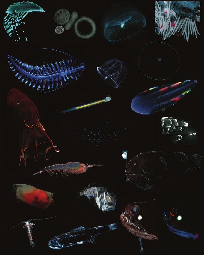
Figure 4
Gallery of marine bioluminescent organisms. (a) Bioluminescent bacteria grown on a petri dish; (b) shallow colonial polycystine
radiolarians; (c) the hydromedusa Aequorea victoria; (d ) red-tipped tentacles and white stinging cells from the siphonophore Erenna sp.;
(e) pelagic polychaete Tomopteris, which emits yellow luminescence; ( f ) planktonic larva of the acorn worm Ptychodera flava;
( g) fluorescence showing the marginal photophores of Aequorea coerulescens; (h) vampire squid Vampyroteuthis internalis with light organs
on its arm tips; (i) the bioluminescent chaetognath Caecosagitta macrocephala; ( j) ctenophore Beroe forskalii, showing a rainbow of
structural colors, not bioluminescence; (k) bioluminescence emission from light organs of the squid Abraliopsis sp.;
(l) intensified image of luminescent frenzy from Beroe forskalii; (m) krill Thysanoessa sp.; (n) live photo of anglerfish Chaenophryne
longiceps; (o) ostracod Conchoecia sp. that uses coelenterazine instead of typical ostracod luciferin; ( p) hatchetfish with an overlay of
ventral photophores shown by their blue fluorescence; (q) large copepod Gaussia princeps; (r) myctophid lampfish with species-specific
pattern of photophores, and white “sternchaser” organ; (s) Tactostoma sp., in white light, and (t) under fluorescent illumination, showing
the red and green photophores. All images show animals illuminated by a white-light strobe except a, k, and l, which record
bioluminescent light, and g, p, and t, which use fluorescence illumination to reveal photophore patterns. (Photos: S. Haddock).ANRV399-MA02-17 ARI 13 November 2009 18:11
a b c d
e f g
h
i
j
k l
m n
o p
q
r s tANRV399-MA02-17 ARI 13 November 2009 18:11
organisms, including Renilla, store coelenterazine in a stabilized enol-sulfate form (Cormier et al.
1970), which may not be detected in standard assays (Thomson et al. 1997).
Cnidarians appear to be unable to synthesize coelenterazine (Haddock et al. 2001), and its
mode of biosynthesis is yet to be determined. Like ostracod luciferin, coelenterazine is thought to
derive from cyclization of a tripeptide precursor, in this case, Phe-Tyr-Tyr (Ward et al. 1994). The
strongest evidence for its natural origin is from experiments on Systellaspis debilis, where isolated
eggs showed increasing levels of luciferin despite being dissociated from any potential maternal
contribution (Thomson et al. 1995). It also persists in captive copepods (Barnes & Case 1972,
Buskey & Stearns 1991), indicating that crustaceans are the most likely, but perhaps not exclusive,
source of coelenterazine in the food chain.
Coelenterazine occurs naturally in conjunction with both photoproteins and luciferases. Several
luciferases have been cloned from the copepods Pleuromamma, Metridia, and Gaussia (Markova
et al. 2004, Takenaka et al. 2008, Szent-Gyorgyi et al. 2003), the decapod shrimp Oplophorus (Inouye
et al. 2000, Inouye & Sasaki 2007), the sea pen Ptilosarcus gurneyi, and two species of Renilla (Lorenz
et al. 1991, Szent-Gyorgyi et al. 2003). Where it has been examined, scyphozoan jellyfish also use
luciferases. The most thoroughly studied is from the coronate Periphylla (Shimomura & Flood
1998), but it has not yet been successfully cloned.
Hydrozoans, ctenophores, and radiolarians use coelenterazine in conjunction with photopro
teins. The first photoprotein, aequorin, was originally discovered, isolated, and characterized
(Shimomura et al. 1962) from the hydromedusa Aequorea victoria (Figure 4c) and subsequently
cloned (Inouye et al. 1985, Prasher et al. 1986). This is the same species that was the source
of the original green fluorescent protein, through a parallel research track (Shimomura 2005).
Several other photoproteins have been cloned from hydromedusae, including Mitrocoma (Fagan
et al. 1993), Clytia (Inouye & Tsuji 1993, Inouye & Sasaki 2007), and Obelia (Illarionov et al. 1995,
Markova et al. 2002). The tertiary structure of these proteins has been solved (Head et al. 2000,
Liu et al. 2000), and their properties have been re-engineered for research uses (e.g., Frank et al.
2008, Dikici et al. 2009). A photoprotein from the squid Symplectoteuthis, known as symplectin,
has also been purified (Fujii et al. 2002, Isobe et al. 2008).
Organisms with a luciferase instead of a photoprotein control light emission by either seques
tering the two compounds separately, or by using a luciferin-binding protein to control exposure
of the luciferin to oxidation. The coelenterazine-binding protein of the sea pansy has been recently
characterized from both R. reniformis (Inouye 2007) and R. mulleri (Titushin et al. 2008). In these
proteins, the coelenterazine is caged within a pocket of helices and escapes through a hole that
opens upon the binding of calcium ions (Stepanyuk et al. 2009).
Other and Novel Luciferins
Despite the prevalence of the four major marine luciferins, there are other light emitters known
from additional taxa. The light-producing chemistry is well known for the bivalve Pholas (Dunstan
et al. 2000), and for some polychaetes, including the polynoiid scale worms (Bassot & Nicolas
1995), chaetopterid tube worms, and syllid fireworms (Shimomura 2006). Thanks to the work of
Shimomura and colleagues, many of these chemicals have been known for decades, so it is rare
for an entirely novel luciferin to be discovered and elucidated. A notable addition to the list of
marine luciferins came with the elucidation of the chemistry of the hemichordate Ptychodera flava
(Kanakubo & Isobe 2005). The hypothesized light emitter, which operates with the involvement
of peroxide and riboflavin, has a unique simple symmetrical structure (Figure 5). There are
many other luciferins still to be discovered, especially among vermiform phyla, echinoderms, and
molluscs (denoted by [X] in Figure 1).ANRV399-MA02-17 ARI 13 November 2009 18:11
Br Br
HO OH
Br Br
Figure 5
The luciferin of the hemichordate worm Ptychodera flava.
FLUORESCENCE AND BIOLUMINESCENCE
The mechanism of light production through a chemical reaction distinguishes bioluminescence from other natural
optical phenomena such as fluorescence and phosphorescence. Fluorescent molecules do not produce their own
light; they absorb photons, which temporarily excite electrons to a higher energy state. As these electrons rapidly
relax to their ground state, they rerelease their energy, usually at a longer wavelength. Because the excitation and
relaxation occur almost immediately (within picoseconds to microseconds), fluorescent light is only seen while the
specimen is being illuminated. Examples of fluorescent molecules found in nature are chlorophyll, phycobiliproteins,
and the green fluorescent proteins (GFPs). The term phosphorescence technically refers to a special case of optically
excited light emission in which the relaxation occurs gradually over a long period of time, and photon emission
persists for seconds to minutes. This phenomenon is seen naturally in some minerals and photosynthetic systems.
In the early literature and for many years thereafter, it was common to use the term “phosphorescence” (perhaps
poetically) to describe bioluminescence (Huxley 1898, Darwin 1909).
The technical distinction between bioluminescence and fluorescence is sometimes blurred in a practical context,
for several reasons. First, compounds that are bioluminescent may also be autofluorescent, and thus photophores
can often be visualized through their fluorescence under short-wavelength or UV illumination (Figure 4g,p,t).
Examples are the arm-tip light organs of the vampire squid Vampyroteuthis infernalis (Robison et al. 2003), the pho
tophores of hatchetfishes and the midshipman Porichthys notatus, the fin photophores of the chaetognath Caecosagitta
macrocephala, and the “lures” of the siphonophore Erenna (Haddock et al. 2005b). Some of these photophores and
their associated chemical compounds become fluorescent only after they have reacted to produce light (e.g., Inouye
2004). Another link is that the fluorescence emission spectrum of a molecule may match its luminescence emission
spectrum, since the same molecules are participating in the excitation-emission process. Because other natural ma
terials can also be fluorescent (chitin, calcium phosphate), care must be taken when inferring the involvement of
bioluminescence from the presence of fluorescence (e.g., Stabili et al. 2008).
One other relationship between bioluminescence and fluorescence is that brightly fluorescent proteins like
green fluorescent protein (GFP) may be colocalized with their bioluminescent counterparts. For example, in the
hydromedusa Aequorea victoria and several other bioluminescent cnidarians, their bioluminescence reaction would
normally emit blue light at around 470 nm. Because of the tight association between the light-emitting chemicals and
a separate green fluorescent protein, the energy that would be radiated as blue light instead excites the fluorescent
pigment and is emitted as green photons (Figure 6). As a result of these linkages, it is often possible to learn about
the bioluminescence of an organism by examining its fluorescent properties, even though fluorescence does not
automatically indicate the presence of luminescence.ANRV399-MA02-17 ARI 13 November 2009 18:11
100 Figure 6
Green fluorescent pro
80
tein.
Fluorescence:
emission of light 60
derived from the
energy of an absorbed 40
photon
20 Photoprotein Fluorescent
GFP: green protein
fluorescent protein; its
fluorescent 425 450 475 500 525 550
chromophore is Wavelength (nm)
generated
autocatalytically from
peptides within the
translated protein; can DIVERSITY OF BIOLUMINESCENCE
occur in association
The most comprehensive list of bioluminescent genera was compiled by Herring (1987).
with bioluminescent
proteins Supplemental Table 1 lists bioluminescent genera discovered since or omitted from that com
pilation (Follow the Supplemental Material link from the Annual Reviews home page at
http://www.annualreviews.org). Here we detail some of the predominant bioluminescent groups
in which there has been recent research.
Bacteria
Bioluminescent bacteria are common in the ocean, especially in temperate to warmer waters
(Dunlap & Kita-Tsukamoto 2006). They may be cultured from almost any piece of detritus or
tissue found on the beach, and even from uncooked seafood, which has been known to glow after
being left for a time. Most bioluminescent animals do not get their luminescence from bacterial
symbionts, but this continues to be a persistent misconception (e.g., Sinniger et al. 2008). These
mutualistic associations are known mainly from a variety of marine fish and squid species, although
the terrestrial pathogen Photorhabdus can infect nematode worms (Forst et al. 1997), and even
human tissue (Peel et al. 1999). Bacteria are not luminous until they have reached sufficiently
high concentrations to initiate quorum sensing (Waters & Bassler 2005, Nealson & Hastings
2006), and once induced, they glow continuously in the presence of oxygen rather than producing
discrete flashes. These properties are specific to bacteria, which makes them uniquely suitable
as photogenic symbionts and can lead to spectacular marine phenomena such as bioluminescent
milky seas (see “Milky Seas” sidebar on page 472).
Among prokaryotes, light production is known only from the so-called eubacteria, specifically
Gram-negative γ-proteobacteria, and not from Archaea. Names applied to the genera of lumi
nescent bacteria often vary with the time-period and researchers’ preference. The best-studied
symbiotic bacteria are in the genus Vibrio (Figure 4a), including the predominantly free-living
species V. harveyi (sometimes called Beneckea harveyi), although the genus Shewanella also in
cludes a bioluminescent species (Makemson et al. 1997). It was recently shown that many new
strains of luminous bacteria, some related to what has traditionally been called Photobacterium
phosphoreum, are present in the deep sea (Gentile et al. 2008), and many of the current species
actually represent diverse assemblages (Dunlap & Ast 2005). Although there are many exceptions,
Vibrio fischeri (often called Photobacterium) is part of the species-complex typically involved in sym
biosis with sepiolid and loliginid squid and monocentrid fishes, while Photobacterium leiognathi
and relatives are primarily symbionts for leiognathid, apogonid, and morid fishes (Kaeding et al.
2007).ANRV399-MA02-17 ARI 13 November 2009 18:11
The participants in the mutualism between bacterium and host were thought to share paral
lel phylogenies, as might be expected (Nishiguchi et al. 1998). Unexpectedly, however, broader
surveys found that light organs include a complement of bacterial populations in both squid
(Guerrero-Ferreira & Nishiguchi 2007, Wollenberg & Ruby 2009) and fishes (Dunlap et al.
2007). So the evolutionary dynamics are relatively fluid and opportunistic, and several strains
make suitable symbionts for light-organ colonization. Nonetheless, there are close evolutionary
ties, and in the best-studied mutualism, that between the bobtail squid Euprymna scolopes and Vibrio
fischeri, the presence of the bacteria actually induces the morphological development of the squid
light organ (McFall-Ngai & Ruby 1998). The host squid also monitors the luminescent perfor
mance of the symbionts and strains that fail to maintain adequate light production are rejected by
a yet unknown mechanism (Visick et al. 2000, Nyholm et al. 2004). A whole-genome sequence of
V. fischeri provides robust support for the idea that the same mechanisms that allow disease-causing
enteric Vibrionaceae to infect human hosts (e.g., V. cholerae, V. parahaemolyticus) may be at work in
establishing beneficial symbioses with marine species (Ruby et al. 2005). In fact, one researcher is
said to have infected himself for a period of months when working with luminous Photobacterium
leiognathi (Campbell 2008).
Dinoflagellates
Next to fireflies, dinoflagellates are the most commonly encountered bioluminescent organism.
They typically cause the sparkling lights in the water seen by sailors, swimmers, and beachgoers,
and they produce the “bioluminescent bays” which are tourist destinations in Puerto Rico and
Jamaica. These protists can be autotrophic (photosynthetic) or heterotrophic, feeding on other
phytoplankton and prey. In large numbers, some species may form potentially toxic red tides,
typically during a stratified calm period after an influx of nutrients (discussed below). There are
at least 18 luminous genera (Baker et al. 2008), including Gonyaulax (=Lingulodinium), Noctiluca,
Protoperidinium, and Pyrocystis. Dinoflagellates invest heavily in their ability to luminesce, and
allocate energy to bioluminescence before growth, although luminescence comes second to the
ability to swim (Latz & Jeong 1996).
Radiolarians
Radiolarians are ameboid protists whose skeletal elements, when present, are made from amor
phous silica. The classification of this nominal group in the sense of Haeckel (1887) is in a state of
flux, and phylogenetic studies have shown it to include several independent lineages, primarily the
Polycystinea (Figure 4b) and Phaeodarea (Polet et al. 2004, Kunitomo et al. 2006). Within the
polycystines, one order, the shallow-living Collodaria, is known to be bioluminescent. It contains
the genera Collozoum and Thalassicola, which use coelenterazine bound to calcium-activated photo-
proteins (Herring 1979, Latz et al. 1991). It might seem that, as protists, radiolarians would be un
likely to have a way to acquire coelenterazine through their diet, but many of them actually consume
or digest larger prey. The other major lineage of radiolarians lies within the Cercozoa and does not
form a monophyletic group with the polycystines in general, nor the Collodaria in particular (Polet
et al. 2004, Yuasa et al. 2006). Nonetheless, members of this predominantly deep-sea group, includ
ing Aulosphaera spp. and Tuscaridium cygneum, are also bioluminescent (Ling & Haddock 1997).
Ctenophores
Bioluminescence is very well represented in the comb jellies, where more than 90% of planktonic
genera (and none of the benthic species) are known to produce light (Haddock & Case 1995).ANRV399-MA02-17 ARI 13 November 2009 18:11
Ctenophores use calcium-activated proteins and coelenterazine, some of which have been recently
cloned (S.V. Markova, unpublished data; S.H.D. Haddock, unpublished data). Their luminescence
can be internally expressed, sometimes in cascading waves as with Beroe forskalii (Figure 4i,l),
but some species like Euplokamis stationis, Mertensia ovum, and Eurhamphaea vexilligera also emit
glowing particles as part of an escape response (e.g., Widder et al. 1992). At least one species
of bioluminescent ctenophore also contains a green fluorescent protein (S.H.D. Haddock and
N. Mastroianni, unpublished paper).
Cnidarians
Bioluminescence is found in both benthic and planktonic cnidarians, the group that includes corals,
anemones, hydroids, medusae, and siphonophores. As far as is known, the luminous species all use
coelenterazine as their light-emitting substrate.
Luminous hydrozoans include both hydromedusae and siphonophores. Most of the plank
tonic forms are bioluminescent, including 91% of planktonic siphonophore genera (Figure 2b),
while for unknown reasons it is rare among certain other groups, like the species of benthic
hydroids that do not produce medusae. Most famous of the luminescent hydrozoans, and ar
guably of all bioluminescent invertebrates, is the shallow-living hydromedusa Aequorea victoria
(Figure 4c,g), which provided the original source material for research on photoproteins and the
Nobel Prize–winning GFP (Prasher et al. 1985, 1992; Shimomura 2005). Most hydrozoans likely
use bioluminescence for defensive or warning purposes, but siphonophores also use luminescence
(Haddock et al. 2005b) and fluorescence (Pugh & Haddock 2009) to attract prey directly to their
stinging tentacles (Figure 4d).
Two orders of scyphozoans contain luminous members, including nearly all of the deep-sea
coronates such as Atolla spp. and Periphylla periphylla, and some of the semaeostomes such as Pelagia
noctiluca, Phacellophora, and the deep-sea Poralia (Haddock & Case 1999). Rhizostome species are
not known to be luminescent. Scyphozoans were among the first bioluminescent animals recorded
in the literature, dating back to Pliny the Elder in the first century A.D. Periphylla, Atolla, and
other coronates produce cascading waves of light and can also exude luminous particles (Herring
& Widder 2004).
Several luminous anthozoans are found within the octocorals (Alcyonaria), including sea pens
and sea pansies. The first luminous soft coral Eleutherobia grayi was only recently discovered in
the South Pacific (Williams 2001). There are many luminous octocorals found in shallow sandy
bottoms (Renilla, Ptilosarcus), and in the deep sea (Stylatula, Halipterus, Anthomastus). Although
the hard corals and anemones (Hexacorallia) are now famous for the possession of fluorescent
proteins (e.g., Shagin et al. 2004), they are not usually bioluminescent. Among the hexacorals,
one prominent luminous species is the epibiotic parasite Parazoanthus, which is unique in that its
zooids form colonies growing over sea fans and sponges. Deep-living bamboo corals (Isidids) are
also well known for their luminescence, and new species continue to be discovered (Etnoyer 2008).
Annelids
There are several different bioluminescent lineages among marine polychaetes, yet the chemi
cal mechanisms of light production have not been fully determined for most species. Shimomura
(2006) summarizes what is known in this regard. Some of the terrestrial annelids have been chemi
cally characterized (e.g., Petushkov & Rodionova 2007), but there do not seem to be many parallels
between the groups, and luminescence has several independent origins just within the annelids.
The life cycles of the famous syllid fireworms, including Odontosyllis, have been thoroughly studiedANRV399-MA02-17 ARI 13 November 2009 18:11
through the years (Fischer & Fischer 1995). This normally benthic species produces a spawning
stage near the time of the full moon. Females produce luminescent secretions that attract the males
to swarm around them. Although these polychaetes use bioluminescence during spawning, like
Pelagic: living in the
most organisms they will also produce internal luminescence in response to physical disturbance water column, as
(Fischer & Fischer 1995, Deheyn & Latz 2009). In Eusyllis, fragments can continue luminescing opposed to benthic
¨
for weeks, even without the head attached (Zorner & Fischer 2007). Such defensive responses are
also common in other polychaete lineages, where there is no evidence of a function during mating.
Although luminescence is often expressed by planktonic species or life-history stages, there are
several benthic scale-worms (Polynoidae) that emit light using a protein triggered with superoxide
radicals (Bassot & Nicolas 1995). The scales in Polynoidae are shed into the water where, released
from nervous inhibition, they can glow and flash as a distractive decoy for minutes. In other
benthic species, the function of luminescence is not as clear. The tube-dwelling chaetopterid
Chaetopterus and the terebellid Polycirrus both produce light at around 440 nm (Huber et al.
1989). Glowing particles are exuded from their tubes when the worms are disturbed. It has been
suggested that short-wavelength luminescence is an aposematic signal advertising distastefulness,
or that light production drives off commensals that would otherwise take up residence in the
worms’ tubes (Morin 1983), although no experiments have been conducted to test this. Benthic
Chaetopterus make light using a unique photoprotein (Shimomura 2006) five times as large as
cnidarian photoproteins. Luminescence is also present in a recently discovered planktonic species
of Chaetopterus (Osborn & Rouse 2008).
While terebellids fall at the short-wavelength end of bioluminescence spectra, another poly
chaete emits at the other extreme, with long-wavelength luminescence. Among the planktonic
polychaetes, species of Tomopteris (Figure 4e) produce a golden yellow light with unknown
chemistry. Many planktonic members of the Flabelligeridae, including Poeobius meseres and Flota
vitjasi, are luminescent (S.H.D. Haddock, unpublished data), and a newly discovered group of
deep-sea polychaetes carries luminescent “bombs” that it drops when disturbed (Osborn et al.
2009). These are families in which planktonic existence is a derived trait (Osborn & Rouse 2008),
suggesting another independent origin of luminescence correlated with animals moving up into
the water column.
Other Worms
Bioluminescence makes scattered appearances among the many other wormlike phyla. Several
species of acorn worms (Hemichordata) are luminescent, including Balanoglossus and the plank
tonic tornaria larvae of Ptychodera flava (Figure 4f ), whose luciferin was recently characterized
(Kanakubo & Isobe 2005).
In the ribbon worms (Phylum Nemertea), there is only one known luminous species (Kanda
1939), but there are many deep-sea pelagic species that have only been examined preliminarily
( J.L. Norenburg & P. Roe, pers. comm.). In 1939, Nemertea was the last phylum found to be
luminescent until the discovery of the first bioluminescent chaetognath (Figure 4h) (Haddock &
Case 1994). Chaetognaths (arrow worms) use a luciferase+coelenterazine chemistry and shed a
cloud of glowing particles in conjunction with an escape response.
Molluscs
Luminous marine molluscs include a few unusual gastropods like the whelk Planaxis and the
spectacular pelagic nudibranch Phylliroe. One of the longest-known and best studied luminous
molluscs is the bivalve Pholas. It seems somewhat unexpected for a clam to be luminous, but thisANRV399-MA02-17 ARI 13 November 2009 18:11
was the species used in pioneering experiments by Dubois in 1887 that established the existence of
a luciferin+luciferase reaction. More than 100 years later, the photoprotein pholasin was cloned
and characterized (Dunstan et al. 2000).
The most prominent of the bioluminescent marine molluscs are the cephalopods. Among the
squids alone, there are at least 70 luminous genera (Herring 1977). Bacterial symbionts produce
luminescence for several genera in the families Sepiolidae and Loliginidae (Ruby & McFall-Ngai
1992; Jones & Nishiguchi 2004; Nyholm et al. 2004, 2009). The rest of the squids, though, have
intrinsic bioluminescence, using a luciferin along with their individual luciferase. Some of these
have been chemically characterized. For example, Symplectoteuthis has a photoprotein that operates
with dehydro-coelenterazine (Takahashi & Isobe 1994, Isobe et al. 2008). In Watasenia scintillans,
the luciferase reacts with coelenterazine-disulfate and also has a requirement for ATP and Mg2+
as cofactors, which is unusual for coelenterazine-based luminescence (Tsuji 2002, 2005). Known
in Japan as hotaru-ika or the firefly squid, Watasenia is the subject of a popular festival each spring.
Light organs cover the ventral mantle with bright organs near the eyes and at the tips of the arms—
typical for many other kinds of squids. Cranchiids also have well-developed ocular photophores,
and many squid combine their complex iridescent reflectors with their light-emitting apparatus
(Herring et al. 2002).
Squids can produce an impressive variety of luminescent displays. Many use their ventral
photophores for counterillumination (Figure 4k) (Herring et al. 1992). The deep-sea vampire
squid Vampyroteuthis (Figure 4j) is sufficiently distinct to have been classified in its own order.
In addition to two large mantle photophores, and small light organs scattered across its body,
it can release glowing particles from special light organs on its arm tips, apparently to distract
predators (Robison et al. 2003). Octopoteuthis takes on a variety of coloration patterns, and will
drop its arms when disturbed, leaving the glowing arm tips as decoys (Bush et al. 2009). Another
cephalopod with light organs at its arm tips is Taningia danae. This highly active squid has clawlike
hooks instead of suckers, and large (up to 2 cm) light organs at its arm tips. They are thought to
use luminescence both for intraspecific communication and potentially to stun prey. Using high-
definition cameras deployed in combination with bait and glowing lights, Kubodera et al. (2007)
obtained incredible in situ video (available at doi:10.1098/rspb.2006.0236) of Taningia seemingly
signaling to the artificial light sources using long glows. It also produced short, bright flashes from
its arm-tip photophores as it attacked the bait.
Females of the pelagic deep-sea octopods Japetella and Eledonella have a greenish-yellow ring
around their mouth which is only periodically luminous (Robison & Young 1981), indicating a
potential role in reproduction (Herring 2007). Stauroteuthis and other genera of deep-sea cirrate
octopods were long suspected to have glowing suckers (Chun 1910), but this was only recently
confirmed to be bioluminescence ( Johnsen et al. 1999). The lights lining the arms are thought to
attract prey within the webbed canopy characteristic of this slow-moving cephalopod group.
Given the diversity of ways that cephalopods produce bioluminescence, it is likely that the
number of independent evolutionary origins in this group, and in Mollusca generally, is much
higher than estimated.
Crustaceans
Many types of planktonic crustaceans are bioluminescent, and they use species-specific luciferases
with at least three different types of luciferins. With regard to the number of times they have
reinvented bioluminescence, crustaceans rival molluscs and polychaetes. Euphausiids, or krill
(Figure 4m), use the same luciferin as dinoflagellates (Nakamura et al. 1989, Shimomura 1995b),ANRV399-MA02-17 ARI 13 November 2009 18:11
strongly suggesting a dietary connection. They have light organs along the lower surface of their
body, which they use for counterillumination, and some species also have two small light organs
on their eyestalks. These might serve as feedback mechanisms for determining how well their
ventral photophores are matching background light. Like most photophores, these are under ner
¨
vous control, involving serotonin moderated by nitric oxide (Kronstr ¨ et al. 2007). Krill also use
om
physical mechanisms to contract and dilate photophores to regulate light production (Kronstr ¨ ¨
om
et al. 2009).
In shallow reefs of the tropical Atlantic, dramatic displays of luminescence at dusk are often
attributable to ostracods (Figure 4o). Ostracods can eject luciferin and luciferase through nozzles
near their mouth, leaving discrete puffs of light in the water. Cypridinids provide the best marine
examples of complex use of luminescence during mating, analogous in complexity and subtlety to
the situation seen in fireflies (e.g., Buck & Case 2002). Shallow Caribbean ostracods, including
many new species, have been extensively studied by Morin and colleagues (Cohen & Morin 1990;
Morin & Cohen 2010; Torres & Cohen 2005; Rivers & Morin 2008, 2009).
Copepods, as one of the most abundant marine invertebrates, are also one of the most abun
dant bioluminescent groups in the sea, although the widespread genus Calanus is not luminescent.
Common luminous genera include Pleuromamma, Metridia, Oncaea, and Gaussia. Bioluminescence
involves coelenterazine, exhibited either as intracellular flashes or emitted into the water as part
of an escape response (Widder et al. 1999). The giant (1 cm) deep-sea copepod Gaussia prin
ceps (Figure 4q) is a particularly good model for laboratory study of bioluminescence, behavior,
and neurobiology (Bowlby & Case 1991, Weatherby et al. 2000, Fields et al. 2002). GFP-like
fluorescent proteins have been cloned from copepods (Shagin et al. 2004, Masuda et al. 2006),
but surprisingly they have only been found in nonluminous species. Many of the bioluminescent
copepods are predatory and live moderately deep, although the tiny Poecilostomatoid copepod
Oncaea is one example that is shallow and pseudoplanktonic (Bottger-Schnack & Schnack 2005).
It occurs in the water column in association with larvacean houses (Ohtsuka et al. 1996), marine
snow (Green & Dagg 1997), and other organisms (Gasca et al. 2007).
Decapod shrimp use light in several ways: Sergestids counterilluminate with photophores that
tilt to maintain their downward orientation no matter which way the animal is swimming (Latz
1995). Oplophorids, including the genera Systellaspis, Acanthephyra, and Oplophorus, can disgorge
large volumes of luminous fluid (Inouye et al. 2000) as part of a distress response. The visual systems
of many of these crustaceans have been recently investigated in relation to their bioluminescent
ability and their vertical migration behaviors. In both temporal and spectral properties, the visual
systems of adults are tuned to detect bioluminescence (Frank & Widder 1999, Frank 1999), while
juveniles may also be attuned to shorter wavelengths related to the detection of downwelling
surface light (Frank et al. 2009).
Although many amphipods living in the water column are parasites on gelatinous plankton, sev
eral of these are nonetheless bioluminescent. Scina is a genus that emits uniquely short-wavelength
luminescence (λmax ∼ 440 nm) from its antennae and legs (Bowlby & Case 1991). The visual sys
tem of this species, however, is not especially sensitive to those wavelengths (Cohen & Frank
2007), and the function of the luminescence is not known. Nonluminous amphipods will track
blue light sources (Land et al. 1995), and it is suggested that they use this in hunting for the biolu
minescent jellies they parasitize. Although the group that includes Scina and Proscina is not usually
thought to be parasitic, examples of associations between Proscina and gelatinous hosts have been
found (Gasca et al. 2007). In addition to producing typical blue-green light, some individuals of
Cyphocaris, a genus of gammarid, can emit orange light (595 nm) through an unknown mechanism
(Bowlby & Case 1991).ANRV399-MA02-17 ARI 13 November 2009 18:11
Echinoderms
Bioluminescence is found in most of the major groups of echinoderms: brittle stars (Ophiuroidea),
sea stars (Asteroidea), sea cucumbers (Holothuroidea), and even crinoid sea lilies (reviewed by
Herring & Cope 2005). Much of the recent work on echinoderms has focused on ophiuroid
(brittle star) behavior and neurophysiology (e.g., Deheyn et al. 2000, Dewael & Mallefet 2002). A
complex system of neurotransmitters modulates light output in these groups, and light originates
from both an unknown photoprotein and a coelenterazine+luciferase reaction, depending on the
species (Shimomura 2006).
In echinoderms other than brittle stars, luminescence is more common among deep-sea taxa
(Brisingidae and Paxillosida for sea stars, Pannychia, Peniagone, and Scotoanassa for the holothuri
ans, and Thaumatocrinus and Annacrinus among crinoids). As is typical for bioluminescence, it
is also disproportionately represented in the pelagic holothurians such as Enypniastes eximia
(Robison 1992) and Pelagothuria. New luminous ophiuroid species continue to be discovered
(Mallefet et al. 2004), and undoubtedly the diversity of bioluminescent echinoderms as a whole
will continue to increase as more deep-sea species are examined in good condition.
Tunicates
Bioluminescence is not as prevalent in planktonic colonial tunicates (thaliaceans: salps and do
liolids) as in other plantonic groups. Although the colonial salp Pyrosoma has been renowned for
its bioluminescence for centuries, other salps have not been confirmed as bioluminescent. Py
rosome luminescence has two unusual properties: It comes as a long, steady glow, and it can be
induced upon illumination by light. The duration of the glow and the presence of bacteria-like
particles suggest that bacteria are involved (Haygood & Prince 1993), but this is still the subject
of a long-running debate (Godeaux et al. 1998).
Two other urochordate groups, long thought to be nonluminous, have been found to have lumi
nous members. Doliolids are similar to salps, and nearly all species are not luminescent. However,
a deep-sea bioluminescent doliolid was recently described (Robison et al. 2005), and biolumi
nescence has also been seen in Paradoliopsis harbisoni and Pseudusa bostigrinus (S.H.D. Haddock,
unpublished data). Although most benthic tunicates are not luminescent, intrinsic biolumines
cence was discovered in the shallow-living benthic ascidian Clavelina miniata (Aoki et al. 1989).
This species produces light of 535 nm from phagocytes in the tunic (Chiba et al. 1998), and the
glow may persist for 10 to 30 seconds (Hirose 2009).
Bioluminescence is well represented in the planktonic larvaceans (Appendicularia). Most known
genera have bioluminescence, including Mesochordaeus (Renaux & Youngbluth 1990, Hopcroft &
Robison 1999), Bathochordaeus (Hamner & Robison 1992), and about half of the Oikopleura species
(Galt & Flood 1998). Larvaceans secrete luminous inclusions into their mucus and cellulose
feeding filters (Galt & Sykes 1983) and can leave behind many bioluminescent “houses” each
day with the potential to contribute a disproportionately large amount of luminescence (1014 to
1016 photons · m−3 ) to the water column (Galt & Flood 1998). Oikopleura labradoriensis uses a
coelenterazine+luciferase system (Galt & Flood 1998), which is curious since their diet is unlikely
to include crustaceans and thus suggests another potential source for coelenterazine.
Fish
Bioluminescence is found in at least 42 families across 11 orders of bony fishes (compiled from
Suntsov et al. 2008), in addition to one family of sharks. In contrast with invertebrate taxa,You can also read