Elucidation of SIRT-1/PGC-1 α-associated mitochondrial dysfunction and autophagy in nonalcoholic fatty liver disease
←
→
Page content transcription
If your browser does not render page correctly, please read the page content below

Jiang et al. Lipids in Health and Disease  (2021) 20:40
https://doi.org/10.1186/s12944-021-01461-5

 RESEARCH                                                                                                                                            Open Access

Elucidation of SIRT-1/PGC-1α-associated
mitochondrial dysfunction and autophagy
in nonalcoholic fatty liver disease
Yan Jiang1,2† , Duankai Chen2†, Qiming Gong2†, Qunqing Xu2, Dong Pan2, Feiyan Lu2 and Qianli Tang1,2*

  Abstract
  Background: Nonalcoholic fatty liver disease (NAFLD) can lead to chronic liver diseases associated with
  mitochondrial damages. However, the exact mechanisms involved in the etiology of the disease are not clear.
  Methods: To gain new insights, the changes affecting sirtuin 1 (SIRT-1) during liver fat accumulation was
  investigated in a NAFLD mouse model. In addition, the in vitro research investigated the regulation operated by
  SIRT-1 on mitochondrial structures, biogenesis, functions, and autophagy.
  Results: In mice NAFLD, high-fat-diet (HFD) increased body weight gain, upregulated serum total cholesterol,
  triglycerides, aspartate aminotransferase, alanine aminotransferase, blood glucose, insulin levels, and liver
  malondialdehyde, and decreased liver superoxide dismutase activity. In liver, the levels of SIRT-1 and peroxisome
  proliferator-activated receptor-gamma coactivator -1α (PGC-1α) decreased. The expression of peroxisome
  proliferator-activated receptor-α and Beclin-1 proteins was also reduced, while p62/SQSTM1 expression increased.
  These results demonstrated SIRT-1 impairment in mouse NAFLD. In a well-established NAFLD cell model, exposure
  of the HepG2 hepatocyte cell line to oleic acid (OA) for 48 h caused viability reduction, apoptosis, lipid
  accumulation, and reactive oxygen species production. Disturbance of SIRT-1 expression affected mitochondria. Pre-
  treatment with Tenovin-6, a SIRT-1 inhibitor, aggravated the effect of OA on hepG2, while this effect was reversed
  by CAY10602, a SIRT-1 activator. Further investigation demonstrated that SIRT-1 activity was involved in
  mitochondrial biogenesis through PGC-1α and participated to the balance of autophagy regulatory proteins.
  Conclusion: In conclusion, in high-fat conditions, SIRT-1 regulates multiple cellular properties by influencing on
  mitochondrial physiology and lipid autophagy via the PGC-1α pathway. The SIRT-1/PGC-1α pathway could be
  targeted to develop new NAFLD therapeutic strategies.
  Keywords: Sirtuin 1, Peroxisome proliferator-activated receptor-gamma coactivator -1a, Mitochondrial physiology,
  Mitochondrial autophagy, Mitochondrial dysfunction; Lipid autophagy, Nonalcoholic fatty liver disease

* Correspondence: htmgx@163.com
†
 Yan Jiang, Duankai Chen and Qiming Gong contributed equally to this
work.
1
 Medical College of Guangxi University, Nanning 530004, Guangxi, China
2
 YouJiang Medical University for Nationalities, Baise 533000, Guangxi, China

                                         © The Author(s). 2021 Open Access This article is licensed under a Creative Commons Attribution 4.0 International License,
                                         which permits use, sharing, adaptation, distribution and reproduction in any medium or format, as long as you give
                                         appropriate credit to the original author(s) and the source, provide a link to the Creative Commons licence, and indicate if
                                         changes were made. The images or other third party material in this article are included in the article's Creative Commons
                                         licence, unless indicated otherwise in a credit line to the material. If material is not included in the article's Creative Commons
                                         licence and your intended use is not permitted by statutory regulation or exceeds the permitted use, you will need to obtain
                                         permission directly from the copyright holder. To view a copy of this licence, visit http://creativecommons.org/licenses/by/4.0/.
                                         The Creative Commons Public Domain Dedication waiver (http://creativecommons.org/publicdomain/zero/1.0/) applies to the
                                         data made available in this article, unless otherwise stated in a credit line to the data.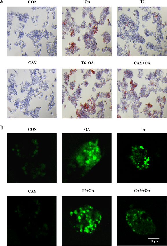
Jiang et al. Lipids in Health and Disease   (2021) 20:40                                                              Page 2 of 12

Background                                                          environment and fed a normal diet for 1 week. Then, the
Nonalcoholic fatty liver disease (NAFLD) is the most com-           six-week-old mice were randomly distributed between the
mon chronic liver disease, with a severity ranging from sim-        control group (CON) and the HFD group, each group gath-
ple steatosis, steatohepatitis, fibrosis, to cirrhosis, and leads   ering eight animals. The mice were provided food and
to all-cause and liver-related mortality [1]. So far, the work-     drinking water ad libitum. The mice in the CON group
ing model of NAFLD proposes the “two-hit hypothesis”. In-           continued being fed an ordinary diet, while the mice in the
sulin resistance, which increases diet intake and hepatic           HFD group were fed a HFD for 8 weeks. The ordinary diet
lipogenesis, may cause the accumulation of triglycerides            was composed of (expressed as % total calories) 65% carbo-
(TG) and free fatty acids in the liver. However, lipid peroxi-      hydrates, 11% fat, 24% proteins, with a total caloric value of
dation, mitochondrial dysfunction, and inflammation may             3.84 kcal/g, while HFD was composed of 18% carbohy-
eventually cause hepatocyte damages and liver fibrosis. Re-         drates, 62% fat, 24% proteins, with a total caloric value of
cently, some experts in the field proposed that in essence,         5.49 kcal/g. The specific components of the HFD were
NAFLD may be a mitochondrial disease [1].                           diversified and were produced by Jiangsu Synergetic
   It is well established that a high-fat diet (HFD) causes         Pharmaceutical Bioengineering Co., Ltd. (Jiangsu, China).
an abnormal accumulation of TG and imbalances mito-
chondrial function in the liver [2–5]. In progressive liver         Collection and biochemical analysis of serum and liver
diseases, mitochondrial dysfunction produces excessive              tissue
reactive oxygen species (ROS) and cytokines, which                  After 8 weeks of normal or HFD food intake, the 14-
leads to hepatic inflammation and injury. Furthermore,              week-old mice were fasted for 12 h and anesthetized
mitochondrial dysfunction disturbs fat homeostasis in               using 7% chloral hydrate. Blood samples were with-
hepatic cells and eventually causes lipid accumulation,             drawn from the retro-orbital cavity. The serum was
which leads to lipotoxicity [6]. It has recently been sug-          prepared by centrifugation at 5000×g for 10 min at
gested that imbalanced activity of the transcription fac-           4 °C and then stored at − 80 °C for biochemical ana-
tors sterol regulatory element binding protein-1c                   lyses. The liver was harvested, rinsed in PBS, and
(SREBP-1c) and peroxisome proliferator-activated                    wiped with filter paper. Part of the liver was fixed in
receptor-α (PPAR-α) causes mitochondrial dysfunction,               formalin at 4 °C, while the remaining part was stored
which in turn induces liver steatosis [7, 8]. In addition,          at − 80 °C for subsequent analyses, after being quickly
HFD (such as palmitic acid) intake is a high-risk inducer           frozen in liquid nitrogen. The concentrations of total
of SREBP-1c/PPAR-α ratio imbalance [9].                             cholesterol (TC), TG, aspartate aminotransferase
   Sirtuin 1 (SIRT-1), a master metabolic mediator, is a            (AST), alanine aminotransferase (ALT) and blood glu-
NAD-dependent protein deacetylase [10] involved in the              cose in serum were detected using biochemical re-
control of lifespan extension and lipid metabolism, includ-         agent kits (Nanjing Jiancheng Bioengineering Institute,
ing the processes of fatty acid synthesis, oxidation, and adi-      Nanjing, China). Biochemical assays were also per-
pocyte generation [11, 12]. SIRT-1 activation has a                 formed to probe SOD activity and MAD level in liver
beneficial effect against NAFLD through inhibiting lipogen-         tissues. Serum insulin was measured by the enzyme-
esis, by phosphorylation of SREBP-1c [11]. Studies have             linked immunosorbent assay (ELISA) (Beyotime, Nan-
demonstrated that SIRT-1 reduces oxidative stress [13, 14].         jing, China) methods using a TriStar LB941 system
In HFD-induced obese mice, the expression of SIRT-1 is              (Berthold, Wildbad, Germany).
inhibited, resulting in liver metabolic damages [15]. Yet,
how SIRT-1 regulates mitochondrial biology in high fat-             Histopathological examination
induced livers remains poorly understood.                           The right lobe of the livers were collected for histo-
   In the present study, the mechanisms underlying the              pathological examination. The liver tissues fixed in for-
effects of HFD on SIRT-1 in liver were investigated                 malin were cut into 5-μm sections and stained with
in vivo. The present study also tested if SIRT-1 could              hematoxylin and eosin dyes. The livers sections frozen
modulate mitochondrial physiology, and investigated the             in liquid nitrogen were cut into 5-μm using a frozen
mechanisms involving SIRT-1 regulation in oleic acid                slicer (Thermo, Waltham, USA). The sections were
(OA)-treated hepatocytes in vitro.                                  stained with oil red O solution and hematoxylin. Histo-
                                                                    pathological structures were observed under a light
Materials and methods                                               microscope (BX53, Olympus, Tokyo, Japan).
Animal experiments
Animal models                                                       RNA extraction and analysis
Four- to five-week-old C57BL/6 mice were purchased from             Total RNA was extracted using TRIzol reagent (CoWin
Changsha Tianqin Biotechnology Co., Ltd. (Changsha,                 Biosciences, Beijng, China) and transcribed into cDNA
China). The mice were housed in a specified constant                with a Reverse Transcription Kit (Thermo Fisher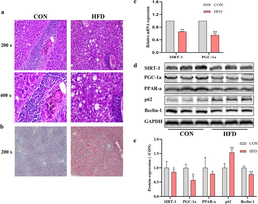
Jiang et al. Lipids in Health and Disease   (2021) 20:40                                                     Page 3 of 12

Scientific, Waltham, USA), according to the manufac-          after removing the cell culture medium. Then, the cells
tures’ instructions. Then, real-time quantitative PCR         were stained with 5 mg/mL of oil red O solution for 30
(RT-qPCR) was performed using SYBR Green Master               min at room temperature. The cells were washed with
Mix (CoWin Biosciences, Beijng, China). The reaction          PBS, and then stained with hematoxylin for 1 min. After
conditions were: pre-denaturation at 95 °C for 10 min,        rinsing, the cells were observed and imaged under a light
then 40 cycles of denaturation at 95 °C for 15 s and an-      microscope (BX53, Olympus, Tokyo, Japan).
nealing at 60 °C for 30 s. The level of GAPDH cDNA (F:
5′-CCTCGTCCCGTAGACAAAATG-3′, R: 5′-TGAG                       Flow cytometric analysis
GTCAATGAAGGGGTCGT-3′) was used as internal                    The apoptosis rate in HepG2 cells was measured using
reference to quantify the level of SIRT-1 (F: 5′-             the Annexin V-fluorescein Isothiocyanate/ propidium iod-
TTCAGAACCACCAAAGCGGA-3′, R: 5′-TCCCACAG                       ide (Annexin V-FITC/PI) apoptosis kit. Cells were rinsed
GAGACAGAAACCC-3′) and PGC-1ɑ (F: 5′-CGAGAA                    with PBS, digested, and centrifuged. The cell density was
GCGGGAGTCTGAAAG-3′, R: 5′-GAGCAGCGAA                          adjusted to 1 × 10⁶ /mL. Then, the cells were incubated
AGCGTCACA-3′) cDNA. The relative mRNA expres-                 with Annexin V-FITC and PI. Flow cytometric data were
sion of SIRT-1 and PGC-1ɑ was calculated using the            collected on a flow cytometer from BD Biosciences (FACS
2-ΔΔCt formula. All results were normalized to the            Verse, BD Biosciences, Franklin, USA) and analyzed using
GAPDH expression level.                                       FlowJo software (v10.0; BD Biosciences, Franklin, USA).

Cell experiments                                              Immunofluorescence analysis
Culture conditions for the HepG2 hepatocyte cell line         Cells were seeded into glass-bottom cell culture dishes.
The HepG2 cell line was purchased from the Cell Bank          The cells were co-transfected with Ad-GFP-LC3 and
of China Academy of Sciences. Cells were maintained in        Ad-HBmTur-Mito (Hanbio, Shanghai, China, catalogue
Dulbecco’s Modified Eagle’s Medium (DMEM, 4.5 g/L             #: HBAD-1006, 1014). Following stimulation, the
glucose, Gibco, Carlsbad, CA, USA) supplemented with          localization of mitochondrial autophagy (MA) in cells
8.0% fetal bovine serum, penicillin (100 U/mL) and            was monitored under confocal laser scanning micro-
streptomycin (100 μg/mL).                                     scope (FV3000, Olympus, Tokyo, Japan). In another ex-
                                                              periment, after removing the cell culture medium, the
Cell treatment                                                cells were incubated with a ROS fluorescent probe from
Cells were treated with OA (Sigma-Aldrich. St. Louis,         an assay kit for 45 min at 37 °C. Finally, the cells were
MO, USA, catalogue #: O1008), and SIRT-1 induction            washed and analyzed with the confocal laser scanning
was monitored by comparing levels in unpretreated and         microscope.
pretreated cultures. Briefly, after inoculation, HepG2
cells were grown for 24 h and treated with 1.5 mM OA          Transmission electron microscopy analysis
for 48 h to induce NAFLD in cell model. The SIRT-1 in-        After treatment as described above, the culture medium
hibitor Tenovin-6, referred to as T6 (MCE, New Jersey,        was discarded, and 2.5% glutaraldehyde was quickly
USA, catalogue #: HY-15510), was used at a concentra-         added to fix the cells. Cells were collected, centrifuged,
tion of 2 μM. The SIRT-1 activator CAY-10602, referred        re-fixed, dehydrated, infiltrated, and embedded. Finally,
to as CAY (MCE, New Jersey, USA, catalogue #: HY-             the ultrathin sections were stained with uranium and
104073), was used at a concentration of 20 μM. Cells          lead double solution and analyzed using a transmission
were incubated with T6 or CAY for 2 h, and subse-             electron microscope (HT7700, HITACHI, Tokyo, Japan).
quently stimulated with OA for 48 h. The drug-
containing medium contained 12.0% of serum.                   Western blotting analysis
                                                              The total proteins were extracted on ice from the cells
Cell viability assay                                          and liver tissues by ultrasonic lysis in RIPA buffer con-
After 48 h of treatment, the viability of the HepG2 cells     taining 1 mM PMSF and 1% phosphatase inhibitor cock-
was assessed with the CCK-8 kit (ABMole, Houston,             tail. The lysates were centrifuged at 12000 ×g for 15 min
USA, catalogue #: M4839). In brief, 10 μL of CCK-8 so-        at 4 °C. The protein concentration in the supernatant of
lution was added to each well and incubated for 1 h. The      the lysates was determined with a BCA protein quantita-
optical density (OD) was measured at 450 nm using the         tive kit. Next, the samples were normalized, mixed with
TriStar LB941 microplate reader.                              loading buffer, and denatured by heating at 95 °C for 5
                                                              min. Aliquots of 50 mg of proteins were loaded onto 8,
Oil red O staining                                            10, or 12.5% SDS-PAGE gels. The proteins were trans-
Oil red O was used to stain intracellular lipids. The cells   ferred onto 0.45 μm PVDF membranes. The membranes
were fixed with 10% neutral formaldehyde for 20 min           were blocked and incubated overnight at 4 °C with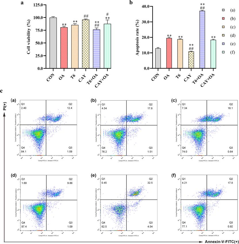
Jiang et al. Lipids in Health and Disease         (2021) 20:40                                                                 Page 4 of 12

primary monoclonal antibodies against SIRT-1, PGC-1α,                         serum TC, TG, AST, ALT, blood glucose, insulin levels
Cytochrome C oxidase IV (COX-IV), PPAR-ɑ, Mito-                               and liver MDA content, and reduction of SOD activity
chondrial fission factor (MFF), Mitofusin-1 (MFN1),                           (Table 1C-D).
p62/SQSTM1 (p62), Light chain 3B (LC3B), Beclin-1,
and GAPDH (all from Affinity Biosciences, OH, USA).                           HFD leads to aggregated lipids synthesis, decreased
The membranes were washed with TBST and incubated                             expression of SIRT-1 and PGC-1α, and alteration of
with secondary antibodies at room temperature for 40                          autophagy-related proteins
min, and after washing them five times, an enhanced                           As shown in Fig. 1, large amounts of fat forming vacuoles
chemiluminescence kit was used to detect the proteins                         were observed in the livers of mice fed HFD (Fig. 1a). The
on the membranes. The bands were quantified using the                         oil red O staining also confirmed that lipid droplets accu-
ImageJ system (v1.8.0, NIH, Bethesda, USA).                                   mulated in the livers of the mice from the HFD group
                                                                              (Fig. 1b). SIRT-1 and PGC-1α expression was quantified
Statistical analysis                                                          in liver tissues by RT-qPCR and western blotting analysis.
The data were analyzed using IBM SPSS statistics 23.0                         After 8 weeks of HFD regimen, liver SIRT-1 and PGC-1α
software. Paired two-tailed t-test and one-way analysis of                    expression declined (Fig. 1c-e). The expression of PPAR-ɑ
variance (ANOVA) were used to assess the significance                         and Beclin-1 proteins also decreased, while p62 protein
of differences observed between groups. P < 0.05 was                          expression increased (Fig. 1d-e).
considered statistically significant. All data are expressed
as the mean ± standard deviation (M ± SD).                                    Effect of SIRT-1 on OA-induced cell viability in hepG2 cells
                                                                              To determine treatment toxicity, HepG2 cell viability
Results                                                                       was assessed by CCK-8 assay. Compared with untreated
HFD exacerbates obesity-related parameters in mice                            CON cultures, OA and T6 treatments decreased cell via-
The mice fed HFD had a significant increase of total                          bility significantly. The combination of the two sub-
body weight, net weight gain and liver weight compared                        stances aggravated the cell loss. CAY had no effect on
with the mice of the control group (Table 1A). Energy                         cell viability and rescued the low viability induced by
intake was increased in HFD mice compared to the con-                         OA (Fig. 2a).
trols (Table 1B), concomitantly with marked increase of
                                                                              Effect of SIRT-1 on OA-induced cell apoptosis in hepG2
Table 1 General and hepatic parameters in control mice (CON)
                                                                              cells
and high-fat-diet (HFD) mice
                                                                              Cell death includes apoptosis and necrosis. The apop-
                                   CON                     HFD
                                                                              tosis provoked by the different treatments of HepG2
A. General and hepatic parameters
                                                                              cells was assessed by flow cytometry. Upon OA and
  Initial body weight (g)          14.49 ± 0.55            14.56 ± 0.41       T6 treatments, apoptosis increased and was even
  Final body weight (g)            29.38 ± 5.58            43.45 ± 5.22**     more drastic in the T6-pretreated T6 + OA cultures.
  Net weight gain (g)              14.89 ± 5.60            28.89 ± 4.97**     CAY reduced cell apoptosis and rescued OA-induced
  Liver weight (g)                 1.20 ± 0.16             2.08 ± 0.47**      apoptosis (Fig. 2b-c).
B. Food and Energy intake
                                                                              Effect of SIRT-1 on OA-induced intracellular lipid in hepG2
  Dietary intake (g/day)           3.64 ± 0.37             3.66 ± 0.41
                                                                              cells
  Energy intake (kcal/day)         14.64 ± 0.68            18.29 ± 1.83**     The level of intracellular lipids was revealed by oil red O
C. Serum parameters                                                           staining. OA or T6 treatment led to lipid accumulation
  TC (mM)                          1.39 ± 0.16             2.98 ± 0.38**      manifesting as intracellular red oil droplets. This
  TG (mM)                          1.64 ± 0.23             3.95 ± 0.54**      phenomenon was more pronounced with the combin-
                                                                              ation of OA and T6. CAY treatment attenuated the lipid
  AST (U/L)                        148.14 ± 13.40          180.24 ± 10.08**
                                                                              accumulation induced by OA. The oil droplets were not
  ALT (U/L)                        64.14 ± 5.56            74.84 ± 9.05*
                                                                              clearly visible in the CON and CAY groups (Fig. 3a).
  Blood glucose (mg/dL)            122.68 ± 12.30          174.83 ± 19.31**
  Insulin (μU/mL)                  5.90 ± 1.95             26.61 ± 3.67**     Effect of SIRT-1 on OA-induced ROS in hepG2 cells
D. Enzyme activity in liver                                                   For the evaluation of mitochondrial damage, intracellular
  MDA (nmol/g)                     37.40 ± 7.10            74.92 ± 9.59  **   ROS was visualized by fluorescence. Mitochondrial ROS
                                                                              was significantly increased by OA and T6, and further
  SOD (U/g)                        78.92 ± 7.75            46.32 ± 6.50**
                                                                              enhanced by a combined treatment. In contrast, CAY
Values are presented as mean ± SD (n = 6). Data were analyzed by one-way
analysis ANOVA. *, ** represents significance from the control group at
                                                                              did not impacted ROS whereas it alleviated OA-induced
P < 0.05, P < 0.01                                                            ROS production (Fig. 3b).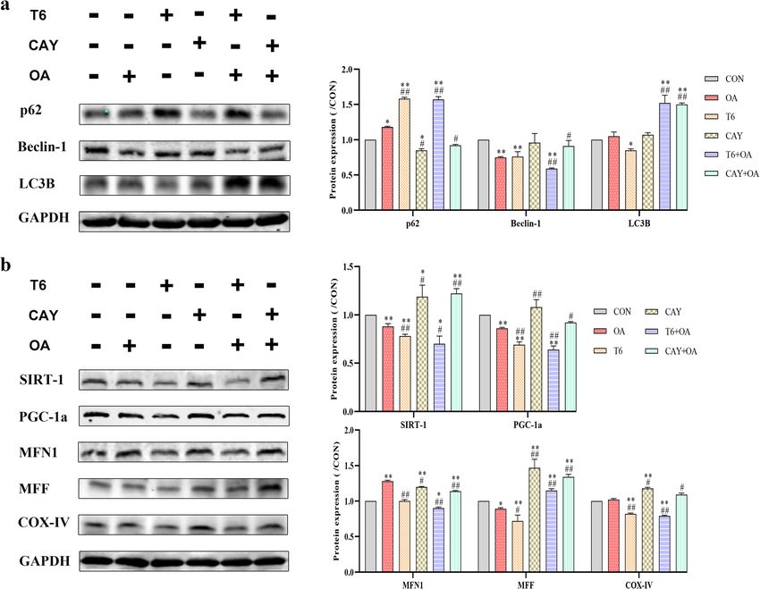
Jiang et al. Lipids in Health and Disease    (2021) 20:40                                                                               Page 5 of 12

 Fig. 1 Effect of HFD for 8 weeks on liver steatosis, SIRT-1 and PGC-1ɑ mRNA level and protein expression of SIRT-1, PGC-1ɑ, PPAR-ɑ, p62, Beclin-1.
 a The liver pathology of mice in the two groups. b Lipid deposition in liver tissues. c The mRNA expression level of SIRT-1 and PGC-1ɑ. d The
 protein expression of SIRT-1, PGC-1a, PPAR-ɑ, p62 and Beclin-1 was analyzed and quantified. *P < 0.05, **P < 0.01, vs. CON. Original magnification:
 × 200, × 400

Effect of SIRT-1 on OA-induced mitochondrial autophagy                      organelles, double-membrane structures represent
in hepG2 cells                                                              autophagosomes, and single-membrane structures are
Mitochondrial autophagy was identified by staining for                      autolysosomes [16].
the autophagy protein LC3 (in green, Fig. 4a) and                             As it can be seen from the micrographs, OA- and T6-
mitochondria (in red, Fig. 4a). Mitochondrial autoph-                       treated cells have high numbers of vacuole (va), lipid drops
agy (in yellow/ orange, Fig. 4a) was detected in OA,                        (LD), swollen and ruptured mitochondria (SM) and autolyso-
CAY, T6 + OA, and CAY + OA treated cells using the                          somes (ASS). Lipid drops are surrounded by autolysosomes
confocal laser scanning microscope. T6 pretreatment                         and undergo lipophagy (LA, Fig. 4b). Several autolysosomes
severely aggravated the increase of mitochondrial                           are observed with the highest numbers being in T6 + OA
autophagy provoked by OA, while CAY pretreatment                            cells. Moreover, these structures are more frequent in T6-
appropriately increased the effect of OA (Fig. 4a).                         treated cells, compared to other cultures (Fig. 4b).
                                                                              Western blotting analysis showed that OA decreased
SIRT-1 mediated the effects of OA on the substructures of                   Beclin-1 protein expression to levels similar to T6. CAY
hepG2 cells                                                                 treatment reversed OA effect. Meanwhile, other
HepG2 hepatocyte substructures were analyzed by trans-                      autophagy-related protein markers were analyzed in the
mission electron microscopy. At different stages and ac-                    cells receiving different treatments. The level of p62 was
cording to membrane enclosing cytoplasm or damaged                          increased in the cells receiving the OA or T6 different
Jiang et al. Lipids in Health and Disease (2021) 20:40 Page 6 of 12
Fig. 2 SIRT-1 regulates HepG2 cell viability and apoptosis crosstalk with OA. The cells were pretreated with T6 (2 μM) or CAY (20 μM) 2 h, followed
incubated with OA (1.5 mM) for 48 h. a The cell viability was estimated using CCK-8 kit. b, c Flow Cytometry analysis of apoptosis and apoptosis
rate quantitative analysis in cells. *P < 0.05, **P < 0.01, vs. CON group; #P < 0.05, ##P < 0.01, vs. OA group
treatments but was the highest in T6 + OA treatmed cells. were verified (Fig. 5b). Besides, the protein PGC-1α was
Furthermore, LC3B protein level increased abnormally quantified as measurement of mitochondrial biogenesis.
after stimulated by CAY or T6 pretreatment in the OA- The results showed that PGC-1α level decreased signifi-
induced cells (Fig. 5a). cantly after OA, T6 and combined treatment. The SIRT-1
activator CAY significantly promoted PGC-1α expression
Effect of SIRT-1 induction upon OA treatment on hepG2 compared to OA and T6. To further monitor the changes
cells mitochondrial biogenesis in mitochondrial biogenesis, the expressions of specific
To assess mitochondrial biogenesis, the effects of SIRT in- mitochondrial regulatory proteins was quantified by west-
hibitor and activator on mitochondrial protein expression ern blotting analysis. The results indicated that OAJiang et al. Lipids in Health and Disease (2021) 20:40 Page 7 of 12 Fig. 3 SIRT-1 regulates HepG2 intracellular lipid and ROS crosstalk with OA. The cells were pretreated with T6 (2 μM) or CAY (20 μM) 2 h, followed incubated with OA (1.5 mM) for 48 h. a The cells were stained oil red O. b Immunofluorescence detected intracellular ROS. Scale bars are 50 μm and 10 μm respectively induced the mitochondrial fusion protein MFN1 but induced fission. Moreover, CAY and CAY + OA upregu- inhibited the expression of the fission protein MFF. T6 lated the expression of the mitochondrial respiratory pro- inhibited MFF protein. However, CAY promoted both tein COX-IV (Fig. 5b). mitochondrial fusion and fission, alleviated OA-promoted fusion, and rescued the inhibition operated by OA on Discussion mitochondrial fission. T6 pretreatment inhibited OA- SIRT-1 is a sensor of cell metabolic reactions such as induced mitochondrial fusion, while increasing OA- stress, starvation, and caloric restriction. Target genes of
Jiang et al. Lipids in Health and Disease   (2021) 20:40                                                                            Page 8 of 12

 Fig. 4 SIRT-1 involved in regulation of mitochondrial autophagy and lipophages in HepG2 cells. The cells were pretreated with T6 (2 μM) or CAY
 (20 μM) 2 h, followed incubated with OA (1.5 mM) for 48 h. a Mitochondrial autophagosome was determined. Red: mitochondria; green: LC3;
 yellow/ orange: merge. Scale bars are 10 μm respectively. b Representative electron micrographs of cells. Mit: mitochondria; SM: swollen and
 ruptured mitochondria; ER: endoplasmic reticulum; ASS: autophagolysosome; PG: phagophore; va: vacuole; LD: lipid drops; LA: lipid component of
 autolysosome. Scalar bar = 2.0 μm and 1.0 μm
Jiang et al. Lipids in Health and Disease   (2021) 20:40                                                                          Page 9 of 12

 Fig. 5 Effect of intervene in SIRT-1 and combine with OA on p62, Beclin-1, LC3B, SIRT-1, PGC-1a, MFN1, MFF and COX-IV protein expression in
 HepG2 cells. HepG2 cells were treated with 1.5 mM OA for 48 h, following T6 (2 μM) or CAY (20 μM) 2 h. a The p62, Beclin-1 and LC3B protein
 expression was detected by Western blotting. b The same method was used to detect the SIRT-1, PGC-1a, MFN1, MFF and COX-IV protein
 expression. The bands were normalized using GAPDH. The images were quantified with ImageJ. Data are presented as the mean ± SD; *P < 0.05,
 **P < 0.01 compared with CON group; #P < 0.05, ##P < 0.01 compared with OA group

mitochondrial biogenesis can be regulated by SIRT-1 to                   produced during mitochondrial dysfunction is associ-
maintain energy and metabolism stability [17]. New                       ated with the development of NAFLD and obesity [1].
studies have regarded SIRT-1 as an important target for                  This pathological mechanism is associated with
diseases associated with mitochondrial dysfunction [18].                 changes in NF-κB and PGC-1α activities [19, 20].
In this experiments in mice, long-term high-fat regimen                  Furthermore, redundant ROS is responsible for cell
reduced the activity of SIRT-1 protein, and simultan-                    apoptosis [21]. This study also proved that ROS accu-
eously, adverse changes occurred in liver structure and                  mulation in hepatocyte cell lines treated with OA or
metabolites, such as increased levels of TG, TC, AST,                    with a SIRT-1 inhibitor decreases cell viability and
ALT and MDA, and reduction of SOD activity. In                           induces apoptosis. The activation of SIRT-1 prior to
addition, the interaction between SIRT-1 and autophagy                   OA stimulation prevents this injurious effect. These
responses in mitochondria under lipid accumulation                       results suggest that SIRT-1 is involved in the regula-
process has not been explored. Further, this study deep-                 tion of cell viability, apoptosis, and mitochondrial
ened the insights on the effect of SIRT-1 on mitochon-                   ROS.
drial biology such as autophagy, structure, biosynthesis,                  Autophagy refers to the degradation of non-specific
and function.                                                            cytoplasmic proteins, organelles, and other substrates,
  Suitable oxidation of mitochondria plays a major                       and represents a mechanism of cell death regulation, dif-
role in energy metabolism. However, excessive ROS                        ferent from apoptosis and pyroptosis. Autophagy resists
Jiang et al. Lipids in Health and Disease   (2021) 20:40                                                     Page 10 of 12

cell pressure and promotes cell viability [22], while ex-     adverse effects on cells, such as decrease of viability, or
cessive autophagy adversely causes cell death. The gold       defective mitochondrial structure and metabolism.
standard markers to assess autophagy pathways are               Mitochondrial fusion and fission regulate mitochondrial
LC3B, Beclin-1, and p62. Especially, LC3B, an early           homeostasis, quantity, and even function [32]. Disrupted
marker of autophagy [20], induces autophagosome               mitochondrial fission/fusion reduce energy production, in-
membrane expansion and fusion [22]. Besides, Beclin-1         crease oxidative stress, and even cell death [3, 32]. Mito-
promotes the initiation of autophagy by mediating the         chondrial fusion is mainly regulated by MFN1 (fusion
formation of autophagosomes. However, p62 aggregates          protein, a dynamic-associated GTPase), which fuses the
outside the lysosomes and is involved in selective au-        outer membranes of mitochondria [3]. MFF plays a role in
tophagy [23], in which the first steps of autophagy are       mitochondrial fission. The decrease of SIRT-1 directly
impaired [24]. This knowledge was used to speculate on        lowered mitochondrial fission by reducing MFF protein
the trend of autophagy in cells, by detecting the changes     levels. This was reversed by SIRT-1 activation. OA treat-
in p62, Beclin-1, and LC3B protein expression. Experi-        ment increased mitochondrial fusion, and decreased fis-
ments in mice showed that HFD increased the expres-           sion. Taken together, the analysis of these phenotypes
sion of p62 and reduced that of Beclin-1. These changes       indicates that mitochondria may become larger by a com-
led to decreased autophagy, were not conducive to             pensatory fusion to adapt to excessive lipid metabolism in-
homeostatic liver lipid metabolism, and resulted in liver     duced by OA.
lipid accumulation, visible as numerous lipid droplets in       It has been reported that hindered mitochondrial dy-
liver tissues. Moreover, the same changes in p62 and          namics (fusion or fission) can lead to increased levels of
Beclin-1 protein expression occurred in OA-stimulated         some triacylglycerols, while autophagy can reverse this
cells. The ultrastructure analyses showed few autophago-      effect [33]. Together with this report, the present data
somes. Additionally, lipid droplets were present in the       indicate that lipid excess reduces SIRT-1, and that lipo-
intracellular space.                                          phages are not activated to a corresponding degree to
   Moderate mitochondrial autophagy plays a crucial role      degrade excessive lipids. This lack of regulation eventu-
in mitochondrial homeostasis by removing damaged              ally leads to lipid accumulation and cell lipotoxicity.
mitochondria and contributing to cell metabolism. Ab-           Disruption of mitochondrial membranes, increase of
normal mitochondria lead to imbalanced mitochondrial          ROS, fragmentation of mitochondria, and abnormal
homeostasis and induction of mitochondrial autophagy.         mitochondrial structure all promoted the initiation of
When mitochondria undergo autophagy, they usually             mitochondrial autophagy [34]. The present findings
show multilayered structures. In vitro, the present find-     demonstrate that excess of lipids and low expression of
ings showed that excess of fat induced oil particle accu-     SIRT-1 lead to PGC-1α imbalance, and result in disor-
mulation in the hepatocytes, and resulted in                  dered mitochondrial biosynthesis and excess of ROS. Ul-
mitochondrial swelling, and even fracture. Second, the        timately, mitochondrial autophagy is initiated.
expression of the fission protein MFF diminished signifi-
cantly, but that of the fusion protein MFN1 increased,        Study strengths and limitations
both phenomena inducing abnormal mitochondrial                In the present study, there were several strengths. First,
structure. Putative mechanisms behind these changes           although mitochondrial disease was considered as the
were the increase of anomalous mitochondria to adapt          essence of NAFLD, few studies have performed the
to the over-accumulation of lipids. Moreover, the activa-     regulatory relationship between SIRT-1/PGC-1a and
tion of SIRT-1 effectively promoted mitochondrial             mitochondrial biology in the disease. Second, to further
autophagy, which plays an important role in maintaining       explore whether mitochondrial and lipid autophagy were
a dynamic balance in mitochondria.                            regulated by SIRT-1, the molecular mechanism and sub-
   PGC-1α is a marker of mitochondrial biogenesis, and        cellular structures influenced by SIRT-1 were included
as a transcriptional coactivator that enhances fatty acids    in this study. However, there are some limitations in this
oxidation and glycogenesis [25], it improves intracellular    paper. The SIRT-1 related mechanisms were solely veri-
energy utilization [26]. The stability of PGC-1α is related   fied in vitro experiments. Therefore, in vivo studies are
to mitochondrial content [27, 28]. The lack of PGC-1α         required to verify the finding.
reduces the reversion of mitochondria, increases defect-
ive mitochondria, and eventually leads to cell death [29].    Conclusion
In addition, SIRT-1/PGC-1α have been shown to be part         In conclusion, excess of fat downregulates SIRT-1/PGC-
of a network regulating mitochondrial biogenesis in skel-     1α pathway and disturbs mitochondria’s biological activ-
etal muscles [30]. In this regard, the changes operated by    ity, damaging them. It also decreases lipophages, causing
SIRT-1 on PGC-1α activity are consistent with previous        lipid accumulation in cells and toxicity that ultimately
reports [30, 31]. The reduction of SIRT-1 has various         leads to cell apoptosis. These results may point SIRT-1/
Jiang et al. Lipids in Health and Disease          (2021) 20:40                                                                                         Page 11 of 12

PGC-1α pathway as a good target for further exploration                            6.    Paradies G, Paradies V, Ruggiero FM, Petrosillo G. Oxidative stress,
of adjuvant therapy in NAFLD treatment.                                                  cardiolipin and mitochondrial dysfunction in nonalcoholic fatty liver
                                                                                         disease. World J Gastroenterol. 2014;20(39):14205–18. https://doi.org/10.3
Abbreviations                                                                            748/wjg.v20.i39.14205.
NAFLD: Nonalcoholic fatty liver disease; HFD: High-fat-diet; OA: Oleic acid;       7.    Pettinelli P, Del Pozo T, Araya J, Rodrigo R, Araya AV, Smok G, et al.
ROS: Reactive oxygen species; TC: Total cholesterol; TG: Triglycerides;                  Enhancement in liver SREBP-1c/PPAR-alpha ratio and steatosis in obese
AST: Aspartate aminotransferase; ALT: Alanine aminotransferase;                          patients: correlations with insulin resistance and n-3 long-chain
MDA: Malondialdehyde; SOD: Superoxide dismutase; SIRT-1: Sirtuin 1; PGC-                 polyunsaturated fatty acid depletion. Biochim Biophys Acta. 2009;11:1080–6.
1ɑ: Peroxisome proliferator-activated receptor-gamma coactivator -1a; COX-         8.    Valenzuela R, Videla LA. Impact of the Co-Administration of N-3 Fatty Acids
IV: Cytochrome c oxidase IV; p62: p62/SQSTM1, sequestosome 1; PPAR-                      and Olive Oil Components in Preclinical Nonalcoholic Fatty Liver Disease
ɑ: Peroxisome proliferator-activated receptor-ɑ; MFF: Mitochondrial fission              Models: A Mechanistic View. Nutrients. 2020;12(2):499.
factor; MFN1: Mitofusin-1; LC3: Light chain 3; LC3B: Light chain 3B;               9.    Hernández-Rodas MC, Valenzuela R, Echeverría F, Rincón-Cervera MÁ,
MA: Mitochondrial autophagy; PG: Phagophores; va: Vacuole; LD: Lipid drops;              Espinosa A, Illesca P, Muñoz P, Corbari A, Romero N, Gonzalez-Mañan D,
SM: Swollen and ruptured mitochondria; ASS: Autolysosomes; LA: Lipophagy;                Videla LA. Supplementation with docosahexaenoic acid and extra virgin
T6: Tenovin-6; CAY: CAY-10602                                                            olive oil prevents liver steatosis induced by a high-fat diet in mice through
                                                                                         PPAR-α and Nrf2 upregulation with concomitant SREBP-1c and NF-kB
Acknowledgements                                                                         downregulation. Mol Nutr Food Res. 2017;61(12). https://doi.org/10.1002/
The authors are grateful for technical support and scientific suggestion of              mnfr.201700479.
the researchers of science lab center at YouJiang Medical University for           10.   Wang Y, Li X, He Z, Chen W, Lu J. Rapamycin attenuates palmitate-induced
Nationalities.                                                                           lipid aggregation by up-regulating sirt-1 signaling in AML12 hepatocytes.
                                                                                         Pharmazie. 2016;71(12):733–7. https://doi.org/10.1691/ph.2016.6695.
Authors’ contributions                                                             11.   Li J, Liu M, Yu H, Wang W, Han L, Chen Q, et al. Mangiferin improves
Yan Jiang, Duankai Chen and Qiming Gong conducted the experiments, and                   hepatic lipid metabolism mainly through its metabolite-Norathyriol by
drafted the article. Qunqing Xu, Dong Pan and Feiyan Lu helped to revise                 modulating SIRT-1/AMPK/SREBP-1c signaling. Front Pharmacol. 2018;9:201–
the manuscript. Qianli Tang participated in its design and coordination. All             14. https://doi.org/10.3389/fphar.2018.00201.
authors read and approved the final manuscript.                                    12.   Khader A, Yang WL, Godwin A, Prince JM, Nicastro JM, Coppa GF, et al.
                                                                                         Sirtuin 1 stimulation attenuates ischemic liver injury and enhances
Funding                                                                                  mitochondrial recovery and autophagy. Crit Care Med. 2016;44:651–63.
This study was supported by Guangxi Clinical Medical Research Center of            13.   Mahmoud AR, Ali FEM, Abd-Elhamid TH, Hassanein EHM. Coenzyme Q10
hepatobiliary Diseases (No. AD17129025), Special Funding for Guangxi                     protects hepatocytes from ischemia reperfusion-induced apoptosis and oxidative
Special Experts (No. GRCT[2019]13) and Guangxi Medical High-level Leading                stress via regulation of Bax/Bcl-2/PUMA and Nrf-2/FOXO-3/Sirt-1 signaling
Talents Training “139” Project (No. GWKJ[2018]22).                                       pathways. Tissue Cell. 2019;60:1–13. https://doi.org/10.1016/j.tice.2019.07.007.
                                                                                   14.   Tang Q, Len Q, Liu Z, Wang W. Overexpression of miR-22 attenuates
Availability of data and materials                                                       oxidative stress injury in diabetic cardiomyopathy via Sirt 1. Cardiovasc Ther.
Not applicable.                                                                          2018;36(2). https://doi.org/10.1111/1755-5922.12318.
                                                                                   15.   Liou CJ, Lee YK, Ting NC, Chen YL, Shen SC, Wu SJ, et al. Wen-Chung
Declarations                                                                             Huang protective effects of Licochalcone a ameliorates obesity and non-
                                                                                         alcoholic fatty liver disease via promotion of the Sirt-1/AMPK pathway in
Ethics approval and consent to participate                                               mice fed a high-fat diet. Cells. 2019;8:e447.
The protocol was approved by the Ethics Committee of YouJiang Medical              16.   Oami T, Watanabe E, Hatano M, Teratake Y, Fujimura L, Sakamoto A, et al.
University for Nationalities (Code: 20210129001).                                        Blocking liver autophagy accelerates apoptosis and mitochondrial injury in
                                                                                         hepatocytes and reduces time to mortality in a murine Sepsis model. Shock.
Consent for publication                                                                  2018;50(4):427–34. https://doi.org/10.1097/SHK.0000000000001040.
Not applicable.                                                                    17.   Dusabimana T, Kim SR, Kim HJ, Park SW, Kim H. Nobiletin ameliorates
                                                                                         hepatic ischemia and reperfusion injury through the activation of SIRT-1/
Competing interests                                                                      FOXO3a-mediated autophagy and mitochondrial biogenesis. Exp Mol Med.
The authors declare that they have no competing interests.                               2019;51(4):1–16. https://doi.org/10.1038/s12276-019-0245-z.
                                                                                   18.   Rigotti M, Cerbaro AF, da Silva IDR, Agostini F, Branco CS, Moura S, et al.
Received: 9 January 2021 Accepted: 1 April 2021                                          Grape seed proanthocyanidins prevent H2O2-induced mitochondrial
                                                                                         dysfunction and apoptosis via SIRT 1 activation in embryonic kidney cells. J
                                                                                         Food Biochem. 2020;44(3):e13147. https://doi.org/10.1111/jfbc.13147.
References                                                                         19.   Ortiz M, Soto-Alarcón SA, Orellana P, Espinosa A, Campos C, López-Arana S,
1. Hu Y, Yin F, Liu Z, Xie H, Xu Y, Zhu B. Acerola polysaccharides ameliorate            et al. Suppression of high-fat diet-induced obesity-associated liver
    high-fat diet-induced non-alcoholic fatty liver disease through reduction of         mitochondrial dysfunction by docosahexaenoic acid and hydroxytyrosol co-
    lipogenesis and improvement of mitochondrial functions in mice. Food                 administration. Dig Liver Dis. 2020;52(8):895–904. https://doi.org/10.1016/j.
    Funct. 2020;11(1):1037–48. https://doi.org/10.1039/C9FO01611B.                       dld.2020.04.019.
2. Ji J, Qin Y, Ren J, Lu C, Wang R, Dai X, et al. Mitochondria-related miR-141-   20.   Echeverría F, Valenzuela R, Espinosa A, Bustamante A, Álvarez D, Gonzalez-
    3p contributes to mitochondrial dysfunction in HFD-induced obesity by                Mañan D, et al. Reduction of high-fat diet-induced liver proinflammatory
    inhibiting PTEN. Sci Rep. 2015;5:1–12.                                               state by eicosapentaenoic acid plus hydroxytyrosol supplementation:
3. Ruan XH, Ma T, Fan Y. Ablation of TMEM126B protects against heart injury              involvement of resolvins RvE1/2 and RvD1/2. J Nutr Biochem. 2019;63:35–
    via improving mitochondrial function in high fat diet (HFD)-induced mice.            43. https://doi.org/10.1016/j.jnutbio.2018.09.012.
    Biochem Biophys Res Commun. 2019;515(4):636–43. https://doi.org/10.1016/       21.   Ghazipour AM, Shirpoor A, Ghiasi R, Pourheydar B, Khalaji N, Naderi R.
    j.bbrc.2019.05.084.                                                                  Cyclosporine a induces testicular injury via mitochondrial apoptotic
4. Wohua Z, Weiming X. Weiming, Glutaredoxin 2 (GRX2) deficiency                         pathway by regulation of mir-34a and sirt-1 in male rats: the rescue effect
    exacerbates high fat diet (HFD)-induced insulin resistance, inflammation and         of curcumin. Chem Biol Interact. 2020;327:e109180.
    mitochondrial dysfunction in brain injury: a mechanism involving GSK-          22.   Deng JS, Jiang WP, Chen CC, Lee LY, Li PY, Huang WC, et al. Cordyceps
    3beta. Biomed Pharmacother. 2019;118:e108940.                                        cicadae mycelia ameliorate Cisplatin-induced acute kidney injury by
5. Ferey JLA, Boudoures AL, Reid M, Drury A, Scheaffer S, Modi Z, et al. A               suppressing the TLR4/NF-kB/MAPK and activating the HO-1/Nrf2 and Sirt-1/
    maternal high-fat, high-sucrose diet induces transgenerational cardiac               AMPK pathways in mice. Oxidative Med Cell Longev. 2020;2020:e7912763.
    mitochondrial dysfunction independently of maternal mitochondrial              23.   Huo Y, Chen W, Zheng X, Zhao J, Zhang Q, Hou Y, et al. The protective
    inheritance. Am J Physiol Heart Circ Physiol. 2019;316:1202–10.                      effect of EGF-activated ROS in human corneal epithelial cells by inducing
Jiang et al. Lipids in Health and Disease (2021) 20:40 Page 12 of 12
mitochondrial autophagy via activation TRPM2. J Cell Physiol. 2020;235(10):
7018–29. https://doi.org/10.1002/jcp.29597.
24. Carotti S, Aquilano K, Zalfa F, Ruggiero S, Valentini F, Zingariello M, et al.
Lipophagy impairment is associated with disease progression in NAFLD.
Front Physiol. 2020;11:850. https://doi.org/10.3389/fphys.2020.00850.
25. Keinicke H, Sun G, Mentzel CMJ, Fredholm M, John LM, Andersen B, et al.
FGF21 regulates hepatic metabolic pathways to improve steatosis and
inflammation. Endocr Connect. 2020;9(8):755–68. https://doi.org/10.1530/
EC-20-0152.
26. Inata Y, Kikuchi S, Samraj RS, Hake PW, O'Connor M, Ledford JR, et al.
Autophagy and mitochondrial biogenesis impairment contribute to age-
dependent liver injury in experimental sepsis: dysregulation of AMP-
activated protein kinase pathway. FASEB J. 2018;32(2):728–41. https://doi.
org/10.1096/fj.201700576R.
27. Miller KN, Clark JP, Anderson RM. Mitochondrial regulator PGC-1a-
modulating the modulator. Curr Opin Endocr Metab Res. 2019;5:37–44.
https://doi.org/10.1016/j.coemr.2019.02.002.
28. Zou P, Liu L, Zheng LD, Payne KK, Manjili MH, Idowu MO, et al. Coordinated
Upregulation of Mitochondrial Biogenesis and Autophagy in Breast Cancer
Cells: The Role of Dynamin Related Protein-1 and Implication for Breast
Cancer Treatment. Oxid Med Cell Longev. 2016;2016:4085727. https://doi.
org/10.1155/2016/4085727.
29. Vainshtein A, Desjardins EM, Armani A, Sandri M, Hood DA. PGC-1α
modulates denervation-induced mitophagy in skeletal muscle. Skelet
Muscle. 2015;5(1):9. https://doi.org/10.1186/s13395-015-0033-y.
30. Mohamed JS, Hajira A, Pardo PS, Boriek AM. MicroRNA-149 inhibits PARP-2
and promotes mitochondrial biogenesis via SIRT-1/PGC-1a network in
skeletal muscle. Diabetes. 2014;63(5):1546–59. https://doi.org/10.2337/
db13-1364.
31. Akhtar S, Siragy HM. Pro-renin receptor suppresses mitochondrial biogenesis
and function via AMPK/SIRT-1/ PGC-1α pathway in diabetic kidney. PLoS
One. 2019;14(12):e0225728. https://doi.org/10.1371/journal.pone.0225728.
32. Li J, Zhang B, Chang X, Gan J, Li W, Niu S, et al. Silver nanoparticles
modulate mitochondrial dynamics and biogenesis in HepG2 cells. Environ
Pollut. 2020;256:e113430.
33. Haeussler S, Köhler F, Witting M, Premm MF, Rolland SG, Fischer C, et al.
Autophagy compensates for defects in mitochondrial dynamics. PLoS
Genet. 2020;16(3):e1008638. https://doi.org/10.1371/journal.pgen.1008638.
34. Hasnat M, Yuan Z, Naveedb M, Khan A, Raza F, Xu D, et al. Drp1-associated
mitochondrial dysfunction and mitochondrial autophagy: a novel
mechanism in triptolide-induced hepatotoxicity. Cell Biol Toxicol. 2018;35:
267–80.
Publisher’s Note
Springer Nature remains neutral with regard to jurisdictional claims in
published maps and institutional affiliations.You can also read