Zinc finger protein 384 enhances colorectal cancer metastasis by upregulating MMP2
←
→
Page content transcription
If your browser does not render page correctly, please read the page content below
ONCOLOGY REPORTS 47: 49, 2022

                          Zinc finger protein 384 enhances colorectal
                          cancer metastasis by upregulating MMP2
                    ZAIHUA YAN1,2*, YU ZHOU1,2*, YANG YANG3*, CHONGPU ZENG4, PEIDONG LI1,2,
                           HONGPENG TIAN1,2, XUEGUI TANG5 and GUANGJUN ZHANG1,2

            1
            Second Department of Gastrointestinal Surgery; 2Institute of Hepatobiliary, Pancreatic and Intestinal Disease,
      The Affiliated Hospital of North Sichuan Medical College, North Sichuan Medical College, Nanchong, Sichuan 637000;
      3
        Department of Dermatology, Xining First People's Hospital, Xining, Qinghai 810000; 4Department of General Surgery,
              Wangcang County People's Hospital, Guangyuan, Sichuan 628200; 5Anorectal Department of Integrated
               Traditional Chinese and Western Medicine, The Affiliated Hospital of North Sichuan Medical College,
                              North Sichuan Medical College, Nanchong, Sichuan 637000, P.R. China

                                    Received September 28, 2021; Accepted December 15, 2021

                                                     DOI: 10.3892/or.2022.8260

Abstract. Zinc finger proteins (ZNFs) serve key roles in             increased the levels of CRC cell invasion and migration,
tumor formation and progression; however, the functions and          whereas ZNF384 knockdown inhibited CRC development.
underlying mechanisms of dysregulated ZNF384 in colorectal           Moreover, the results of the present study demonstrated
cancer (CRC) are yet to be fully elucidated. Therefore, the          that ZNF384 mediated the expression of MMP2. MMP2
present study initially aimed to investigate the expression          knockdown inhibited ZNF384‑mediated CRC cell invasion
levels of ZNF384 in CRC samples. Moreover, lentiviral                and migration, whereas MMP2 overexpression ameliorated
ZNF384 overexpression and ZNF384 knockdown models                    ZNF384 knockdown‑induced inhibition of CRC progression.
were established in CRC cells. Transwell, wound healing and          In addition, the results of the present study demonstrated that
in vivo tail vein metastasis assays were carried out to evaluate     hypoxia‑inducible factor 1α (HIF‑1α) had the ability to bind
the effects of ZNF384 on CRC cell metastasis. Furthermore,           to the ZNF384 promoter, thereby initiating ZNF384 expres‑
reverse transcription‑quantitative PCR, western blotting, serial     sion. In human‑derived CRC samples, the expression levels
deletion, site‑directed mutagenesis, dual‑luciferase reporter        of ZNF384 were positively correlated with both MMP2 and
and chromatin immunoprecipitation assays were conducted to           HIF‑1α expression. Collectively, these findings highlighted
investigate the potential underlying mechanisms. The results         that ZNF384 may act as a prognostic marker and regulator of
of the present study demonstrated that ZNF384 expression             CRC metastasis.
was markedly increased in CRC samples and this was associ‑
ated with a poor prognosis. Notably, ZNF384 overexpression           Introduction

                                                                     In women and men, colorectal cancer (CRC) is the second
                                                                     and third most prevalent type of cancer, respectively (1). High
                                                                     CRC‑associated mortality rates are attributed to the high
Correspondence to: Dr Xuegui Tang, Anorectal Department              levels of metastasis and recurrence (2). Advances in techno‑
of Integrated Traditional Chinese and Western Medicine, The
                                                                     logical and genetic analyses have improved CRC diagnosis
Affiliated Hospital of North Sichuan Medical College, North
                                                                     and therapy (3); however, the mortality rate of CRC remains
Sichuan Medical College, 1 Maoyuan South Road, Nanchong,
Sichuan 637000, P.R. China                                           at a high level (4). Further investigations into the underlying
E‑mail: txg668nc@sohu.com                                            molecular mechanisms involved in CRC progression are
                                                                     required to identify potential prognostic markers.
Dr Guangjun Zhang, Second Department of Gastrointestinal
                                                                         The C2H2‑type zinc finger protein (ZNF), which is
Surgery, The Affiliated Hospital of North Sichuan Medical College,
North Sichuan Medical College, 1 Maoyuan South Road, Nanchong,
                                                                     encoded by the ZNF384 gene, is a transcription factor involved
Sichuan 637000, P.R. China                                           in the transcription of extracellular matrix genes (5). Coupling
E‑mail: zhanggj1977@126.com                                          of ZNF384 with TET family genes, such as the TATA box
                                                                     binding protein‑associated factor, transcription factor 3 and
*
    Contributed equally                                              Ewing sarcoma breakpoint region 1 gene, serves vital roles in
                                                                     acute lymphocytic leukemia (6,7). Moreover, transactivating
Key words: colorectal cancer, zinc finger protein 384, MMP2,         characteristics of the fusion protein have been reported
hypoxia‑inducible factor 1α, migration, invasion                     in NIH3T3 cells, highlighting the oncogenic potential of
                                                                     ZNF384 as a fusion protein (8). In melanoma cells, ZNF384
                                                                     overexpression was shown to promote metastasis (9). Although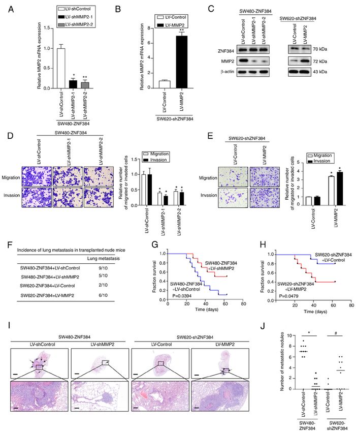
2                       YAN et al: ZINC FINGER PROTEIN 384 ENHANCES COLORECTAL CANCER METASTASIS

previous studies have indicated that ZNF384 may act as an            subsequently incubated for 30 min at room temperature in the
oncogenic factor that promotes cancer progression and metas‑         presence of 0.3% hydrogen peroxide, and blocked for 1 h using
tasis, the specific expression levels and functions of ZNF384        10% BSA (Sangon Biotech Co., Ltd.) at room temperature.
in human CRC are yet to be fully elucidated.                         Sections were incubated overnight at 4˚C with the following
    The present study aimed to examine the expression levels         antibodies: Rabbit anti‑ZNF384 (1:50; cat. no. ab251673;
of ZNF384 in CRC tissues and cells, and to determine its prog‑       Abcam), rabbit anti‑HIF‑1α (1:100; cat. no. ab243860; Abcam)
nostic value in patients with CRC. Additionally, the effects of      and rabbit anti‑MMP2 (1:100; cat. no. ab235167; Abcam).
ZNF384 on CRC cell invasion and migration, as well as the            Subsequently, sections were incubated with an HRP‑conjugated
underlying mechanism, were assessed.                                 secondary antibody (1:200; Goat Anti‑Rabbit IgG H&L;
                                                                     cat. no. ab205718; Abcam) at 37˚C for 2 h. The tissues were
Materials and methods                                                then stained at room temperature for 1 h using DAB (OriGene
                                                                     Technologies, Inc.) and counterstained for 1 min at room
Cell culture. The normal colonic epithelial cell line FHC, 293T      temperature using hematoxylin. All sections were dehydrated
cells and six human CRC cell lines (SW480, Caco‑2, SW620,            and sealed. Visualization and imaging were carried out using
HT29, LoVo and HCT116) were purchased from the American              a light microscope (Carl Zeiss AG), and sections incubated
Type Culture Collection. The 293T and CRC cell lines were            with Rabbit IgG (1:50; cat. no. A7016; Beyotime Institute of
seeded in DMEM (Gibco; Thermo Fisher Scientific, Inc.)               Biotechnology) acted as negative controls (NCs). Scoring
with 10% FBS (Gibco; Thermo Fisher Scientific, Inc.), and            was based on the ratio of positively stained cells, namely: 0,
the FHC cell line was seeded in DMEM:F12 (cat. no. D8437;            0‑5; 1, 6‑35; 2, 36‑70; and 3, >70%. Staining intensities were
Sigma‑Aldrich; Merck KGaA) with 10% FBS. Cells were incu‑            as follows: 3, strong; 2, moderate; 1, weak and 0, no staining.
bated in a humidified atmosphere containing 5% CO2 at 37˚C.          Final score determination was carried out by multiplying the
For cell culture under 0.5% O2 tension, the cells were incubated     scores of the percentage of positive cells with those of the
for 0‑24 h in a humidified atmosphere at 37˚C in a multi‑gas         staining intensities. Final scores were defined as: ‑, 0‑1; +, 2‑3;
CO2‑O2 incubator (NuAire) equilibrated with 0.5% O2, 5% CO2          ++, 4‑6; and +++, >6. Low expression levels were determined
and 94.5% N2. The cell suppliers stated that the identity of the     by a total score
ONCOLOGY REPORTS 47: 49, 2022                                                     3

Western blotting and reverse transcription‑quantitative (RT‑q)     5 min; followed by 35 cycles at 94˚C for 40 sec, 60˚C for
PCR were used to confirm successful transduction.                  30 sec and 72˚C for 60 sec, and a final extension step at 72˚C
                                                                   for 7 min. Experiments were carried out for three biological
In vivo metastasis assay. A total of 80 BALB/C nude female         repeats.
mice (age, 6 weeks; weight ~20 g) were purchased from
Shanghai SLAC Laboratory Animal Co., Ltd. Animals were             Cell proliferation analysis. An MTT assay was performed
housed in a 60% humidified atmosphere at 24˚C, under a 12‑h        to assess the levels of cell proliferation. Briefly, 100 µl trans‑
light/dark cycle with free access to purified drinking water       fected cells (SW480 or SW620; 5x10³ cells/well) were plated
and food. The Experimental Animal Ethics Committee of              in 96‑well plates. Following incubation for 24, 48, 72 or 96 h,
North Sichuan Medical College approved the present study           5 mg/ml MTT solution (20 µl) was added to each well and incu‑
(approval no. 20200908). For the in vivo tail vein metastasis      bated for a further 4 h at 37˚C. Subsequently, the MTT solution
assay, mice (n=10/group) were inoculated with 5x10⁶ cells in       was removed and 150 µl DMSO was added. Absorbance was
100 µl PBS through the tail vein. The mice were divided in to      measured at a wavelength of 490 nm using a SpectraMax M5
the following groups: SW480 (LV‑Control and LV‑ZNF384),            microplate reader (Molecular Devices, LLC). Five biological
SW620 (LV‑shControl and LV‑shZNF384), SW480‑ZNF384                 repeats were performed.
(LV‑shControl and LV‑shMMP2) and SW620‑shZNF384
(LV‑Control and LV‑MMP2). Mice survival was recorded               Transwell migration and invasion assays. Transwell inserts
daily for 9 weeks, after which, animals were sacrificed to         with polycarbonate membranes (pore size, 8.0 µm) were placed
remove the lung tissues. Lung tissues were collected, fixed        in 24‑well plates. The invasion assay was performed after
with 4% paraformaldehyde for 12 h at room temperature and          precoating the upper chamber with 50 µl Matrigel (Corning,
embedded in paraffin for subsequent pathological examina‑          Inc.) for 30 min at 37˚C, followed by overnight drying. For
tion. Paraffin‑embedded tissues were sectioned into 4‑µm           the migration and invasion assays, 1x10 4 and 1x10⁵ cells,
slices. Mice were sacrificed via an intraperitoneal administra‑    respectively, were seeded in the top chamber. Subsequently,
tion of sodium pentobarbital (200 mg/kg). Finally, a dissection    complete medium (600 µl) was added to the lower chamber and
microscope (SZX7; Olympus Corporation) was used to count           incubated for 24 h. Cells on the upper surface were removed
the tumor metastases formed in the lungs.                          by swabbing. Cells on the lower surface were fixed in 10%
                                                                   formalin at 25˚C for 20 min, followed by staining with crystal
Hematoxylin and eosin (H&E) staining. Lung tissues sections        violet (0.1%) at 25˚C for 5 min and counted using an inverted
were de‑paraffinized in two changes of xylene, followed by         light microscope (magnification, x20; Olympus Corporation).
rehydration in two changes of absolute ethanol, and two changes    Three biological repeats were performed.
of 95 and 70% ethanol. Tissue was washed briefly in deionized
water and stained with Harris hematoxylin (Thermo Fisher           Wound healing assay. Cells were plated into 6‑well plates and
Scientific, Inc.). Slides were then processed in 0.25% acid        cultured under standard conditions until 100% confluence was
alcohol, blued in lithium carbonate and counterstained with        reached. Subsequently, a 1‑ml pipette tip was used to scratch the
eosin solution (Thermo Fisher Scientific, Inc.). Tissues were      cell monolayer to generate a linear cell wound and the floating
dehydrated in two changes of 95% and absolute ethanol, and         cells were gently washed twice with DMEM. Cells were
cleared in xylene. Photomicrographs were captured using an         cultured at 37˚C in DMEM (containing 1% FBS) for 24 h. The
Olympus BH‑2 light microscope with DP70 camera operating           cells migrating into the wounded areas were captured using a
with DPS‑BSW v3.1 software (Olympus Corporation).                  light microscope (magnification, x20) at 0 and 48 h. Wound
                                                                   healing was assessed using MShot Image Analysis System
Chromatin immunoprecipitation (ChIP) assay. The ChIP               1.3.10 (Guangzhou Mingmei Photoelectric Technology Co.,
assay was conducted using the Magna ChIP G Assay kit               Ltd.). Wound closure was assess using the following equation:
(MilliporeSigma) according to the manufacturer's protocol.         Wound closure (%)=(area at T0‑area at T48)/area at T0 x100.
Briefly, crosslinking of the transfected SW480 cells was carried   Three biological repeats were performed.
out for 10 min using 1% formaldehyde at 37˚C, followed by
quenching with glycine. Co‑immunoprecipitation of bound            RT‑qPCR. Total RNA was extracted from tissues and cells
DNA from sonicated (VCX750; Sonics & Materials, Inc.;              using TRIzol® reagent (Invitrogen; Thermo Fisher Scientific,
frequency: 20 kHz; 25% power; 4.5S impact; 9S gap; 14 times        Inc.). PrimeScript RT Reagent kit (Takara Bio, Inc.) was used
in total) cell lysates (six rounds of 15 sec on, 90 sec off) was   for cDNA synthesis according to the manufacturer's protocol.
carried out following incubation with primary antibodies           To determine the mRNA expression levels of ZNF384 and
against HIF‑1α (rabbit; 1:100; cat. no. 36169; Cell Signaling      MMP2, qPCR was performed using SYBR Premix Ex Taq II
Technology, Inc.), ZNF384 (rabbit; 1:200; cat. no. ab251673;       (Takara Bio, Inc.) on the ABI 7500 Real‑Time PCR system
Abcam) and normal IgG (rabbit; 1:100; cat. no. 3900; Cell          (Applied Biosystems; Thermo Fisher Scientific, Inc.). The
Signaling Technology, Inc.) overnight at 4˚C. Amplification of     qPCR thermocycling conditions were as follows: 95˚C for
the corresponding promoter binding sites was carried out using     10 min; followed by 40 cycles at 95˚C for 30 sec, 60˚C for
PCR, and the corresponding primer sequences are displayed in       30 sec and 72˚C for 30 sec; followed by a final extension step
Table SII. PCR was performed using Taq DNA polymerase              at 72˚C for 2 min. Samples without a cDNA template were
(cat. no. EP0405; Thermo Fisher Scientific, Inc.) and a PCR        used as the NC. Analysis of amplification curves was carried
system (Takara Biotechnology Co., Ltd.). The thermocycling         out using SDS 1.9.1 software (Applied Biosystems; Thermo
conditions were as follows: Initial denaturation at 94˚C for       Fisher Scientific, Inc.). The expression levels of target genes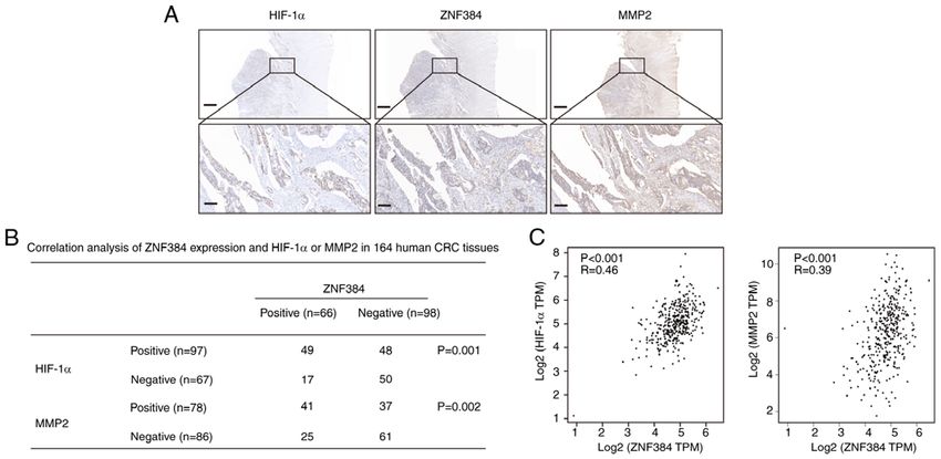
4                       YAN et al: ZINC FINGER PROTEIN 384 ENHANCES COLORECTAL CANCER METASTASIS

in the cell lines were determined using the 2‑ΔΔCq method (10)        Mutagenesis kit (Stratagene; Agilent Technologies, Inc.).
using the following equations: ΔCq=ΔCqtarget‑ΔCqGAPDH and             Vector construction was verified using first DNA sequencing
ΔΔCq=ΔCqexpression vector‑ΔCqcontrol vector. Expression levels were   (Sangon Biotech Co., Ltd.) and HIF‑1α promoter vectors were
normalized to matched control cells, which were set to 1.0.           designed in the same way.
In clinical tissue samples, fold changes in expression levels
of target genes were also determined using the 2‑ΔΔCq method,         Transient transfection and luciferase assay. ZNF384 and
as per the following equations: ΔCq=ΔCqtarget‑ΔCq GAPDH,              HIF‑1α expression plasmids were generated by cloning
and ΔΔCq= ΔCq tumor‑ ΔCq nontumor or ΔΔCq= ΔCq lymph node             ZNF384 or HIF‑1α DNA into pCMV‑tag2A vectors (Agilent
metastatic
           ‑ΔCqnontumor. Expression levels were normalized to         Technologies, Inc.). SW480 or SW620 cells were cultured in
healthy colorectal tissues, which were also set to 1.0. Primer        a 24‑well plate at 1x10⁵ cells/well. After 12‑24 h incubation,
sequences are displayed in Table SII.                                 cells were co‑transfected with expression plasmids [0.6 µg;
                                                                      pCMV‑ZNF384, pCMV‑HIF‑1α or the control (pCMV‑Tag)],
Western blotting. Total proteins were extracted from                  reporter plasmids (0.18 µg) and pRL‑TK plasmids (0.02 µg)
cells using RIPA lysis buffer (Beijing Solarbio Science               (Promega Corporation) using Lipofectamine 3000 reagent. A
& Technology Co., Ltd.) supplemented with proteinase                  total of 5 h post‑transfection, cells were washed and placed
inhibitors. The BCA method was used to assess protein                 in fresh medium containing 1% FBS for 48 h to recover.
concentrations. Proteins (50 µg/lane) were separated by               Cells were subsequently serum‑starved for assaying. A
SDS‑PAGE on 10% gels and were subsequently transferred                Dual‑Luciferase Assay kit (Promega Corporation) was used
to PVDF membranes. After blocking for 1.5 h with 5% skim              to detect luciferase activities according to the manufac‑
milk at room temperature, the membranes were incubated                turer's protocol. Lysed transfected cells were centrifuged
overnight at 4˚C in the presence of primary antibodies                at 72,000 x g for 120 min at 4˚C in Eppendorf microcentrifuge
against ZNF384 (rabbit; 1:1,000; cat. no. ab251673; Abcam),           tubes. A Modulus™ TD20/20 luminometer (Turner Designs)
MMP2 (rabbit; 1:1,000; cat. no. ab235167; Abcam), HIF‑1α              was used to determine relative luciferase activities. Luciferase
(rabbit; 1:2,000; cat. no. ab243860; Abcam) and the control           activity was normalized to Renilla luciferase activity. Three
β‑actin (mouse; 1:500; cat. no. ab8226; Abcam). Subsequently,         biological repeats were performed.
membranes were incubated with anti‑rabbit (HRP‑conjugated;
1:5,000; cat. no. sc‑2357; Santa Cruz Biotechnology, Inc.)            Bioinformatics analysis. UALCAN (11) (http://ualcan.path.
and anti‑mouse (HRP‑conjugated; 1:10,000; cat. no. sc‑2005;           uab.edu/) is an online interactive resource, which also provides
Santa Cruz Biotechnology, Inc.) secondary antibodies for 1 h          easy access to publicly available cancer omics data [The Cancer
at 37˚C. Visualization of protein bands was carried out using         Genome Atlas (TCGA), Metastasis 500, Clinical Proteomic
electrochemiluminescence reagent (cat. no. WBKLS0500;                 Tumor Analysis Consortium (CPTAC) and Children's Brain
MilliporeSigma). Semi‑quantification of protein expression            Tumor Tissue Consortium]. UALCAN was used to deter‑
levels was carried out using ImageJ software (version 1.8.0;          mine the mRNA and protein expression levels of ZNF384 in
National Institutes of Health), with β‑actin used as the loading      primary colon adenocarcinoma cases using data obtained from
control. Experiments were carried out for three biological            TCGA and CPTAC. Gene Expression Profiling Interactive
repeats.                                                              Analysis (GEPIA; http://gepia2.cancer‑pku.cn; version, 2) is an
                                                                      open‑access online tool for the interactive evaluation of RNA
Plasmid construction. Plasmid vectors were established using          sequencing data from 9,736 tumors and 8,587 healthy samples
standard procedures. Primers used in the present study are            in TCGA and Genotype‑Tissue Expression programs (12).
displayed in Table SII. PCR was used to amplify the ZNF384            GEPIA2 was also used to evaluate the association between
promoter sequence (‑1,996/+115) from human genomic DNA                ZNF384 expression levels, and HIF‑1α and MMP2 expression
extracted from SW480 cells using a genomic DNA extrac‑                levels in colon or rectum adenocarcinoma. The University of
tion kit (cat. no. ab156900; Abcam). PCR was performed                California Santa Cruz (UCSC) Genome Browser (13) (http://
using Takara LA Taq polymerase and PCR system (Takara                 genome.ucsc.edu) is a popular web‑based tool for quickly
Biotechnology Co., Ltd.). The thermocycling conditions were           displaying a requested portion of a genome at any scale,
as follows: Initial denaturation at 94˚C for 5 min; followed by       accompanied by a series of aligned annotation ‘tracks’. UCSC
35 cycles at 94˚C for 30 sec, 60˚C for 30 sec and 72˚C for            Genome Browser was used to find the promoter sequence of
40 sec, and final extension at 72˚C for 5 min. This sequence is       MMP2 and ZNF384. JASPAR (14) website (https://jaspar.
localized at the transcriptional start site position (‑1,996/+115)    genereg.net/) was used to analyze the MMP2 and ZNF384
in the 5'‑flanking region of the human ZNF384 gene. Vector            promoter (accessed on 10 October 2019). The relative profile
construction was carried out by integrating both forward and          score threshold was set at 85%. Data were output after calcula‑
reverse primers in the 5'‑ and 3'‑ends of KpnI and HindIII sites,     tion through the JASPAR website.
respectively. Insertion of PCR products between the digested
HindIII and KpnI sites of the pGL3‑Basic vector (Promega              RT‑PCR array. Total RNA was extracted from SW480‑ZNF384
Corporation) was performed. Moreover, the 5'‑flanking region          and SW480‑Control cells using TRIzol according to the
deletion mutants of the ZNF384 promoter [(‑1,996/+115)                manufacturer's protocol and reverse transcribed to cDNA
ZNF384; (‑1,752/+115) ZNF384; and (‑317/+115) ZNF384]                 using PrimeScript RT Reagent kit (Takara Bio, Inc.) according
were established using the (‑1,996/+115) ZNF384 vector as the         to the manufacturer's protocol. Subsequently, RT‑PCR was
template. Mutations in the HIF‑1α‑binding sites in the ZNF384         carried out using the Human Tumor Metastasis RT2 Profiler
promoter were made using the QuikChange II Site‑Directed              PCR array (SuperArray Bioscience) in an ABI PRISM7900
ONCOLOGY REPORTS 47: 49, 2022 5
system (Applied Biosystems; Thermo Fisher Scientific, Inc.), lymph node metastasis and an increased American Joint
according to the manufacturer's instructions. The PCR cycling Committee on Cancer stage (15), and elevated ZNF384 expres‑
conditions were set as follows: 95˚C for 5 min; followed by sion levels were an independent risk factor for CRC progression
40 cycles at 95˚C for 15 sec, 60˚C for 15 sec and 72˚C for (Tables I and II). Patients with high expression of ZNF384
20 sec, and a final extension step at 72˚C for 5 min. The results exhibited markedly poor survival outcomes compared with
were analyzed using the ∆∆Cq method as aforementioned. patients with low expression of ZNF384 (Fig. 1G). In addition,
patients with high expression of HIF‑1α and MMP2 exhibited
Statistical analysis. Continuous data are displayed as the markedly poor survival outcomes compared with patients with
mean ± standard deviation, and TCGA and CPTAC data low expression of HIF‑1α and MMP2 (Fig. S2). These results
are displayed as median and interquartile range. The χ2 test suggested that ZNF384 expression levels were elevated in
was used to analyze categorical data. Comparison of means CRC and may be associated with poor prognostic outcomes.
between and among groups was carried out using Student's
t‑tests and one‑way ANOVA followed by Tukey's or Dunnett's ZNF384 enhances CRC cell invasion and metastasis. The
post hoc tests, respectively. Comparison of means among results of the present study demonstrated that ZNF384
50 matched primary CRC, lymph node metastatic and healthy expression was closely associated with tumor metastasis.
tissue samples was carried out using repeated measures SW480 and SW620 represented cells with low and high
ANOVA followed by Tukey's post hoc tests. Based on vari‑ metastatic ability, respectively, and ZNF384 was differ‑
ables from univariate analyses, determination of independent entially expressed in these two cell lines (Fig. 1H and I).
factors influencing survival was carried out using the Cox Therefore, the present study selected these two cell lines
proportional hazards model. Kaplan‑Meier was used for to study the effect of ZNF384 on the proliferation of
survival analysis following surgery, and the log‑rank test was colorectal cancer cells. Notably, ZNF384 expression exerted
performed for comparisons of survival outcomes. Correlations no significant effect on the proliferation of SW480 and
between ZNF384 and MMP2 or HIF‑1α expression levels in SW620 CRC cells (Fig. S3). Thus, the effect of ZNF384
CRC samples were assessed using Spearman's rank correlation expression on the migratory and invasive capacity of CRC
analysis. SPSS software (version, 19; IBM Corp.) was used for cells was investigated. Both the mRNA and protein expres‑
statistical analyses, and GraphPad Prism (version, 9; GraphPad sion levels of ZNF384 were measured in various CRC cell
Software, Inc.) was used for graph preparation. P6 YAN et al: ZINC FINGER PROTEIN 384 ENHANCES COLORECTAL CANCER METASTASIS Figure 1. High levels of ZNF384 are predictors for poor prognostic outcomes in patients with CRC. (A) Representative data from TCGA datasets displaying the mRNA expression levels of ZNF384 in colon cancer tissues compared with in healthy tissues. Box‑and‑whisker plots represent medians (horizontal lines), interquartile ranges (boxes), and minimum and maximum values (whiskers). (B) mRNA expression levels of ZNF384 in normal, primary CRC and lymph node metastatic tissues. Expression levels of ZNF384 mRNA were determined by RT‑qPCR and normalized to GAPDH. (C) Western blot analyses were conducted using human CRC and adjacent normal tissues. (D) ZNF384 protein expression levels in different tumor types and corresponding healthy tissues from UALCAN. (E) Immunohistochemical staining for the detection of ZNF384 in CRC tissues. Scale bars, 100 µm (upper panel) and 50 µm (lower panel). (F) Results of the Kaplan‑Meier analysis demonstrated the association between ZNF384 mRNA expression levels and overall survival for patients with COAD in TCGA dataset. (G) Results of the Kaplan‑Meier survival curves demonstrated the association between ZNF384 expression and overall survival outcomes in patients with CRC. (H and I) RT‑qPCR and western blot analysis of ZNF384 expression levels in various CRC cell lines. *P
ONCOLOGY REPORTS 47: 49, 2022                                                   7

Table I. Associations between ZNF384 expression and clinicopathological parameters in 164 patients with colorectal cancer.

                                                      ZNF384 expression
	-----------------------------------------------------------------------
Clinicopathological variables Number (n=164)   High (n=66)          Low (n=98)                                            P‑value

Age, years				                0.101
8 YAN et al: ZINC FINGER PROTEIN 384 ENHANCES COLORECTAL CANCER METASTASIS
Table II. Univariate and multivariate analysis of factors associated with survival in patients with colorectal cancer.
Univariate analysis Multivariate analysis
Characteristics HR (95% CI) P‑value HR (95% CI) P‑value
Age, years
(≥60 vs.ONCOLOGY REPORTS 47: 49, 2022 9 Figure 2. ZNF384 enhances CRC cell invasion and metastasis. (A‑C) Reverse transcription‑quantitative PCR and western blotting were carried out to verify ZNF384 overexpression in SW480 and Caco‑2 cells, and ZNF384 knockdown in SW620 and LoVo cells. (A) **P
10                        YAN et al: ZINC FINGER PROTEIN 384 ENHANCES COLORECTAL CANCER METASTASIS

Figure 3. MMP2 is a direct transcriptional target of ZNF384. (A and B) MMP2 mRNA expression levels in CRC cells. (A) **P
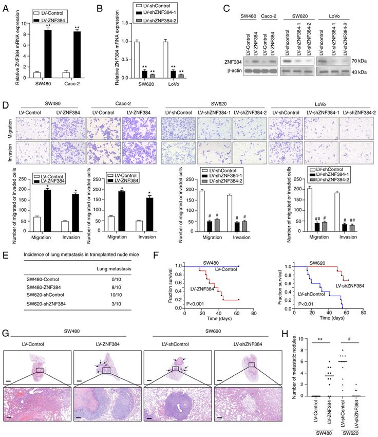
ONCOLOGY REPORTS 47: 49, 2022                                                                    11

Figure 4. MMP2 is crucial for ZNF384‑mediated CRC migration and metastasis. (A‑C) Reverse transcription‑quantitative PCR and western blotting were
carried out to determine MMP2 expression in cells. (A) *P
12                         YAN et al: ZINC FINGER PROTEIN 384 ENHANCES COLORECTAL CANCER METASTASIS

Figure 5. ZNF384 is a direct target gene for HIF‑1α. CRC cells were cultured in a hypoxic atmosphere (0.5% O2) for the indicated time intervals, after
which ZNF384 expression was evaluated by (A) RT‑qPCR and (B) western blot analysis. *P
ONCOLOGY REPORTS 47: 49, 2022                                                               13

Figure 6. ZNF384 expression levels are positively correlated with MMP2 and HIF‑1α expression levels in CRC. (A) Representative immunohistochemical
staining images for ZNF384, HIF‑1α and MMP2 in human CRC tissues. Scale bars, 200 µm (low magnification) and 50 µm (high magnification). (B) Association
between ZNF384 expression levels, and HIF‑1α and MMP2 expression levels in human CRC tissues. (C) Correlation between ZNF384 expression, and HIF‑1α
or MMP2 expression was analyzed by Gene Expression Profiling Interactive Analysis in colon or rectum adenocarcinoma. ZNF384, zinc finger protein 384;
CRC, colorectal cancer; HIF‑1α, hypoxia‑inducible factor 1α.

    MMPs are involved in cancer invasion and metastases (29).                 Compared with the mutant plasmid, the wild‑type plasmid had
Among the MMPs, MMP2, also referred to as gelatinase A,                       almost the same structure except the mutant base, which can
plays critical roles in malignant cell migration, which is attrib‑            also exclude the influence of nucleosomes. Therefore, these
uted to its ability to degrade type IV collagen (30). MMP2                    findings indicated that ZNF384 increased CRC metastasis by
is a potential prognostic biomarker (31). Elevated MMP2                       transactivating MMP2.
expression in cancer cells has been reported to be a significant                  The mechanisms through which ZNF384 and MMP2
predictive factor for poor survival outcomes in CRC (31,32).                  were dysregulated in CRC were also investigated. The asso‑
Kostova et al (33) demonstrated a significant positive corre‑                 ciation between hypoxia and MMPs, such as MMP2, has
lation between MMP2 tissue expression and the presence of                     previously been verified (24). However, the role of hypoxia
nodal metastasis in CRC. Increased expression levels of MMP2                  in ZNF384‑associated MMP2 dysregulation has yet to be
have also been shown to be correlated with poor overall and                   fully elucidated in human CRC. The results of the present
progression‑free survival in patients with CRC (34). In CRC,                  study demonstrated two potential HIF‑1α‑binding sites in
increased levels of MMP2 have been revealed to be associated                  the ZNF384 promoter. Further analyses revealed that HIF‑1α
with microvascular angiogenesis and apoptotic resistance,                     transactivated ZNF384 by binding both HIF‑1α‑binding sites
and increased levels of MMP2 could elevate the levels of                      within the ZNF384 promoter. Moreover, HIF‑1α expression
cell adhesion, and promote invasion and metastasis (35).                      was correlated with ZNF384 expression levels. However, the
The results of intrasplenic injection assays carried out in a                 results of the present study demonstrated that the trend in
previous study demonstrated that, in nude mice, the metastatic                HIF‑1α expression in CRC cells under conditions of normoxia
potential of CRC cell lines was associated with the levels of                 were inconsistent with the expression levels of ZNF384 and
secreted MMP2 (36). These findings highlighted that MMP2                      MMP2. Thus, the ZNF384/MMP2 axis may not be regulated
may be a crucial oncogene in CRC; however, the mechanisms                     by HIF‑1α under conditions of normoxia. Results of previous
through which MMP2 is dysregulated in CRC are yet to be                       studies have demonstrated that hypoxia induces the Warburg
fully elucidated. The results of the present study demonstrated               effect, which is key in the development of cancer and may alter
that MMP2 was a direct functional target of ZNF384, which                     glucose metabolism (22,23). In the present study, the role of
transactivates MMP2 expression by binding its promoter.                       the HIF‑1α/ZNF384/MMP2 axis in CRC progression has yet
Suppression of MMP2 markedly inhibited ZNF384‑mediated                        to be fully elucidated; thus, further investigations are required.
CRC migration, invasion and lung metastasis, whereas MMP2                     The aforementioned results of the present study highlighted
overexpression reversed the ZNF384 knockdown‑induced                          that the hypoxia‑associated protein, HIF‑1α, is a transcrip‑
suppression of CRC cell malignancy. Moreover, ZNF384                          tional regulator of ZNF384, which may enhance both MMP2
expression exhibited a positive correlation with MMP2                         expression and CRC cell metastasis. These results highlight
expression. In the present study, in addition to using serial                 that under hypoxic conditions, human CRC progression may
deletion, site‑directed mutagenesis was used as a control.                    be promoted by dysregulated MMP2.
14 YAN et al: ZINC FINGER PROTEIN 384 ENHANCES COLORECTAL CANCER METASTASIS
In conclusion, ZNF384, a direct HIF‑1α target, was revealed 5. Nakamoto T, Yamagata T, Sakai R, Ogawa S, Honda H, Ueno H,
Hirano N, Yazaki Y and Hirai H: CIZ, a zinc finger protein that
to be markedly elevated in CRC and to be associated with poor interacts with p130(cas) and activates the expression of matrix
prognostic outcomes. Furthermore, ZNF384 overexpression metalloproteinases. Mol Cell Biol 20: 1649‑1658, 2000.
enhanced CRC cell metastasis by transactivating MMP2 6. Zhong CH, Prima V, Liang X, Frye C, McGavran L, Meltesen L,
Wei Q, Boomer T, Varella‑Garcia M, Gump J and Hunger SP:
expression. Therefore, these findings indicated that ZNF384 E2A‑ZNF384 and NOL1‑E2A fusion created by a cryptic t(12;19)
may act as a potential prognostic factor and therapeutic target (p13.3; p13.3) in acute leukemia. Leukemia 22: 723‑729, 2008.
for CRC. 7. Do Amaral A: Complexities of nomenclature in biology.
Gender of generic names ending in ops z, n, (s) 1572. Mem Inst
Butantan 39: 27‑36, 1975 (In Portuguese).
Acknowledgments 8. Lilljebjörn H and Fioretos T: New oncogenic subtypes in pedi‑
atric B‑cell precursor acute lymphoblastic leukemia. Blood 130:
1395‑1401, 2017.
Not applicable. 9. Sakuma T, Nakamoto T, Hemmi H, Kitazawa S, Kitazawa R,
Notomi T, Hayata T, Ezura Y, Amagasa T and Noda M: CIZ/
Funding NMP4 is expressed in B16 melanoma and forms a positive feed‑
back loop with RANKL to promote migration of the melanoma
cells. J Cell Physiol 227: 2807‑2812, 2012.
The present study was supported by the Sichuan Youth Science 10. Livak KJ and Schmittgen TD: Analysis of relative gene expres‑
and Technology Foundation (grant no. 2017JQ0039). sion data using real‑time quantitative PCR and the 2(‑Delta Delta
C(T)) method. Methods 25: 402‑408, 2001.
11. Chandrashekar DS, Bashel B, Balasubramanya SAH,
Availability of data and materials Creighton CJ, Ponce‑Rodriguez I, Chakravarthi BVSK and
Varambally S: UALCAN: A portal for facilitating tumor
subgroup gene expression and survival analyses. Neoplasia 19:
The datasets used and/or analyzed during the present study are 649‑658, 2017.
available from the corresponding author on reasonable request. 12. Tang Z, Kang B, Li C, Chen T and Zhang Z: GEPIA2: An
enhanced web server for large‑scale expression profiling and
interactive analysis. Nucleic Acids Res 47: W556‑W560, 2019.
Authors' contributions 13. Kent WJ, Sugnet CW, Furey TS, Roskin KM, Pringle TH,
Zahler AM and Haussler D: The human genome browser at
ZY, YZ and YY designed the study, analyzed and interpreted UCSC. Genome Res 12: 996‑1006, 2002.
14. Castro‑Mondragon JA, Riudavets‑Puig R, Rauluseviciute I,
the data, and wrote the manuscript. CZ analyzed and inter‑ Berhanu Lemma R, Turchi L, Blanc‑Mathieu R, Lucas J,
preted the data. PL and HT analyzed and interpreted the data, Boddie P, Khan A, Manosalva Pérez N, et al: JASPAR 2022: The
and wrote the manuscript. XT and GZ interpreted the data and 9th release of the open‑access database of transcription factor
binding profiles. Nucleic Acids Res 30: gkab1113, 2021.
confirm the authenticity of all the raw data. All authors read 15. Edge SB and Compton CC: The American Joint Committee on
and approved the final manuscript. Cancer: The 7th edition of the AJCC cancer staging manual and
the future of TNM. Ann Surg Oncol 17: 1471‑1474, 2010.
16. Khemlina G, Ikeda S and Kurzrock R: The biology of
Ethics approval and consent to participate Hepatocellular carcinoma: Implications for genomic and immune
therapies. Mol Cancer 16: 149, 2017.
The present study was approved by the Medical Ethics 17. Fan Z, Tardif G, Hum D, Duval N, Pelletier JP and
Martel‑Pelletier J: Hsp90{beta} and p130(cas): Novel regulatory
Committee of North Sichuan Medical College [approval factors of MMP‑13 expression in human osteoarthritic chondro‑
no. 2021ER(A)007]. All patients provided written informed cytes. Ann Rheum Dis 68: 976‑982, 2009.
consent for participation in the present study. The animal 18. Torrungruang K, Alvarez M, Shah R, Onyia JE, Rhodes SJ and
Bidwell JP: DNA binding and gene activation properties of the
experiments were approved by the Experimental Animal Nmp4 nuclear matrix transcription factors. J Biol Chem 277:
Ethics Committee of North Sichuan Medical College (approval 16153‑16159, 2002.
no. 20200908). 19. Keith B, Johnson RS and Simon MC: HIF1α and HIF2α: Sibling
rivalry in hypoxic tumour growth and progression. Nat Rev
Cancer 12: 9‑22, 2011.
Patient consent for publication 20. Finger EC and Giaccia AJ: Hypoxia, inflammation, and the tumor
microenvironment in metastatic disease. Cancer Metastasis
Rev 29: 285‑293, 2010.
Not applicable. 21. Yoshimura H, Dhar DK, Kohno H, Kubota H, Fujii T, Ueda S,
Kinugasa S, Tachibana M and Nagasue N: Prognostic impact of
Competing interests hypoxia‑inducible factors 1alpha and 2alpha in colorectal cancer
patients: Correlation with tumor angiogenesis and cyclooxy‑
genase‑2 expression. Clin Cancer Res 10: 8554‑8560, 2004.
The authors declare that they have no competing interests. 22. Renga G, Oikonomou V, Moretti S, Stincardini C, Bellet MM,
Pariano M, Bartoli A, Brancorsini S, Mosci P, Finocchi A, et al:
Thymosin β4 promotes autophagy and repair via HIF‑1α stabi‑
References lization in chronic granulomatous disease. Life Sci Alliance 2:
e201900432, 2019.
1. Siegel RL, Miller KD and Jemal A: Cancer statistics, 2018. CA 23. Xu WL, Wang SH, Sun WB, Gao J, Ding XM, Kong J, Xu L
Cancer J Clin 68: 7‑30, 2018. and Ke S: Insufficient radiofrequency ablationinduced autophagy
2. Fakih MG: Metastatic colorectal cancer: Current state and future contributes to the rapid progression of residual hepatocellular
directions. J Clin Oncol 33: 1809‑1824, 2015. carcinoma through the HIF‑1α/BNIP3 signaling pathway. BMB
3. Punt CJ, Koopman M and Vermeulen L: From tumour heteroge‑ Rep 52: 277‑282, 2019.
neity to advances in precision treatment of colorectal cancer. Nat 24. Wang J, Ni Z, Duan Z, Wang G and Li F: Altered expression of
Rev Clin Oncol 14: 235‑246, 2017. hypoxia‑inducible factor‑1α (HIF‑1α) and its regulatory genes in
4. Sato J, Nakamura M, Watanabe O, Yamamura T, Funasaka K, gastric cancer tissues. PLoS One 9: e99835, 2014.
Ohno E, Miyahara R, Kawashima H, Goto H and Hirooka Y: 25. Osinsky SP, Ganusevich II, Bubnovskaya LN, Valkovskaya NV,
Prospective study of factors important to achieve observation Kovelskaya AV, Sergienko TK and Zimina SV: Hypoxia level and
of the entire colon on colon capsule endoscopy. Therap Adv matrix metalloproteinases‑2 and ‑9 activity in Lewis lung carci‑
Gastroenterol 10: 20‑31, 2017. noma: Correlation with metastasis. Exp Oncol 27: 202‑205, 2005.ONCOLOGY REPORTS 47: 49, 2022 15
26. Ryu J, Vicencio AG, Yeager ME, Kashgarian M, Haddad GG and 32. Leeman MF, Curran S and Murray GI: New insights into the
Eickelberg O: Differential expression of matrix metalloprotein‑ roles of matrix metalloproteinases in colorectal cancer develop‑
ases and their inhibitors in human and mouse lung development. ment and progression. J Pathol 201: 528‑534, 2003.
Thromb Haemost 94: 175‑183, 2005. 33. Kostova E, Slaninka‑Miceska M, Labacevski N, Jakovski K,
27. Mori S, Takeuchi T, Ishii Y and Kukimoto I: Identification of Trojachanec J, Atanasovska E, Janevski V, Jovanovik R and
APOBEC3B promoter elements responsible for activation Janevska V: Expression of matrix metalloproteinases 2, 7 and 9 in
by human papillomavirus type 16 E6. Biochem Biophys Res patients with colorectal cancer. Vojnosanit Pregl 71: 52‑59, 2014.
Commun 460: 555‑560, 2015. 34. Shi M, Yu B, Gao H, Mu J and Ji C: Matrix metalloproteinase
28. He L, Fan X, Li Y, Chen M, Cui B, Chen G, Dai Y, Zhou D, Hu X 2 overexpression and prognosis in colorectal cancer: A meta‑
and Lin H: Overexpression of zinc finger protein 384 (ZNF 384), analysis. Mol Biol Rep 40: 617‑623, 2013.
a poor prognostic predictor, promotes cell growth by upregu‑ 35. Egeblad M and Werb Z: New functions for the matrix metallopro‑
lating the expression of Cyclin D1 in Hepatocellular carcinoma. teinases in cancer progression. Nat Rev Cancer 2: 161‑174, 2002.
Cell Death Dis 10: 444, 2019. 36. Shah V, Kumar S and Zirvi KA: Metastasis of human colon tumor
29. Shay G, Lynch CC and Fingleton B: Moving targets: Emerging cells in vivo: Correlation with the overexpression of plasminogen
roles for MMPs in cancer progression and metastasis. Matrix activators and 72 kDa gelatinase. In Vivo 8: 321‑326, 1994.
Biol 44‑46: 200‑206, 2015.
30. Nagase H and Woessner JF Jr: Matrix metalloproteinases. J Biol This work is licensed under a Creative Commons
Chem 274: 21491‑21494, 1999.
Attribution-NonCommercial-NoDerivatives 4.0
31. Zucker S and Vacirca J: Role of matrix metalloproteinases
(MMPs) in colorectal cancer. Cancer Metastasis Rev 23: 101‑117, International (CC BY-NC-ND 4.0) License.
2004.You can also read