Immune profiling and immunotherapeutic targets in pancreatic cancer
←
→
Page content transcription
If your browser does not render page correctly, please read the page content below
Original Article
                                                                                                                                           Page 1 of 15

Immune profiling and immunotherapeutic targets in pancreatic
cancer
Felicia L. Lenzo1, Shumei Kato2, Sarabjot Pabla1, Paul DePietro1, Mary K. Nesline1, Jeffrey M. Conroy1,3,
Blake Burgher1, Sean T. Glenn1,3,4, Boris Kuvshinoff5, Razelle Kurzrock2, Carl Morrison1,3,6,7
1
OmniSeq, Inc., Buffalo, NY, USA; 2Center for Personalized Cancer Therapy, Moores Cancer Center, La Jolla, CA, USA; 3Center for Personalized
Medicine, Roswell Park Comprehensive Cancer Center, Buffalo, NY, USA; 4Department of Molecular and Cellular Biology, Roswell Park
Comprehensive Cancer Center, Buffalo, NY, USA; 5Department of Surgery, Roswell Park Comprehensive Cancer Center, Buffalo, NY, USA;
6
    Department of Pathology, Roswell Park Comprehensive Cancer Center, Buffalo, NY, USA; 7Cancer Genetics and Genomics, Roswell Park
Comprehensive Cancer Center, Buffalo, NY, USA
Contributions: (I) Conception and design: All authors; (II) Administrative support: All authors; (III) Provision of study materials or patients: All
authors; (IV) Collection and assembly of data: All authors; (V) Data analysis and interpretation: All authors; (VI) Manuscript writing: All authors; (VII)
Final approval of manuscript: All authors.
Correspondence to: Carl Morrison. Roswell Park Comprehensive Cancer Center, Elm & Carlton Streets, Buffalo, NY 14263, USA.
Email: carl.morrison@omniseq.com.

                 Background: Immunotherapeutic approaches for pancreatic ductal adenocarcinoma (PDAC) are less
                 successful as compared to many other tumor types. In this study, comprehensive immune profiling was
                 performed in order to identify novel, potentially actionable targets for immunotherapy.
                 Methods: Formalin-fixed paraffin embedded (FFPE) specimens from 68 patients were evaluated for
                 expression of 395 immune-related markers (RNA-seq), mutational burden by complete exon sequencing of
                 409 genes, PD-L1 expression by immunohistochemistry (IHC), pattern of tumor infiltrating lymphocytes
                 (TILs) infiltration by CD8 IHC, and PD-L1/L2 copy number by fluorescent in situ hybridization (FISH).
                 Results: The seven classes of actionable genes capturing myeloid immunosuppression, metabolic
                 immunosuppression, alternative checkpoint blockade, CTLA-4 immune checkpoint, immune infiltrate,
                 and programmed cell death 1 (PD-1) axis immune checkpoint, discerned 5 unique clinically relevant
                 immunosuppression expression profiles (from most to least common): (I) combined myeloid and metabolic
                 immunosuppression [affecting 25 of 68 patients (36.8%)], (II) multiple immunosuppressive mechanisms
                 (29.4%), (III) PD-L1 positive (20.6%), (IV) highly inflamed PD-L1 negative (10.3%); and (V) immune
                 desert (2.9%). The Wilcoxon rank-sum test was used to compare the PDAC cohort with a comparison
                 cohort (n=1,416 patients) for the mean expressions of the 409 genes evaluated. Multiple genes including
                 TIM3, VISTA, CCL2, CCR2, TGFB1, CD73, and CD39 had significantly higher mean expression versus
                 the comparison cohort, while three genes (LAG3, GITR, CD38) had significantly lower mean expression.
                 Conclusions: This study demonstrates that a clinically relevant unique profile of immune markers can
                 be identified in PDAC and be used as a roadmap for personalized immunotherapeutic decision-making
                 strategies.

                 Keywords: Precision immunotherapy; PD-L1; immune suppression; immune activation; checkpoint inhibitors

                 Submitted Jan 28, 2020. Accepted for publication Sep 29, 2020.
                 doi: 10.21037/atm-20-1076
                 View this article at: http://dx.doi.org/10.21037/atm-20-1076

© Annals of Translational Medicine. All rights reserved.                      Ann Transl Med 2021;9(2):119 | http://dx.doi.org/10.21037/atm-20-1076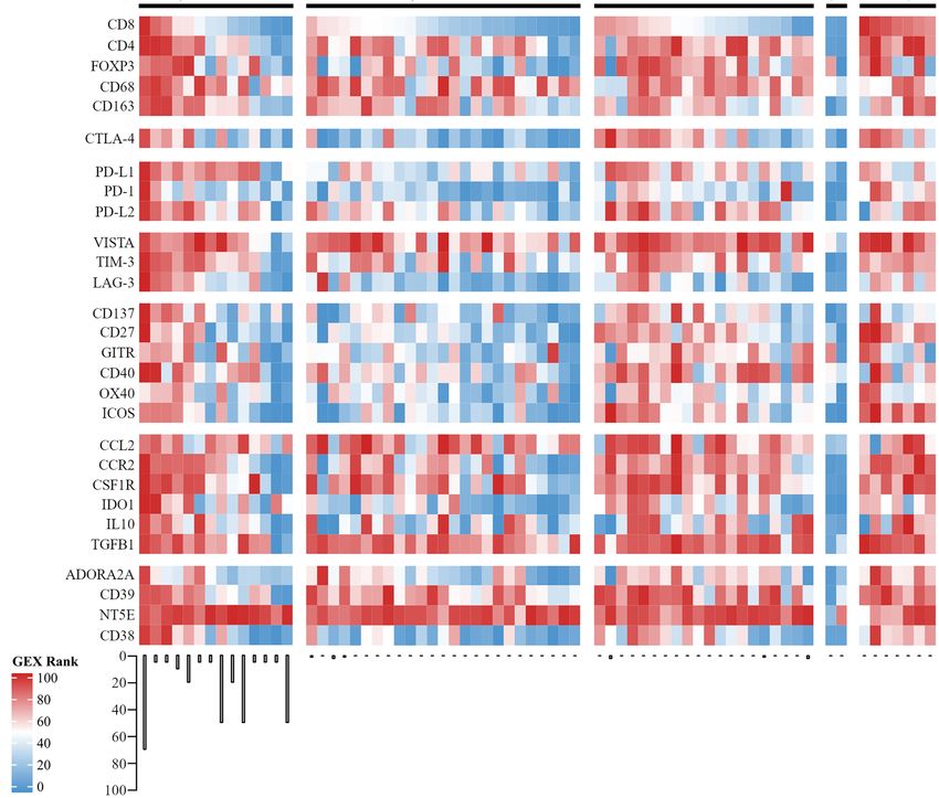
Page 2 of 15                                                                 Lenzo et al. Immune profiling in pancreatic cancer

Introduction                                                   CCR2 is unknown. This constitutes an obvious obstacle to
                                                               the design of novel therapeutic regimens aimed at reversing
Data from recent clinical trials suggest that immune
                                                               immunosuppression beyond checkpoint blockade for the
checkpoint inhibitors targeting cytotoxic T-lymphocyte
                                                               treatment of PDAC.
associated protein 4 (CTLA4), programmed cell death 1
                                                                  Here, we demonstrate that comprehensive immune
(PDCD1; best known as PD-1), or CD274 (best known
                                                               profiling, simultaneously assessing PD-L1 activation
as PD-L1) are poorly effective as stand-alone therapeutic
                                                               status by immunohistochemistry (IHC) and fluorescence
interventions in individuals with pancreatic cancer
                                                               in situ hybridization (FISH), CTL infiltration by IHC,
(1-3). The lack of efficacy of these immunotherapeutics
                                                               tumor mutational burden (TMB), microsatellite instability
may reflect the fact that pancreatic oncogenesis is
                                                               (MSI), and the expression levels of immunological
generally accompanied by the establishment of a
                                                               relevant transcripts can provide an initial overview of the
strongly immunosuppressive tumor microenvironment,
                                                               immunological landscape in patients with PDAC with a
as well as by a robust stromal reaction (the so-called
                                                               particular focus on actionable pathways (18). The overall
“desmoplastic stroma”) that is expected to counteract
                                                               objective of this study is to generate awareness of which
tumor infiltration by immune effector cells (4-6). Thus,
                                                               immunosuppressive pathways are likely to be activated
pancreatic ductal adenocarcinoma (PDAC) are generally
                                                               in PDAC and enable the rational design of clinical trials
abundant in CD4+FOXP3+ regulatory T-cells (TREG), M2-
                                                               targeting such pathways.
like tumor-associated macrophages (TAMs), myeloid-
                                                                  We present the following article in accordance with
derived suppressor cells (MDSCs), and FAP + cancer-
                                                               the MDAR reporting checklist (available at http://dx.doi.
associated fibroblasts, which together generate a local
                                                               org/10.21037/atm-20-1076).
microenvironment that inhibits innate and adaptive
immunity (7). Moreover, several proteins involved in
metabolic immunosuppression, such as ectonucleoside            Methods
triphosphate diphosphohydrolase 1 (ENTPD1; best
                                                               Patients
known as CD39), an extracellular nucleotidase that
precipitates the conversion of immunostimulatory ATP           Formalin-fixed paraffin embedded (FFPE) blocks or
into immunosuppressive adenosine, as well as adenosine         slides from sixty-eight (68 PDAC patients (34 primary, 34
A2a receptor (ADORA2A), which transduces such                  metastatic) were evaluated to identify actionable targets
immunosuppressive effects, are overexpressed in the PDAC       for immunomodulatory immunotherapeutic drugs using
microenvironment (8-11).                                       comprehensive immune profiling via Immune Report
   A potential approach to treat patients with PDAC is         Card® (IRC). IRC is a commercially available test approved
to combine checkpoint inhibitors, chemotherapy, and/           for clinical use by the New York State Clinical Laboratory
or radiation therapy with small molecules designed to          Evaluation Program (NYS CLEP). Testing was performed
reverse immunosuppression beyond checkpoint blockade,          in a CLIA certified laboratory at OmniSeq, Inc. (Buffalo,
potentially enabling CD8+ cytotoxic T lymphocyte (CTL)-        NY). An additional 1,416 non-PDAC tumor samples that
dependent tumor eradication (12,13). Promising work in         also underwent IRC testing were used as the comparison
this area comes from a single institution Phase 2 clinical     cohort to determine which genes were differentially
trial combining a C-C motif chemokine receptor 2 (CCR2)        expressed within PDAC. All tumor samples were
inhibitor with the FOLFIRINOX (folinic acid plus               collected in the context of patient clinical care. Associated
5-fluorouracil plus irinotecan plus oxaliplatin) regimen in    deidentified molecular data was considered non-human
patients with borderline resectable and locally advanced       subjects research under IRB approved BDR #073166 at
PDAC, resulting in local tumor control in 97% of cases         Roswell Park Comprehensive Cancer Center (Buffalo, NY)
(n=32) (14,15). This reflects the ability of C-C motif         and in accordance with the Helsinki declaration (as revised
chemokine ligand 2 (CCL2) produced by PDAC cells               in 2013).
to attract immunosuppressive CCR2 + TAMs, ultimately
resulting in CTL exclusion and the establishment of a
                                                               TMB
“cold tumor” microenvironment (16,17). However, the
true incidence of patients with PDAC that overexpress          DNA was extracted from each sample and evaluated for
immunosuppressive cytokines including (but not limited to)     TMB by whole-exon DNAseq as previously described

© Annals of Translational Medicine. All rights reserved.      Ann Transl Med 2021;9(2):119 | http://dx.doi.org/10.21037/atm-20-1076
Annals of Translational Medicine, Vol 9, No 2 January 2021 Page 3 of 15
with minor modifications (19,20). TMB qualified, non- detection of product size by capillary electrophoresis
synonymous somatic variants are reported as mutations per (GeneMapper software version 5.0) using fluorescently-
megabase (Mut/Mb). A high TMB cutoff was defined as the labeled primers for amplification of five markers including
top tenth percentile of values (7 mutations per megabase two mononucleotide repeat markers (BAT-25, BAT-26)
of DNA or higher) observed in this study specific to and three dinucleotide repeat markers (D2S123, D5S346,
PDAC (21). Cases with less than 20% neoplastic nuclei (n=4) and D17S250). MSI at ≥2 loci was defined as MSI-H
were excluded from the analysis. (high), instability at a single locus was defined as MSI-L
(low), and no instability at any of the loci tested was
defined as microsatellite stable (MSS). For the remaining
Immunohistochemical studies
cases, MSI analysis was performed using next generation
The expression of PD-L1 on the surface of tumor cells sequencing of a panel targeting 29 homopolymer regions.
was assessed in all samples via the Dako Omnis platform Briefly, TruSeq Custom Amplicon libraries (Illumina) were
(Agilent, Santa Clara, CA) using the anti-PD-L1 22C3 generated to carry out multiplexed targeted sequencing of
pharmDx antibody (Agilent, Santa Clara, CA), as per the 29 homopolymer loci, clustered and then sequenced
manufacturers protocol (22). Expression levels were on a MiSeq system (Illumina). A pipeline was created to
scored as per published guidelines (23). Additional serially read “.fastq” files and conduct sequence alignment, variant
sectioned tissue was evaluated for lymphocyte infiltration calling, and indel extraction to determine MSI or MSS
using the anti-CD8 antibody C8/144B (Agilent, Santa status.
Clara, CA) at a dilution ratio of 1:75 and assigned a
qualitative score of non-infiltrated, infiltrated, or excluded.
Immune expression analysis
Non-infiltrated referred to a sparse number of CD8+ T-cells
that infiltrate nests of neoplastic cells in a non-overlapping Multiplexed gene-specific primer pairs and next generation
fashion and with less than 5% of the tumor showing sequencing were used to assess expression of 394 immune-
an infiltrating pattern. Infiltrated represents frequent related genes (https://cdn.amegroups.cn/static/public/
CD8+ T-cells that infiltrate nests of neoplastic cells in an ATM-20-1076-Supplementary.pdf) from RNA isolated
overlapping fashion at least focally and in more than 5% from FFPE slides (19,20). Transcript abundance was
of the tumor. Excluded represents restriction of more than normalized and compared to an internal reference
95% of all CD8+ T-cells in a tumor to the periphery or population encompassing 338 patients from twenty-nine
interstitial stromal areas and not actively invading nest or solid tumor types. The reference population represented
groups of neoplastic cells. a broad dynamic range of expression values which were
used to rank gene expression in test samples, as previously
described (19). Rank values were set on a scale of 1 to 100
PD-L1/2 copy number
and stratified into “High” [85–100], “Moderately high”
Fluorescence in situ hybridization (FISH) was performed [75–84], “Moderate” [50–74], “Moderately low” [25–49],
as previously described (24). Briefly, locus specific bacterial and “Low” [0–24].
artificial chromosomes (BAC) for PD-L1/2 at 9p24.1 were
labeled with SpectrumOrange and a commercially available
Statistical analysis
SpectrumGreen chromosome 9 centromeric probe (Abbott
Molecular/Vysis, Des Plaines, Illinois) were hybridized The expression levels of 23 genes that code for targets
to test samples and not less than 60 neoplastic cells were of immunomodulatory immunotherapeutic agents
evaluated for copy number gain as defined by CAP ASCO currently in clinical development were used for precision
(College of American Pathologists; American Society of immunotherapy evaluation (Table 1). Prevalence of gene
Clinical Oncology) HER2 guidelines (25). expression rank for each actionable target was calculated as
the mean percentage of PDAC cases that ranked moderately
high, high, or very high (expression rank ≥75) as compared
Microsatellite instability (MSI)
to the ranks for all tumor types in the reference population.
MSI analysis, for a subset of cases, was performed using Wilcoxon Rank-Sum tests were performed to compare the
polymerase chain reaction (PCR) amplification with mean expression ranks of unique genes in the pancreatic
© Annals of Translational Medicine. All rights reserved. Ann Transl Med 2021;9(2):119 | http://dx.doi.org/10.21037/atm-20-1076Page 4 of 15 Lenzo et al. Immune profiling in pancreatic cancer
Table 1 Differential expression of immunotherapeutic targets
Mean expression Comparison cohort mean
Target Prevalence* (%), n=68 P value
rank PDAC [95% CI], n=68 expression rank [95% CI], n=338
PD-1 axis checkpoint
PD-L1 18 52 [46.44–58.0] 51 [49.75–52.92] 0.975904
PD-1 6 34 [28.44–40.24] 36 [34.18–37.04] 0.9845348
CTLA-4 immune checkpoint activation
CTLA4 18 40 [32.58–47.21] 40 [38.71–41.93] 0.989908
Alternative immune checkpoint
LAG3 9 36 [29.79–42.92] 49 [47.23–50.27] 0.0028822↓
TIM3 29 58 [51.64–64.03] 46 [44.26–47.38] 0.003997663↑
VISTA 63 76 [70.49–80.78] 56 [54.29–57.22] 6.97566E-08↑
Immune checkpoint activation
CD137 16 43 [36.08–49.21] 46 [44.06–47.09] 0.595649
CD27 21 46 [39.2–52.27] 48 [46.13–49.25] 0.6473552
CD40 31 56 [49.6–62.9] 49 [47.25–50.22] 0.0745479
GITR 15 46 [39.14–52.09] 55 [53.02–56.25] 0.028381153↓
OX40 7 43 [36.84–48.45] 48 [46.31–49.22] 0.3213262
ICOS 22 45 [37.28–51.95] 47 [45.64–49.05] 0.5369071
Myeloid immunosuppression
CCR2 41 58 [50.43–65.01] 48 [46.64–49.83] 0.0315833↑
CCL2 47 68 [62.1–74.02] 52 [50.26–53.3] 4.16123E-05↑
IDO1 13 43 [35.77–49.58] 51 [49.64–53.02] 0.0479018
TGFB1 71 77 [71.54–81.58] 63 [61.18–64.03] 0.000172118↑
TNF 40 58 [51.55–65.28] 59 [57.7–60.75] 0.8878454
CSF1R 50 66 [58.92–72.64] 58 [56.37–59.55] 0.0898868
Metabolic immunosuppression
CD38 10 38 [30.99–45.27] 48 [46.71–49.95] 0.018921692↓
ADORA2A 13 47 [40.56–52.85] 47 [45.81–48.85] 0.975904
CD73 90 86 [83.34–89.6] 69 [67.5–70.29] 1.0509E-06↑
CD39 47 67 [61.25–72.9] 53 [51.49–54.33] 0.000129827↑
Immune infiltrate
CD8 15 42 [35.1–48.22] 44 [42.87–45.82] 0.585528
*, prevalence was the percent of patients with gene mean expression rank ≥75 compared to the total cohort (n=68); ↑/↓, statistically
significant expression ranks in pancreatic patients (n=68) that were higher (red arrow) or lower (blue arrow) compared to the expression
rank of the comparison cohort (n=1,416). PDAC, pancreatic ductal adenocarcinoma.
© Annals of Translational Medicine. All rights reserved. Ann Transl Med 2021;9(2):119 | http://dx.doi.org/10.21037/atm-20-1076Annals of Translational Medicine, Vol 9, No 2 January 2021 Page 5 of 15 cohort (n=68) to the remaining tested patients (n=1,416) Supplementary2.pdf), while a minority were considered across all disease sites, statistical significance if P
Page 6 of 15 Lenzo et al. Immune profiling in pancreatic cancer
H&E PD-L1 22c3 CD8
A B C
D E F
G H I
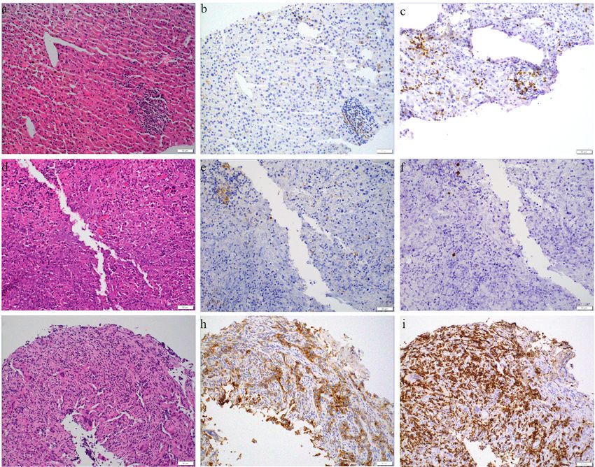
Figure 1 Tumor-infiltrating CD8+ CTLs in PDAC. Immunohistochemistry for CD8 and H/E-stained sections show a highly inflamed and
a non-inflamed case. Scale bar = 100 µm. PDAC, pancreatic ductal adenocarcinoma.
in the net signal of suppression versus T-cell activation, to effector T-cells. Unlike CTLA-4, the expression of PD-1
respectively. The majority (61.8%; 42/68) of the PDAC and ligands is not limited to T-cells and APC, but are also
samples evaluated for CTLA-4 and CD28 expression expressed on B-cells, myeloid cells, and neoplastic cells.
by RNA-seq showed a higher level of the latter receptor The majority (69.1%; 47/68) of PDAC samples evaluated
indicating an overall T-cell activation process through for PD-L1 expression by IHC had no staining in neoplastic
this pathway. Additionally, CTLA-4 was at maximal levels cells, with the remaining samples having staining in 1%
of moderately high in only 12 of 68 (17.6%) of PDAC or more of neoplastic cells. Among PD-L1 IHC positive
samples, for which all but three showed very similar levels cases, four showed a strong PD-L1 IHC result, i.e., greater
of CD28. The overall results support that CTLA-4 immune than 50% staining in neoplastic cells. These IHC results
checkpoint is an immunosuppressive mechanism in PDAC, were confirmed by RNA-seq analysis for PD-L1 showing
but in a minority of cases. a less than high level of expression for the majority (44/47;
93.6%) of IHC negative cases and a high expression for
the majority (9/14; 64.3%) of cases with 5% or more of
PD-1 Axis immune checkpoint
neoplastic cells staining. The discordant results between
PD-1, and its ligands PD-L1 and PD-L2 members of the IHC and RNA-seq for these other five cases is not readily
B7/CD28 family of receptors deliver a co-inhibitory signal explained other than perhaps a lack of sensitivity of the
© Annals of Translational Medicine. All rights reserved. Ann Transl Med 2021;9(2):119 | http://dx.doi.org/10.21037/atm-20-1076Annals of Translational Medicine, Vol 9, No 2 January 2021 Page 7 of 15
22C3 clone for PDAC (27). These results suggest that differentiation or function of CD4+CD25+FOXP3+ TREG
PD-1 axis checkpoint blockade is a factor in a subset of cells and subsequent myeloid suppression (32-35).
PDAC, and while staining in 1% or more of neoplastic cells In this series of PDAC the prevalence of over-expression
can be considered a positive result there are undoubtedly of the immune checkpoint activation receptors was
an estimated 5% or so of cases with very high PD-L1 somewhat common (CD137 16.2%, CD27 20.6%, CD40
expression. 30.9%, ICOS 22.1%; Table 1) and at least one of these
genes was highly or moderately highly expressed in more
than half of all cases (36/68; 52.9%). While over-expression
Alternative immune checkpoint
of at least one co-stimulatory T-cell signal was quite
In addition to PD-1 and CTLA-4 immune checkpoint common this finding was not remarkably different from the
receptors, there are additional inhibitory receptors (LAG3, comparison cohort (Table 1), with the exception that GITR
TIM3, VISTA) preferentially expressed on either T- cells had significantly lower expression in PDAC (P=0.0283).
(LAG3, TIM3) or myeloid cells (VISTA). Animal models As will be shown later in categories of immunosuppression
have shown enhanced antitumor immune responses after in PDAC, the over expression of co-stimulatory T-cell
blockade of these additional inhibitory receptors when signals is common in some groups, but uncommon in
combined with blockade of PD-1 or CTLA-4 and more a unique group with combined metabolic and myeloid
recently human clinical trial reports for LAG3 inhibition immunosuppression. To summarize, immune checkpoint
in melanoma have shown encouraging results (28-30). The activation by specific co-stimulatory receptors, including
prevalence of moderately high or high expression for either CD27, CD137, OX40, CD40, and ICOS, but not GITR, is
TIM3 or VISTA was 29.4% (20/68) and 63.2% (43/68), a frequent event in certain subgroups of PDAC.
respectively, while LAG3 was only highly expressed in six
cases (6/68; 8.8%) (Table 1). The lack of expression of LAG3
Myeloid immunosuppression
and its nearly exclusive expression on T-cells is consistent
with the prior similar results for CTLA-4. This compares to While there are many types of myeloid cells in the tumor
the expression pattern of VISTA on myeloid cells, as well as microenvironment, most notable are TAMs, which
pancreatic ducts, implying the significant role that myeloid consist of inflammatory (M1) and immunosuppressive
suppression has in PDAC (31). The overall results support (M2) phenotypes. These two types of TAMs have distinct
alternative immune checkpoint as having a role in PDAC, patterns of cytokine expression with M1 TAMs noted for
but primarily via TIM3 or VISTA and not LAG3. secretion of interferon gamma (IFNγ), IL-6, and chemokine
(C-X-C motif) ligand 10 (CXCL10). While M2 TAMs
secrete transforming growth factor beta 1 (TGFB1; best
Immune checkpoint activation
known as TGF-β1), interleukin 10 (IL10), chemokine
CTL-dependent tumor eradication requires the TCR- (CC motif) ligand 22 (CCL22), and promote indoleamine
dependent recognition of a tumor-associated antigen 2,3-dioxygenase 1 (IDO1) production (36). Additionally,
(TAA) or neoantigen presented by dendritic cells (DCs) colony stimulating factor 1 receptor (CSF1R), CCR2,
or other antigen-presenting cells (APCs) in the context of and CCL2 produced by M2 macrophages and neoplastic
MHC Class I molecules, along with activation signals via cells, promote the recruitment of additional monocytes
one or multiple co-stimulatory ligand and receptor pairs to the tumor microenvironment, their proliferation,
expressed by CTLs and associated APCs, respectively, and differentiation towards the M2 immunosuppressive
including CD137 ligand (CD137L) and CD137, TNFSF7 phenotype, in a self-propagating process (37).
(CD27 Ligand/CD27L; also known as CD70) and CD27, In this series of PDAC, the mean rank expression value
TNFSF18 (GITR Ligand/GITRL) and GITR, CD40 of 61.4 for M2 markers (TGBF1, IL10, CCL22, IDO1,
ligand (CD40L) and CD40, OX40 ligand (OX40L) and CSF1R, CCR2, CCL2) was nearly double the value of 38.3
OX40, and inducible costimulator-ligand (ICOSL) and for M1 markers (IFN gamma, IL-6, CXCL10) supporting
ICOS. Activation of specific co-stimulatory receptors, the significant role that myeloid suppression has in PDAC.
including CD27, CD137, OX40 and GITR have been Among this list of myeloid immunosuppressive markers,
shown to promote the differentiation of CD4 + TH1 or several are the specific target of immunomodulatory
TH9 T cells with anticancer activity while suppressing the immunotherapeutics including IL-10, TGFB1, CSF1R,
© Annals of Translational Medicine. All rights reserved. Ann Transl Med 2021;9(2):119 | http://dx.doi.org/10.21037/atm-20-1076Page 8 of 15 Lenzo et al. Immune profiling in pancreatic cancer
and CCR2/CCL2. Among this list, CRR2 and its ligand this list of metabolic immunosuppressive markers
CCL2, CSF1R, and TGFB1 were moderately-high to ADORA2A and CD38 are targets of immunomodulatory
highly expressed in more than one-half of all cases, with the immunotherapeutics. However, ADORA2A and CD38
latter (TGFB1) being the most frequently over expressed in were infrequently over-expressed (9/68; 13.2% and 7/68;
comparison to the reference population. Using a Wilcoxon 10.3%, respectively) relegating much of the potential
rank-sum test to compare the PDAC patient cohort with clinical efficacy of targeting this immunosuppressive
the reference population for the expression rank of all pathway to the upstream ectonucleotidases CD39 and
genes evaluated, multiple M2 markers were expressed CD73. The uniqueness of these two therapeutic targets is
significantly higher in PDAC samples as compared to the shown by the significant difference of CD39 (P=0.0001)
remaining tumor types tested including CCR2 (P=0.0316), and CD73 (P=1.0509E-06) expression as compared to the
CCL2 (P=4.16123E-05), and TGFB1 (P=0.0001) (Table 1). reference population (Table 1) supports that the canonical
As a group at least one gene from this list of M2 markers adenosinergic pathway is more commonly used in PDAC.
was moderately-high to highly expressed for all cases.
Expression values for macrophage content (CD68, CD163)
TMB, microsatellite instability, and PD-L1/2 copy number
paralleled this over expression of myeloid suppressive
cytokines and chemokines being higher than CD8 values TMB was available for 64 of 68 cases with four cases
for all but nine of sixty-four cases. Overall, myeloid having a low neoplastic cell content (Annals of Translational Medicine, Vol 9, No 2 January 2021 Page 9 of 15
ve
si
s res
io lic
sm p
ni su
ss o
tiv d
re tab
n
ha no
ga me
e
ec u
up e
rt
e
ne la
m imm
os & m
se
tiv
1 inf
de
si
-L y
po
le
un d
l
PD igh
e
m loi
tip
un
1
Im ye
H
ul
-L
m
M
M
PD
Im
CD8
CD4
Immune infiltrate
FOXP3
CD68
CD163
CTLA-4 CTLA-4 immune checkpoint
PD-L1
PD-1 PD-1 axis immune checkpoint
PD-L2
VISTA
TIM-3 Alternative Immune Checkpoint
LAG-3
CD137
CD27
GITR
Immune Checkpoint Activation
CD40
OX40
ICOS
CCL2
CCR2
CSF1R Myeloid Immunosuppression
IDO1
IL10
TGFB1
ADORA2A
CD39
Metabolic Immunosuppression
NT5E
CD38
GEX rank 0
100 20
80
40
60
40 60
20 80
0 100
PD-L1 TPS
Figure 2 Clinically relevant immune gene expression profiles in PDAC. Expression rank of 23 genes in 68 PDAC patients as compared
to a comparison cohort expression database of multiple tumor types (n=1,416). Seven different classes of actionable genes including
immune infiltrate, CTLA-4 immune checkpoint, PD-1 axis immune checkpoint, alternative immune checkpoint, immune checkpoint
activation, myeloid immunosuppression, and metabolic immunosuppression were used to classify cases into a clinically relevant framework.
The clinically relevant immunosuppression expression profiles were combined myeloid metabolic immunosuppression and multiple
immunotherapy mechanisms representing slightly more than two-thirds of all cases, followed by PD-1 axis driven, highly inflamed and
immune desert. PDAC, pancreatic ductal adenocarcinoma.
© Annals of Translational Medicine. All rights reserved. Ann Transl Med 2021;9(2):119 | http://dx.doi.org/10.21037/atm-20-1076Page 10 of 15 Lenzo et al. Immune profiling in pancreatic cancer
absent from this group was over expression of the co- immunotherapeutic target. One sample showed moderately
stimulatory T-cell signals CD27, CD137, OX40, CD40, high or greater expression of CD73 and GITR (Figure 2).
ICOS, and GITR.
Conclusions
Multiple immunosuppressive mechanisms
The second most common group (20/68; 29.4%), cases Immune checkpoint inhibitors targeting CTLA4, PD-
with multiple mechanisms of immunosuppression typically 1, and PD-L1 employed as standalone interventions have
showed a modest number of CD8+ T-cells, a relatively much currently provided limited clinical benefit to subjects with
higher number of CD4+FOXP3+ T-cells, high macrophage PDAC (1,2,42). Three phase 1 or 2 trials reported the
content, alternative checkpoint blockade, co-stimulatory investigational use of either ipilimumab, pembrolizumab,
T-cell signaling, a dominant myeloid suppression coupled or the investigational PD-L1 inhibitor BMS-936559 with
with metabolic adenosinergic immunosuppression, and total enrollment of less than 60 PDAC patients (1,2,42).
less than significant PD-1 axis checkpoint blockade. These No responders were identified after treatment with
cases are best described as having multiple mechanisms of ipilimumab or the investigational PD-L1 inhibitor BMS-
immunosuppression (Figure 2) and while this supports more 936559, although three partial responders from seventeen
than one immunotherapy approach, the optimal sequential patients were identified in the pembrolizumab study and
order of combination immunotherapies for this group is for which PD-L1 expression was not reported. This should
somewhat difficult to interpret. not be interpreted to mean that single agent PD-1 axis
intervention has no role in future PDAC immunotherapy
PD-L1 positive but, rather, that identifying specific patients with a PD-L1-
PD-L1 positive, representing a minority of cases (14/68; driven phenotype may be necessary to derive benefit. In our
20.6%) showed 5% or greater expression of PD-L1 by case series, one-fifth of PDAC patients showed evidence
IHC in neoplastic cells. The number of CD8 and CD4 of PD-1 axis-driven immunosuppression by 5% or greater
T-cells for each case was highly variable in this group, with expression of PD-L1 by IHC in neoplastic cells that could
low to moderately low numbers of both of these important be favorable for response to targeting PD-L1 or PD-1. The
subsets of TILs in a substantial number of cases (4/14) RNA-seq analysis for these cases showed that greater than
representing what could be referred to as non-inflamed, one-fourth of these PD-L1 positive PDAC were either non-
or cold, PD-L1 positive PDAC. As expected, expression of inflamed or at most weakly inflamed. At the current time
other immunotherapeutic targets in this group more closely the impact of number of TILs in PD-L1 positive PDAC
aligned with the number of TILs and not with the level of to response to ICIs is unknown, but is likely important.
PD-L1 IHC expression. Nonetheless, our series of 68 PDAC shows approximately
10 to 15% of cases are inflamed PD-L1 positive and should
Highly inflamed PD-L1 negative be considered as a unique group for future trial enrollment.
This group, characterized by an abundance of both CD8 Our study supports immunotherapeutic interventions
and CD4 T-cells and negative PD-L1 by IHC, were that target myeloid and metabolic immunosuppression
uncommon (7/68; 10.3%), but still represented a substantial should lead to clinical benefit for a proportion of PDAC
number of all cases (Figure 2). Similar to the cases with patients. There are (at the time of writing) more than
multiple mechanisms of immunosuppression there was twenty clinical trials targeting the myeloid-associated
over expression of multiple immunotherapeutic targets. targets of CCL2/CCR2, CSF1R, TGFB1, IDO1, or
Overall these cases represented a highly inflamed tumor TNF, and are ongoing and/or enrolling patients with
microenvironment that is unusual for PDAC and often seen pancreatic cancer (Table 2; source https://clinicaltrials.
in more immunogenic tumor types such as melanoma or gov). Prior results in PDAC point to the CCL2/CCR2 axis
lung cancer. as a promising actionable pathway in this setting. Results
from at least one single institution Phase I clinical trial
Immune desert focused on CCR2 blockade in this oncological indication is
There were two cases that met the definition of a available (43). In this study, the CCR2 inhibitor PF-
low number of both CD8 and CD4 T-cells with low 04136309 was given in combination with FOLFIRINOX,
or moderately-low expression of not more than one achieving 42% objective response rate (n=33) as
© Annals of Translational Medicine. All rights reserved. Ann Transl Med 2021;9(2):119 | http://dx.doi.org/10.21037/atm-20-1076Annals of Translational Medicine, Vol 9, No 2 January 2021 Page 11 of 15
Table 2 Examples of immunotherapeutic targets beyond PD-L1 and associated interventions and clinical trials with eligibility of advanced solid
tumors or PDAC
Mechanism of
Function Target Drug Clinical trial
action
Alternative immune LAG3 Relatlimab; BI 754111; BMS-986213; Antagonist NCT02460224, NCT02720068,
checkpoint FS118; INCAGN02385; LAG525; NCT03005782, NCT03156114,
MGD013; MK-4280; REGN3767; NCT03219268, NCT03250832,
Sym022; TSR-033 NCT03335540, NCT03440437,
NCT03459222, NCT03489369,
NCT03538028, NCT03607890
TIM3 BMS-986258; LY3321367; MBG453; Antagonist NCT02608268, NCT02791334,
Sym023; TSR-022 NCT02791334, NCT02817633,
NCT03099109, NCT03446040,
NCT03489343, NCT03652077
VISTA CA-170 Antagonist NCT02812875
Checkpoint activation CD137 Urelumab; Utomilumab; PRS-343 Agonist NCT02554812, NCT03217747,
NCT03330561, NCT03431948
CD27 Varlilumab Agonist No trials for solid tumor or pancreatic
CD40 Selicrelumab; ABBV-428; ABBV-927; Agonist NCT02304393, NCT02376699,
ADC-1013; APX005M; CDX-1140; NCT02588443, NCT02665416,
MEDI5083; SEA-CD40 NCT02829099, NCT02955251,
NCT02988960, NCT03089645,
NCT03193190, NCT03214250,
NCT03329950, NCT03502330
GITR BMS-986156; GWN 323; Agonist NCT01239134, NCT02628574,
INCAGN01876; MK-4166; OMP- NCT02697591, NCT02740270,
336B11; TRX518 NCT03126110, NCT03295942,
NCT03335540
OX40 ABBV-368; BMS-986178; Agonist NCT02554812, NCT02737475,
GSK3174998; INCAGN01949; NCT03217747, NCT03447314
MEDI0562; PF-04518600
ICOS BMS-986226; GSK3359609; Agonist NCT02904226, NCT03251924
JTX-2011
Myeloid suppression CCR2/CCL2 BMS-813160 Antagonist NCT03184870, NCT03496662
IDO1 Epacadostat; Indoximod; BMS- Antagonist NCT02658890, NCT02867007,
986205; KHK2455; LY3381916; MK- NCT03164603, NCT03217669,
7162; NLG802 NCT03322384, NCT03335540,
NCT03343613, NCT03364049,
NCT03459222, NCT03589651
TGFB1 Galunisertib; M7824; NIS793; SAR- Antagonist NCT02423343, NCT02517398,
439459 NCT02947165, NCT03192345,
NCT03436563, NCT02734160
TNF Certolizumab; Lenalidomide; Antagonist NCT01661400
Thalidomide
CSF1R Cabiralizumab; Emactuzumab; Antagonist NCT02554812, NCT02829723,
AMG 820; ARRY-382; BLZ945; JNJ- NCT03193190, NCT03238027,
40346527; LY3022855; PD-0360324; NCT03335540, NCT03336216,
SNDX-6352 NCT03431948, NCT03502330,
NCT03599362
Table 2 (continued)
© Annals of Translational Medicine. All rights reserved. Ann Transl Med 2021;9(2):119 | http://dx.doi.org/10.21037/atm-20-1076Page 12 of 15 Lenzo et al. Immune profiling in pancreatic cancer
Table 2 (continued)
Mechanism of
Function Target Drug Clinical trial
action
Metabolic suppression ADORA2A/ AZD4635; CPI-444; NIR178; PBF- Antagonist NCT02740985, NCT03207867,
CD39/CD73 509; MEDI9447; BMS-986179; CPI- NCT02503774, NCT02754141,
006 NCT03454451, NCT03334617,
NCT03381274, NCT03611556
Immune deserts CD8 Etirinotecan Pegol; ALKS 4230; ALT- Immunostimulator NCT02799095
803; NKTR-214
PDAC, pancreatic ductal adenocarcinoma.
compared to no objective responses in patients receiving expressed on the surface of T-cells (47). A recent study
FOLFIRINOX alone (n=5) (NCT01413022), and to the using an azido-labeled bioorthogonal chemical reporter
historic response rate of FOLFIRINOX alone (31%) (44). to metabolically label N-linked glycoproteins on the
In the context of this novel treatment regimen, PF-04136309 surface of pancreatic cancer cell lines to identify potential
efficiently disrupted CCL2/CCR2 signaling and inhibited tumor-specific targets identified CD73 as a leading
the intra-tumoral accumulation of immunosuppressive candidate (48). Additionally, there are more than twenty
CCR2+ TAMs, which was paralleled by an increase in CD8+ clinical trials targeting alternative checkpoint blockade
T-cell infiltration (recapitulating preclinical data with PF- (TIM3, LAG3, VISTA) with eligibility of advanced solid
04136309) (15,16,45). Importantly, in our study CCR2 and tumors applicable to PDAC, with one prior study supporting
CCL2 were significantly over expressed in PDAC compared that within this group of targets that VISTA is over expressed
to the comparison cohort. in comparison to a more immunogenic (melanoma) tumor
In a comparable fashion, the TGFB1 inhibitor type (melanoma) (49). Our results support this conclusion
galunisertib has shown a survival advantage combined with 43 of 68 PDAC patients (63%) demonstrating
with gemcitabine as front line therapy. In a recently overexpression of VISTA, and with much lower rates for
reported phase 1b/2 study patients were randomised TIM3 or LAG3. What was not shown in this prior study,
2:1 to galunisertib-gemcitabine (n=104) or placebo- but identified in our results is that over expression of VISTA
gemcitabine (n=52) with median survival times of 8.9 and is rarely if ever a singular event and typically seen in cases
7.1 months for galunisertib and placebo, respectively (46). with a predominant myeloid and metabolic or multiple
More importantly, when galunisertib-gemcitabine treated mechanisms of immunosuppression. In these scenarios,
patients were evaluated for TGFB1 response there was a the use of VISTA antagonists will likely be complicated by
greater difference in survival of 10.1 months for responders other immunosuppressive mechanisms, although there were
and 6.7 months for non-responders. To summarize, instances where this target was over expressed in PD-1 axis
previously completed trials in PDAC and our current driven tumors and the dual PD-1/VISTA inhibitor CA-170
study would support that targeting myeloid and metabolic could be more uniquely considered.
immunosuppression should lead to clinical benefit for a This study with 68 patients was comparable in size
proportion of subjects with PDAC. to some prior studies of mRNA expression profiling
Our findings also point to CD39 and CD73, which are in PDAC, but considerably smaller than prior studies
overexpressed by TREG and malignant cells, respectively, performed by The Cancer Genome Atlas Project. Our
as potential actionable targets. CD39 and CD73 are study in comparison to these prior studies is unique in its
ectonucleotidases that through the conversion of ADP/ focus on immunotherapeutics that are currently FDA-
ATP to AMP and AMP to adenosine, respectively, result in approved or in human clinical trials. Prior classification
a shift from an ATP-driven pro-inflammatory environment schemas such as the four-group classification of squamous,
to an anti-inflammatory state. Adenosine suppresses immunogenic, pancreatic progenitor, or aberrantly
antitumor immunity by binding to adenosine receptors differentiated exocrine of Bailey et al., the three-group
(A1, A2a, A2b, A3), among which A2aR, or ADORA2A, classification (classical, quasimesenchymal, or exocrine-like)
is the predominant cell surface immune checkpoint of Collisson et al., and the two-group classification of basal-
© Annals of Translational Medicine. All rights reserved. Ann Transl Med 2021;9(2):119 | http://dx.doi.org/10.21037/atm-20-1076Annals of Translational Medicine, Vol 9, No 2 January 2021 Page 13 of 15
like or classical of Moffitt et al. (50), do not readily lend Conflicts of Interest: All authors have completed the ICMJE
themselves to clinical applications (40,51). Gene targets uniform disclosure form (available at http://dx.doi.
will evolve as new immunotherapeutic agents enter into org/10.21037/atm-20-1076). FLL, SP, PDP, MN, JC, BB,
human clinical trials, but the overall approach of a clinically SG, and CM are all employees of OmniSeq, Inc. (Buffalo,
directed comprehensive immune profiling will remain NY) and hold restricted stock in OmniSeq, Inc. JC, SG, BK,
the same. Nonetheless, the overall findings of this study and CM are employees of Roswell Park Comprehensive
are consistent with the available literature, which reports Cancer Center (Buffalo, NY). Roswell Park Comprehensive
the overexpression of ectonucleotidases, such as CD39 Cancer Center is the majority shareholder of OmniSeq,
and CD73, as well as the presence of various biomarkers Inc. RK receives research funding from Incyte, Genentech,
of myeloid suppression, i.e., CSF1R, and a prominent Merck, Serono, Pfizer, Sequenom, Foundation Medicine,
macrophage component, in PDAC (11). It should also and Guardant, as well as consultant fees from X Biotech,
be noted that while uncommon, there are examples of Loxo, NeoMed, and Actuate Therapeutics, speaker fees from
inflamed PD-1 axis checkpoint blockade-driven PDAC. Roche, and has an ownership interest in Curematch Inc.
These specific cases, which can only be properly classified Dr. Nesline reports other from OmniSeq, Inc., outside the
by a comprehensive approach as performed in this study, submitted work. Dr. Kato reports other from null, during the
are likely similar to more immunogenic tumor types like conduct of the study. Dr. Pabla reports other from Omniseq,
melanoma and lung cancer. This would imply a subset of outside the submitted work; In addition, Dr. Pabla has a
PDAC do exist that are potentially responsive to checkpoint patent 20200048717 pending, and a patent 20180107786
inhibitors and could readily be enrolled in PD-1 axis pending. Dr. Kurzrock reports other from Dr. Kurzrock has
checkpoint blockade clinical trials. the following disclosure information: Stock and Other Equity
To summarize, the strength of this study is demonstration Interests. (IDbyDNA, CureMatch, Inc., and Soluventis);
that a unique profile of immune markers can be identified in Consulting or Advisory Role (Gaido, LOXO, X-Biotech,
PDAC and used to direct personalized immunotherapeutic Actuate Therapeutics, Roche, NeoMed, Soluventis, Pfizer,
decision-making strategies. Themes that emerge in the and Merck); Speaker’s fee (Roche); Research Funding (Incyte,
current study in patients with PDAC include myeloid Genentech, Merck Serono, Pfizer, Sequenom, Foundation
and metabolic suppression, and expression of a variety of Medicine, Guardant Health, Grifols, Konica Minolta,
checkpoints. This study demonstrates that a unique profile DeBiopharm, Boerhringer Ingelheim, and OmniSeq [All
of immune markers can be identified in PDAC and used to institutional]); Board Member (CureMatch, Inc., and
direct personalized immunotherapeutic decision-making CureMetrix, Inc.)., during the conduct of the study. The
strategies. While some types of immune profiles may be other authors have no conflicts of interest to declare.
more or less common, ultimately, optimal therapeutic
targeting should be based on the immune profile of the Ethical Statement: The authors are accountable for all
individual patient to be treated, a concept that is central to aspects of the work in ensuring that questions related
the tenets of personalized cancer therapy. to the accuracy or integrity of any part of the work are
appropriately investigated and resolved. OmniSeq’s analysis
utilized deidentified data that was considered non-human
Acknowledgments
subjects research in accordance with institutional local IRB
Funding: The research was funded by OmniSeq, Inc. requirements (IRB-approved protocol BDR #073166) and
(Buffalo, NY). the Helsinki declaration (as revised in 2013) at Roswell Park
Comprehensive Cancer Center (Buffalo, NY).
Footnote
Open Access Statement: This is an Open Access article
Reporting Checklist: The authors have completed the MDAR distributed in accordance with the Creative Commons
reporting checklist. Available at http://dx.doi.org/10.21037/ Attribution-NonCommercial-NoDerivs 4.0 International
atm-20-1076 License (CC BY-NC-ND 4.0), which permits the non-
commercial replication and distribution of the article with
Data Sharing Statement: Available at http://dx.doi. the strict proviso that no changes or edits are made and the
org/10.21037/atm-20-1076 original work is properly cited (including links to both the
© Annals of Translational Medicine. All rights reserved. Ann Transl Med 2021;9(2):119 | http://dx.doi.org/10.21037/atm-20-1076Page 14 of 15 Lenzo et al. Immune profiling in pancreatic cancer
formal publication through the relevant DOI and the license). Immunogenic cell death induction by anticancer
See: https://creativecommons.org/licenses/by-nc-nd/4.0/. chemotherapeutics. Oncoimmunology 2017;6:e1386829.
15. Nywening TMM, Wang-Gillam A, Sanford DEE, et al.
Targeting tumour-associated macrophages with CCR2
References
inhibition in combination with FOLFIRINOX in patients
1. Royal RE, Levy C, Turner K, et al. Phase 2 Trial of Single with borderline resectable and locally advanced pancreatic
Agent Ipilimumab (Anti-CTLA-4) for Locally Advanced cancer: a single-centre, open-label, dose-finding, non-
or Metastatic Pancreatic Adenocarcinoma. J Immunother randomised, phase 1b trial. Lancet Oncol 2016;17:651-62.
2010;33:828-33. 16. Sanford DE, Belt BA, Panni RZ, et al. Inflammatory
2. Brahmer JR, Tykodi SS, Chow LQM, et al. Safety Monocyte Mobilization Decreases Patient Survival in
and Activity of Anti-PD-L1 Antibody in Patients with Pancreatic Cancer: A Role for Targeting the CCL2/CCR2
Advanced Cancer. N Engl J Med 2012;366:2455-65. Axis. Clin Cancer Res 2013;19:3404-15.
3. Patel SP, Kurzrock R. PD-L1 Expression as a Predictive 17. Iovanna JL, Closa D. Factors released by the tumor
Biomarker in Cancer Immunotherapy. Mol Cancer Ther far microenvironment are decisive for pancreatic
2015;14:847-56. adenocarcinoma development and progression.
4. Vonderheide RH, Bayne LJ. Inflammatory networks and Oncoimmunology 2017;6:e1358840.
immune surveillance of pancreatic carcinoma. Curr Opin 18. Khagi Y, Kurzrock R, Patel SP. Next generation predictive
Immunol 2013;25:200-5. biomarkers for immune checkpoint inhibition. Cancer
5. Clark CE, Beatty GL, Vonderheide RH. Metastasis Rev 2017;36:179-90.
Immunosurveillance of pancreatic adenocarcinoma: 19. Conroy JM, Pabla S, Glenn ST, et al. Analytical Validation
Insights from genetically engineered mouse models of of a Next-Generation Sequencing Assay to Monitor Immune
cancer. Cancer Lett 2009;279:1-7. Responses in Solid Tumors. J Mol Diagn 2018;20:95-109.
6. Dougan SK. The Pancreatic Cancer Microenvironment. 20. Chaudhary R, Bishop J, Broomer A, et al. Estimating
Cancer J 2017;23:321-5. tumor mutation burden using next-generation sequencing
7. Galluzzi L, Zitvogel L, Kroemer G. Immunological assay. J Clin Oncol 2017;35:e14529.
Mechanisms Underneath the Efficacy of Cancer Therapy. 21. Yan L, Zou L, Zhao W, et al. Clinicopathological significance
Cancer Immunol Res 2016;4:895-902. of c-KIT mutation in gastrointestinal stromal tumors: a
8. Bantug GR, Galluzzi L, Kroemer G, et al. The spectrum systematic review and meta-analysis. Sci Rep 2015;5:13718.
of T cell metabolism in health and disease. Nat Rev 22. Dako. PD-L1 IHC 22C3 pharmDx - Rx Only - SK006 -
Immunol 2018;18:19-34. 50 tests for use with Autostainer Link 48. 2019;1-38.
9. Galluzzi L, Baehrecke EH, Ballabio A, et al. Molecular 23. Dako. PD-L1 IHC 28-8 pharmDx - Interpretation Manual
definitions of autophagy and related processes. EMBO J (Non-Squamous Non-Small Cell Lung Cancer) Dako
2017;36:1811-36. Education Guides. Santa Clara, CA: Dako, 2016.
10. Buqué A, Bloy N, Aranda F, et al. Trial Watch: 24. Shen H, Morrison CD, Zhang J, et al. 6p22.3 amplification
Immunomodulatory monoclonal antibodies for oncological as a biomarker and potential therapeutic target of advanced
indications. Oncoimmunology 2015;4:e1008814. stage bladder cancer. Oncotarget 2013;4:2124-34.
11. Künzli BM, Berberat PO, Giese T, et al. Upregulation of 25. Singh K, Tantravahi U, Lomme MM, et al. Updated
CD39/NTPDases and P2 receptors in human pancreatic 2013 College of American Pathologists/American
disease. Am J Physiol Gastrointest Liver Physiol Society of Clinical Oncology (CAP/ASCO) guideline
2007;292:G223-30. recommendations for human epidermal growth factor
12. Vacchelli E, Bloy N, Aranda F, et al. Trial Watch: receptor 2 (HER2) fluorescent in situ hybridization (FISH)
Immunotherapy plus radiation therapy for oncological testing increase HER2 positive and HER2 e. Breast
indications. Oncoimmunology 2016;5:e1214790. Cancer Res Treat 2016;157:405-11.
13. Feig C, Jones JO, Kraman M, et al. Targeting CXCL12 26. Chen DS, Mellman I. Elements of cancer immunity and
from FAP-expressing carcinoma-associated fibroblasts the cancer-immune set point. Nature 2017;541:321-30.
synergizes with anti-PD-L1 immunotherapy in pancreatic 27. Conroy JM, Pabla S, Nesline MK, et al. Next generation
cancer. Proc Natl Acad Sci 2013;110:20212-7. sequencing of PD-L1 for predicting response to immune
14. Garg AD, More S, Rufo N, et al. Trial watch: checkpoint inhibitors. J Immunother Cancer 2019;7:18.
© Annals of Translational Medicine. All rights reserved. Ann Transl Med 2021;9(2):119 | http://dx.doi.org/10.21037/atm-20-1076Annals of Translational Medicine, Vol 9, No 2 January 2021 Page 15 of 15
28. Woo S-R, Turnis ME, Goldberg MV, et al. Immune 2016;531:47-52.
Inhibitory Molecules LAG-3 and PD-1 Synergistically 41. Cancer Genome Atlas Research Network. Electronic
Regulate T-cell Function to Promote Tumoral Immune address: andrew_aguirre@dfci.harvard.edu; Cancer
Escape. Cancer Res 2012;72:917-27. Genome Atlas Research Network. Integrated Genomic
29. Okudaira K, Hokari R, Tsuzuki Y, et al. Blockade of B7- Characterization of Pancreatic Ductal Adenocarcinoma.
H1 or B7-DC induces an anti-tumor effect in a mouse Cancer Cell 2017;32:185-203.e13.
pancreatic cancer model. Int J Oncol 2009;35:741-9. 42. Weiss GJ, Blaydorn L, Beck J, et al. Phase Ib/II study
30. Ascierto PA, Bono P, Bhatia S, et al. LBA18Efficacy of gemcitabine, nab-paclitaxel, and pembrolizumab in
of BMS-986016, a monoclonal antibody that targets metastatic pancreatic adenocarcinoma. Invest New Drugs
lymphocyte activation gene-3 (LAG-3), in combination with 2018;36:96-102.
nivolumab in pts with melanoma who progressed during 43. Xue CB, Wang A, Han Q, et al. Discovery of INCB8761/
prior anti-PD-1/PD-L1 therapy (mel prior IO) in all-comer PF-4136309, a Potent, Selective, and Orally Bioavailable
and biomarker-enriched. Ann Oncol 2017;28:v611-2. CCR2 Antagonist. ACS Med Chem Lett 2011;2:913-8.
31. Byers JT, Paniccia A, Kaplan J, et al. Expression of the 44. Conroy T, Desseigne F, Ychou M, et al. FOLFIRINOX
Novel Costimulatory Molecule B7-H5 in Pancreatic versus Gemcitabine for Metastatic Pancreatic Cancer. N
Cancer. Ann Surg Oncol 2015;22:S1574-9. Engl J Med 2011;364:1817-25.
32. Pedroza-Gonzalez A, Zhou G, Singh SP, et al. GITR 45. Hutchinson L. Disrupting the chemokine axis in PDAC.
engagement in combination with CTLA-4 blockade Nat Rev Clin Oncol 2016;13:330.
completely abrogates immunosuppression mediated by 46. Melisi D, Garcia-Carbonero R, Macarulla T, et al.
human liver tumor-derived regulatory T cells ex vivo. Galunisertib plus gemcitabine vs. gemcitabine for first-line
Oncoimmunology 2015;4:e1051297. treatment of patients with unresectable pancreatic cancer.
33. He LZ, Prostak N, Thomas LJ, et al. Agonist Anti-Human Br J Cancer 2018;119:1208-14.
CD27 Monoclonal Antibody Induces T Cell Activation 47. Antonioli L, Pacher P, Vizi ES, et al. CD39 and CD73
and Tumor Immunity in Human CD27-Transgenic Mice. in immunity and inflammation. Trends Mol Med
J Immunol 2013;191:4174-83. 2013;19:355-67.
34. Akhmetzyanova I, Zelinskyy G, Littwitz-Salomon E, et 48. Haun RS, Quick CM, Siegel ER, et al. Bioorthogonal
al. CD137 Agonist Therapy Can Reprogram Regulatory labeling cell-surface proteins expressed in pancreatic
T Cells into Cytotoxic CD4 + T Cells with Antitumor cancer cells to identify potential diagnostic/therapeutic
Activity. J Immunol 2016;196:484-92. biomarkers. Cancer Biol Ther 2015;16:1557-65.
35. Piconese S, Valzasina B, Colombo MP. OX40 triggering 49. Blando J, Sharma A, Higa MG, et al. Comparison of
blocks suppression by regulatory T cells and facilitates immune infiltrates in melanoma and pancreatic cancer
tumor rejection. J Exp Med 2008;205:825-39. highlights VISTA as a potential target in pancreatic cancer.
36. Schupp J, Krebs FK, Zimmer N, et al. Targeting myeloid Proc Natl Acad Sci U S A 2019;116:1692-7.
cells in the tumor sustaining microenvironment. Cell 50. Moffitt RA, Marayati R, Flate EL, et al. Virtual
Immunol 2019;343:103713. microdissection identifies distinct tumor- and stroma-
37. Cannarile MA, Weisser M, Jacob W, et al. Colony- specific subtypes of pancreatic ductal adenocarcinoma. Nat
stimulating factor 1 receptor (CSF1R) inhibitors in cancer Genet 2015;47:1168-78.
therapy. J Immunother Cancer 2017;5:53. 51. Collisson EA, Sadanandam A, Olson P, et al. Subtypes
38. Horenstein AL, Quarona V, Toscani D, et al. Adenosine of pancreatic ductal adenocarcinoma and their differing
Generated in the Bone Marrow Niche Through a CD38- responses to therapy. Nat Med 2011;17:500-3.
Mediated Pathway Correlates with Progression of Human
Myeloma. Mol Med. 2016;22:694-704.
39. Cekic C, Day YJ, Sag D, et al. Myeloid Expression of
Cite this article as: Lenzo FL, Kato S, Pabla S, DePietro P,
Adenosine A2A Receptor Suppresses T and NK Cell
Nesline MK, Conroy JM, Burgher B, Glenn ST, Kuvshinoff
Responses in the Solid Tumor Microenvironment. Cancer
B, Kurzrock R, Morrison C. Immune profiling and
Res 2014;74:7250-9.
immunotherapeutic targets in pancreatic cancer. Ann Transl
40. Bailey P, Chang DK, Nones K, et al. Genomic analyses
Med 2021;9(2):119. doi: 10.21037/atm-20-1076
identify molecular subtypes of pancreatic cancer. Nature
© Annals of Translational Medicine. All rights reserved. Ann Transl Med 2021;9(2):119 | http://dx.doi.org/10.21037/atm-20-1076You can also read