Immune system challenge improves recognition memory and reverses malaria induced cognitive impairment in mice
←
→
Page content transcription
If your browser does not render page correctly, please read the page content below
www.nature.com/scientificreports
OPEN Immune system challenge
improves recognition memory
and reverses malaria‑induced
cognitive impairment in mice
Luciana Pereira de Sousa1,5, Flávia Lima Ribeiro‑Gomes1,5, Roberto Farina de Almeida2,4,
Tadeu Mello e Souza2, Guilherme Loureiro Werneck3, Diogo Onofre Souza2 &
Cláudio Tadeu Daniel‑Ribeiro1*
The immune system plays a role in the maintenance of healthy neurocognitive function. Different
patterns of immune response triggered by distinct stimuli may affect nervous functions through
regulatory or deregulatory signals, depending on the properties of the exogenous immunogens. Here,
we investigate the effect of immune stimulation on cognitive-behavioural parameters in healthy mice
and its impact on cognitive sequelae resulting from non-severe experimental malaria. We show that
immune modulation induced by a specific combination of immune stimuli that induce a type 2 immune
response can enhance long-term recognition memory in healthy adult mice subjected to novel object
recognition task (NORT) and reverse a lack of recognition ability in NORT and anxiety-like behaviour
in a light/dark task that result from a single episode of mild Plasmodium berghei ANKA malaria. Our
findings suggest a potential use of immunogens for boosting and recovering recognition memory that
may be impaired by chronic and infectious diseases and by the effects of ageing.
Following antigenic or sensory stimulation, vertebrates undergo changes in the cellular connections of their
immune and nervous systems. Therefore, the immune and nervous systems may both be categorized as plastic
cognitive systems due to their capacity to recognize real world objects, including microbes, and to their abil-
ity to adapt through experience. Interactions between these two systems exist1–3, and immunomodulation of
the nervous system can occur through either physiological or pathological mechanisms. Previous studies have
demonstrated that; (i) exogenous immune stimuli may have positive or negative effects on neuronal plasticity
and cognitive performance, depending on the nature and intensity of the immune response e licited4–6 and (ii)
neurocognitive dysfunction may occur in both human and experimental models of infectious diseases such as
malaria7,8.
Cerebral malaria (CM), the most severe complication of human malaria caused by Plasmodium falciparum9,
can result in neurocognitive sequelae, including motor deficits, behavioural alterations and severe learning
difficulties7,10–12. Some of these sequelae are also observed in Plasmodium berghei ANKA (PbA) infected C57BL/6
mice, a well-studied model of experimental CM (ECM)8,13,14. Cognitive impairment has also been reported in
residents of endemic regions presenting with non-severe m alaria15–17. More recently, we adapted the ECM model
in order to assess neurocognitive alterations in mice, and observed neurocognitive impairment following a short-
term episode of non-severe malaria treated before brain i mpairment18,19.
Given the known effect of the immune system on neurocognitive function, we hypothesized that immune
stimulation may affect cognitive performance. Here, we use PbA infected C57BL/6 mice to evaluate the effects of
immune stimuli on behaviour such as memory and anxiety during homeostasis or following a single episode of
1
Laboratório de Pesquisa em Malária, Instituto Oswaldo Cruz & Centro de Pesquisa, Diagnóstico e Treinamento
em Malária (CPD-Mal) of Fundação Oswaldo Cruz (Fiocruz) and of Secretaria de Vigilância em Saúde (SVS),
Ministério da Saúde, Fiocruz. Av. Brasil 4365, Manguinhos, Rio de Janeiro, RJ CEP 2104‑360, Brazil. 2Departamento
de Bioquímica, Universidade Federal do Rio Grande do Sul (UFRGS), Porto Alegre, Rio Grande do Sul,
Brazil. 3Departamento de Epidemiologia of Instituto de Medicina Social, Universidade do Estado do Rio de
Janeiro and Instituto de Estudos de Saúde Coletiva da Universidade Federal do Rio de Janeiro, Rio de Janeiro,
Brazil. 4Present address: Programa de Pós‑Graduação em Ciências Biológicas, Instituto de Ciências Exatas e
Biológicas, Universidade Federal de Ouro Preto, Minas Gerais, Brazil. 5These authors contributed equally: Luciana
Pereira de Sousa and Flávia Lima Ribeiro-Gomes. *email: malaria@fiocruz.br
Scientific Reports | (2021) 11:14857 | https://doi.org/10.1038/s41598-021-94167-8 1
Vol.:(0123456789)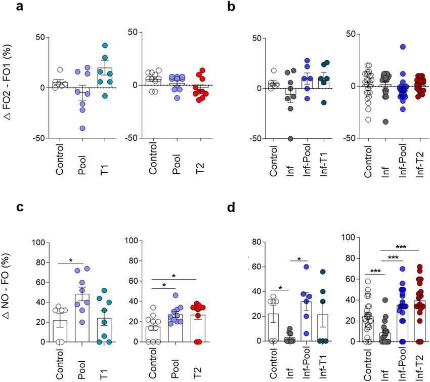
www.nature.com/scientificreports/
Figure 1. Flowchart of experiments. Groups of mice were infected with Plasmodium berghei ANKA (PbA), or
mock infected with PBS (Control mice), and treated with chloroquine (25 mg/kg) for 7 days via gavage from the
fourth day post-infection. After 14 days, the animals were subdivided into groups of mice immune stimulated
with different immunization strategies or non-immune stimulated. Subsequently, mice were evaluated via
behavioural tasks for locomotivity, memory and anxiety phenotype. The immune response of randomly chosen
mice was evaluated.
mild malaria. Our results show a beneficial effect of immune stimulation on cognitive-behavioural parameters
in healthy mice and a reversal of cognitive impairment caused by malaria parasite infection.
Methods
Mice and parasite. The Instituto de Ciência e Tecnologia em Biomodelos of the Fundação Oswaldo Cruz
(ICTB-Fiocruz, Brazil) provided seven-week-old female C57BL/6 mice weighing 20–25 g. Mice were housed in
racks with an air filtration system in a room maintained at 25 °C and light/dark cycles of 12 h in cages contain-
ing five animals with free access to food and water. All procedures were carried out in accordance with animal
welfare practices approved by the Ethical Committee on the Use of Laboratory Animals of Instituto Oswaldo
Cruz under CEUA-IOC: L-010/2015 concession. The study was carried out in compliance with the ARRIVE
guidelines. Plasmodium berghei ANKA (PbA) infections were carried out using a stable transfected strain of PbA
expressing a green fluorescent protein (PbA-GFP) generated as described p reviously20.
Infection and treatment of experimental groups. Mice were infected intraperitoneally (ip) with
150 μl of thawed cryopreserved PbA-infected red blood cells. Five days after infection, total blood was collected,
adjusted to 1 × 10⁶ parasitized erythrocytes in 100 μl of PBS and injected ip to mice in experimental groups. Para-
sitaemia was monitored by flow cytometry, based on the percentage of GFP+ erythrocytes. In this experimental
model, the establishment of cerebral malaria (CM) occurs between the fifth and sixth day of infection19. Mice
were treated on the fourth day of infection (mean parasitaemia 2.5%) with 25 mg/kg of chloroquine (CQ) by
gavage for seven days8, before any clinical signs of CM. All groups were similarly manipulated. Previous experi-
ments carried out with groups of uninfected mice comparatively treated with CQ or PBS have previously shown
that the CQ treatment does not influence the performance of mice in behavioural tasks and/or their anxiety
phenotypes18.
Experimental description. Mice were divided into groups of PbA-infected and control animals (non-
infected) and both were treated with chloroquine (CQ) for 7 days from the fourth day of infection. Thirteen
days after treatment, mice from respective groups were subdivided into non-immune stimulated and immune
stimulated groups (Fig. 1). The following vaccines and antigens were used for immune stimulation: Diphthe-
ria and Tetanus toxoids (dT) vaccine for adults, Influenza vaccine, Plasmodium falciparum Merozoite Surface
Protein 3 (PfMSP-3 recombinant protein), White chicken egg ovalbumin (OVA) and Lipopolysaccharide of
Escherichia coli (EcLPS). Three different immune stimulation strategies were performed: a combination of all
antigens and vaccines described above (henceforth referred to as ‘Pool’); a combination of antigens and vac-
cines (Influenza vaccine and EcLPS) that trigger, preferentially, a type 1 pattern of immune response (hence-
forth denominated ‘T1’); and a combination of antigens and vaccines (dT vaccine, PfMSP-3 recombinant pro-
tein and OVA) that preferentially trigger a type 2 immune response (henceforth called ‘T2’). Mouse groups
were named: Control (non-infected/non-immune stimulated); Pool (non-infected/Pool-immune stimulated);
T1 (non-infected/T1-Immune stimulated); T2 (non-infectedimmune stimulated); Inf (infected/non-immune
stimulated); Inf-Pool (infected/Pool-immune stimulated); Inf-T1 (infected/T1-immune stimulated) and Inf-T2
(infected/T2-immune stimulated). All control groups were treated as the experimental groups: they were age-
matched, mock-immune stimulated, mock-infected, and treated with CQ whenever appropriate. Subsequently,
mice behavioural performance was assessed by open field task (OFT), novel object recognition task (NORT) and
light–dark task (Fig. S1). About 300 mice were used for these experimental strategies in five consecutive sessions.
Scientific Reports | (2021) 11:14857 | https://doi.org/10.1038/s41598-021-94167-8 2
Vol:.(1234567890)
www.nature.com/scientificreports/
Immune system stimuli. Immune stimulation was initiated 14 days after the end of CQ treatment, and
was performed in the course of the following 62 days (Fig. S1). Antigens and/or vaccines were administered by
different routes and in different regions of the animal’s body (Table S1). In view of the originality of our approach
and the lack of information on immunization procedures influencing mice behaviour, immunogens were chosen
according to the pattern of immune response induced and to the accessibility of experimental or human vac-
cines to our laboratory. The doses administrated were defined based on dose–response protocols available in the
literature capable of stimulating the murine immune system without the risk of d eath21–25.
Immune stimulation with Plasmodium falciparum Merozoite Surface Protein 3 (PfMSP‑3 recombinant pro-
tein). Mice were challenged with 10 μg of PfMSP-3/mouse recombinant protein (in collaboration with Clinical
Trials of Malaria Vaccines, Vac4All Initiative, Paris, France) adsorbed on 70% adjuvant solution MONTANIDE
ISA 50 V2W/O (SEPPIC. Air Liquide—Healthcare), in 100 μl of PBS. Three subcutaneous injections were per-
formed at the tail region with a twenty-day interval between immune s timulations23 (Fig. S1, Table S1).
Immune stimulation with Tetanus‑Diphtheria and influenza vaccines. The vaccines used in this study were:
Tetanus-Diphtheria (dT) double bacterial (Biological E Limited—BE, Telangana—India, Lot. 34005815), in col-
laboration with the Division of Health Surveillance—CAP 3.1 of the Fundação Oswaldo Cruz (Rio de Janeiro,
Brazil); and Trivalent Influenza granted by the Technological Development and Production Division of Instituto
Butantan (São Paulo, Brazil, Lot. 160034). Mice received 100 μl (1/5 of the human dose) of dT and Influenza vac-
cines by subcutaneous (dorsal region) and intramuscular (left quadriceps region) routes, respectively (Table S1).
Three inoculations with a twenty-day interval between immune stimulations were p erformed21 (Fig. S1).
Immune stimulation with ovalbumin (allergen). Mice received 50 μg/mice of white chicken egg ovalbumin
(SIGMA-ALDERICH, Cod. A5503-50 g) adsorbed onto aluminum hydroxide [Al (OH) 3] in a final volume of
200 μl per animal in three inoculations. The first inoculation was performed at the dorsal region by subcutane-
ous injection and the following (second and third inoculation) by ip route (Table S1) with 6 days between them24
(Fig. S1).
Immune stimulation with lipopolysaccharide from Escherichia coli (EcLPS). Mice were challenged with 0.1 mg/
kg of EcLPS O111: B4 (SIGMA-ALDERICH, L2630-10MG, Lot 025M4040V12140701) diluted in phosphate-
buffered saline (PBS). Two ip inoculations were performed (Table S1) with a range of 9 days between the immune
stimulations25 (Fig. S1).
Evaluation of the immune response. Following stimulation of the immune system, mice were ran-
domly selected and sacrificed for individual withdrawal of whole blood, via cardiac puncture, and spleen at
day 84 after the end of CQ treatment. Serum samples were preserved at − 70 °C. Total IgG antibody response
to PfMSP-3 recombinant protein, Tetanus-Diphtheria toxoids (dT) and Influenza vaccines; the serum cytokine
profile; the splenic lymphocyte subpopulations; and the response to Ovalbumin sensitization were evaluated to
confirm the effectiveness of immune stimuli.
Specific antibody responses. The antibody response against PfMSP-3 recombinant protein and Influenza vac-
cine were determinate by conventional Enzyme-Linked immunosorbent Assay—ELISA9,10, and the antibody
response against dT vaccine was determined by Toxin Binding Inhibition—ToBI26.
Cytokine profile. Cytokines in the serum samples were measured with Cytometric Bead Array (CBA) Mouse
Th1/Th2/Th17 (BD Biosciences) according to the manufacturer’s instructions. The data were collected on a FAC-
SCANTO II flow cytometer (BD Bioscience) and analysed by FCAP Array Software 3.0 (BD Bioscience).
Splenic lymphocyte subpopulations. Individual spleens were removed and mechanically dissociated using a
syringe plunger above 70 μm-pore size Falcon cell strainer (BD Biosciences). Red blood cells were lysed using
ACK lysing buffer (Sigma). Single-cell suspensions were counted and incubated with anti-Fcγ III/II (CD16/32)
receptor Ab (2.4G2) in PBS containing 3% FCS for 15 min, and immunolabelled for 30 min at 4 °C in the
dark with the following fluorochrome-conjugated antibodies: PE-Cy7 anti-mouse CD8 (53–6.7), PerCP-Cy5.5
anti-CD3 (145-2c11), APC-H7 anti-mouse CD4 (GK1.5), APC anti-mouse B220 (RA3-6B2), BB515 anti-mouse
CD62L (MEL14), APC anti-mouse CD44 (IM7) and/or PE anti-mouse CD25 (7D4). For Treg cells analyses,
cells were fixed and permeabilized, after staining for surface markers, with eBioscience™ Foxp3/Transcription
Factor Staining Buffer Set according to the manufacturer instructions and incubated with the antibody Alexa
Flour 647 anti-Foxp3 (R16715). All antibodies were from BD Biosciences. Data were collected using FACSDiva
software on a FACSCANTO II flow cytometer (BD Biosciences), and analysed using FlowJo software 10.0 (BD
Biosciences).
Intradermal skin test. In the footpad of the left paw, 3 µg of OVA, diluted in 30 µl of PBS, were injected in each
animal. After 30 min, the plantar thickness (mm) was measured using a digital caliper. Oedema formation was
expressed as the difference of the pad thickness measured before and after the inoculation of O VA24.
Behavioural analysis. The schedule of the behavioural tasks is shown in Supplementary Fig. S1. Mice
were individually submitted to different behavioural paradigms to evaluate their exploratory and locomotor
Scientific Reports | (2021) 11:14857 | https://doi.org/10.1038/s41598-021-94167-8 3
Vol.:(0123456789)www.nature.com/scientificreports/
activity, cognitive abilities, and parameters involved in anxiety-like behaviour from day 88 to 92 post-infection
(77 to 81 days after the complete parasitological cure of animals obtained with CQ treatment). The beginning of
behavioural tests corresponded to 22 days after the last stimulation with PfMSP-3 recombinant protein, Tetanus-
Diphtheria, and Influenza vaccines, 7 days after the last injection of ovalbumin, and 2 days after the LPS final
inoculation. The same cohort of mice was used in all tasks (Fig. S1). All experiments were carried out with an
incandescent light source of 200 lx of intensity in the evening period. Animals were acclimatized in the experi-
mental room for at least two hours before the experimental sessions. Behaviour was captured by a video camera
positioned above the task apparatus. Locomotion in the open field and the object recognition task was analyzed
by the AnyMaze software 5.1 (Stoelting Co., Wood Dale, IL, USA), while a trained blind-to-treatment researcher
evaluated other behavioural parameters by video analysis. In all behavioural tests, mice were individually placed
on the apparatus, which was previously cleaned with 70% alcohol and dried.
Open field task (OFT). To address the effect of immune stimuli on locomotion and on long-term habituation,
mice were individually submitted to the OFT with a training (OFT1) and a test (OFT2) session 24 h apart, as
described elsewhere18. In each OFT session, mice were individually allowed to freely explore a grey acrylic square
box, dimensions (50 × 50 × 50 cm, length × width × height), for 10 min. In OFT1, locomotor activity (measured
by the total distance travelled) and the time and total distance travelled in the centre zone were evaluated during
the entire session. In OFT2, we evaluated the total distance travelled during the session.
Novel object recognition task (NORT). To evaluate long-term memory for object recognition, a NORT was
carried out in the OFT apparatus, 24 h after the test session18. In the training session, mice were exposed to
two identical familiar objects (FO), for which similar exploratory activity was expected27. The test session was
carried out 24 h later when mice were exposed to a new object (NO) and to one of the FO previously exposed.
Memory expression is indicated by the tendency of the animal to spend more time exploring the NO rather
O18,27. Animals were individually placed in the periphery of the box with the objects in a session for
that the F
10 min. Exploration was recorded only when the animals touched the objects, located in opposite and symmetri-
cal corners of the box, with their nose or mouth. The time of exploration of each object was recorded, and its
percentage of the time of exploration of both objects was calculated. The object recognition index is calculated
as the percentage of time spent on each object (referred to the total time spent on both objects). The difference
between the time spent with the NO and the FO is expressed as a delta value obtained with the subtraction of
the indexes of each object.
Light/dark task. The light/dark task, a conflict avoidance test, was carried out as described by Almeida et al.28.
The apparatus was a rectangular acrylic box (50 × 30 × 30 cm, height × length × width) with two sides coloured
white and black, separated by a wall (5 × 5 cm) with an opening at the level of the base of the apparatus joining
both sides. A white 100 W lamp, placed 60 cm above the centre of the apparatus, illuminated the white side of the
apparatus, while the black side was kept closed without illumination. The mice were individually placed in the
light compartment for free exploration of the apparatus for five minutes. The following behavioural parameters
were analysed: the time spent in the light compartment and the number of transitions between the compart-
ments (light and dark).
Statistical analysis. All statistical analyses were performed using a Prism statistical software 7.0 (Graph-
Pad). The data were extracted from the AnyMaze software 5.1. To analyse OFT and light/dark task, we used the
absolute data. The time in each object in NORT was transformed into a percentage, from which the delta was
extracted based on the subtraction: OF1 − OF2 (training session) and NO − FO (test session). The two-way
ANOVA with Bonferroni correct were used to analyse OFT. One-way ANOVA were used to compare multiple
groups in the NORT and light–dark task and Student’s t-test for immune response analyses. Data are presented
as mean ± standard error. P < 0.05 was considered statistically significant.
Results
Type 2 immune stimuli enhance long‑term recognition memory in healthy mice. To study
the effect of immune stimuli on behavioural paradigms, immunogens were chosen according to the pattern of
immune response induced. Three immune stimulation strategies were used: T1 and T2 strategies employed well-
known antigens able to induce type 1 and type 2 immune responses (T1—influenza vaccine and LPS; T2—dT
vaccine, PfMSP3 and OVA), respectively21–23,29. A Pool strategy was created by the combination of T1 and T2
strategies. Briefly, mice were infected with PbA, treated with chloroquine (CQ) and immune stimulated with
different strategies according to Fig. 1.
The effects of immune responses on locomotion and long-term spatial habituation were assessed in mice
subjected to two different sessions of the open field task (OFT), with training (OF1) and test (OF2) sessions
24 h apart18. At the training session, a high rate of locomotor activity is commonly observed. Surprisingly, mice
immune stimulated with Pool or T1 strategies showed reduced total OF1 locomotion when compared to non-
immune stimulated mice (Fig. S2A).
Commonly, after the training session [first OFT (OF1), exposure], exploratory behaviour decreases as the
stress related to novelty disappears, and is usually significantly lower after 10 min of task p erformance18,28.
Both non-immune stimulated (Control group) and immune stimulated (Pool, T1 and T2 groups) mice showed
decreased locomotion in the test session (Fig. S2A) indicating that immune stimulation did not affect long-term
habituation memory.
Scientific Reports | (2021) 11:14857 | https://doi.org/10.1038/s41598-021-94167-8 4
Vol:.(1234567890)www.nature.com/scientificreports/
Figure 2. Immune stimulation enhances long-term memory performance in healthy and PbA-infected mice.
Healthy or PbA-infected (and treated) mice were immune stimulated, or not, with the Pool, T1 or T2 strategies.
Behavioural tasks were performed from day 88 to 92 post-infection (77 to 81 days after CQ treatment).
Differences of time, in percentage of total time, spent on each object during the training (a,b) and test (c,d)
sessions of the new object recognition task (NORT) in healthy (a,c) and infected (b,d) mice. FO familiar
object; NO new object. Experimental groups: Control [non-infected/non-immune stimulated mice; n = 6, 10
and 25 (representative of different experiment); Pool (non-infected/Pool-immune stimulated mice, n = 8–10
representative of different experiment); T1 (non-infected/T1-immune stimulated mice, n = 8); T2 (non-
infected/T2-immune stimulated mice, n = 10); Inf (infected/non-immune stimulated mice, n = 6–17); Inf-Pool
(infected/Pool-immune stimulated mice, n = 6—20); Inf-T1 (infected/T1-immune stimulated mice, n = 6);
Inf-T2 (infected/T2-immune stimulated mice, n = 8—18). Data are expressed as mean and s.e.m. ***P < 0.001;
**P < 0.01; *P < 0.05; One-Way ANOVA was used [C, F (2,19) = 4.370 and F (2,27) = 3.416; D, F (3,22) = 3.705 and
F (3,76) = 12.72]. Data shown represent one of two to five independent experiments (Control, Pool, T1, T2, Inf,
Inf-Pool, Inf-T1); and a pool of two independent experiments (Control, Inf, Inf-Pool, Inf-T2).
Next, mice were subjected to the novel object recognition task (NORT) in the same open field arena. The
known possible interference of anxiety on memory evaluation is relieved in the NORT sessions by previous
exposure in the open field twice on 2 consecutive days. During the training session, a similar exploratory activity
of familiar objects (FO1 and FO2) is expected and was observed in all groups of mice (Control, Pool, T1 and T2)
(Figs. 2A and S3A), with a mean exploration of 25 s (data not shown). Remarkably, mice immune stimulated
with the Pool or T2 strategies presented significantly higher recognition memory performance in relation to the
Control group during the test session, reflected by the increased exploitation of the new object in relation to the
familiar object, 24 h later. Mice submitted to the T1 strategy did not differ from the Control group (Figs. 2C and
S3C). These data indicate that immune stimulation with immunogens that induce type 2 immune responses may
enhance long-term recognition memory in healthy mice.
Immune stimulation of healthy mice did not generate an anxiety‑like state. In addition to
exploratory activity, the OFT also allows the evaluation of phenotypes related to anxiety-like behaviour through
analysis of the dwell time or the locomotion rate in the centre of the open field arena during the first exposure
to the apparatus. Immune stimulated mice (Pool, T1 and T2 groups) showed no difference in dwell time in the
centre of the open field arena (data not shown) but presented significantly reduced locomotion in relation to the
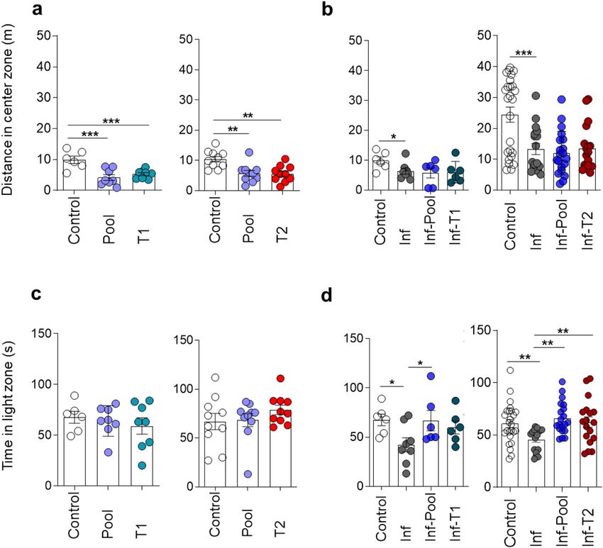
non-immune stimulated mice (Control group) (Fig. 3A). It seems, however, that this observation which is con-
sistent with an anxiety-like state, may have been influenced by the total reduced locomotion observed in animals
submitted to Pool and T1 strategies (Fig. S2A). In the Light–Dark specific task, however, immune stimulated
mice (Pool, T1 and T2 groups) clearly behaved similarly to mice of the Control group, remaining an equal time
in the light zone (Fig. 3C), thus implying that immune stimulation did not generate an anxiety-like state.
Exposure to type 2 immune stimuli may reverse cognitive‑behavioural damage induced by
non‑severe Plasmodium berghei ANKA infection. About 98% of the world’s malaria cases are due to
Scientific Reports | (2021) 11:14857 | https://doi.org/10.1038/s41598-021-94167-8 5
Vol.:(0123456789)www.nature.com/scientificreports/
Figure 3. Immune stimulation attenuates the anxiety-like behaviour observed in PbA-infected mice. Healthy
or PbA-infected (and treated) mice were immune stimulated, or not, with the Pool, T1 or T2 strategies.
Behavioural tasks were performed from day 88 to 92 post-infection (77 to 81 days after CQ treatment). Distance
travelled in the centre of the arena during the training session of the open field task (OFT) by healthy (a) and
infected (b) immune stimulated mice. Time in the light zone of the Light/Dark apparatus by healthy (c) and
infected (d) immune stimulated mice. For definition of experimental groups see Legend of Fig. 2. Data are
expressed as mean and s.e.m. ***P < 0.001; **P < 0.01; *P < 0.05; One-way ANOVA was used [A, F (2,19) = 11.01
and F (2,27) = 8.901; B, F (3,22) = 3.147 and F (3,76) = 9.175; D, F (3,22) = 3.184 and F (3,76) = 4.727]. Data shown
represent one of two to five independent experiments (Control, Pool, T1, T2, Inf, Inf-Pool, Inf-T1); and a pool of
two independent experiments (Control, Inf, Inf-Pool, Inf-T2).
Plasmodium falciparum, 1 to 2% of which progress to CM. Therefore, up to 97% of all malaria cases globally are
caused by this lethal parasite, but occur without apparent clinical c omplications9, but may, nevertheless, may
impair the cognitive development of children15–17.
The experimental model we employed uses PbA-infected C57BL/6 mice treated at day 4 post-infection, before
the appearance of any clinical signs of CM19. Its main advantage is that it best corresponds to the large majority
of human malaria cases in the world; non-severe falciparum malaria9. In fact, both parasite-host pairs involve the
potential of CM development that can be avoided with timely drug treatment. Using this model, we have been
able to observe long-term cognitive-behavioural impairment related to memory and anxiety as late as 92 days
post-infection (82 days after the end CQ treatment), when no parasites are present in the blood18.
Given the beneficial effect of immune stimulation on long-term memory in healthy mice described above,
we evaluated the effect of the same immune stimuli in mice with behavioural alterations caused by non-severe
malaria infection. PbA-infected and treated mice (from here on referred to as the “Infected group”), did not
display reduced total locomotion in the training session of the OFT when compared to healthy mice (Fig. S2B).
However, infected and immune stimulated animals (Inf-Pool and Inf-T2 groups) showed a significant reduction
in locomotion in the OF1 when compared to healthy mice (Fig. S2B). Control, infected and infected-immune-
stimulated groups (Inf-Pool and Inf-T2, but not Inf-T1) displayed normal behaviour with a significant decrease
in locomotion in the test session as compared to the training session of the OFT (Fig. S2B).
As expected, there was no object preference in the NORT training session (Figs. 2B and S3B). Infected mice
consistently presented long-term recognition memory sequelae that manifested as similar exploration of the
familiar object (FO) and new object (NO) in the NORT. This impairment disappeared following stimuli with
Pool or T2 immunization (Figs. 2D and S3D), pointing to a beneficial effect of immune stimulation triggered by
type 2 immunogens in reversing the cognitive deficits associated with malaria parasite infection.
The main advantage of the NORT protocol over other rodent memory tasks, for the analysis of cognitive
ability is that it relies on the mouse’s natural proclivity for exploring novelty in a well-recognized apparatus. This
feature ensures that NORT is much less stressful in comparison with others tasks; and that anxiety does not,
therefore, impact NORT p erformance27,30. In this context, the OFT paradigm followed by NORT protocol has
been extensively used in preclinical studies to assess different aspects involved in learning and memory, allowing
the detection of neuropsychological changes following pharmacological, biological, or genetic m anipulations31
and the results provided using both of these behavioural paradigms are usually reliable.
Scientific Reports | (2021) 11:14857 | https://doi.org/10.1038/s41598-021-94167-8 6
Vol:.(1234567890)www.nature.com/scientificreports/
Plasmodium berghei ANKA infection in mice induces an anxiety‑like behaviour that is reversed
by immune stimulation with type 2 immunogens. The distance travelled in the periphery and in
the centre of the open field arena are inversely related. Since the latter was decreased in PbA-infected mice
(Fig. 3B) and no change in the locomotion during the training session (OF1) occurred among Control and
Infected groups (Fig. S2B), the decrease may be interpreted as the expression of an anxiety-like behaviour. This
behaviour was confirmed by the observation of a reduction in time spent by infected mice in the light zone of
the light–dark task, a more sensitive and widely used test to evaluate anxiety-related parameters in rodents.
Anxiety-like behaviour was reversed by Pool and T2, but not by T1 strategies of immune stimulation (Fig. 3D).
Immune stimulation procedures and non‑severe Plasmodium berghei ANKA malaria elicit
immune responses. Evaluation of specific responses to the immunogens in the Pool, T1 and T2 strategies
(tetanus toxoid, influenza, PfMSP3 and OVA) confirmed the effectiveness of the stimuli (Fig. S4A–D) and the
known low immunogenicity of diphtheria toxoid in mice32.
An effort was also made to characterize systemic inflammatory status at the time of cognitive-behavioural
evaluation, through the measurement of TNF-α, INF-γ, IL-6, IL-4, and IL-10 levels in the serum. At the time of
immune response evaluation, 84 days after the end of CQ treatment (Fig. 1), non-immune stimulated infected
animals did not present increased levels of serum cytokines when compared to the Control group (Fig. S5A–D).
However, higher levels of TNFα, IFNγ, IL-6, IL-10 and/or IL-4 were detectable in all groups of immune stimu-
lated mice with T1, T2, or Pool strategies (Fig. S5A–D), ratifying the immune stimulation by the different
strategies used. Only IL-10 was consistently increased among healthy and infected mice stimulated with Pool
or T2 strategies, although statistical significance was not achieved between Pool and Control groups (Fig. S5E).
We evaluated the splenic immune response of healthy and infected mice exposed to Pool and T2 strategies,
since only these approaches were able to immunomodulate cognitive behaviour in mice. Only immune stimu-
lated mice showed increased spleen weight and total number of splenocytes (Fig. S6A–C). Healthy mice immune
stimulated with either Pool or T2 strategies presented similar patterns of modulation of different immune com-
ponents. An increase in the frequency of splenic B cells (Fig. S7B), CD4 and CD8 T cells with central memory
phenotype (Fig. S8D,G) and CD4 T cells with regulatory function (Treg cells) (Fig. S7E) were recorded in both
Pool and T2 immune stimulated groups when compared to non-immune stimulated animals. A reduction in the
frequency of CD8 T cells was also observed in mice immune stimulated with the T2 strategy when compared
to the Control group (Fig. S7D).
PbA-infected mice had higher frequencies of B cells, total CD4 and CD8 T cells (Fig. S7A–C), CD4 and CD8
T cells with naïve and central memory phenotypes (Fig. S8A,B,D,E,G) and reduced frequencies of effector/
effector memory CD8 T cells (Fig. S8F) when compared to healthy mice (Control group). The frequency of Treg
cells, however, was similar between infected and healthy mice (Fig. S7E). Immune stimulation of PbA-infected
mice with Pool or T2 strategies induced comparable increases in the frequencies of splenic B cells, Treg cells
(Fig. S7A,B,E), effector/effector memory CD4 T cells and central memory CD8 T cells (Fig. S8A,C,G), and
reduction in the frequencies of total CD8 T cells when compared to non-immune stimulated infected mice
(Fig. S7A,D).
In summary, independent of the health status of the mice, immune stimulation with type 2 immunogens
reduces the frequency of CD8 T cells and increases the percentage of Treg cells in the spleen, as well as the serum
levels of IL-10.
Discussion
We demonstrate a clear positive influence of immune responses induced by strategies involving type 2 stimuli on
the long-term recognition memory of healthy mice and confirm our previous demonstration of late neurocogni-
tive behavioural dysfunction following a single episode of non-severe m alaria18. Additionally, our data reveal
that immune stimulation with type 2 immunogens subsequent to infection can reverse the malaria induced
cognitive impairment.
Stimulation of the immune system can trigger both beneficial or deleterious effects on brain f unctionality4–6.
Remarkably, we observed a positive effect of immune stimulation on reversing the cognitive-behavioural impair-
ment associated with non-severe malaria. Mice treated with CQ 4 days after infection by PbA and immune
stimulated with T2 and Pool strategies did not display deficits in object recognition that are recorded late after
infection without subsequent immune stimulation. We also observed reversal of anxiety-like behaviour in a
light–dark task, following immune stimulation of infected mice.
The impairment of recognition (non-spatial) memory, as measured through NORT following a single epi-
sode of non-severe malaria, cannot be extrapolated to other memory categories (spatial, emotional, associative)
that were not studied here. However, although habituation in the open field task (OFT) is not as robust as the
water maze task to evaluate spatial memory, no alteration was recorded in the OFT in the experimental groups
evaluated.
Mice presented a clear anxiety-like behaviour in the light–dark task 90 days after a treated episode of non-
severe malaria. This result, together with the exploratory behaviour in the OFT centre zone, corroborates those
of Guha et. al. (2014)33, who detected anxious behaviour in another experimental model also considered as
non-severe malaria (C57BL/6 mice infected with Plasmodium chabaudi adami). It is known that the emotional
state of animals may influence exploratory behaviour in tests used to evaluate memory mechanisms. Some
acquired concepts weaken the possibility that this may have influenced our results: (a) beyond the investigation
of habituation memory in the OFT, it is noteworthy that mice submitted to two OFT sessions before NORT had
the anxiety-like phenotype interference mitigated in NORT sessions, as adopted in our protocol and described
previously27,30,31; (b) the decrease in the total distance travelled in the OFT1 central area by PbA-infected mice
Scientific Reports | (2021) 11:14857 | https://doi.org/10.1038/s41598-021-94167-8 7
Vol.:(0123456789)www.nature.com/scientificreports/
is no longer observed in the OFT2 exposition (data not shown), reinforcing that in the second OFT session the
anxiety emotional state was abolished; and (c) although locomotion in OFT (centre zone) alone does not directly
reflect anxiety behaviour, it may be considered indirect evidence of this emotional condition. In this respect,
T1-, but not T2-, treated infected animals showed decreased locomotor activity in the OFT, without presenting
memory deficits in the NORT.
It is thought that certain infectious diseases may induce cognitive and behavioural deficits34–36. Malaria is
a systemic disorder that can affect the central nervous system and the diseases caused by Plasmodium species
capable of affecting the brain can affect the cerebellum, hippocampus, olfactory bulb and the cerebral cortex, both
in humans37–39 and experimental models40–43. The hippocampus is involved in memory consolidation and anxi-
ety. Additionally, the perirhinal cortex is the centre of familiarity and recency discrimination s ystem44. Further
studies may clarify whether these or even other brain structures are involved in the present results.
Although no breakdown of the blood–brain barrier is observed on the fourth day of PbA infection, brain
histopathologic analysis reveals minimal haemorrhage or oedema restricted to focal areas, with the adherence
of low numbers of l eukocytes19. Expression of immunological markers is also recorded 3 days after infection of
C57BL/6 mice with PbA manifested as increased expression of PD-1 (programmed cell death protein) in the
cerebellum, CTLA-4 (cytotoxic T lymphocyte associated with protein 4) and LAG-3 (lymphocyte activation
gene 3: protein encoded by the LAG3 gene) in the hippocampus, as well as reduced expression of CXCL-4 in
the hippocampus37. These data indicate that the systemic inflammation already in progress at days 3 and 4 of
infection45,46 could modulate the brain function leading to cognitive behavioural changes detectable even in the
absence of clinical signs of patent cerebral commitment.
T cells are essential for normal neurogenesis and cognition2,47,48 and may influence the CNS via the production
of cytokines. It has been shown that proinflammatory cytokines impair cerebral function and cognition at high
pathological concentrations, as those induced during some infections2. An exacerbated peripheral inflamma-
tory response may cause M1 microglial activation and provoke the production of proinflammatory cytokines
such as TNF-α and IL1-β that may impair cognitive f unction49. Elevated levels of anti-inflammatory/regulatory
cytokines such as IL-4 and IL-10 may have the opposite effect, inducing M2 microglial activation and positively
influencing cognition50,51.
Microglia activation is recorded even before the overwhelming cerebral inflammation and development of
the clinical signs of CM, at day 4 post-PbA infection19,52. The serum levels of proinflammatory cytokines also
increase around 3–4 days after PbA infection in C75BL/6 mice45,46. The observation of early behavioural changes
(12 days after malaria treatment), points to a potential reversible (and not preventable) effect of the immune
stimuli (unpublished observations). This is in accordance with the concept that sufficiently severe systemic
inflammation can damage brain functions in the case of individual vulnerabilities or p redispositions53 even
before the appearance of clinical CM. It is possible, therefore, that the late cognitive deficit observed in our studies
results from the early activation of immune cells in the central nervous system (CNS) during malaria infection.
Treg cells are a subset of T cells with immunomodulatory function, important for immune and neuronal
homeostasis under physiological conditions, and for the control of pathological immune responses54–56 They
perform their function mainly via secretion of IL-10 and TGFβ, anti-inflammatory/regulatory c ytokines54–56 as
documented in rodent models45,46. The neuroprotective activity of Treg cells, through a mechanism that may
involve IL-10 secretion, has been described in murine models of Parkinson’s disease, HIV-1-associated neu-
rodegeneration, amyotrophic lateral sclerosis and s troke57–60. In the present study, healthy and infected mice
stimulated with type 2 immunogens significantly increased the number of splenic Treg cells and IL-10 levels
in the serum. As Treg cells and IL-10 can restrict n euroinflammation45,61, it is reasonable to assume that these
immunogens may enhance cognitive function by promoting balanced cross-talk between the immune system
and the CNS mediated through Treg cells and IL-10. The mechanism by which immune stimulation with type-2
immunogens benefits cognition is presently under investigation.
Conclusion
Our data demonstrate the positive influence of immune responses induced by type 2-inducing stimuli on the
long-term recognition memory of healthy mice, confirm our previous demonstration of late neurocognitive
behavioural dysfunction following a single episode of non-severe m alaria18, and demonstrate the recovery of
this deficit upon immune stimulation with type 2 immunogens following infection. Taken together, these results
offer a new paradigm for the design of memory enhancement strategies and suggest that vaccination procedures
may provide benefits additional to the prevention of infection, offering a potential approach for boosting and
recovering recognition memory that may have been impaired by chronic and infectious diseases, including
malaria, and by the effects of ageing.
Received: 23 February 2021; Accepted: 5 July 2021
References
1. Cohen, I. R. The cognitive paradigm and the immunological homunculus. Immunol. Today. 13(12), 490–494 (1992).
2. Kipnis, J., Gadini, S. & Derecki, N. Pro-cognitive properties of T cells. Nat. Rev. Immunol. 12(9), 663–669 (2012).
3. Nataf, S. Autoimmunity as a driving force of cognitive evolution. Front. Neurosci. 11, 582 (2017).
4. Schwartzer, J. J. et al. Behavioural impact of maternal allergic-asthma in two genetically distinct mouse strains. Brain Behav. Immun.
63, 99–107 (2017).
5. Yang, J. et al. Neonatal BCG vaccination of mice improves neurogenesis and behavior in early life. Brain Res. Bull. 120, 25–33
(2016).
Scientific Reports | (2021) 11:14857 | https://doi.org/10.1038/s41598-021-94167-8 8
Vol:.(1234567890)www.nature.com/scientificreports/
6. Bossu, P. et al. A single intraperitoneal injection of endotoxin in rats induces long-lasting modifications in behavior and brain
protein levels of TNF-α and IL-18. J. Neuroinflamm. 9, 101 (2012).
7. Odera, V. M., Snow, R. W. & Newton, C. R. J. C. The burden of the neurocognitive impairment associated with Plasmodium falci-
parum malaria in sub-Saharan Africa. Am. J. Trop. Med. Hyg. 71(2 Suppl), 64–70 (2004).
8. Reis, P. A. et al. Cognitive dysfunction is sustained after rescue therapy in experimental cerebral malaria, and is reduced by additive
antioxidant therapy. PLoS Pathog. 6(6), e1000963 (2010).
9. World Health Organization. World Malaria Report (WHO, 2019).
10. Boivin, M. J. et al. Cognitive impairment after cerebral malaria in children: A prospective study. Pediatrics 119(2), e360–e366
(2007).
11. Bangirana, P. et al. Cognition, behaviour and academic skills after cognitive rehabilitation in Uganda children surviving severe
malaria: A randomised trial. BMC Neurol. 11, 96 (2011).
12. Holmberg, D. et al. Cerebrospinal fuid kynurenine and kynurenic acid concentrations are associated with coma duration and
long-term neurocognitive impairment in Ugandan children with cerebral malaria. Malar. J. 16(1), 303 (2017).
13. Desruisseaux, M. S. et al. Cognitive dysfunction in mice infected with Plasmodium berghei strain ANKA. J. Infect. Dis. 197(11),
1621–1627 (2008).
14. Reverchon, F. et al. IL-33 receptor ST2 regulates the cognitive impairments associated with experimental cerebral malaria. PLoS
Pathog. 13(4), e1006322 (2017).
15. Fernando, S. D. et al. The impact of repeated malaria attacks on the school performance of children. Am. J. Trop. Med. Hyg. 69(6),
582–588 (2003).
16. Vitor-Silva, S., Reyes-Lecca, R. C., Pinheiro, T. R. A. & Lacerda, M. V. G. Malaria is associated with poor school performance in
endemic area of the Brazilian Amazon. Malar. J. 8, 230 (2009).
17. Tapajós, R. et al. Malaria impact on cognitive function of children in a peri-urban community in the Brazilian Amazon. Malar. J.
18(1), 173 (2019).
18. de Sousa, L. P. et al. Long-term effect of uncomplicated Plasmodium berghei ANKA malaria on memory and anxiety-like behaviour
in C57BL/6 mice. Parasites Vectors 11(1), 191 (2018).
19. Potter, S. et al. Perforin mediated apoptosis of cerebral microvascular endotelial cells during experimental cerebral malaria. Int. J.
Parasitol. 36(4), 485–496 (2006).
20. Franke-Fayard, B. et al. A Plasmodium berghei reference line that constitutively expresses GFP at a high level throughout the
complete life cycle. Mol. Biochem. Parasitol. 137(1), 23–33 (2004).
21. Castilho, S. F. et al. A Bordetella pertussis proteoliposome induces protection in mice without affecting the immunogenicity of
diphtheria and tetanus toxoids in a trivalent formulation. Clin. Exp. Vaccine Res. 5(2), 175–178 (2016).
22. Goodier, M. R. et al. Influenza vaccination generates cytokine-induced memory-like NK cells: Impact of human cytomegalovirus
infection. J. Immunol. 197(1), 313–325 (2016).
23. Oeuvray, C. et al. A novel merozoite surface antigen of Plasmodium falciparum (MSP-3) identified by cellular-antibody cooperative
mechanism antigenicity and biological activity of antibodies. Mem. Inst. Oswaldo Cruz. 89(2), 77–80 (1994).
24. Lima, M. C. R. et al. Studies on the mechanisms involved in antigen-evoked pleural inflammation in rats: Contribution of IgE and
complement. J. Leukoc. Biol. 61(3), 286–292 (1997).
25. Couch, Y. et al. Low-dose lipopolysaccharide (LPS) inhibits aggressive and augments depressive behaviours in a chronic mild
stress model in mice. J. Neuroinflamm. 13(1), 108 (2016).
26. Matos, D. C. S. et al. Immunogenicity test of tetanus component in adsorbed vaccines by toxin binding inhibition test. Mem. Inst.
Oswaldo Cruz. 97(6), 909–913 (2002).
27. Leger, M. et al. Object recognition test in mice. Nat. Protoc. 8(12), 2531–2537 (2013).
28. Almeida, R. F. et al. Guanosine anxiolytic-like effect involves adenosinergic and glutamatergic neurotransmitter systems. Mol.
Neurobiol. 54(1), 423–436 (2017).
29. Comoy, E. E., Capron, A. & Thyphronitis, G. In vivo induction of type 1 and 2 immune responses against protein antigens. Int.
Immunol. 9(4), 523–531 (1997).
30. Lueptow, L. M. Novel object recognition test for the investigation of learning and memory in mice. J. Vis. Exp. 126, 55718 (2017).
31. Sik, A., Nieuwehuyzen, P. V., Prickaerts, J. & Blokland, A. Performance of different mouse strains in an object recognition task.
Behav. Brain Res. 147(1–2), 49–54 (2003).
32. Maheshwari, S. C., Sbarma, S. B., Ahujat, S. & Saxena, S. N. Development of a mouse model to estimate the potency of the diphtheria
toxoid component of diphtheria-tetanus and diphtheria-tetanus-pertussis vaccines. J. Biol. Stand. 16(3), 139–146 (1988).
33. Guha, S. K. et al. Single episode of mild murine malaria induces neurinflammation, alters microglial profile, impairs adult neuro-
genesis, and causes deficits in social and anxiety-like behavior. Brain Behav. Immun. 42, 123–137 (2014).
34. Ruggieri, A., Gagliardi, M. C. & Anticoli, S. Sex-dependent outcome of hepatitis B and C viruses infections: Synergy of sex hor-
mones and immune responses?. Front. Immunol. 9, 2302 (2018).
35. Martin, E. et al. Verbal and spatial working memory among drug-using HIV-infected men and women. J. Neurovil. 24(4), 488–497
(2018).
36. Kannan, G. & Pletnikov, M. V. Toxoplasma gondii and cognitive deficits in schizophrenia: An animal model perspective. Schizoph.
Bull. 38(6), 1155–1161 (2012).
37. Taylor, T. E. et al. Differentiating the pathologies of cerebral malaria by postmortem parasite counts. Nat. Med. 10, 143–145 (2004).
38. Dorovini-Zis, K. et al. The neuropathology of fatal cerebral malaria in Malawian children. Am. J. Pathol. 178, 2146–2158 (2011).
39. Maude, R. J. et al. Magnetic resonance imaging of the brain in adults with severe falciparum malaria. Malaria J. 13, 177 (2014).
40. Khandare, A. V. et al. Expression of negative immune regulatory molecules, pro-inflammatory chemokine and cytokines in
immunopathology of ECM developing mice. Acta Trop. 172, 58–63 (2017).
41. Ma, N., Harding, A. J., Pamphlett, R., Chaudhri, G. & Hunt, N. H. Increased c-fos expression in the brain during experimental
murine cerebral malaria: Possible association with neurologic complications. J. Infect. Dis. 175(6), 1480–1489 (1997).
42. Oliveira, K. R. H. M. et al. Cerebral malaria induces electrophysiological and neurochemical impairment in mice retinal tissue:
Possible effect on glutathione and glutamatergic system. Malaria J. 16, 1–10 (2017).
43. Zhao, H. et al. Olfactory plays a key role inspatiotemporal pathogenesis of cerebral malaria. Cell Host Microbe 15, 551–563 (2014).
44. Brown, M. W. & Aggleton, J. P. Recognition memory: What are the roles of the perirhinal cortex and hippocampus?. Nat. Rev.
Neurosci. 2, 51–61 (2001).
45. Hanum, S., Hayano, M. & Kojima, S. Cytokine and chemokine responses in a cerebral malaria-susceptible or-resistant strain of
mice to Plasmodium berghei ANKA infection: Early chemokine expression in the brain. Int. Immunol. 15(5), 633–640 (2003).
46. Hansen, D. S., Siomos, M. A., Buckingham, L., Scalzo, A. A. & Schofield, L. Regulation of Murine Cerebral Malaria Pathogenesis
by CD1d-restricted NKT cells and the natural killer complex. Immunity 18(3), 391–402 (2003).
47. Ziv, Y. et al. Immune cells contribute to the maintenance of neurogenesis and spatial learning abilities in adulthood. Nat. Neurosci.
9(2), 268–275 (2006).
48. Radjavi, A., Smirnov, I. & Kipnis, J. Brain antigen-reactive CD4+ T cells are sufficient to support learning behavior in mice with
limited T cell repertoire. Brain Behav. Immun. 35, 58–63 (2014).
49. Hoogland, I. C. M., Houbolt, C., Westerloo, D. J. V., Gool, W. A. V. & Beek, D. V. Systemic inflammation and microglial activation:
Systematic review of animal experiments. J. Neuroinflamm. 12, 114 (2015).
Scientific Reports | (2021) 11:14857 | https://doi.org/10.1038/s41598-021-94167-8 9
Vol.:(0123456789)www.nature.com/scientificreports/
50. Tang, Y. & Le, W. Differential roles of M1 and M2 microglia in neurodegenerative diseases. Mol. Neurobiol. 53(2), 1181–1194
(2016).
51. Qi, F. et al. Combined effect of BCG vaccination and enriched environment promote neurogenesis and spatial cognition via a shift
in meningeal macrophage M2 polarization. J. Neuroinflamm. 14(1), 32 (2017).
52. Pais, T. F. & Chatterjee, S. Brain macrophage activation in murine cerebral malaria precedes accumulation of leukocytes and CD8+
T cell proliferation. J Neuroimmunol. 163(1–2), 73–83 (2005).
53. Cunninghan, C. Microglia and neurodegeneration: The role of systemic inflammation. Glia 61, 71–79 (2013).
54. O’Garra, A. & Vieira, P. Regulatory T cells and mechanisms of immune system control. Nat. Med. 10(8), 801–805 (2004).
55. Anderson, K. M. et al. Dual destructive and protective roles of adaptive immunity in neurodegenerative disorders. Transl. Neuro-
degener. 3(1), 25 (2014).
56. Evans, F. L., Dittmer, M., de la Fuente, A. G. & Fitzgerald, D. C. Protective and regenerative roles of T cells in central nervous
system disorders. Front Immunol. 10, 2171 (2019).
57. Reynolds, A. D., Banerjee, R., Liu, J., Gendelman, H. E. & Mosley, R. L. Neuroprotective activities of CD4+CD25+ regulatory T
cells in an animal model of Parkinson’s disease. J. Leukoc. Biol. 82(5), 1083–1094 (2007).
58. Liu, J. et al. Neuromodulatory activities of CD4+CD25+ regulatory T cells in a murine model of HIV-1-associated neurodegenera-
tion. J. Immunol. 182(6), 3855–3865 (2009).
59. Liesz, A. et al. Regulatory T cells are key cerebroprotective immunomodulators in acute experimental stroke. Nat. Med. 15(2),
192–199 (2009).
60. Banerjee, R. et al. Adaptive immune neuroprotection in G93ASOD1 amyotrophic lateral sclerosis mice. PLoS ONE 3(7), e2740
(2008).
61. Ito, M. et al. Brain regulatory T cells suppress astrogliosis and potentiate neurological recovery. Nature 565(7738), 246–250 (2019).
Acknowledgements
LPS is grateful to the Programa de Pós-Graduação em Biologia Parasitária of IOC, Fiocruz for the Doctoral fel-
lowship. The authors are grateful to Professor Richard Culleton for carefully reading the final version of this
manuscript and for the valuable comments and suggestions. We do also thank Doctor Leonardo Carvalho for
his critical review and welcome discussions and Mrs (MSc) Pamela Rosa Gonçalves for considerations on the
dynamics of cognitive-behavioural deficits in malarious mice. We are in debt to the Doctors Andréa Luca,
Daniella Mendes Arêas, Denise Cristina Matos, Dyna Raposo, Márcia Coronha Ramos Lima, Marco Aurélio
Martins, Maria de Lourdes de Sousa Maia, Tatiana Ferreira and Mr(s) Alessandro Fonseca, Camilla Bayma,
Luana Santos and Thalita Ferraz (from Fiocruz); Drs Jorge Kalil and Paulo Lee Ho and Mrs Aline Abrantes
(from Instituto Butantan) and Dr. Pierre Duiilhe (from Vac4all) for reagent supply and/or study of the immune
responses to vaccines.
Author contributions
L.P.S. realized all experiments (including infection and treatment; immune stimulation, and conduction and
data collection/systematization of cognitive tests and immune response analyses in mice), helped in the analysis
and interpretation of tests and drafted the manuscript. F.L.R.G. followed all stages of the experiment, discussed
the protocols and the project, was in charge of the analyses and discussion of immune response data and helped
in drafting the manuscript. R.F.A. and T.M.S. helped in systematization of data concerning behavioral tests
and analyzed and interpreted the cognitive data. G.L.W. proposed and performed the statistical analyses of
data. D.O.G.S. discussed the project since its conception and helped in designing the experiments. C.T.D.R. is
responsible for conception and design of the study, and helped in data analyses, interpretation and drafting and
finalizing the manuscript together with L.P.S. and F.L.R.G. All authors read, reviewed and approved the final
manuscript. L.P.S. and F.L.R.G. contributed equally to the work.
Funding
The work received financial support from the IOC’s POM, Fiocruz. This work is part of LPS’s PhD research
supported by Capes (Brazil) and by Faperj (RJ, Brazil) fellowships. FLRG, GLW, DOS and CTDR receive a Pro-
ductivity Research Fellowship CNPq, Brazil and GLW and CTDR are “Cientistas do Nosso Estado” (Faperj). The
Laboratório de Pesquisa em Malária (LPM, IOC, Fiocruz) and the Departamento de Bioquímica of UFRGS are
National Institutes of Science & Technology (INCT) Associated Laboratories. The LPM is also an Associated
Laboratory of the Neuroinflammation Network (Faperj).
Competing interests
The authors declare no competing interests.
Additional information
Supplementary Information The online version contains supplementary material available at https://doi.org/
10.1038/s41598-021-94167-8.
Correspondence and requests for materials should be addressed to C.T.D.-R.
Reprints and permissions information is available at www.nature.com/reprints.
Publisher’s note Springer Nature remains neutral with regard to jurisdictional claims in published maps and
institutional affiliations.
Scientific Reports | (2021) 11:14857 | https://doi.org/10.1038/s41598-021-94167-8 10
Vol:.(1234567890)www.nature.com/scientificreports/
Open Access This article is licensed under a Creative Commons Attribution 4.0 International
License, which permits use, sharing, adaptation, distribution and reproduction in any medium or
format, as long as you give appropriate credit to the original author(s) and the source, provide a link to the
Creative Commons licence, and indicate if changes were made. The images or other third party material in this
article are included in the article’s Creative Commons licence, unless indicated otherwise in a credit line to the
material. If material is not included in the article’s Creative Commons licence and your intended use is not
permitted by statutory regulation or exceeds the permitted use, you will need to obtain permission directly from
the copyright holder. To view a copy of this licence, visit http://creativecommons.org/licenses/by/4.0/.
© The Author(s) 2021
Scientific Reports | (2021) 11:14857 | https://doi.org/10.1038/s41598-021-94167-8 11
Vol.:(0123456789)You can also read