Imaging Arm Regeneration: Label-Free Multiphoton Microscopy to Dissect the Process in Octopus vulgaris
←
→
Page content transcription
If your browser does not render page correctly, please read the page content below
BRIEF RESEARCH REPORT
                                                                                                                                         published: 04 February 2022
                                                                                                                                       doi: 10.3389/fcell.2022.814746

                                           Imaging Arm Regeneration:
                                           Label-Free Multiphoton Microscopy to
                                           Dissect the Process in Octopus
                                           vulgaris
                                           Pamela Imperadore 1,2*, Roberta Galli 3,4, Martin J. Winterhalder 5, Andreas Zumbusch 5 and
                                           Ortrud Uckermann 6,7
                                           1
                                             Department of Biology and Evolution of Marine Organisms, Napoli, Italy, 2Association for Cephalopod Research—CephRes,
                                           Napoli, Italy, 3Clinical Sensoring and Monitoring, Anesthesiology and Intensive Care Medicine, TU Dresden, Dresden, Germany,
                                           4
                                             Medical Physics and Biomedical Engineering, Faculty of Medicine Carl Gustav Carus, TU Dresden, Dresden, Germany,
                                           5
                                             Department of Chemistry, University of Konstanz, Konstanz, Germany, 6Department of Neurosurgery, University Hospital Carl
                                           Gustav Carus and Faculty of Medicine, TU Dresden, Dresden, Germany, 7Division of Medical Biology, Department of Psychiatry
                         Edited by:        and Psychotherapy, University Hospital Carl Gustav Carus and Faculty of Medicine, TU Dresden, Dresden, Germany
                  Edwina Mcglinn,
         Monash University, Australia
                                           Cephalopod mollusks are endowed with an impressive range of features that have
                          Reviewed by:
                      Toshio Takahashi,
                                           captured the attention of scientists from different fields, the imaginations of artists, and
Suntory Foundation for Life Sciences,      the interests of the public. The ability to spontaneously regrow lost or damaged structures
                                  Japan
                                           quickly and functionally is among one of the most notable peculiarities that cephalopods
                      Tony De Tomaso,
University of California, Santa Barbara,   possess. Microscopical imaging techniques represent useful tools for investigating the
                           United States   regenerative processes in several species, from invertebrates to mammals. However,
                 *Correspondence:          these techniques have had limited use in cephalopods mainly due to the paucity of
                 Pamela Imperadore
            pamela.imperadore@szn.it
                                           specific and commercially available markers. In addition, the commonly used
                     p_imperadore@         immunohistochemical staining methods provide data that are specific to the antigens
             cephalopodresearch.org
                                           studied. New microscopical methods were recently applied to vertebrates to investigate
                                           regenerative events. Among them, multiphoton microscopy appears promising. For
                    Specialty section:
         This article was submitted to     instance, it does not depend on species-related epitopes, taking advantage of the
      Morphogenesis and Patterning,        specific characteristics of tissues and allowing for its use in a species-independent
               a section of the journal
  Frontiers in Cell and Developmental      way. Here, we illustrate the results obtained by applying this label-free imaging
                               Biology     technique to the injured arm of Octopus vulgaris, a complex structure often subject to
      Received: 14 November 2021           injury in the wild. This approach allowed for the characterization of the entire tissue arm
        Accepted: 03 January 2022
                                           architecture (muscular layers, nerve component, connective tissues, etc.) and elements
       Published: 04 February 2022
                                           usually hardly detectable (such as vessels, hemocytes, and chromatophores). More
                             Citation:
                Imperadore P, Galli R,     importantly, it also provided morpho-chemical information which helped decipher the
  Winterhalder MJ, Zumbusch A and          regenerative phases after damage, from healing to complete arm regrowth, thereby
   Uckermann O (2022) Imaging Arm
Regeneration: Label-Free Multiphoton       appearing promising for regenerative studies in cephalopods and other non-model
Microscopy to Dissect the Process in       species.
                    Octopus vulgaris.
    Front. Cell Dev. Biol. 10:814746.      Keywords: spontaneous functional regeneration, vibrational spectroscopy, label-free imaging, cephalopod
     doi: 10.3389/fcell.2022.814746        mollusks, hemocytes, chromatophores

Frontiers in Cell and Developmental Biology | www.frontiersin.org                 1                                        February 2022 | Volume 10 | Article 814746

                                                               Konstanzer Online-Publikations-System (KOPS)
                                                      URL: http://nbn-resolving.de/urn:nbn:de:bsz:352-2-1ls0sh46jhgyf1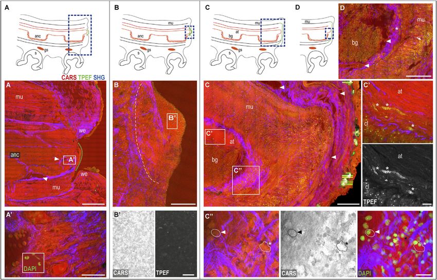
Imperadore et al.                                                                                                 Label-Free Imaging in Regeneration

INTRODUCTION                                                               standard histopathology; for instance, it has been proven
                                                                           suitable both for ex vivo and in vivo samples (Evans et al.,
Arm and tentacles in cephalopod mollusks are structures lacking            2005; Bocklitz et al., 2020). One of the potentially most useful
fluid-filled cavities and hard skeletal support. These animals               applications of this technique is in the evaluation of mammalian
utilize their appendages for environmental exploration, prey               disease states, including the production of high-resolution
manipulation, mating, and communication (for a review, see,                imaging of myelin sheets in physiological and pathological
for example, Villanueva et al., 2017). O. vulgaris arms have been          conditions (Huff and Cheng, 2007), and the identification of
subject to particularly detailed investigation because their               vessel tissue components to monitor the onset and progression of
peculiar architecture empowers possessing animals with high                arterial diseases, such as atherosclerosis or aneurysms (Wang
degrees of freedom in movement, including fine manipulation                 et al., 2008; Sehm et al., 2020). This approach has proved to be
abilities and muscular softening–stiffening control. The                   exceptionally versatile; it has been applied to the study of axon
architecture of O. vulgaris arms, in turn, has inspired the                regeneration after spinal cord or peripheral nerve lesions in
construction of robotic models (Cianchetti et al., 2011) for               mammals (Morisaki et al., 2013), amphibians (Uckermann
medical applications (e.g., minimally invasive surgical systems;           et al., 2019) and even invertebrates (Imperadore et al., 2018),
Cianchetti et al., 2014) and underwater exploration and sampling           facilitating comparison among animal species because it does not
(Calisti et al., 2015). Such an extensive use of appendages makes          rely on species-related epitopes.
these structures susceptible to a high risk of damage.                        Recently, we applied CARS microscopy in combination with
    It has been estimated that, in Octopus digueti, around 26% of          TPEF and SHG on cephalopods for the first time, using the
the population presents an arm injury (Voight, 1992), an                   regenerating pallial nerve of O. vulgaris as case study. We
incidence reaching 51% in O. vulgaris (Florini et al., 2011). A            highlighted structures, tissues, and cells implicated in
similar frequency was recently also confirmed by Voss and Mehta             regeneration and degeneration by evaluating the status of
(2021), who reported a 59.8% of incidence of injury in one or              axons and cells involved in debris removal as well as the
more arms in museum specimens of various octopus species                   connective tissues driving neural fibers. Such evidence would,
(i.e., O. bimaculatus, O. bimaculoides, and O. rubescens).                 otherwise, have proven hardly detectable with classical staining
    Moreover, the capacity to quickly heal and regenerate these            methods; at the very least, they would have required several
structures, even after severe injury or complete loss, is a peculiar       techniques in order to be revealed (Imperadore et al., 2018).
feature of octopuses that has been under investigation since                  In the current study, we applied multiphoton microscopy to
scientists first reported it in 1856 (Steenstrup, 1856).                    the octopus’ arm, a structure with a high level of structural
    The majority of studies examining the regenerative capacities          complexity. The arm is composed of nervous, muscular,
of appendages in cephalopods are, however, mostly descriptive              endothelial, vascular, and other tissues that, after severe
and focused on macroscopical events; only in recent years has              damage or complete loss, regenerate, resuming full
attention to the cellular and biological machinery of regeneration         functionality and complexity of the uninjured arm. By
begun to escalate (Fossati et al., 2013; Fossati et al., 2015; Zullo       comparing multiphoton microscopy images with classical
et al., 2017). One of the main issues hindering an in-depth                histological staining and immunohistochemistry (IHC), we
examination of regenerative processes remains the limited                  highlighted phases and key events during stump healing and
number of markers that are commercially available and                      regeneration and detected tissues and cells involved.
specifically designed for these organisms (Wollesen et al.,
2009; Imperadore et al., 2018; Zullo et al., 2020), thereby
reducing the potential for direct imaging.                                 MATERIALS AND METHODS
    Recently, new microscopical methods have been applied to
vertebrate models and may help resolve the issue of marker                 Ethical Statement
paucity in cephalopods and other non-model invertebrates.                  Cephalopods are included in the Directive 2010/63/EU and, thus,
Vibrational spectroscopy, the collective term used to describe             regulated for their use in scientific research (Fiorito et al., 2014;
the analytical techniques of infrared and Raman spectroscopy,              Fiorito et al., 2015). Experiments included in this study were
appears to be extremely helpful in this sense. Vibrational                 carried out in 2018 on tissue samples originating from wild
spectroscopy is a label-free technique for probing vibrational             animals. This study has been granted an ethical clearance for
energy levels associated with chemical bonds in a non-destructive          “label-free multiphoton microscopy for the investigation of the
and non-invasive manner, allowing the collection of                        process of arm regeneration in Octopus vulgaris” by the
comprehensive information about sample composition. In                     institutional AWB (OBA: case 4/2021/ec AWB-SZN -28 June
turn, coherent anti-Stokes Raman scattering (CARS)                         2021).
microscopy is a non-linear variant of Raman spectroscopy that
provides intensity information about single-molecular vibration            Animals, Surgery, and Sample Collection
modes at sub-micrometer resolution. In combination with                    This study was carried out on recently deceased Octopus vulgaris
endogenous two-photon excited fluorescence (TPEF) and                       (N = 6; four males, two females, body weight: 194–402 g) collected
second harmonic generation (SHG), CARS generates large                     from fishermen (Bay of Naples, Mediterranean Sea, Italy) during
datasets about the tissue under examination and allows for the             spring (seawater temperature range: 15–20°C). The animals were
acquisition of morpho-chemical information comparable to                   selected for the presence of one or more damaged arms in the

Frontiers in Cell and Developmental Biology | www.frontiersin.org      2                                  February 2022 | Volume 10 | Article 814746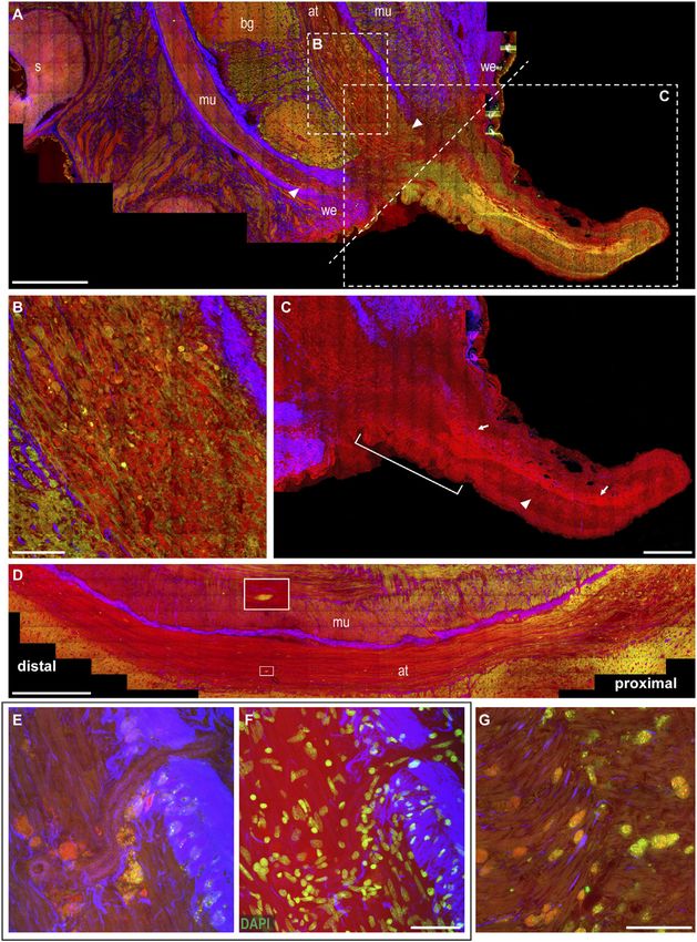
Imperadore et al.                                                                                                 Label-Free Imaging in Regeneration

phase of healing or regeneration (following stages reviewed in the          signals in the forward direction were collected using an air
study by Imperadore and Fiorito (2018)).                                    condenser (NA 0.4, Leica Microsystems, Mannheim,
   In the cases of octopuses still showing signs of life, the animals       Germany). Forward CARS (2850cm−1, CH2-stretch vibration)
were euthanized (3.5% MgCl2 in seawater, > 30 min), and death               was spectrally filtered by a short-pass filter SP750, a beam splitter
confirmed by transection of the dorsal aorta (Fiorito et al., 2015).         BS560, and a band-pass filter BP670/125. SHG was detected in
   Quality of tissues was assessed through classical histological           parallel and spectrally filtered by a short-pass filter SP750, a beam
methods and was found suitable for immunohistochemistry and                 splitter BS560, and a band-pass filter BP465/170. Signals in the
multiphoton microscopy imaging.                                             epi-direction were spectrally separated in SHG in the range from
   Damaged and corresponding contralateral uninjured arms                   400 to 510 nm and TPEF from 515 to 640 nm. All z-stacks were
(control) were harvested (~3 cm in length) for a total number               recorded with a voxel size of 0.2 µm × 0.2 µm × 3.0 µm. Z-stacks
of 23 samples. For the control, the arm tip (i.e., the most distal          range from 100 to 130 µm in height.
part) and a piece of arm at around 50% of its length (proximal)                 The resulting multimodal RGB images are represented as
were also collected.                                                        follows: red channel = CARS, green channel = TPEF, and blue
   The dissected samples were immediately processed, following              channel = SHG.
the study by Imperadore et al. (2017). In brief, the tissues were               The images were processed with LAS X (Leica Microsystems,
fixed in 4% PFA in seawater (3 h), followed by PBS (pH 7.4)                  Mannheim, Germany) and Zen Blue Edition (Carl Zeiss, AG,
washes, and immersion in sucrose 30% (in PBS) until sinking.                Jena, Germany) software.
The samples were then embedded in freezing and blocking
medium (OCT; Leica Biosystems) and stored at −80°C until                    Light Microscopy
use. Cryostat (Leica CM3050 S) sections (either 30 or 150 µm)               Following multiphoton imaging, the coverslip was carefully
were mounted on SuperFrost Plus glass slides.                               removed in PBS and slides used for immunohistochemistry or
   Two additional control arm tips were harvested, fixed, and                stained with hematoxylin and eosin (H&E). The H&E staining
stored in PBS to image the whole mount sample.                              protocol consisted of a 2-min bath in Meyer hematoxylin
                                                                            followed by a 5-min step in tap water and 20 s in eosin.
Multiphoton Microscopy                                                          IHC was performed as previously described (Imperadore et al.,
The cryostat sections were air-dried for 30 min, rehydrated in              2017). In brief, after blocking in normal goat serum (5% NGS, in
PBS, and covered with a glass coverslip. Imaging was performed              PBT: PBS Tween 0.1%) for 1 h in RT, the sections were incubated
with an optical microscope Axio Examiner Z.1 coupled to a laser             overnight with primary antibody (i.e., anti–acetylated tubulin,
scanning module LSM 7 (Carl Zeiss AG, Jena, Germany)                        SIGMA T6793, dilution 1:1,000; anti–phospho-Histone H3,
equipped with non-descanned detectors. An erbium fiber laser                 Sigma H9908, dilution 1:600) in PBT and NGS 1% at 4°C.
(Femto Fiber pro NIR from Toptica Photonics AG, Munich,                     Following washes in PBT, the sections were incubated with
Germany) provides excitation for TPEF and SHG by emitting at                secondary antibodies [1:250, Alexa Fluor goat anti-mouse IgG
781 nm with a pulse length of 1.2 ps and a maximum nominal                  (H + L) 488 and Alexa Fluor goat anti-rat IgG (H + L) 594] for 1 h
power of 100 mW. The TPEF signal was acquired in the spectral               at room temperature. DAPI (14.3 μmol L−1 in PBT) was used
range 500–550 nm, while the SHG signal was retrieved using a                after IHC or on unstained sections to counterstain nuclei.
band-pass filter centered at 390 nm. CARS excitation needed a                    The sections following IHC protocol were mounted in PBS
second laser source (i.e., the Femto Fiber pro TNIR from Toptica            and imaged again for multiphoton microscopy; H&E sections
Photonics AG) which is tunable in the range 850–1,100 nm and                were, instead, dehydrated in an ethanol series, cleared in xylene,
has a pulse length of 0.8 ps. In all CARS experiments, the                  coverslipped using DePex, and imaged using either Axio
wavelength was set to 1,005 nm (emitted power 1.5 mW) in                    Examiner Z.1 (Carl Zeiss AG) equipped with the camera
order to resonantly excite the symmetric stretching vibration               AxioCam or Axio Scope A1 (Carl Zeiss AG) equipped with
of methylene groups at 2,850 cm−1. CARS, TPEF, and SHG were                 the camera Canon DS126231. The images were processed with
simultaneously excited and acquired with a W Plan-Apochromat                Zen Blue Edition (Carl Zeiss, AG, Jena, Germany) software.
20x/1.0 (Carl Zeiss AG) (for a schematic diagram of the
system used for multiphoton microscopy, see Supplementary
Figure S1).                                                                 RESULTS
   For multimodal imaging of thicker slices (150 µm thickness)
and whole arm tips, a Leica SP8 CARS microscope with SRS                    Octopus appendages have sophisticated architecture. The major
upgrade (special part request, Leica Microsystems GmbH,                     structures of focus in this study are as follows: 1) the skin,
Mannheim, Germany) was used. A picoEmerald S Optical                        covering the arm (as well as the entire animal’s body; Packard,
Parametric Oscillator (APE Angewandte Physik und                            1988); 2) the intrinsic musculature, comprising a three-muscular
Elektronik GmbH, Berlin, Germany) provides a Stokes beam                    bundle (oblique, longitudinal, and transverse) (for a review, see
at 1,031 nm and a tunable pump beam in the range of                         Kier, 1988) arranged around a 3) central nerve cord, running
720–970 nm. The two pulse trains (pulse duration 1–2 ps)                    longitudinally along the entire arm and connecting centrally to
were spatially and temporally overlapped. The images were                   the sub-esophageal mass (in the brain) (Graziadei, 1971).
acquired using a ×25 water objective (HCX IRAPO L ×25/NA                       CARS, TPEF, and SHG during multimodal multiphoton
0.95/water, Leica Microsystems, Mannheim, Germany), and                     imaging on rehydrated cryosections and arm tip whole

Frontiers in Cell and Developmental Biology | www.frontiersin.org       3                                 February 2022 | Volume 10 | Article 814746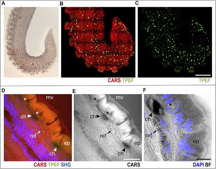
Imperadore et al.                                                                                                                            Label-Free Imaging in Regeneration

 FIGURE 1 | Arm skin structure and reflective elements. An isolated ex vivo arm tip imaged before fixation (A) with visible chromatophores. The same sample was
 imaged in whole mount through multiphoton microscopy. (B) Entire arm structure is shown in CARS (red), while chromatophores appear in TPEF (green spots in B,C).
 (D,E) Imaging of thin sections (30 μm, sagittal plane) highlighted the presence of microvilli (mv) covering the epidermis (ep); mucous cells (asterisks) are found distributed
 in the epidermis. Reflective elements (arrows) are identified as round granules in the dermal layer close to chromatophores (arrowheads). (F) Bright-field (BF)
 imaging of the same section counterstained with DAPI showed chromatophores (arrowhead) and reflective elements (arrow) underneath the epidermal layer, where
 mucous cells (asterisk) are identified based on morpholgy and position. Scale bars: 150 µm in (B,C), 50 µm in (D,E), and 20 µm in (F). Abbreviations: ch,
 chromatophores; ep, epidermis; mv, microvilli; ref, reflective elements.

mounts revealed the architecture of the intact octopus’                                     over the entire arm structure, which, in turn, appears in red
appendage, highlighting the entire tissue composition. Injured                              (CARS) in multichannel images (Figures 1B,C, whole mount
and healing arms were also imaged, allowing for the identification                           sample).
of main phases of regeneration, including at the levels of cells and                           Higher-magnification imaging of sagittal thin sections of the
tissues.                                                                                    arm allowed for identification of other distinctive elements of the
                                                                                            skin and surrounding tissues. The epidermis appeared covered in
Control Uninjured Arm                                                                       microvilli, characterized by an intense CARS signal; mucous cells
The skin. Octopus skin contains various organs and elements                                 were identified as negative imprints in the epidermal layer
(i.e., chromatophores, iridophores, leucophores, and papillae)                              (Figures 1D–F, asterisks); and close to chromatophores
that can be finely controlled to change the animal’s skin tone                               (Figures 1D–F, arrowheads), reflective elements appeared as
and texture, thereby providing the animals with extraordinary                               round granules just underneath the epidermis (CARS, Figures
camouflaging and interspecific communication (Borrelli et al.,                                1D–F, arrows). Bright-field imaging (i.e., transmitted white light)
2006; How et al., 2017; for a review, see Packard and Hochberg,                             of the same section highlighted the nuclear components of the
1977). Chromatophores, in particular, are sacculus organs                                   epidermis (DAPI counterstain in blue in Figure 1F), confirming
responsible for color change, owing to the presence of diverse                              the identity of mucous cells by morphology and nuclear position.
pigment granules controlled by muscle bundles that are radially                                The muscular tissue. A schematic drawing of the arm
organized to open and close the sacculus (Messenger, 2001).                                 morphology in the transverse plane is included in Figure 2A
    In the skin covering the arm (Figure 1A), chromatophores                                to facilitate structural identification (Supplementary Figure
were easily identified as bright green spots (TPEF) distributed                              S2A). The three muscle groups belonging to the intrinsic

Frontiers in Cell and Developmental Biology | www.frontiersin.org                       4                                           February 2022 | Volume 10 | Article 814746
Imperadore et al. Label-Free Imaging in Regeneration
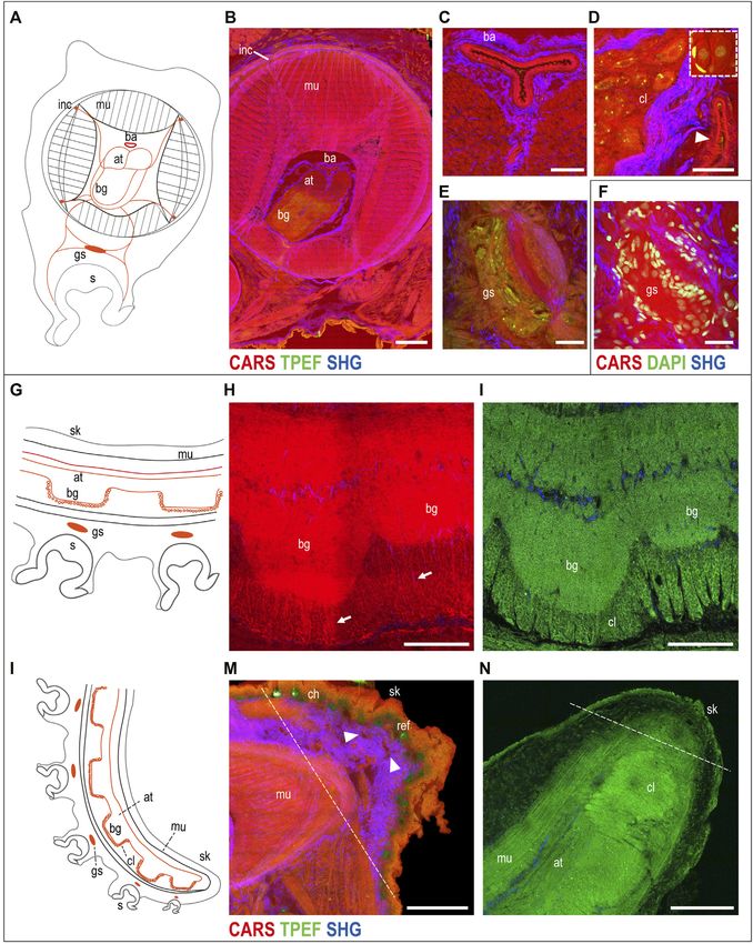
FIGURE 2 | Uninjured arm. Schematic drawings of octopus arm morphology in transverse (A) and sagittal (G,L) planes. (B) Multiphoton microscopy image of an
arm transverse section showing the three muscle bundles belonging to the intrinsic musculature of the arm (CARS) and the connective tissue sheaths enveloping them
(SHG). The axial nerve cord (comprising two axonal tracts on the dorsal side and brachial ganglia on the ventral side) and the four intramuscular nerve cords are clearly
identified (CARS and TPEF). (C) Above the axonal tracts, the brachial artery is visible (CARS) surrounded by connective tissue (SHG). (D) Outer cellular layer of a
brachial ganglion appears comprising small and big neurons emitted in CARS and TPEF. DAPI counterstaining (white dotted rectangle) highlights neuron nuclei and
supporting cells surrounding them. The arrowhead points a blood vessel around the nerve cord. (E) Ganglion of the suckers comprising a central neuropil and
(Continued )
Frontiers in Cell and Developmental Biology | www.frontiersin.org 5 February 2022 | Volume 10 | Article 814746Imperadore et al. Label-Free Imaging in Regeneration
FIGURE 2 | surrounding neurons. (F) Neuron nuclei are counterstained in DAPI for further confirmation. (H,I) Imaging of the axial nerve cord in the thick sagittal section
(150 µm). (H) Fibers from the brachial ganglia descend from the neuropil (arrows), passing through the (I) cellular layer. (M) Most distal part of the arm tip (delimited by a
dotted line) presents numerous blood vessels (arrowheads). (N) Single plane from arm tip whole-mount imaging (in TPEF). Dotted line delimits the most distal part of the
arm tip. Scale bars: 500 µm in (B), 100 µm in (C,E,F,H,I), 50 µm in (D), and 200 µm in (M,N). Abbreviations: at, axonal tract; ba, brachial artery; bg, brachial ganglion; ch,
chromatophores; cl, cellular layer; gs, sucker ganglion; inc, intramuscular nerve cord; mu, muscles; ref, reflective elements; s, sucker; sk, skin.
musculature of the arm were visualized in CARS (Figure 2B). In Compared to more aboral arm portions, in the tip, we
particular, the i) tightly packed transverse muscle bundles observed a greater area occupied by the axial nerve cord,
running perpendicular to the arm long axis, which elongate reducing the space for muscles; brachial ganglia get closer to
through structures called trabeculae, were visible among the ii) each other (Figures 2H,I,N) as suckers get smaller and closer.
longitudinal muscles and the iii) three bundles of oblique muscles Fibers from the brachial ganglia descend from the neuropil
(see Supplementary Figure S2 for more details). Connective (CARS, Figure 2H, arrow), passing through the cellular layer
tissue sheaths, highlighted in SHG, appear to envelop the (TPEF, Figure 2I).
different muscle layers of the intrinsic musculature (Figure 2B). Tissues and structures at the most distal part of the tip
CARS imaging also enabled identification of the intrinsic (delimited by a dotted line in Figures 2M,N) appear less
musculature of the sucker (data not shown) and the organized and differentiated compared to all the other
acetabulo-brachial muscles (Supplementary Figure S2), which neighboring areas (Supplementary Figure S3). The tip
connect the intrinsic muscles of the arm and the intrinsic muscles appears characterized by a thick layer of connective tissue
of the suckers. (SHG) (Figure 2M), which appears in between the epidermis
The neural structures. The neural control for these sets of and the muscular layer covering the nerve cord. CARS also
muscles is provided by six main nerve centers per arm, that is, a highlighted the presence of numerous blood vessels in this
central axial nerve cord connected to four intramuscular nerve zone (Figure 2M, arrowhead).
cords and to sucker ganglia. The axial nerve cord comprises two
axonal tracts (dorsal) and several brachial ganglia (ventral), facing Healing Arm
and innervating suckers (Figures 2A,B; Supplementary Figure The wounded skin. The regenerative process of a damaged arm
S2). Running longitudinally to the axonal tracts, the main blood in O. vulgaris is always initiated by wound healing, with the
vessel supplying hemolymph to the arm (brachial artery) is dermis wound edges closing around the lesion. This process
shown by CARS and is surrounded by connective tissue generally requires between 0 and 5 days, depending on several
(SHG) (Figures 2B,C; Supplementary Figure S2B). factors, such as temperature, animal age and sexual maturity, and
Each brachial ganglion comprises an inner neuropil and an health status (for a review, see Imperadore and Fiorito, 2018). To
outer cellular layer (Figures 2B,D; Supplementary Figures facilitate readers, the main phases of the healing process are
S2B–D). The cellular layer contains many small and some sketched in Figures 3A–D.
big neurons, with nuclei ranging from less than 5–20 µm The wounded dermis contracts and forms a rim that starts
(Young 1963). These cells emit both in CARS and TPEF, covering the wound to form a first protective layer for the exposed
with the latter mainly highlighting their cytoplasm, giving a tissues (Figure 3A). The connective tissue within it appears
strong signal of granular structures contained in the cells involved in the process, narrowing around the muscular
(Figure 2D; Supplementary Figure S2C) that could be partly tissues of the stump (SHG, Figure 3A) and contributing to
due to lipofuscin. DAPI counterstaining confirmed these results wound closure.
and highlighted the presence of supporting cells around the The central portion of the damaged arm (i.e., the internal
neurons, which were not detected with multiphoton microscopy muscles and axial nerve cord) remains exposed until a clot of
alone (see the dotted white rectangle in Figure 2D). Axons in agglutinated blood corpuscles start depositing over it (green
the intricate neuropil of the brachial ganglia are also highlighted dotted line in Figure 3A). Blood vessels, which are observed
in CARS and TPEF (Figure 2B; Supplementary Figures in great number in the arm stump (Figure 3A, arrowheads),
S2B,C). DAPI counterstaining and acetylated tubulin represent the origin of these cells (Figures 3A9, see also inset in
immunoreactivity confirmed these results (Supplementary Figure 3A9).
Figure S2D). The clot then increases in size, covering the whole exposed
Some of the nerves departing from the brachial ganglia are tissue in between the wound epithelium (Figure 3B) and forming
linked to the four intramuscular nerve cords (CARS; Figure 2B; a dense and fine network of interdigitated cells called primary
Supplementary Figure S2B), and to the ganglion of the sucker blastema (Lange, 1920). The cells in this blastema appear full of
(CARS and TPEF; Figures 2E,F). This ganglion comprises a dense granules highlighted in CARS and TPEF (Figure 3B9). A
central neuropil and neurons surrounding it (TPEF and DAPI) boundary line of connective tissue separates this blastema from
(Figures 2E,F). the underlying and well-differentiated tissues (SHG, Figure 3B).
Imaging of the arm tip allowed visualization of the The primary blastema finally never casts off, but rather is retained
abovementioned anatomical structures (a schematic drawing in and eventually completely covered by the regenerating
the sagittal section is reported to facilitate structure identification, epithelium, the latter appearing highly vascularized
Figures 2G,L). (Figure 3C, arrowheads).
Frontiers in Cell and Developmental Biology | www.frontiersin.org 6 February 2022 | Volume 10 | Article 814746Imperadore et al. Label-Free Imaging in Regeneration
FIGURE 3 | Phases of the arm healing process. Schematic drawings of an arm in the sagittal section (A–D) Describing healing phases imaged with multiphoton
microscopy. (A) Wounded dermis forms a rim contracting around the wound. The connective tissue (SHG) narrows around the muscular tissues of the stump
contributing to wound closure. A clot of agglutinated blood cells deposits over it (green dotted line) originating from blood vessels in the stump (arrowheads). (A9)
Hemocytes released in the stump. DAPI counterstaining shows the peculiar u-shaped nucleus of the hemocytes, which occupies most of the cytoplasm. (B) Clot
covers the whole exposed tissue in between the wound epithelium. A boundary line (white dotted line) of the connective tissue (SHG) separates the clot from the
underlying and well-differentiated tissues of the stump. (B9) Clot appears as a dense and fine network of interdigitated cells, (C) Regenerating epithelium and highly
vascularized (arrowheads) covers the clot. Damaged muscles and axonal tracts degenerate (C9) (asterisks). (C99) Hemocytes are released from the blood stream. They
change their appearance from circulating round-shaped cells with a small cytoplasm and u-shaped nuclei (dotted line with arrowhead) into amebocyte-like cells with a
large and granular cytoplasm, intensely emitted in CARS and TPEF (dotted line with asterisks). DAPI counterstaining shows the nuclei of these cells. (D) Hemocytes are
released from the vessels around the nerve cord (arrowheads) into the connective tissue around it (asterisk), then, invade all muscle layers below the wounded epithelium
(arrow). Scale bars: 500 µm in (A), 50 µm in (A9), 20 µm enlargement in (A9), 200 µm in (B–D), and 20 µm in (B9,C9,C99). Abbreviations: at, axonal tract; bg, brachial
ganglion; cl, cellular layer; gs, sucker ganglion; s, sucker; we, wound epithelium.
The muscular and neural tissues. At this stage, the damaged (Figure 3D, arrowheads) are first released into the connective
muscles and nerve tissues (i.e., the axonal tracts) show evident tissue around it (Figure 3D, asterisk) and then invade all muscle
signs of degeneration (i.e., swelling and fragmentation). layers below the wounded epithelium (Figure 3D, arrow).
Degenerating tissues, highlighted in CARS and TPEF
(Figure 3C9, asterisks) are not observed in control tissues. Regenerating Arm
The hemocytes. The muscular layers in the healed stump are The wounded skin. From the healed skin, a little knob appears,
invaded by many cells (Figure 3C), identified as hemocytes, regenerating an arm from the dorsal side of the stump. The
which change their appearance once released from blood resulting arm is initially much thinner than the original stump
vessels. They indeed transform from circulating, round-shaped (Figure 4A). The wound epithelium, narrowing around the
cells with a small cytoplasm and u-shaped nuclei (Figure 3C99, original site of the lesion, is still visible and characterized by
dotted line with arrowhead) into amebocyte-like cells with a large thick connective tissue (SHG, Figures 4A,C).
and granular cytoplasm, intensely emitting in CARS and TPEF The muscular and neural tissues. Degeneration in this phase
(Figure 3C99, dotted line with asterisk). DAPI counterstaining involves greater areas in the muscular tissues and the nerve cord
confirmed the cellular nature of these structures (Figure 3C99). of the stump (Figure 4A). Degeneration is particularly evident in
Hemocytes released from the vessels around the nerve cord the axonal tracts of the nerve cord, close to the original site of the
Frontiers in Cell and Developmental Biology | www.frontiersin.org 7 February 2022 | Volume 10 | Article 814746Imperadore et al. Label-Free Imaging in Regeneration
FIGURE 4 | Arm regeneration. (A) Little knob regenerates from the dorsal side of the stump. The wound epithelium narrows around the original site of the lesion,
characterized by thick connective tissue. White dotted line marks the original site of the lesion dividing the stump from the regenerating tip. (B) Degeneration is evident in
the axonal tracts of the nerve cord, where fibers appear swollen and break into lumps (CARS and TPEF). (C) Regenerating fibers appear among degenerating lumps in
the axonal tract (highlighted in CARS) and can be followed providing innervation in the newly forming tip (arrows); thin processes can be seen descending
perpendicularly to these fibers (arrowhead). Suckers develop at the base of the regenerating arm (white bar). (D) Degeneration can be followed along the axonal tracts: it
involves a great number of fibers proximal to the lesion; few degenerating fibers can be detected moving distally (see enlargement). (E,F) Large number of cells, whose
cytoplasm is rich in granules emitting in CARS and TPEF, are imaged accumulating close to blood vessel walls. DAPI counterstaining highlighted cell nuclei. (G) Cells rich
in granules emitted in CARS and TPEF invade muscle layers in the stump. Scale bars: 500 µm in (A,D), 100 µm in (B,G), 250 µm in (C), and 50 µm in (E,F).
Abbreviations: at, axonal tract; bg, brachial ganglion; mu, muscles; s, sucker; we, wound epithelium.
lesion, where fibers appear swollen and broken into lumps (CARS in the newly forming arm tip (Figure 4C, arrows). The
and TPEF, Figure 4B). Degeneration can be followed along the regenerating tip is mostly occupied by the newly forming
axonal tract, with the number of fibers involved decreasing when nerve cord (Figure 4C, arrows), and thin processes can be
farther from the site of the lesion. Distal to this site, fewer seen descending from it toward the ventral site, where new
degenerative events are found using multiphoton imaging suckers will later form (Figure 4C, arrowhead). Suckers start
(Figure 4D). to develop in the forming arm, close to the stump (Figure 4C).
Among the degenerating lumps in the axonal tract, The hemocytes. The tissues in the stump are invaded by cells
regenerating fibers also appear (highlighted by strong CARS whose cytoplasm is rich in small granules, strongly emitting in
signal, Figure 4B) and can be followed providing innervation CARS and TPEF (Figures 4E–G). They mainly invade muscles
Frontiers in Cell and Developmental Biology | www.frontiersin.org 8 February 2022 | Volume 10 | Article 814746Imperadore et al. Label-Free Imaging in Regeneration
around the nerve cord and the axonal tracts of the latter (see also contributing to better compliance with the 3R Principle
Supplementary Figure S4A, arrowheads), but are never observed (Fiorito et al., 2014; https://nc3rs.org.uk/the-3rs).
in the neuropil of the brachial ganglia. These cells also reach This approach could be extended to other lines of cephalopod
muscles in the regenerating arm tip, very close to the site of the research and to different non-mammalian animal species,
lesion (Supplementary Figure S4A), but are not found distant to enabling data collection without having to focus on one or a
this site or in any other tissue of this new structure. few proteins, as is usually the case with IHC approaches.
These cells appear to be released by blood vessels close to the
injury site (Supplementary Video S1).
Areas invaded by these structures are also characterized by DATA AVAILABILITY STATEMENT
numerous proliferating cells (Supplementary Figure S4B).
The original contributions presented in the study are included in
the article/Supplementary Material, further inquiries can be
DISCUSSION directed to the corresponding author.
Multiphoton imaging has been successfully applied to several
species to investigate a number of biological processes (Zipfel ETHICS STATEMENT
et al., 2003) including healing and regeneration.
Multimodal images (CARS, TPEF, and SHG) of O. vulgaris The animal study was reviewed and approved by the Organismo
uninjured and damaged arms allowed for the identification of the per il Benessere Animale (AWB-SZN) della Stazione Zoologica
cellular and structural elements characterizing the parts and Anton Dohrn, Napoli Italia.
contributing to appendage regeneration, helping in dissecting
this complex phenomenon in the absence of specific markers
available for the taxon. In particular, chromatophores—skin AUTHOR CONTRIBUTIONS
element key for body patterning—and muscular
bundles—contributing to motor patterns of the arm and the PI designed the experiments, carried out the sample collection
main neural components—were detected (Figures 1, 2; and processing, imaged the thin sections, analyzed the data, and
Supplementary Figure S2). drafted the manuscript; OU and RG contributed to multiphoton
Wound healing is a phenomenon with widespread occurrence imaging; MW and AZ performed multiphoton imaging on whole
among both vertebrates (e.g., Ambystoma mexicanum and Danio mounts and thick sections. All authors contributed to the final
rerio) and invertebrates (e.g., Caenorhabditis elegans and writing of the manuscript.
Drosophila melanogaster), also occurring in mammals. This
involves the activation of the immune response and the
remodeling of the extracellular matrix (Arenas Gómez et al., FUNDING
2020), with regenerative species sharing impressive similarities in
the process. This study was initiated through a Travelling Fellowship of The
In octopus, healing is marked by dermis contraction, which Company of Biologists’—Journal of Experimental Biology
eventually covers the clot of agglutinated corpuscles depositing (JEB—180806, https://journals.biologists.com/jeb/)—granted to
over the exposed tissue to form the blastema. Hemocytes invade PI to visit Uckermann’s laboratory.
the stump, changing their appearances from circulating, round-
shaped cells to amebocyte-like cells (Figure 3). These latter cells
resemble vertebrate macrophages (Aurora and Olson, 2014; ACKNOWLEDGMENTS
Uckermann et al., 2019), thus suggesting their involvement in
debris removal. The authors acknowledge COST and the COST Action FA1301
After complete healing, a little tip regenerates from the “A network for improvement of cephalopod welfare and
octopus arm stump with new fibers innervating it. Cells rich husbandry in research, aquaculture, and fisheries
in small granules (CARS and TPEF), likely hemocytes, are (CephsInAction)” for support and networking capabilities.
found to invade muscles and nerve tissues which are also Finally, the authors are grateful to Dr. Giovanna Ponte for
characterized by intense proliferation (Figure 4; continuous advice and to Dr. Kathryn Maxson Jones for
Supplementary Figure S4). proofreading the article.
Here, we imaged structures and cells involved in arm
regeneration in the octopus, bypassing the need for staining or
markers, enabling the collection of voluminous data in a short SUPPLEMENTARY MATERIAL
period of time. Additionally, scanned samples are suitable for
further processing, for instance IHC and staining, allowing for The Supplementary Material for this article can be found online at:
amplified saving of time and resources and reducing the number https://www.frontiersin.org/articles/10.3389/fcell.2022.814746/
of samples and experimental animals needed, thereby full#supplementary-material
Frontiers in Cell and Developmental Biology | www.frontiersin.org 9 February 2022 | Volume 10 | Article 814746Imperadore et al. Label-Free Imaging in Regeneration
REFERENCES Messenger, J. B. (2001). Cephalopod Chromatophores: Neurobiology and Natural
History. Cephalopod chromatophores: Neurobiol. Nat. Hist. Biol. Rev. 76 (4),
473–528. doi:10.1017/s1464793101005772
Arenas Gómez, C. M., Sabin, K. Z., and Echeverri, K. (2020). Wound Healing Morisaki, S., Ota, C., Matsuda, K.-i., Kaku, N., Fujiwara, H., Oda, R., et al. (2013).
across the Animal Kingdom: Crosstalk between the Immune System and the Application of Raman Spectroscopy for Visualizing Biochemical Changes
Extracellular Matrix. Dev. Dyn. 249 (7), 834–846. doi:10.1002/dvdy.178 during Peripheral Nerve Injury in Vitro and in Vivo. J. Biomed. Opt. 18
Aurora, A. B., and Olson, E. N. (2014). Immune Modulation of Stem Cells and (11), 116011. doi:10.1117/1.jbo.18.11.116011
Regeneration. Cell Stem Cell 15 (1), 14–25. doi:10.1016/j.stem.2014.06.009 Packard, A., and Hochberg, F. G. (1977). “Skin Patterning in octopus and Other
Bocklitz, T., Silge, A., Bae, H., Rodewald, M., Legesse, F. B., Meyer, T., et al. (2020). Genera,” in The Biology of Cephalopods. Editors M Nixon and JB Messenger
Non-invasive Imaging Techniques: From Histology to In Vivo Imaging: (London: Academic press), 191–231.
Chapter of Imaging In Oncology. Recent Results Cancer Res. 216, 795–812. Packard, A. (1988). “The Skin of Cephalopods (Coleoids): General and Special Adaptations,”
doi:10.1007/978-3-030-42618-7_25 in The Mollusca, Form and Function. Editors ER Trueman and MR Clarke (New York:
Borrelli, L., Ghirardi, F., and Fiorito, G. (2006). A Catalogue of Body Patterning in Academic Press), 37–67. doi:10.1016/b978-0-12-751411-6.50010-2
Cephalopods. Napoli: Stazione Zoologica A. Dohrn; Firenze University Press. Sehm, T., Uckermann, O., Galli, R., Meinhardt, M., Rickelt, E., Krex, D., et al.
Calisti, M., Corucci, F., Arienti, A., and Laschi, C. (2015). Dynamics of Underwater (2020). Label-free Multiphoton Microscopy as a Tool to Investigate Alterations
Legged Locomotion: Modeling and Experiments on an Octopus-Inspired of Cerebral Aneurysms. Sci. Rep. 10, 12359. doi:10.1038/s41598-020-69222-5
Robot. Bioinspir. Biomim. 10 (4), 046012. doi:10.1088/1748-3190/10/4/046012 Steenstrup, J. J. (1856). Hectocotyldannelsen Hos Octopodslaegterne Argonauta
Cianchetti, M., Ranzani, T., Gerboni, G., Nanayakkara, T., Althoefer, K., Dasgupta, Og Tremoctopus, Oply St Ved Lagttagelse of Lignende Dannelser Hos
P., et al. (2014). Soft Robotics Technologies to Address Shortcomings in Today’s Blaekspruntterne I Almindelighed. K danske Vidensk Selsk Skr 5R (4), 186–215.
Minimally Invasive Surgery: The Stiff-Flop Approach. Soft robotics 1 (2), Uckermann, O., Hirsch, J., Galli, R., Bendig, J., Later, R., Koch, E., et al. (2019).
122–131. doi:10.1089/soro.2014.0001 Label-free Imaging of Tissue Architecture during Axolotl Peripheral Nerve
Cianchetti, M., Arienti, A., Follador, M., Mazzolai, B., Dario, P., and Laschi, C. Regeneration in Comparison to Functional Recovery. Sci. Rep. 9 (1), 12641.
(2011). Design Concept and Validation of a Robotic Arm Inspired by the doi:10.1038/s41598-019-49067-3
octopus. Mater. Sci. Eng. C. 31 (6), 1230–1239. doi:10.1016/j.msec.2010.12.004 Villanueva, R., Perricone, V., and Fiorito, G. (2017). Cephalopods as Predators: A
Evans, C. L., Potma, E. O., Puoris’haag, M., Cote, D., Lin, C. P., and Xie, X. S. Short Journey Among Behavioral Flexibilities, Adaptions, and Feeding Habits.
(2005). Chemical Imaging of Tissue In Vivo with Video-Rate Coherent Anti- Front. Physiol. 8, 598. doi:10.3389/fphys.2017.00598
stokes Raman Scattering Microscopy. Proc. Natl. Acad. Sci. 102 (46), Voight, J. R. (1992). Movement, Injuries and Growth of Members of a Natural
16807–16812. doi:10.1073/pnas.0508282102 Population of the pacific Pygmy Octopus, Octopus diguenti. J. Zoolog. 228 (2),
Fiorito, G., Affuso, A., Basil, J., Cole, A., de Girolamo, P., D’Angelo, L., et al. (2015). 247–264. doi:10.1111/j.1469-7998.1992.tb04606.x
Guidelines for the Care and Welfare of Cephalopods in Research -a Consensus Voss, K. M., and Mehta, R. S. (2021). Asymmetry in the Frequency and Proportion
Based on an Initiative by Cephres, Felasa and the Boyd Group. Lab. Anim. 49 (2 of Arm Truncation in Three Sympatric california Octopus Species. Zoology 147,
Suppl. l), 1–90. doi:10.1177/0023677215580006 125940. doi:10.1016/j.zool.2021.125940
Fiorito, G., Affuso, A., Anderson, D. B., Basil, J., Bonnaud, L., Botta, G., et al. (2014). Wang, H.-W., Le, T. T., and Cheng, J.-X. (2008). Label-free Imaging of Arterial
Cephalopods in Neuroscience: Regulations, Research and the 3rs. Invert Cells and Extracellular Matrix Using a Multimodal Cars Microscope. Opt.
Neurosci. 14 (1), 13–36. doi:10.1007/s10158-013-0165-x Commun. 281 (7), 1813–1822. doi:10.1016/j.optcom.2007.07.067
Florini, M., Fiorito, G., Hague, T., and Andrews, P. (2011). Monco": A Natural Wollesen, T., Loesel, R., and Wanninger, A. (2009). Pygmy Squids and Giant
Model for Studying Arm Usage and Regeneration in octopus Vulgaris. Brains: Mapping the Complex Cephalopod CNS by Phalloidin Staining of
J. Shellfish Res. 30 (3), 1002. Vibratome Sections and Whole-Mount Preparations. J. Neurosci. Methods 179
Fossati, S. M., Candiani, S., Nödl, M.-T., Maragliano, L., Pennuto, M., (1), 63–67. doi:10.1016/j.jneumeth.2009.01.021
Domingues, P., et al. (2015). Identification and Expression of Young, J. Z. (1963). The Number and Sizes of Nerve Cells in octopus. Proc. Zoolog.
Acetylcholinesterase in Octopus vulgaris Arm Development and Soc. Lond. 140 (2), 229–254. doi:10.1111/j.1469-7998.1963.tb01862.x
Regeneration: A Conserved Role for AchE? Mol. Neurobiol. 52 (1), 45–56. Zipfel, W. R., Williams, R. M., and Webb, W. W. (2003). Nonlinear Magic:
doi:10.1007/s12035-014-8842-2 Multiphoton Microscopy in the Biosciences. Nat. Biotechnol. 21 (11),
Fossati, S. M., Carella, F., De Vico, G., Benfenati, F., and Zullo, L. (2013). Octopus 1369–1377. doi:10.1038/nbt899
Arm Regeneration: Role of Acetylcholinesterase during Morphological Zullo, L., Bozzo, M., Daya, A., Di Clemente, A., Mancini, F. P., Megighian, A., et al.
Modification. J. Exp. Mar. Biol. Ecol. 447, 93–99. doi:10.1016/j.jembe.2013.02.015 (2020). The Diversity of Muscles and Their Regenerative Potential across
Graziadei, P. (1971). The Nervous System of the Arm. The Anatomy of the Nervous Animals. Cells 9 (9), 1925. doi:10.3390/cells9091925
System of Octopus vulgaris. London, UK: Oxford University Press, 45–61. Zullo, L., Fossati, S. M., Imperadore, P., and Nödl, M.-T. (2017). Molecular
How, M. J., Norman, M. D., Finn, J., Chung, W.-S., and Marshall, N. J. (2017). Dynamic Determinants of Cephalopod Muscles and Their Implication in Muscle
Skin Patterns in Cephalopods. Front. Physiol. 8, 393. doi:10.3389/fphys.2017.00393 Regeneration. Front. Cel Dev. Biol. 5, 53. doi:10.3389/fcell.2017.00053
Huff, T. B., and Cheng, J. X. (2007). In Vivo coherent Anti-stokes Raman Scattering
Imaging of Sciatic Nerve Tissue. J. Microsc. 225 (Pt 2), 175–182. doi:10.1111/j. Conflict of Interest: The authors declare that the research was conducted in the
1365-2818.2007.01729.x absence of any commercial or financial relationships that could be construed as a
Imperadore, P., Uckermann, O., Galli, R., Steiner, G., Kirsch, M., and Fiorito, G. potential conflict of interest.
(2018). Nerve Regeneration in the Cephalopod Mollusc Octopus vulgaris: Label-
free Multiphoton Microscopy as a Tool for Investigation. J. R. Soc. Interf. 15 Publisher’s Note: All claims expressed in this article are solely those of the authors
(141), 20170889. doi:10.1098/rsif.2017.0889 and do not necessarily represent those of their affiliated organizations, or those of
Imperadore, P., and Fiorito, G. (2018). Cephalopod Tissue Regeneration: the publisher, the editors and the reviewers. Any product that may be evaluated in
Consolidating over a century of Knowledge. Front. Physiol. 9, 593. doi:10. this article, or claim that may be made by its manufacturer, is not guaranteed or
3389/fphys.2018.00593 endorsed by the publisher.
Imperadore, P., Shah, S. B., Makarenkova, H. P., and Fiorito, G. (2017). Nerve
Degeneration and Regeneration in the Cephalopod Mollusc Octopus vulgaris: Copyright © 2022 Imperadore, Galli, Winterhalder, Zumbusch and Uckermann.
the Case of the Pallial Nerve. Sci. Rep. 7, 46564. doi:10.1038/srep46564 This is an open-access article distributed under the terms of the Creative Commons
Kier, W. M. (1988). “The Arrangement and Function of Molluscan Muscle,” in The Attribution License (CC BY). The use, distribution or reproduction in other forums is
Mollusca, Form and Function. Editors ER Trueman and MR Clarke (New York: permitted, provided the original author(s) and the copyright owner(s) are credited
Academic Press), 211–252. doi:10.1016/b978-0-12-751411-6.50016-3 and that the original publication in this journal is cited, in accordance with accepted
Lange, M. M. (1920). On the Regeneration and Finer Structure of the Arms of the academic practice. No use, distribution or reproduction is permitted which does not
Cephalopods. J. Exper. Zool. 31, 1–57. doi:10.1002/jez.1400310102 comply with these terms.
Frontiers in Cell and Developmental Biology | www.frontiersin.org 10 February 2022 | Volume 10 | Article 814746You can also read