Influence of Heterologous and Homologous Vaccines, and Their Components, on the Host Immune Response and Protection Against Experimental Caprine ...
←
→
Page content transcription
If your browser does not render page correctly, please read the page content below
ORIGINAL RESEARCH
                                                                                                                                         published: 05 January 2022
                                                                                                                                    doi: 10.3389/fvets.2021.744568

                                             Influence of Heterologous and
                                             Homologous Vaccines, and Their
                                             Components, on the Host Immune
                                             Response and Protection Against
                                             Experimental Caprine
                                             Paratuberculosis
                                             Noive Arteche-Villasol 1,2*† , Daniel Gutiérrez-Expósito 1,2† , Natalia Elguezabal 3 ,
                                             Iker A. Sevilla 3 , Raquel Vallejo 1,2 , José Espinosa 1,2 , María del Carmen Ferreras 1,2 ,
                                             Julio Benavides 2 and Valentín Pérez 1,2
                           Edited by:        1
                                              Departamento de Sanidad Animal, Facultad de Veterinaria, Universidad de León, León, Spain, 2 Departamento de Sanidad
            Francisco Javier Salguero,       Animal, Instituto de Ganadería de Montaña (CSIC-ULE), León, Spain, 3 Departamento de Sanidad Animal, NEIKER-Instituto
               Public Health England,        Vasco de Investigación y Desarrollo Agrario, Derio, Spain
                      United Kingdom

                         Reviewed by:
                         Beatriz Vidana,     Vaccination against paratuberculosis, a chronic disease of ruminants caused by
 University of Bristol, United Kingdom       Mycobacterium avium subsp. paratuberculosis (Map), has been considered as the
                        Mourad Tayebi,
 Western Sydney University, Australia
                                             most effective control method. However, protection is incomplete, and the mechanisms
                   *Correspondence:
                                             operating in the response of the animals to vaccination are not fully understood.
                  Noive Arteche-Villasol     Therefore, this study analyzed the immune response and the effects on protection against
                      nartv@unileon.es       Map infection, elicited by paratuberculosis (Silirum® ) and tuberculosis (heat-inactivated
     † These   authors have contributed      M. bovis [HIMB]) vaccines and their components in a caprine experimental model. Fifty
                    equally to this work     goat kids were divided into 10 groups (n = 5) according to their vaccination (Silirum® ,
                                             HIMB and nonvaccinated), immunization (inactivated bacteria or adjuvant), and/or
                    Specialty section:
         This article was submitted to       infection. Oral challenge with Map was performed 45 days postvaccination/immunization
          Veterinary Experimental and        (dpv), and animals were euthanized at 190 dpv. Peripheral immune response and
                 Diagnostic Pathology,
                a section of the journal     proportion of lymphocyte subpopulations were assessed monthly by enzyme-linked
        Frontiers in Veterinary Science      immunosorbent assay and flow cytometry analysis, respectively. Local immune response,
            Received: 20 July 2021           proportion of tissue lymphocyte subpopulations, Map detection (polymerase chain
       Accepted: 02 December 2021
                                             reaction), and histological examination were conducted in gut-associated lymphoid
        Published: 05 January 2022
                                             tissues. All infected groups developed paratuberculosis granulomatous lesions despite
                               Citation:
                     Arteche-Villasol N,     vaccination or immunization. The Silirum® and HIMB-vaccinated groups showed a
  Gutiérrez-Expósito D, Elguezabal N,        considerable lesion reduction consistent with a significant peripheral cellular and humoral
      Sevilla IA, Vallejo R, Espinosa J,
       Ferreras MC, Benavides J and
                                             immune response. Besides, a lower number of granulomas were observed in groups
           Pérez V (2022) Influence of       immunized with inactivated bacteria and adjuvants in comparison to nonvaccinated
      Heterologous and Homologous
                                             and infected group. However, despite not being significant, this reduction was even
 Vaccines, and Their Components, on
     the Host Immune Response and            higher in adjuvant immunized groups, which developed milder granulomatous lesion
      Protection Against Experimental        with no detectable peripheral immune responses associated with immunization. No
             Caprine Paratuberculosis.
             Front. Vet. Sci. 8:744568.
                                             changes in the peripheral and local proportion of lymphocyte subsets or local immune
     doi: 10.3389/fvets.2021.744568          response were detected in relation to either vaccination/immunization or infection.

Frontiers in Veterinary Science | www.frontiersin.org                             1                                        January 2022 | Volume 8 | Article 744568
Arteche-Villasol et al.                                                                                            Vaccination Against Caprine Paratuberculosis

                                             Despite that paratuberculosis and tuberculosis vaccination showed a partial and
                                             cross-protection against Map infection, respectively, only histological examination could
                                             assess the progression of infection in these animals. In addition, the pattern observed
                                             in the reduction of the lesions in adjuvant immunized groups suggests the possible
                                             involvement of a nonspecific immune response that reduces the development of
                                             granulomatous lesions.

                                             Keywords: vaccination, paratuberculosis, tuberculosis, HIMB, adjuvant, cross-protection

INTRODUCTION                                                                       vaccinated animals could remain highly infectious and develop
                                                                                   severe lesions (16, 22, 23).
Paratuberculosis is a chronic and debilitating disease                                 Most paratuberculosis heat-killed vaccines are based on Map
of ruminants caused by Mycobacterium avium subsp.                                  316F strain, which has demonstrated reduced virulence (tissue
paratuberculosis (Map) (1) and responsible for significant global                  bacterial burden) during experimental infection of calves with
economic losses estimated at approximately US $12.61 million                       this live isolate likely due to continuous in vitro passages (24).
and $364.31 million in Spanish and European Union dairy cattle,                    In this sense, subcutaneous immunization of sheep with heat-
respectively (2, 3). Map is a ubiquitous bacterium transmitted                     killed Map 316F strain alone did not show significant cell-
among the livestock at an early age through the fecal–oral route,                  mediated and humoral immune response (25). Supplementation
although clinical manifestations may appear several years after                    with mineral oil adjuvants is a core principle in order to enhance
infection (4). Initial protective immune response against Map                      the immunogenicity of antigens and their continuous liberation
has been related to a strong cell-mediated immune response                         in order to promote a robust immune response through the
characterized by the release of the proinflammatory cytokine                       mount of a strong local inflammation at the site of injection (26).
interferon γ (IFN-γ), the formation of granulomas, and the                         In a previous experiment, significant variations in the immune
clearance of the mycobacteria (5, 6). In this sense, Map-infected                  response were found when paratuberculosis vaccines made with
animals can show a variety of lesions that have been shown                         different adjuvants were subcutaneously administered to sheep
to be closely related to the different phases of the disease.                      (25). On the other hand, the sole administration of adjuvants
Early paratuberculosis lesions are characterized by small and                      has been shown to elicit an unspecific immune response whose
well-demarcated granulomas, named focal or multifocal forms,                       protective effect has not been evaluated in detail (27–29). In the
located within the intestinal lymphoid tissue that can be also                     case of paratuberculosis vaccines, the effect that each component
seen in adult animals, where they are considered as forms of                       could have in a protective response against Map infection has not
latency or resistance; disruption of the protective cell-mediated                  been fully elucidated.
immune response leads to the development of diffuse lesions and                        Besides, Map shares a high number of common antigens
the evolution toward a widespread granulomatous enteritis and                      with related mycobacteria such as Mycobacterium bovis (Mbv) or
the occurrence of clinical signs (7, 8). Pathological methods for                  Mycobacterium caprae, responsible for tuberculosis in ruminants,
paratuberculosis lesion characterization have been successfully                    a fact that is beyond the cross-reactions that appear when
applied in previous works as a reliable indicator of the presence                  immunological-based tests are used for tuberculosis diagnosis
of Map infection and the form of the disease shown by the                          in paratuberculosis infected or vaccinated animals (30, 31).
infected animals (9–11).                                                           This also could be associated with the significant reduction
    The long incubation period and the ability of Map to persist                   of tuberculosis-related lesions achieved after paratuberculosis
in the environment hamper control programs based on hygiene-                       vaccination in goats and calves (32, 33). Despite this fact, the
management measures or early diagnosis and culling of positive                     effect of immunization with tuberculosis-related mycobacteria
animals (12, 13). For this reason, vaccination with commercially                   on the development of paratuberculosis is yet unknown.
available heat-killed vaccines has been considered as the most                         Therefore, the objective of this study was to analyze the
cost-effective measure for the control of paratuberculosis, as its                 effect of homologous or heterologous vaccination of goats, and
use reduces the incidence of the disease within the herd (14, 15).                 a subsequent challenge with Map, on the immune response and
Vaccination leads to a reduction of both the colonization of                       protection and evaluate whether the different components from
intestinal tissues and the number of clinically affected animals,                  these vaccines (inactivated bacteria or adjuvant) could participate
achieving a decrease by approximately 90% in the occurrence of                     in the response of the vaccinated host against Map.
Map-shedding animals (16–18).
    However, the mechanisms that might be involved in
the protection associated with vaccination are yet not fully                       MATERIALS AND METHODS
understood (12, 13). Protection conferred by vaccines against                      Ethics Statement
Map infection has been correlated with an early and strong                         All the experimental procedures were carried out according
cell-mediated immune response (19–21), despite the fact that                       to European (86/609) and Spanish laws (R.D. 223/1988, R.D.
there is a low percentage of vaccination failure, where some                       1021/2005, R.D. 53/2013) and approved by the local government

Frontiers in Veterinary Science | www.frontiersin.org                          2                                      January 2022 | Volume 8 | Article 744568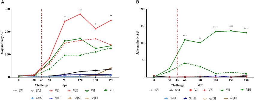
Arteche-Villasol et al.                                                                               Vaccination Against Caprine Paratuberculosis

following the report and suggestions from the Subcommittee               Ganadería de Montaña (IGM-ULE) in León (Spain), goat kids
on Animal Experiments and Welfare of the University of León              were randomly allocated in separated pens, subjected to standard
(ULE) (OEBA-ULE-016-2017).                                               management practices, and their health and welfare status was
                                                                         checked daily.
Vaccines, Immunization Products, and                                        At the beginning of the study, and according to the
Challenge Inoculum                                                       vaccination and/or infection protocol to be followed, goats were
Silirum R commercial vaccine against paratuberculosis and its            classified into 10 groups (n = 5) and distributed in different pens
components (the MontanideTM adjuvant and the Map 316F                    in order to prevent direct contact between groups according to
strain included in the vaccine) were prepared separately and             the following scheme (Figure 1):
shipped by the manufacturer, CZ Vaccines (Porriño, Spain),                  VS: Silirum R vaccinated and noninfected
whereas the heat-inactivated Mbv vaccine (HIMB) and its                     VSI: Silirum R vaccinated and infected
components (a MontanideTM adjuvant and the 1403 Mbv                         VH: HIMB vaccinated and noninfected
strain) were prepared by NEIKER as previously described                     VHI: HIMB vaccinated and infected
(34). Briefly, each dose of HIMB vaccine (1 mL) consisted                   StrSI: immunized with Map 316F Silirum R strain and infected
of an aqueous suspension of heat-inactivated 107 colony-                    StrHI: immunized with Mbv 1403 HIMB strain and infected
forming units (CFUs) of Mbv 1403 NEIKER’s strain (84–85◦ C                  AdjSI: immunized with MontanideTM Silirum R adjuvant
for 45 min) emulsified in MontanideTM ISA 50 V 2 adjuvant                and infected
(Seppic, France). Furthermore, each inactivated bacteria and                AdjHI: immunized with MontanideTM HIMB vaccine
adjuvant immunization products were adjusted to the same dose            adjuvant and infected
administered in Silirum R and HIMB vaccines. Thus, each dose                NV: nonvaccinated and noninfected
(1 mL) of inactivated bacteria (109 CFUs of Map 316F and 107                NVI: nonvaccinated and infected
CFUs of Mbv 1403 strains) was diluted in phosphate-buffered                 No uninfected animals were included in groups immunized
saline (PBS), whereas each dose of MontanideTM adjuvant (1 mL)           with inactivated bacteria or adjuvants, because of the fact that
(used in Silirum R and HIMB vaccines) was emulsified in PBS.             the study was focused on the evaluation of the effect of the
    Besides, bovine Map 764 strain was prepared for the challenge        components of the vaccines on protection, after Map infection,
as previously described by Fernández et al. (11). Briefly, Map           and this would involve the inclusion of a large number of animals
764 strain was grown on Middlebrook 7H9 broth enriched with              and groups that would hinder the study.
10% oleic acid–albumin–dextrose–catalase (OADC) and 2 mg ·                  Vaccination was performed at the beginning of the
L−1 Mycobactin J (7H9 OADC MJ) for 3 weeks at 37 ± 1◦ C.                 experiment, on day 0, by a subcutaneous injection in the
Then, cultures were harvested by centrifugation at 3,000g for            brisket with 1 mL of Silirum R vaccine, 109 Map 316F Silirum R
10 min, and bacterial pellets were washed twice and resuspended          vaccine strain CFUs, 1 mL of MontanideTM Silirum R adjuvant
in PBS. In order to disrupt bacterial clumps, resultant suspension       (CZ Vaccines, Porriño, Spain), 1 mL of HIMB vaccine, 107
was passed up and down through a 27-gauge needle several                 Mbv 1403 HIMB vaccine strain CFUs, and MontanideTM HIMB
times and vortexed. Bacterial concentration was estimated by             vaccine adjuvant, whereas 10 animals remained as the control
optical density (O.D.) and CFU estimation of 10-fold serial              group (nonvaccinated) and were inoculated subcutaneously with
dilutions plated onto agar-solidified 7H9 OADC MJ. Finally,              1 mL of PBS (Figure 1).
suspensions were adjusted to 1.2 × 10¹⁰ CFUs · mL−1 and                     Forty-five days postvaccination (dpv), animals were
maintained at 4◦ C throughout all the challenge period (2 weeks),        challenged orally (Figure 1) using an automatic syringe with a
and bacterial clumps were again disrupted before oral inoculation        total amount of 1.2 × 10¹⁰ Map 764-CFUs diluted in 40 mL of
as mentioned previously.                                                 PBS as previously described by Fernández et al. (11), whereas
                                                                         40 mL of PBS was administered orally to noninfected animals
Experimental Design                                                      at the same time. Throughout the experimental trial, all goats
A total of 50 female Murciano–Granadina breed goat kids of               were monitored daily for clinical signs and sampled every 30
age 1 month were used in this study. Animals were selected               days until sacrifice (Figure 1). At 190 dpv, complete necropsies
from a flock without clinical signs and tested negative to               and postmortem sampling were performed on all goats after
paratuberculosis and tuberculosis in the last 10 years. Moreover,        being humanely euthanized by deep sedation with xylazine
no positive reactors were identified in the annual official              (XILAGESIC R , Laboratorios Calier, Barcelona, Spain) and a
tuberculosis eradication campaigns, based on intradermal skin            subsequent intravenous injection of T61 R (MSD Animal Health,
test, conducted by the regional animal health authorities during         Salamanca, Spain) followed by exsanguination (Figure 1).
the last 5 years. The paratuberculosis- and tuberculosis-free
status of the experimental animals was confirmed using antibody          Sample Collection
enzyme-linked immunosorbent assay (ELISA) against Map (ID                Blood samples were monthly collected from the jugular vein into
Screen R Paratuberculosis indirect, IDVet, Gabrels, France) and          Vacutainer tubes with lithium heparin (Becton Dickinson and
Mbv (INgezim Tuberculosis DR, Eurofins Technology, Madrid,               Company, UK) and without anticoagulant (Becton Dickinson
Spain) and the IFN-γ release test (Bovigam R Mbv IFN-γ test              and Company, UK). Then, heparinized samples were processed
for cattle, Thermo Fisher Scientific, Waltham, USA). After an            immediately for IFN-γ release test in response to protein
adaptation period of 15 days in the facilities of the Instituto de       derivative of avian (PPDa) and bovine (PPDb) antigens at 0,

Frontiers in Veterinary Science | www.frontiersin.org                3                                    January 2022 | Volume 8 | Article 744568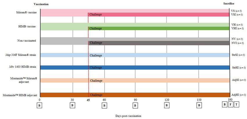
Arteche-Villasol et al.                                                                                                   Vaccination Against Caprine Paratuberculosis

  FIGURE 1 | Experimental design scheme. Goats were divided into seven groups and vaccinated with different components at 0 days postvaccination (dpv). From
  then until sacrifice (190 dpv), blood samples (B) were taken at 30-day interval. Five goats from each group were orally challenged with Map 764 strain at 45 dpv and
  divided into 10 groups: NV, nonvaccinated and noninfected; NVI, nonvaccinated and infected; VS, Silirum® vaccinated and noninfected; VSI, Silirum® vaccinated and
  infected; VH, HIMB vaccinated and noninfected; VHI, HIMB vaccinated and infected; StrSI, Map 316F Silirum® strain immunized and infected; StrHI, Mbv 1403 HIMB
  strain immunized and infected; AdjSI, MontanideTM Silirum® adjuvant immunized and infected; AdjHI, MontanideTM HIMB adjuvant immunized and infected. Finally, at
  190 dpv, feces (F) were collected, and culling and complete necropsies for tissue (T) sample collection were carried out.

30, 60, 90, 120, 150, and 190 dpv (Figure 1). At the same time                         Isolation of PBMCs and Tissue Leukocytes
points, heparinized blood was also collected for peripheral blood                      PBMCs were isolated as previously described (35). Briefly, 30 mL
mononuclear cell (PBMC) isolation (35) and their subsequent                            of heparinized peripheral blood was centrifuged, and PBMCs
characterization by flow cytometry. Besides, nonheparinized                            were isolated by gradient centrifugation using LymphoprepTM
samples were processed for Map (ID Screen R Paratuberculosis                           (STEMCELL Technologies R , Cologne, Germany). Resultant
indirect, IDVet, Gabrels, France) and Mbv-specific (INgezim                            PBMCs were washed three times with PBS, and cell suspensions
Tuberculosis DR, Eurofins Technology, Madrid, Spain) antibody                          were resuspended in supplemented RPMI1640 medium +
determination tests at 0, 30, 60, 90, 120, 150, and 190 dpv                            GlutaMaxTM (Gibco, Paisley, UK) and counted in a Neubauer
(Figure 1).                                                                            chamber and adjusted at a final concentration of 106 cells ·
   Fecal samples from each goat were collected separately into                         mL−1 . Intestinal samples and mesenteric lymph nodes were
disposable plastic gloves directly from the rectum at 190 dpv,                         collected and washed in PBS during the necropsy, put into
prior to the euthanasia of the animal, and frozen at −20◦ C until                      individual falcon tubes containing 30 mL of sterile supplemented
processing for Map detection through bacteriological culture.                          RPMI1640 medium, and processed in the laboratory within
   Animals were euthanized at day 190 dpv and a regulated,                             30 min after collection. For tissue lymphocyte isolation, 5 cm
orderly, and complete necropsy was performed. After gross                              of ileum and jejunal Peyer patches were longitudinally opened
examination of the viscera, samples from ileum (proximal,                              showing the mucosa and washed with PBS until fecal remains
medium, and distal zones), jejunum (proximal, medium, and                              were eliminated, whereas pericapsular fat was removed from the
distal zones), ileocecal valve, and jejunal Peyer patches (at least                    lymph nodes. The excess tissue around each Peyer patch was
three patches from each zone: proximal, medium, and distal),                           removed, and the mucosa from ileum and Peyer patches were
together with mesenteric, jejunal, and ileocecal lymph nodes,                          scraped and minced. Besides, 50 mg of mesenteric lymph node
were taken into buffered formol saline fixative for histological                       tissue was cut into small pieces and chopped using a scalpel blade.
examination. In addition, samples of distal ileum, jejunal Peyer                       Minced tissue was suspended in 11 mL of PBS with EDTA (2 mM)
patches, and mesenteric lymph node were also collected and                             and processed with a stomacher blender (Masticator, IUL) for
stored at −20◦ C for Map isolation by culture and detection                            2.5 min. Then, 10 mL from the upper homogenized portion was
by real-time quantitative polymerase chain reaction (qPCR) as                          passed through 40-µm filter (Thermo Fisher Scientific, Madrid,
well as processed immediately for leukocyte isolation in order to                      Spain), and resultant suspension was layered in an equal volume
characterize lymphocyte subpopulation by flow cytometry and                            of LymphoprepTM and centrifuged at 800 g for 30 min with no
also to perform IFN-γ release test in response to PPDa and                             stop or acceleration. Cells from the interface layer were washed
PPDb antigens.                                                                         three times with PBS/EDTA, counted in a Neubauer chamber and

Frontiers in Veterinary Science | www.frontiersin.org                              4                                          January 2022 | Volume 8 | Article 744568
Arteche-Villasol et al.                                                                                Vaccination Against Caprine Paratuberculosis

resuspended in supplemented RPMI1640 at a final concentration             Extracted DNA was diluted at 50 ng · µL−1 and stored at
of 106 cells · mL−1 .                                                     −20◦ C until qPCR was performed. Genomic DNA from
   Cell viability determined by trypan blue dye exclusion was             2 × 10⁸ Map CFUs and that from 50 mg of tissues of a
usually >90% for both PBMCs and tissue lymphocytes (data                  noninfected animal were extracted and quantified to generate a
not shown).                                                               standard curve.
                                                                             Detection of Map IS900 sequence was performed as previously
Peripheral and Local Cell-Mediated and                                    described by Arteche-Villasol et al. (40). In addition, Map-
Peripheral Humoral Immune Response                                        DNA quantification and qPCR analytical sensitivity were assessed
Whole heparinized peripheral blood samples taken at 0, 30, 60,            by the construction of a 10-fold diluted standard curve
90, 120, 150, and 190 dpv were processed within 3 h of collection.        using Map-genomic DNA ranging from 1,000 pg to 0.001
For each animal, three wells of a 24-well tissue plate (Thermo            pg/reaction mixed with 100 ng/reaction of tissue DNA from a
Fischer Scientific, Rochester, NY) were filled with 1.5 mL of             noninfected animal. Samples were considered as positive when
blood each and were either mixed with 100 µL of sterile PBS               the dissociation peak (Tm) was 89.1 ± 1.5◦ C and threshold
(negative control) or stimulated with 100 µL of PPDa or PPDb              cycles (Ct) were ≤37 (41, 42). The qPCR results were analyzed
(CZ Vaccines, Porriño, Spain) at a final concentration of 30 µg ·         using 7500 Software v2.0.6 (Applied BiosystemsTM ). Map-DNA
mL−1 diluted in sterile PBS (11). After 22 h of stimulation, plates       quantity (pg) of each well was calculated by interpolation
were centrifuged at 750 g for 15 min, and plasma was collected            of their Ct values with the standard curve as previously
and stored at −20◦ C until tested.                                        described (43), and the mean quantity was calculated from
   For the analysis of the cell-mediated local immune response,           both duplicates.
a total of 2 × 10⁶ mononuclear leukocytes isolated from ileum,
Peyer patches, and mesenteric lymph node were seeded per well             Tissue and Fecal Culture of Map
into three wells each of a 24-well tissue plate and stimulated as         Distal ileum, jejunal Peyer patches, and mesenteric lymph node
described for whole blood. After 22 h of stimulation, plates were         from each animal were tested individually, whereas fecal samples
centrifuged at 750 g for 15 min, and supernatants were collected          were pooled into groups of three goats each. Ileum and Peyer
and stored at −20◦ C until tested.                                        patch segments (12 cm) and 2 grams of mesenteric lymph
   IFN-γ production in these assays was assessed by duplicate             node were processed as described for tissue leukocyte isolation
using commercial ELISA for bovine IFN-γ following                         and following methods previously described (44). Briefly, 2 g
manufacturer’s instructions (Bovigam R Mbv IFN-γ test                     of each tissue and fecal samples pools were decontaminated
for cattle, Thermo Fisher Scientific, Waltham, USA), and                  with 38 mL of hexadecylpyridinium chloride and homogenized
absorbance values were measured spectrophotometrically using              in a stomacher blender (Masticator, IUL) for approximately
an ELX800 ELISA reader (Bio-Tek Instruments) at 450 nm. The               15 s. After 18 h of decontamination, 200 µL of the suspension
O.D. values were adjusted by dividing the plasma or supernatant           was used to inoculate two tubes containing Herrold’s egg yolk
O.D. value by the negative control O.D. value of each plate in            medium supplemented with sodium pyruvate and mycobactin
order to prevent interplate variations. Results were expressed as         J (MJ) and two tubes containing 7H9 OADC supplemented
avian and bovine index values by means of the quotient between            with MJ, penicillin, amphotericin, and chloramphenicol. Cultures
the mean O.D. of PPDa or O.D. of PPDb-stimulated plasma,                  were incubated at 37◦ C ± 1◦ C, and growth was checked by
respectively, and the mean O.D. of the negative control plasma            examination under a stereoscopic microscope after 8, 12, 16, and
(11, 36).                                                                 20 weeks postinoculation. Cultures were considered positive if
   Nonheparinized blood samples were allowed to clot and                  one or more characteristic Map colonies were observed in any
retract, and serum was stored at −20◦ C until used. Each serum            tube. Colonies isolated on both mediums were confirmed by
was tested for specific antibodies against Map (37) and Mbv               a real-time multiplex PCR detecting IS900 and ISMap02 Map
(38) using commercial ELISA tests (ID Screen R Paratuberculosis           sequences (45).
indirect, IDVet, Gabrels, France; and INgezim Tuberculosis
DR Eurofins Technology, Madrid, Spain, respectively) following            Flow Cytometric Analysis of PBMCs and
manufacturer’s instructions. Results were expressed as the S/P            Tissue Leukocytes
ratio of Map and Mbv antibodies calculated by dividing the                Single-color flow cytometry analysis was carried out for
corrected O.D. of the sample by the corrected O.D. of the positive        phenotypic characterization of PBMCs isolated at 0, 30, 60, 90,
control and multiplying by 100 (39).                                      120, 150, and 190 dpv and mononuclear leukocytes isolated from
                                                                          distal ileum, jejunal Peyer patches, and mesenteric lymph node.
Real-Time Quantitative Detection of Map                                   A total number of 2 × 10⁵ cells per well were seeded in a 96-
DNA was extracted from 50 mg of distal ileum, jejunal                     well plate (Thermo Fisher Scientific, Roskilde, Denmark) and
Peyer patches, and mesenteric lymph node by using                         incubated with primary antibodies against lymphocyte surface
Maxwell R 16 Tissue DNA Purification Kit (Promega, WI,                    markers detailed in Table 1 for 1 h at 4◦ C. Afterward, cells
USA) with the Maxwell 16 Instrument (Promega) following                   were washed twice with PBS and incubated with appropriate
manufacturer’s instructions. Thereupon, DNA was quantified                conjugated secondary antibodies for 1 h at 4◦ C (Table 1). Finally,
using QuantiFluorTM ONEdsDNA System kit (Promega, WI,                     cells were fixed with 1% of CellFIXTM (Becton Dickinson
USA) and QuantusTM Fluoremeter (Promega, WI, USA).                        and Company, Erembodegem, Belgium) until analyzed. Sample

Frontiers in Veterinary Science | www.frontiersin.org                 5                                    January 2022 | Volume 8 | Article 744568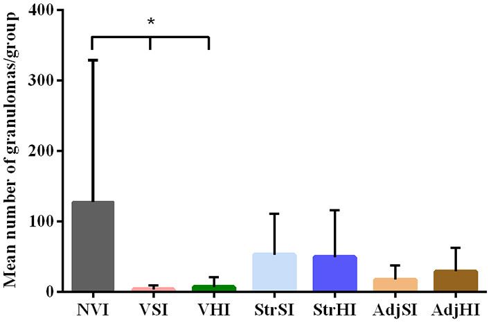
Arteche-Villasol et al.                                                                                              Vaccination Against Caprine Paratuberculosis

TABLE 1 | Primary and secondary antibodies used in flow cytometry analysis of PBMCs and tissue lymphocyte subpopulations.

Target      Specificity                     Monoclonal/polyclonal   Primary          Reference                   Secondary antibody                  Secondary
                                                                    antibody                                                                         antibody
                                                                    dilution                                                                         dilution

CD4         T helper lymphocytes            Monoclonal              1:400            MCA2213GA, BioRad           Polyclonal rabbit anti–mouse        1:50
                                                                                                                 IgG-FITC, F0313, Dako
CD8         Cytotoxic T lymphocytes         Monoclonal              1:400            MCA2216GA, BioRad           Polyclonal rabbit anti–mouse        1:50
                                                                                                                 IgG-FITC, F0313, Dako
WC1         γδ T lymphocytes                Monoclonal              1:200            MCA8586, BioRad             Polyclonal rabbit anti–mouse        1:50
                                                                                                                 IgG-FITC, F0313, Dako
CD21        Naive B lymphocytes             Monoclonal              1:10             MCA1185, BioRad             Polyclonal rabbit anti–mouse        1:50
                                                                                                                 IgG-FITC, F0313, Dako
CD20        Mature B lymphocytes            Polyclonal              1:100            9013-P, Thermo Scientific   Polyclonal goat anti–rabbit         1:2,000
                                                                                                                 IgG-Alexa Fluor® 488,
                                                                                                                 ab150077, Abcam
CD3         T lymphocytes, natural killer   Polyclonal              1:100            A0452, Dako                 Polyclonal goat Anti- rabbit        1:2,000
                                                                                                                 IgG-Alexa Fluor® 488,
                                                                                                                 ab150077, Abcam

acquisition of 10,000 events was performed using a flow                            associated lamina propria or in the mucosa not related to
cytometer (MACSQuant, Miltenyi Biotec R ), where events were                       lymphoid tissue.
gated during the acquisition analysis as previously described
elsewhere (35) to discard the presence of air and doublets. Then,                  Statistical Analysis
analysis of data was carried out using the MACSQuantify10                          Normal distribution of the results from Map- and Mbv-
SoftwareTM (Miltenyi Biotec R ), and results were expressed as                     specific antibodies, local and peripheral IFN-γ production, and
percentage of positive cells.                                                      proportion of peripheral and tissue lymphocyte subpopulations
                                                                                   were assessed for normality using Shapiro–Wilk test. Data
Histopathological Examination                                                      from IFN-γ production and antibody ELISA tests were
Fixed tissues for histopathological examination were                               logarithmically transformed. Then, flow cytometry and ELISA
conventionally processed for paraffin embedding and stained                        results from peripheral IFN-γ and antibody production were
with hematoxylin–eosin and Ziehl–Neelsen technique for                             analyzed using generalized lineal model (GLM) procedure for
acid-fast bacilli detection (46).                                                  evaluation of the main effects of vaccination, challenge, time, and
    Lesions consistent with Map infection were classified as                       its interactions. Subsequently, differences between vaccination
focal, multifocal a and multifocal b, or diffuse forms according                   groups for each time sampled were estimated using Tukey–
to the presence and location of granulomas and following                           Kramer correction for multiple comparisons. Similarly, the
the guidelines previously described for small ruminants (8,                        main effects of vaccination, tissue, and its interactions were
46). Briefly, lesions were characterized as focal forms when                       estimated in the results of local IFN-γ production followed
granulomas were restricted to the lymphoid tissue of the                           by the evaluation of differences between vaccination groups
Peyer patches; multifocal forms when the granulomas were                           and tissues using Tukey–Kramer multiple-comparisons test.
located in the lamina propria adjacent to the lymphoid tissue                      Besides, differences between groups in the number of goats
(multifocal a) or not (multifocal b) and diffuse forms when                        with lesions were evaluated using χ 2 and Fisher exact tests.
granulomas spread to wide areas of the mucosa. Classification                      In addition, after logarithmic transformation, differences in the
of each animal was based on its most severe granulomatous                          granuloma counts between vaccination groups were calculated
lesion. Following histopathological examination, the number                        using the Student t-test. All statistical analyses were carried
of granulomas per tissue section was quantified in all tissue                      out using GraphPad Prism 6.0 software (San Diego, CA, USA)
samples as described elsewhere (11, 46). Three tissue sections                     excluding GLMs that were performed using R software 3.5.3
of each intestinal site (i.e., ileum—proximal, medium, and                         (R Development Core Team, 2019). P < 0.05 was considered
distal zones; jejunum—proximal, medium, and distal zones;                          statistically significant.
ileocecal valve and jejunal Peyer patches—proximal, medium,
and distal zones) and two sections from each lymph node (i.e.,                     RESULTS
mesenteric, jejunal and ileocecal lymph nodes), were selected,
and the mean number of granulomas per animal was recorded                          Peripheral and Local Cell-Mediated
by the same observer (V.P.; diplomat of the European College                       Immune Response (IFN-γ )
of Veterinary Pathologists), distinguishing those granulomas                       Results from GLM showed that avian and bovine index values
located in the lymphoid tissue from those located in the                           reached significant levels from 60 to 190 dpv in the VS, VSI,

Frontiers in Veterinary Science | www.frontiersin.org                          6                                         January 2022 | Volume 8 | Article 744568
Arteche-Villasol et al. Vaccination Against Caprine Paratuberculosis
FIGURE 2 | Kinetics of the IFN-γ production by whole blood stimulated with avian (PPDa) and bovine (PPDb) antigens. Results are expressed as avian (A) and bovine
(B) O.D. index values of each vaccination group (n = 5) (NV, nonvaccinated and noninfected; NVI, nonvaccinated and infected; VS, Silirum® vaccinated and
noninfected; VSI, Silirum® vaccinated and infected; VH, HIMB vaccinated and noninfected; VHI, HIMB vaccinated and infected; StrSI, Map 316F Silirum® strain
immunized and infected; StrHI, Mbv 1403 HIMB strain immunized and infected; AdjSI, MontanideTM Silirum® adjuvant immunized and infected; AdjHI, MontanideTM
HIMB adjuvant immunized and infected) and time sampled (dpv, days postvaccination). Vertical dotted red line represents the time of Map oral challenge (45 dpv).
Significant differences determined by multiple comparisons were represented as *p < 0.05, **p < 0.01, ***p < 0.001, ****p < 0.0001.
VH, and VHI groups (p < 0.001 and p < 0.01 respectively) AdjSI groups (Figure 3A). Peyer patches and mesenteric lymph
(Figures 2A,B). In addition, Silirum R (VS and VSI) and HIMB node from NVI group showed the highest avian index values
vaccine (VH and VHI) immunization exerted a considerable followed by the ileum, Peyer patches, and mesenteric lymph node
effect on avian index values regardless of the infection status from the StrHI group (Figure 3A). However, statistical analysis
(p < 0.01 and p < 0.001, respectively) (Figure 2A), but only showed no significant effect of vaccination or tissue location
the VHI, VH, and StrHI groups showed a significant effect on on the avian or bovine index values (p > 0.05). In addition,
bovine index values (p < 0.01) (Figure 2B). Furthermore, oral no significant differences were observed between the vaccinated
challenge significantly impacted on the avian index levels of the groups in the multiple-comparisons analysis either (p < 0.05).
VSI, VHI (p < 0.001), and NVI (p < 0.05) groups and on the Meanwhile, significant differences were observed in the avian
bovine index values of the VSI, VHI (p < 0.01), and StrHI (p < index values within the NVI group, where IFN-γ production was
0.05) groups. higher in Peyer patches and mesenteric lymph node than in the
Multiple-comparisons analysis showed that the VSI group ileum (p < 0.05) (Figure 3A). Besides, despite the high variability
had higher avian index values than NV, NVI, StrSI, AdjSI, and within groups observed in the bovine index values, the values of
AdjHI at 60 and 120 dpv, whereas the VS group showed these mesenteric lymph node from the VH group were considerably
differences only at 120 dpv (Figure 2A). In contrast, these groups higher than those of the ileum and Peyer patches (p < 0.01)
did not show any significant differences in bovine index levels (Figure 3B).
(Figure 2B). Furthermore, no differences were observed between
VS and VSI either in avian or bovine index values at any time Peripheral Humoral Response
point. On the other hand, the VHI group showed greater avian Results from GLM estimated that both Map- and Mbv-
index values than NVI, NV, StrSI, AdjSI, and AdjHI at 120 dpv specific antibody production reached significant levels from
(Figure 2A). In addition, bovine index values of this group were 60 to 190 dpv (p < 0.001 and p < 0.0001, respectively)
significantly higher than NV, NVI, StrSI, AdjSI, and AdjHI at in Silirum R and HIMB-vaccinated groups (Figures 4A,B).
90, 120, 150, and 190 dpv and greater than VH at 120 dpv In addition, oral challenge boosted Map-specific antibody
(Figure 2B). production in the vaccinated groups VSI, VHI (p < 0.001),
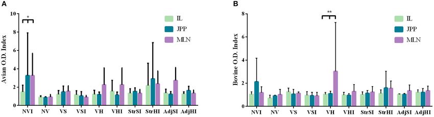
Local IFN-γ production in mononuclear leukocytes purified and NVI (p < 0.05) (Figure 4A) and Mbv-specific antibody
from tissues and stimulated with PPDa and PPDb was lower production in VHI (p < 0.001) (Figure 4B). Besides, multiple-
than that observed in PBMCs. Furthermore, avian index values comparisons analysis showed that only VSI had significantly
were more heterogeneous than bovine index levels on account higher Map-specific S/P values in comparison with the
of the higher individual variability observed in the former groups immunized with adjuvants, inactivated bacteria, and
(Figures 3A,B), specifically at the NVI, VH, VHI, StrHI, and nonvaccinated at 90, 120, 150, and 190 dpv, whereas no
Frontiers in Veterinary Science | www.frontiersin.org 7 January 2022 | Volume 8 | Article 744568Arteche-Villasol et al. Vaccination Against Caprine Paratuberculosis FIGURE 3 | IFN-γ production by leukocytes from ileum (IL), jejunal Peyer patches (JPP), and mesenteric lymph node (MLN) stimulated with avian (PPDa) and bovine (PPDb) antigens. Results are expressed as avian (A) and bovine (B) O.D. index values of each vaccination group (n = 5) (NV, nonvaccinated and noninfected; NVI, nonvaccinated and infected; VS, Silirum® vaccinated and noninfected; VSI, Silirum® vaccinated and infected; VH, HIMB vaccinated and noninfected; VHI, HIMB vaccinated and infected; StrSI, Map 316F Silirum® strain immunized and infected; StrHI, Mbv 1403 HIMB strain immunized and infected; AdjSI, MontanideTM Silirum® immunized and infected; AdjHI, MontanideTM HIMB adjuvant immunized and infected). Bars and vertical lines represent mean values and standard deviations, respectively. Significant differences determined by multiple comparisons were represented as *p < 0.05, **p < 0.01, ***p < 0.001, ****p < 0.0001. FIGURE 4 | Kinetics of peripheral blood serum antibody levels. Results are expressed as S/P ratio of Map (A) and Mbv (B) specific antibodies of each vaccination group (n = 5) (NV, nonvaccinated and noninfected; NVI, nonvaccinated and infected; VS, Silirum® vaccinated and noninfected; VSI, Silirum® vaccinated and infected; VH, HIMB vaccinated and noninfected; VHI, HIMB vaccinated and infected; StrSI, Map 316F Silirum® strain immunized and infected; StrHI, Mbv 1403 HIMB strain immunized and infected; AdjSI, MontanideTM Silirum® immunized and infected; AdjHI, MontanideTM HIMB adjuvant immunized and infected) and time sampled (dpv, days postvaccination). Vertical dotted red line represents the time of Map oral challenge (45 dpv). Significant differences determined by multiple comparisons were represented as *p < 0.05, **p < 0.01, ***p < 0.001, ****p < 0.0001. significant differences were observed in VHI (Figure 4A). Quantitative PCR and Bacteriology of Additionally, no significant differences were observed in the Tissues and Feces Map antibody levels either between Silirum R and HIMB Map detection was confirmed by qPCR (n = 4) and vaccination groups or between infected and noninfected bacteriological culture (n = 3) within infected goats (n = vaccinated groups. 35). Animals positive to qPCR corresponded to (i) two goats Mbv-specific S/P values were significantly higher when from StrHI (mesenteric lymph node and the Peyer patches, comparing the VHI group with the VS, VSI, StrSI, StrHI, AdjSI, respectively), (ii) one from VHI (Peyer patches), and (iii) and AdjHI groups at 60, 90, 120, 150, and 190 dpv (Figure 4B). one from AdjSI (Peyer patches). Besides, animals positive to However, significant differences between VHI and VH were bacteriological culture corresponded to one goat each from observed only at 120, 150, and 190 dpv (Figure 4B). NVI, VHI (ileum, Peyer patches, and mesenteric lymph node Frontiers in Veterinary Science | www.frontiersin.org 8 January 2022 | Volume 8 | Article 744568
Arteche-Villasol et al.                                                                                                           Vaccination Against Caprine Paratuberculosis

positives), and VSI (ileum and mesenteric lymph node positives).                            and results of multiple comparisons of lymphocyte relative
Only the Peyer patches from the goat from VHI showed                                        subpopulations in ileum, Peyer patches, and mesenteric lymph
positive results for both detection techniques. All tissue samples                          node are summarized in Supplementary Tables 2–4.
from noninfected groups were negative to IS900 sequence
amplification by qPCR or bacteriological culture. Bacteriological
culture of Map was negative in every fecal sample analyzed.                                 Pathological Findings
                                                                                            Gross lesions compatible with paratuberculosis were not found
                                                                                            in any animal. Microscopic granulomatous lesions characteristic
Relative Peripheral Blood and Tissue                                                        of Map infection were detected in all infected groups, with
Lymphocyte Subsets                                                                          differences in the severity and distribution. The number of goats
The mean and standard deviations of the relative proportions                                per group with lesions according to the tissue and the number of
of three T lymphocytes (CD4+ , CD8+ , and CD3+ ), one γδ T                                  goats per group classified in terms of severity of the lesions are
lymphocyte, and two B lymphocyte (CD21+ and CD20+ ) surface                                 shown in Table 2. Goats with small and well-defined granulomas
markers were estimated by flow cytometry.                                                   composed of macrophages, with an abundant pale cytoplasm
   Along all groups, the highest relative proportion of positive                            and a large nucleus, escorted by a few lymphocytes located
cells in PBMCs isolated from whole blood corresponded to γδ T                               exclusively in the interfollicular area of the intestinal lymphoid
lymphocytes (30.46% ± 3.55%), whereas the lowest proportion                                 tissue were classified as focal (n = 5) (Figure 5A). Besides, goats
was observed in CD20+ B lymphocytes (5.96 ± 1.48%)                                          with well-defined granulomas in the interfollicular area of the
throughout the study (Supplementary Table 1). However,                                      Peyer patches and also in the lamina propria closely associated
despite statistical analysis showing significant oscillations (p                            with the intestinal lymphoid tissue were categorized as multifocal
< 0.001) of the relative proportions of CD4+ , CD8+ , WC1+ ,                                a (n = 5) (Figure 5B). Finally, the presence of granulomatous
CD21+ , CD3+ , and CD20+ lymphocytes at different samplings,                                lesions not only in the Peyer patches and related lamina propria
no significant differences were found between groups (p > 0.05).                            but also in areas of the mucosa without any association with
   Flow cytometry carried out in cells purified from tissue                                 the lymphoid tissue was considered as multifocal b (n = 13)
samples showed that the highest relative proportion levels                                  (Figure 5C). No diffuse lesions were noticed in the tissues of any
corresponded to CD21+ B lymphocytes in the ileum (39.47 ±                                   goat (n = 0). VSI and VHI groups were the less affected with
4.71%), Peyer patches (26.81 ± 3.59%), and mesenteric lymph                                 only one animal from each group developing granulomatous
node (44.90 ± 3.52%), whereas γδ T lymphocytes were the                                     lesions, showing significant differences in comparison to NVI in
scarcest cell population in these samples (0.99 ± 0.24%, 1.69                               which lesions were present in all animals (p < 0.05). In addition,
± 0.33%, and 0.92 ± 0.26%, respectively). Statistical analysis                              lesions from the VSI goat were classified as focal, whereas the
showed a significant influence of vaccination and immunization                              goat from VHI group showed more severe lesions, categorized as
with inactivated bacteria and adjuvants on the relative proportion                          multifocal b as all goats from the NVI. Besides, most of the goats
of tissue lymphocyte subsets (p < 0.05). However, results from                              immunized with strains (StrSI and StrHI) developed multifocal b
comparisons between groups did not show a clear pattern                                     forms, except for one animal from StrHI with a focal lesion. In
between the relative proportion of lymphocyte subpopulations                                contrast, among the animals immunized with adjuvants (AdjSI
and the different groups likely on account of the individual                                and AdjHI), only one animal from the AdjSI showed multifocal
variability observed within groups. Mean, standard deviation,                               b lesions, whereas focal and multifocal a lesions were found in

TABLE 2 | Number of animals from each group with microscopic lesions consistent with paratuberculosis according to the examined tissue and its severity (focal,
multifocal a, multifocal b, and diffuse).

Group (n = 5)                                           Tissues                                                                      Severity

                          IL     JJ        JPP          ICV       MLN        JLN           ICLN      Focal         Multifocal a        Multifocal b         Diffuse        Total

NVI                       3/5   2/5        5/5          3/5       1/5         1/5           —          —                —                   5/5                —            5/5
VSI                       1/5    —         1/5          —         —           —            1/5         1/5              —                    —                 —           1/5a*
VHI                       1/5    —         1/5          —         —           —             —          —                —                   1/5                —           1/5a*
StrSI                     3/5   2/5        3/5          1/5       3/5         1/5          1/5         —                —                   3/5                —            3/5
StrHI                     1/5    —         4/5          1/5       1/5         1/5           —          1/5              —                   3/5                —            4/5
AdjSI                     1/5    —         3/5          1/5       —           —             —          —                2/5                 1/5                —            3/5
AdjHI                     1/5    —         3/5          1/5       1/5         —             —          3/5              1/5                  —                 —            4/5

IL, Ileum; JJ, jejunum; JPP, jejunal Peyer patches; ICV, ileocecal valve; MLN, mesenteric lymph node; JLN, jejunal lymph node; ICLN, ileocecal lymph node; NVI, Nonvaccinated and
infected; VSI, Silirum® vaccinated and infected; VHI, HIMB vaccinated and infected; StrSI, Map 316F Silirum® strain immunized and infected; StrHI, Mbv 1403 HIMB strain immunized
and infected; AdjSI, MontanideTM Silirum® adjuvant immunized and infected; AdjHI, MontanideTM HIMB adjuvant immunized and infected. Significant differences detected by χ2 and
Fisher exact test are expressed as *p < 0.05, **p < 0.01, ***p < 0.001, and ****p < 0.0001.
— No lesion.
a Significantly different from NVI.

Frontiers in Veterinary Science | www.frontiersin.org                                  9                                             January 2022 | Volume 8 | Article 744568
Arteche-Villasol et al. Vaccination Against Caprine Paratuberculosis
the lowest corresponded to the VSI and VHI groups. In addition,
the number of granulomas observed in the StrSI and StrHI
groups was higher than those observed in AdjSI and AdjHI.
Significant differences in the mean number of granulomas per
group (Figure 6) were observed between NVI (159.60 ± 271.90)
and VSI (4.60 ± 10.29) and VHI (6.20 ± 13.86) groups (p <
0.05). However, no significant differences were found in the StrSI
(57.60 ± 61.54), StrHI (61.40 ± 83.22), AdjSI (20.00 ± 26.82),
and AdjHI (29.20 ± 33.86) groups (p > 0.05).
DISCUSSION
Paratuberculosis vaccination studies conducted in ruminants
have demonstrated worthwhile results in the reduction of the
number of clinically affected animals and the prevalence of the
disease (16, 23). Nevertheless, the inability of the protective
immune response elicited by vaccination to completely prevent
the infection and its progression and spread is alienating
from the term “ideal vaccine” (47, 48). This handicap is
partly due to the lack of knowledge about the peripheral
and local mechanisms related to the success or failure of the
protection against mycobacteria. Here, this work analyzes the
local and peripheral immune response elicited by homologous
(Silirum R ) and heterologous (HIMB vaccine) vaccination and
the immunization with their components separately and its
relation to the protection degree estimated by the evaluation
of the microscopic granulomatous lesions and Map presence in
the tissues.
Despite the identification of distinctive paratuberculosis
granulomatous lesions in all groups of infected goats, regardless
of its vaccination or immunization status, the variation in their
severity mirrors the observations from field and experimental
studies, as lesions observed in paratuberculosis-vaccinated goats
were mild (focal) and scarce, whereas those in nonvaccinated and
infected goats were more severe (multifocal b), proving the high
FIGURE 5 | Types of paratuberculosis granulomatous lesions found in the protective effect of vaccination (16, 22, 41). No diffuse lesions
study. (A) Focal lesion: well-demarcated granulomas composed of small were noticed, possible due to the short incubation period of this
groups of macrophages located in the interfollicular area of the Peyer patches. work (4–5 months), in line with other studies in which multifocal
(B) Multifocal a lesion: small granulomas located in the interfollicular area of the
b lesions were observed up to 7 months postinfection, whereas
lymphoid tissue (asterisk) and in the associated lamina propria (arrowheads).
(C) Multifocal b lesion: granulomas located in the lamina propria in area devoid diffuse-type lesions were not reported until 12 months (46, 49).
of lymphoid tissue. Despite this fact, the results obtained here agree with other
experimental studies in which lesions observed in vaccinated
animals were limited to regressive forms (focal), contained in
the interfollicular area of the intestinal lymphoid tissue, avoiding
the remaining goats. No significant differences were detected their dissemination through the intestine but without preventing
in the number of goats with granulomatous lesions in groups infection as it was confirmed by the detection of Map in tissues
immunized with inactivated bacteria or adjuvants (p > 0.05). from a VSI goat (41, 50, 51).
Granulomatous lesions were localized mainly in the jejunal Peyer To date, in vivo vaccination efficacy studies have been mainly
patches followed by distal ileum, lymph nodes, ileocecal valve, focused on the early and sustained induction of peripheral
and jejunum, although lesions observed in the lymph nodes were IFN-γ (20, 21, 52) as a release of this cytokine has been
always associated with the presence of granulomatous lesions in associated with the activation of antimycobacterial mechanisms
the intestine (Table 3). No microscopic lesions were observed in and the prevention of intracellular bacterial growth (53, 54).
the groups that remained as noninfected. No acid-fast bacilli were Certainly, IFN-γ production dynamics observed in Silirum R -
detected in the lesions from any of the infected goats. vaccinated groups (i.e., VS and VSI) was consistent with
The total number of granulomas per group was summarized other studies conducted using goat kids vaccinated with heat-
in Table 3. The NVI group showed the highest number, whereas killed vaccines (52, 55). In addition to cell-mediated immune
Frontiers in Veterinary Science | www.frontiersin.org 10 January 2022 | Volume 8 | Article 744568Arteche-Villasol et al. Vaccination Against Caprine Paratuberculosis
TABLE 3 | Results of granuloma count. Number of granulomas according to their location for each group and total number and percentage (%) of granulomas per group.
Group (n = 5) IL JJ JPP ICV MLN JLN ICLN Total number
NVI 35 6 690 33 12 22 0 798 (47.10%)
VSI 2 0 12 0 0 0 9 23 (1.36%)
VHI 2 0 30 0 0 0 0 31 (1.83%)
StrSI 39 6 186 7 29 2 19 288 (17.00%)
StrHI 3 0 293 8 3 1 0 308 (18.18%)
AdjSI 4 0 92 4 0 0 0 100 (5.90%)
AdjHI 21 0 83 3 39 0 0 146 (8.63%)
Total 106 12 1,386 55 83 25 28 1,694 (100%)
IL, Ileum; JJ, jejunum; JPP, jejunal Peyer patches; ICV, ileocecal valve; MLN, mesenteric lymph node; JLN, jejunal lymph node; ICLN, ileocecal lymph node; NVI, Nonvaccinated and
infected; VSI, Silirum® vaccinated and infected; VHI, HIMB vaccinated and infected; StrSI, Map 316F Silirum® strain immunized and infected; StrHI, Mbv 1403 HIMB strain immunized
and infected; AdjSI, MontanideTM Silirum® adjuvant immunized and infected; AdjHI, Montanide® HIMB adjuvant immunized and infected.
response, Silirum R -vaccinated animals also showed increased
Map antibody levels. The role of humoral response has been
underestimated as it has been considered ineffective against
intracellular pathogens (56, 57). However, an emerging number
of studies performed under experimental conditions have
suggested the potential positive effect of antibodies in the
control of paratuberculosis, even in relation to vaccination (58–
60). This is supported by the initial strong humoral response
observed in paratuberculosis-vaccinated sheep and the ability
of these animals to modulate that response in later stages to
prevent the progression of infection (60). Both cellular and
humoral responses were significantly enhanced in this study FIGURE 6 | Mean granuloma count from the tissues of vaccinated and
after experimental challenge with Map. These results mimic the infected groups. Data are expressed as mean number of granulomas per
increase in IFN-γ observed in paratuberculosis-vaccinated and group, and standard deviations in each group (n = 5) and significant
naturally exposed goats in field conditions (61), although the differences were represented as *p < 0.05, **p < 0.01, ***p < 0.001, ****p <
0.0001.
significant increase in the antibody production after Map oral
challenge here observed contrasts with previous studies where
experimental oral infection in vaccinated sheep did not exhibit
differences with nonvaccinated animals or even lead to a decrease CD4+ and B lymphocytes at 13 days postchallenge assessed in
in the antibody production (22, 60), although differences in in vitro Map-stimulated PBMCs, but this was not evaluated in
the adjuvant of the vaccine used in that study (i.e., Gudair R ) nonstimulated and freshly isolated cells (22). On the contrary,
or the species (i.e., sheep) may explain these discrepancies. no changes in the relative proportion of either peripheral T
Nevertheless, the heightened cellular and humoral responses lymphocytes (CD4+ , CD8+ , γδ, and CD3+ ) or B lymphocytes
here observed did not prevent the appearance of granulomatous (CD20+ and CD21+ ) were detected in freshly isolated PBMCs
lesions in VSI group. Thus, despite that vaccination might be between any group in this study. In addition, differences in the
limiting the progression and severity of the lesions, as only one tissue relative proportion of lymphocyte subpopulations were
VSI goat developed a low number of focal granulomas, peripheral estimated, but no clear relationship between these results and
cellular and humoral response did not allow predicting the vaccination/immunization and/or infection was observed, likely
presence of lesions or infection in vaccinated animals. due to the high individual variability within tested groups.
In addition, changes in the relative proportion of lymphocyte Similarly, no relation between local production of IFN-γ and
subpopulations have also been connected to mycobacterial development of lesions either in VSI or other infected groups was
infections (62–64). In this sense, cows with clinical disease observed. In this sense, only the Peyer patches and mesenteric
have shown lower proportions of CD4+ , CD8+ , and γδ T lymph node from NVI group produced a slightly higher IFN-
lymphocytes than subclinical cows in freshly isolated PBMCs γ production. This result is consistent with the greater number
(65). Furthermore, in another study, higher percentages of γδ of granulomas detected in this group as an increased number
T and B lymphocytes and a decline in CD4+ were detected of granulomatous lesions have been related to a higher number
in tissues of infected sheep during early Map experimental of IFN-γ-immunolabeled cells (6) and are likely that lesions
infection (10). Regarding the effect of vaccination, it has present in the remaining infected groups might not be sufficient
been reported that infection of Gudair R -vaccinated sheep with to generate a substantial local cell-mediated immune response in
Map leads to a decrease in the proportions of peripheral response to mycobacterial antigens. Therefore, neither peripheral
Frontiers in Veterinary Science | www.frontiersin.org 11 January 2022 | Volume 8 | Article 744568Arteche-Villasol et al. Vaccination Against Caprine Paratuberculosis
cellular and humoral immune responses nor evaluation of the 76). Therefore, the development and use/implementation of
relative proportion of lymphocyte subpopulations in whole blood more specific antigens (DIVA reagents) for both IFN-γ assay
and tissues nor local production of IFN-γ was able to predict and antibody detection are necessary in order to improve the
those animals that remain unprotected early after infection. immunological diagnosis of paratuberculosis and tuberculosis
Besides, vaccination has demonstrated to reduce the number and to overcome the interferences produced by vaccination (31).
of excreted bacteria both in field and experimental conditions Addressing the effect of Silirum R and HIMB inactivated
(17, 21, 37, 66). In this study, Map was not isolated in the feces strains individually, it was observed that goats from StrSI
of any goat. Heightened Map shedding has been detected in and StrHI showed a higher number of granulomatous lesions
relation to the development of severe lesions and clinical signs in affecting a greater number of animals than those of the VSI
naturally and experimentally infected animals (67, 68), although or VHI groups. In addition, despite the fact that the number
it could be intermittently detected in animals with subclinical of granulomas was lower than that in the NVI goats, these
paratuberculosis (53, 69). However, feces were collected only at were also classified as multifocal b, proving the progression
sacrifice (145 days postinfection), so the presence of Map could of infection in these groups. Regarding peripheral immune
have been underestimated because of the short postchallenge responses, only the StrHI group showed high peripheral IFN-
time and the limited time sampled. Moreover, it has been γ levels in response to both mycobacterial antigens but not
observed that fecal shedding is frequently associated with animals the production of peripheral antibodies, which could suggest
with a high number of culture-positive tissues and severe lesions the ability of this strain to stimulate only the peripheral
(70). In addition, the low number of Map-positive tissues cellular immune response. In this sense, in a study conducted
detected either by bacteriological culture or qPCR (7 of 35 in cattle, oral administration of inactivated Mbv 1403 strain
infected goats) was not surprising as in several previous works, suspended in PBS did not show an increased IFN-γ production in
either in natural or experimental cases (46, 71), it has been shown response to bovine antigen, whereas the parenteral immunization
that the bacterial load in animals with lesions similar to those with this strain homogenized in MontanideTM ISA 50 V 2
found in this study was absent or very low. adjuvant (HIMB vaccine) showed an elevation of the IFN-γ
Interestingly, only one goat from the VHI group developed production from 2 weeks postimmunization, although the effect
paratuberculosis granulomatous lesions, albeit classified as of the inactivated bacteria administered parenterally was not
multifocal b, showing the effectiveness of heterologous protection evaluated in absence of the adjuvant (77). On the contrary,
that vaccination for tuberculosis confers against Map infection. no evident peripheral cellular or humoral immune responses
Similar to homologous vaccination, HIMB-vaccinated groups were detected in the StrSI group, although IFN-γ rose at 150
(i.e., VH and VHI) showed a significant IFN-γ and antibody dpv. In this sense, it is feasible that this late increase was the
production as previously described in goats (66, 72), even when effect of Map infection rather than the immunization itself, as
blood was stimulated with PPDa. In addition, boost in the IFN-γ the animals with the highest cell-mediated peripheral immune
production was also noticed in goats vaccinated subcutaneously response corresponded to those with the most numerous
with HIMB vaccine and endobronchially challenged with a granulomatous lesions (data not shown). Thus, this limited
closely related mycobacteria (M. caprae) under experimental peripheral immune response elicited by inactivated bacteria
conditions (66), proving again that the elevation of these might be a consequence of their weak immunogenicity in absence
responses does not ensure the success of vaccination as in the of adjuvants (78, 79).
VHI goat, granulomatous lesions were more severe (multifocal Besides, the immunization with adjuvants (i.e., AdjSI and
b) than those from the VSI group (focal). Cross-protection effect AdjHI) in the absence of bacterial antigens resulted in a lower
related to vaccination has been previously reported as a reduction number of granulomatous lesions than in the StrSI, StrHI, or even
of pulmonary lesions was noticed in paratuberculosis-vaccinated NVI group. Furthermore, these lesions were mainly classified
goats (32) or calves (33) endobronchially infected with M. caprae as focal and multifocal a, with only one animal from the AdjSI
or Mbv, respectively. Besides, despite this beneficial effect, cross- developing multifocal b lesions. In this sense, no significant
reaction against standard mycobacterial antigens was noticed in peripheral cellular or humoral immune responses were detected
IFN-γ release assay and Map-specific antibody determination in adjuvant immunized groups. This is in agreement with a
and entailed a relevant downside for the differentiation between previous study where no specific peripheral immune response
infected and vaccinated animals (DIVA) (31). This cross-reaction was recorded in MontanideTM ISA 50 V 2 adjuvant immunized
has been previously described in response to PPDa and PPDb calves (80). However, only AdjSI showed a later increase in
mycobacterial antigens, although usually this production is peripheral IFN-γ response (150 dpv) and Map-specific antibodies
biased toward the antigen related to the homologous stimulation (190 dpv) possibly associated with the presence of multifocal a
(73–75). It is remarkable that specific antibodies against Mbv and b lesions in contrast to AdjHI that showed mainly focal
were detected only in the VHI group and, for a short while, in the lesions similar to the VSI group, although the main effect of
VH group, whereas specific antibodies against Map were readily adjuvants could not be assessed because of the lack of noninfected
found not only in the VSI group, but also in the VS, VH, and adjuvant immunized groups. This is similar to the later increase
VHI groups. Recent advances in the evaluation of recombinant observed in StrSI, StrHI, and NVI, in which also multifocal b
proteins for Mbv serological tests, as the one used here (MBP83), lesions were detected. Therefore, it is possible that the greater
have made it possible to minimize the cross-reaction induced number and extension of the lesions in these groups were
by paratuberculosis vaccination on tuberculosis diagnosis (38, correlated with a higher response from sensitized lymphocytes
Frontiers in Veterinary Science | www.frontiersin.org 12 January 2022 | Volume 8 | Article 744568You can also read