Lab on a Chip - RSC Publishing
←
→
Page content transcription
If your browser does not render page correctly, please read the page content below

Volume 21
                                                                                                 Number 12
                                                                                                 21 June 2021
                                                                                                 Pages 2297–2478

Lab on a Chip
Devices and applications at the micro- and nanoscale
rsc.li/loc

                                ISSN 1473-0197

                                PAPER
                                Yan Yan Shery Huang et al.
                                On-chip perivascular niche supporting stemness of
                                patient-derived glioma cells in a serum-free, flowable culture
Lab on a Chip
This article is licensed under a Creative Commons Attribution-NonCommercial 3.0 Unported Licence.
View Article Online
PAPER View Journal | View Issue
On-chip perivascular niche supporting stemness
Cite this: Lab Chip, 2021, 21, 2343
of patient-derived glioma cells in a serum-free,
flowable culture†
Open Access Article. Published on 05 May 2021. Downloaded on 7/1/2024 11:47:27 PM.
Magda Gerigk,ab Harry Bulstrode,c HaoTian Harvey Shi, da Felix Tönisen, ea
Camilla Cerutti,f Gillian Morrison,g David Rowitchh and Yan Yan Shery Huang *ab
Glioblastoma multiforme (GBM) is the most common and the most aggressive type of primary brain
malignancy. Glioblastoma stem-like cells (GSCs) can migrate in vascular niches within or away from the
tumour mass, increasing tumour resistance to treatments and contributing to relapses. To study individual
GSC migration and their interactions with the perivasculature of the tumour microenvironment, there is a
need to develop a human organotypic in vitro model. Herein, we demonstrated a perivascular niche-on-a-
chip, in a serum-free condition with gravity-driven flow, that supported the stemness of patient-derived
GSCs and foetal neural stem cells grown in a three-dimensional environment (3D). Endothelial cells from
three organ origins, (i) human brain microvascular endothelial cells (hCMEC/D3), (ii) human umbilical vein
endothelial cells (HUVECs) and, (iii) human lung microvascular endothelial cells (HMVEC-L) formed rounded
microvessels within the extracellular-matrix integrated microfluidic chip. By optimising cell extraction
protocols, systematic studies were performed to evaluate the effects of serum-free media, 3D cell cultures,
and the application of gravity-driven flow on the characteristics of endothelial cells and their co-culture
with GSCs. Our results showed the maintenance of adherent and tight junction markers of hCMEC/D3 in
the serum-free culture and that gravity-driven flow was essential to support adequate viability of both the
microvessel and the GSCs in co-culture (>80% viability at day 3). Endpoint biological assays showed
upregulation of neovascularization-related genes (e.g., angiopoietins, vascular endothelial growth factor
receptors) in endothelial cells co-cultured with GSCs in contrast to the neural stem cell reference that
showed insignificant changes. The on-chip platform further permitted live-cell imaging of GSC –
microvessel interaction, enabling quantitative analysis of GSC polarization and migration. Overall, our
Received 30th March 2021, comparative genotypic (i.e. qPCR) and phenotypic (i.e. vessel permeability and GSC migration) studies
Accepted 3rd May 2021
showed that organotypic (brain cancer cells–brain endothelial microvessel) interactions differed from those
DOI: 10.1039/d1lc00271f
within non-tissue specific vascular niches of human origin. The development and optimization of this on-
chip perivascular niche, in a serum-free flowable culture, could provide the next level of complexity of an
rsc.li/loc in vitro system to study the influence of glioma stem cells on brain endothelium.
Introduction
Glioblastoma multiforme (GBM) is the most common and
aggressive form of primary brain tumour associated with poor
survival. Following the diagnosis of a GBM, the current
standard of care – surgical resection, chemotherapy, and
a
Department of Engineering, University of Cambridge, UK.
E-mail: yysh2@cam.ac.uk
b
radiation – together yield a median patient survival of fewer
The Nanoscience Centre, University of Cambridge, UK
c
Department of Clinical Neuroscience, University of Cambridge, UK
than 15 months.1 Although standard treatments can modestly
d
Department of Mechanical & Industrial Engineering, University of Toronto, Canada extend survival, they fail to address molecular inter- and intra-
e
Department of Cell Biology, Radboud Institute for Molecular Life Sciences, tumour heterogeneity of GBM cells, as well as the dynamic
Radboudumc, Netherlands regulation of the tumour microenvironment (TME) that actively
f
Randall Centre of Cell & Molecular Biophysics, King's College London, UK
g
supports tumour progression and evolves treatment resistance.
Centre for Regenerative Medicine, University of Edinburgh, UK
h
Department of Paediatrics, University of Cambridge, UK
Moreover, tumours exhibit inherent chemo-resistance, which
† Electronic supplementary information (ESI) available. See DOI: 10.1039/ has been attributed to a subpopulation of cancer cells termed
d1lc00271f GBM and glioma stem-like cells (GSCs).2 Additionally,
This journal is © The Royal Society of Chemistry 2021 Lab Chip, 2021, 21, 2343–2358 | 2343
View Article Online
Paper Lab on a Chip
glioblastomas can often grow and progress without endothelial cells (HMVEC-L). Although more complex 3D
angiogenesis (formation of new blood vessels) and thus escape brain vasculature mimicking blood–brain barrier
anti-angiogenic therapies.3 Neovascularization has long been characteristics or 3D organoid models have been
implicated as a key feature of glioblastoma progression and demonstrated,17,18 the focus of our work is to investigate
This article is licensed under a Creative Commons Attribution-NonCommercial 3.0 Unported Licence.
could be achieved through different mechanisms, including fundamentally: (1) Can we establish a serum-free culture
vascular co-option, angiogenesis, vasculogenesis, vascular supporting adequate cell viability and characteristics in a
mimicry, and glioblastoma–endothelial cell microvessel–GSC co-culture system? And (2) To what extent
transdifferentiation.4–6 Therefore, the development of a 3D assay format and tissue-specific vessel–glioma interaction
in vitro model to study the vasculature in the presence of brain matter for creating a perivascular niche for studying GBM?
19–21
cancer cells could potentially aid the discovery of new strategies Validation to the above questions could hold practical
to target GBM neovascularization. importance towards designing biologically-relevant on-chip
Open Access Article. Published on 05 May 2021. Downloaded on 7/1/2024 11:47:27 PM.
Substantial progress has been made recently to develop devices for glioma drug testing applications, balancing
in vitro 3D GBM culture models through organ-on-a-chip biological assay fidelity versus process simplicity,
platforms.7,8 For example, several microfluidic models have standardization and costs. With the above goal, a single
been developed to study brain cancer cells interfacing with channel microvessel-on-chip platform was used to support
one or more elements of the TME (Table S1†). Ayuso et al. GSC–microvessel co-culture in three-dimensional (3D)
have created a microfluidic platform that allowed real-time extracellular matrices (ECM). We demonstrated the in vitro
monitoring of oxygen and glucose levels within the device to model design in a serum-free flowable culture, performed
study their effects on the proliferation of a GBM cell line.9 In biological characterisation of the culture system under
their subsequent work, Ayuso et al. focused on mimicking different experimental conditions, and compared the GSC
the pseudopalisade formation in the GBM microenvironment cellular behaviour for the three vascular niches.
via nutrient starvation.10 Ma et al. recreated a coherent GBM
microenvironment by means of embedding multicellular Experimental
spheroids within a collagen matrix accompanied by a Microfluidic device fabrication
perfused culture.11 While early studies from Ayuso et al.9,10
and Ma et al.11 employed long-term established GBM cell The microfluidic device used to construct the microvessel-on-
lines such as U87 and U251, recent work by Truong et al.12 a-chip experiments was adapted from our previous design22
and Xiao et al.13 demonstrated the potential of using patient- and is schematically shown in Fig. 1. Briefly, the microfluidic
derived glioma cell lines co-cultured with endothelial cells device was designed using AutoCAD software. The design
(HUVECs) in microfluidic settings, to overcome the contains two side channels that have a width of 180 μm and
limitations of the established GBM cell lines such as genetic a height of 100 μm. It also includes three channels in the
drifts14 or unknown origin.15 middle, where two channels have a width of 400 μm and a
To further enhance the biological relevance of a perivascular height of 100 μm, and one channel that has a width of 120
niche model for GBM, it is important to consider, firstly, the use μm and a height of 100 μm. A glass slide was used as a
of serum-free media in the on-chip system, and secondly, how bottom part of the device to provide better image quality.
different organotypic vessels of human origin could impact on The microfluidic master was fabricated using the soft
the GBM in vitro model. With regards to the first point, lithography process. SU-8 negative photoresist (MicroChem)
although glioma had been traditionally cultured in fetal bovine was coated onto a 6″ silicon wafer via spinning coating. Mask
serum (FBS) enriched media, studies of cancer stem cells in was then patterned using standard ultraviolet exposure. The
GBM pointed to the concerning effects of serum on GSC microfluidic channels were fabricated by moulding pre-
differentiation and gene expression changes,16 highlighting the crosslinked PDMS on the master. PDMS resin (Sylgard, 10 : 1
need for optimising a serum-free microvessel co-culture for w/w ratio between elastomer and curing agent) was poured
maintaining GSC stemness which enabling direct observations on the master and desiccated to remove the bubbles formed
on GSC behaviours. Considering the second point, since the during the mixing process. The PDMS was then placed in an
tumour microenvironment in each type of cancer is unique to oven for 3 h at 65 °C until fully cured. Afterwards, the PDMS
the organ, the regulation of vascular niches is likely to be was peeled off, and inlet and outlet holes with diameters of
organ-specific. However, in previous studies involving GSCs 0.75 mm were punched. Foreign particles were removed from
with human endothelial cells,12,13 only HUVECs were used to the PDMS surfaces using adhesive tape, washing with
form the microvessels, which are not derived from the brain, ethanol, and then blow-drying. The 3 mm thick PDMS was
and therefore may not represent organotypic GBM- bound to a round glass coverslip with a diameter of 22 mm
microvasculature crosstalk. by air-plasma treatment (Femto Science; 15 s, 25 sccm, 10
Here, we perform comparative studies on the interactions power), forming the microfluidic device.
between patient-derived glioma stem-like (GSC) cell lines and
flowable microvessels generated from human microvascular Coating of microfluidic channels
endothelial cells (hCMEC/D3), human umbilical vein Plasma treatment conferred hydrophilic properties to the
endothelial cells (HUVECs) and human lung microvascular channel walls, making the subsequent poly-D-lysine (PDL)
2344 | Lab Chip, 2021, 21, 2343–2358 This journal is © The Royal Society of Chemistry 2021
View Article Online

                                                                                                               Lab on a Chip                                                                                                                           Paper
           This article is licensed under a Creative Commons Attribution-NonCommercial 3.0 Unported Licence.
Open Access Article. Published on 05 May 2021. Downloaded on 7/1/2024 11:47:27 PM.

                                                                                                               Fig. 1 a) Schematics demonstrating physical attributes of on-chip perivascular niche with patient-derived glioma cells. b) Microfluidic device
                                                                                                               design and co-culture configuration. The design scheme of the microfluidic device, where the ECM-integrated PDMS channel shelters the quasi-
                                                                                                               cylindrical microvessel formed with various endothelial cells with flow; c) photograph of the actual microfluidics channel fabricated for
                                                                                                               microvessel-on-a-chip implementation; d) cell suspension injection leads to a central 3D ECM cell culture and two vacant side channels, then
                                                                                                               seeding of endothelial cells within a side channel forms a microvessel.

                                                                                                               coating process feasible. The microfluidic channels were                 Incorporation of an extracellular matrix
                                                                                                               coated with PDL (1 mg ml−1 in deionized water (DIW)) for
                                                                                                               5 h at 37 °C. After coating, residual PDL was washed                     Collagen gel was incorporated in the PDMS-based device to
                                                                                                               multiple times using DIW to remove any excess molecules                  establish a 3D extracellular matrix for cancer cell culture.
                                                                                                               that could cause cellular damage. The device was then                    While levels of collagens in the normal adult brain are
                                                                                                               placed at 50 °C for 18 h to recover the surface                          relatively low, in glioma, collagen levels are elevated and play
                                                                                                               hydrophobicity. The hydrophobic recovery was necessary to                an important role in driving the tumour progression.23 The
                                                                                                               successfully insert collagen I gel in the central region of              ECM of brain tumours consists of the basement membrane
                                                                                                               the device, forming vertical barriers facing the cell culture            components, collagen IV, laminin and fibronectin lining the
                                                                                                               channels.                                                                blood vessels, as well as collagen I, tenascin-C, vitronectin

                                                                                                               This journal is © The Royal Society of Chemistry 2021                                                  Lab Chip, 2021, 21, 2343–2358 | 2345
View Article Online
Paper Lab on a Chip
and hyaluronan surrounding the tumour.23–25 Thus, collagen routine maintenance. For the on-chip experiments, cells were
I-based hydrogel, mixed with laminin, and collagen IV grown to confluency. After trypsinization, cells were spun
coating for the outermost side channels were chosen for the down into cell pellet and re-suspended in EGM-2MV media
microfluidic platform constructed. (with or without serum supplement) in ∼500 cells per 5 μl.
This article is licensed under a Creative Commons Attribution-NonCommercial 3.0 Unported Licence.
Meanwhile, collagen/laminin hydrogel was prepared as
Hydrogel injection and side channel coating described in the previous section, which was then mixed with
the cells. 3 μl of cell suspension in ECM hydrogel was then
After restoring hydrophobicity in a PDL-coated device, the
injected through one of the middle access ports. The device
microfluidic chip was glued with a silicon rubber compound
was then placed in an incubator, and drops of cell medium
(RS Components, 692-542) to a new, bottomless polystyrene
were added 10 min later. 5 min after that, the device was
dish, and collagen I gel was prepared and carefully injected
flipped upside down to make sure the cells did not settle
Open Access Article. Published on 05 May 2021. Downloaded on 7/1/2024 11:47:27 PM.
from the access port of one of the channels directly facing the
down on the bottom but suspended across the entire
side channels. In terms of the gel preparation, DIW, collagen
hydrogel layer. 30 min after hydrogel injection, collagen IV
I rat protein (Gibco, A1048301), 10x phosphate-buffered saline
was perfused in the side channels. 1.5 h after collagen IV
(PBS; Gibco) with phenol red and 0.5 N NaOH (Sigma-Aldrich)
coating, the outermost side channels were washed, and the
solutions were kept on ice. By using a wide orifice pipette tip,
device was submerged in the cell culture medium. In parallel,
27.2 μl of collagen was added to a 0.5 ml tube kept on ice. 5 μl
cells were cultured in 3D collagen type I gel droplets in
of 10x PBS with phenol red and 3 μl of 0.5 N NaOH were
μ-slides (ibidi, IB-81506). μ-Slides have small chambers where
added in a new 0.5 ml tube and mixed. 5.4 μl of DIW was then
5 μl of ECM-embedded cells can be cultured with 40 μl of
added and mixed until the gel appeared to be uniform in
medium to sustain the culture.
colour. Following the above procedure, the collagen gel
Patient-derived GBM cell lines. All patient-derived glioma
concentration was 2 mg ml−1 and the pH around 7.5, resulting
stem-like cells (GSCs) were obtained under an MTA, from the
in a pink colour solution. Next, the gel solution was carefully
Glioma Cellular Genetics Resource (www.gcgr.org.uk) funded
injected into the access port of one of the channels directly
by Cancer Research UK, and from the Pollard Lab at the
facing the microvessel channels and interfaces were created
University of Edinburgh. The routine culture maintenance for
between the cell culture channels and the central region of
the GSCs was detailed elsewhere.26 For healthy brain control,
the device. Gel cross-linking was performed at room
the foetal neural stem (FNS) cell line GCGR-NS6FB_A was
temperature for 1 h. The gel was able to distribute in the
used. The FNS cell line was also obtained from the Glioma
central region and in the three channels connected to it,
Cellular Genetics Resource. For incorporating the GSCs or
leaving the two side channels empty for endothelial cell
FNS into the on-chip devices, the same procedure as detailed
seeding. Vertical interfaces could be created at the gaps
for the U87 cell line was employed, apart from the fact that
between the pillars separating the side channels from the
only serum-free medium was used for the GSCs.
central region. 10 min after gel insertion, drops of cell
medium were positioned on top of the access ports of all the
Glioma cell line name Classification
channels, except for the outermost side channels. After gel
cross-linking, collagen IV (Sigma, C5533, 1 mg ml−1 in DIW) GCGR-E13 Classical
GCGR-E17 Classical
was inserted inside the side channels to perform a second GCGR-E28 Classical
coating of the channel surfaces with a substrate suitable for GCGR-E35 Mesenchymal
cell seeding. Cell medium was added to the polystyrene dish, G166 Classical
which surrounds the device to avoid solution evaporation
inside the channels, and the device was left inside an
Endothelial cell cultures. Routine culture and
incubator, at 37 °C, for 1.5 h. After collagen IV coating, the
maintenance of hCMEC/D3 (VH Bio), HUVECs (pooled,
side channels were washed several times with the cell
Promocell) and HMVEC-L (Lonza) were performed with an
medium to remove any excess of uncoated collagen. Next, the
EGM-2MV medium. For hCMEC/D3, culture flasks were
device was completely submerged in the cell medium and
coated with collagen from calfskin (Sigma, C8919; 5% v/v).
stored inside the cell incubator for endothelial cell seeding.
All experiments conducted using HUVECs and HMVEC-L
were of passage 6 or lower.
Cell cultures Microvessel formation on-chip. For the on-chip
U87 – human GBM cell line. The U87 human GBM cell experiments, endothelial cells were grown to confluency.
line was purchased from the American Type Culture After trypsinization, cells were spun down into cell pellet and
Collection (ATCC). Cells were cultured in a serum-containing re-suspended in EGM-2MV media (with or without FBS
or a serum-free media (Dulbecco's modified Eagle's medium supplement). A suspension at a density of 5 × 10⁶ cells per ml
(DMEM, Gibco), 1% penicillin/streptomycin (Sigma), with or was mixed well to avoid cell clumping. Then, 3 μl of the cell
without 10% foetal bovine serum (FBS, Gibco)), in T75 flasks suspension was injected inside the cell culture channel by
at 37 °C, in a humidified 95% air and 5% CO2 atmosphere. manually decreasing the pipette volume; thus, the cell
Cells were passaged after reaching 80–90% confluency for seeding was performed at a low flow rate (∼0.2 μl s−1), which
2346 | Lab Chip, 2021, 21, 2343–2358 This journal is © The Royal Society of Chemistry 2021
View Article Online
Lab on a Chip Paper
was necessary to obtain a uniform distribution of cells inside (either mono- or co-culture) was optimized. Accutase® was
the channel. To avoid the backflow of cell solution inside the used to detach endothelial cells in the outermost side channel.
channels, the device was submerged in a medium. The device PicoPure™ RNA isolation kit (ThermoFisher, KIT0204) was
was then stored inside the incubator for about 2 h to allow used to isolate the RNA from cells extracted from microfluidic
This article is licensed under a Creative Commons Attribution-NonCommercial 3.0 Unported Licence.
the cells to attach to the bottom surface of the channel. Next, devices. First, all culture media were removed from the dish
the second seeding of endothelial cells was performed, and with the microfluidic device of interest. Then 3 μl of Accutase®
the device was turned upside down to allow the cells to was added to the microvessel channel. After that, the dish
attach to the top surface of the side channel. The device was containing the microfluidic device was placed inside the
put back to the upright position after 1 h. incubator for 1 min. After confirming that the cells have
detached using a brightfield microscope, cells were removed
Vessel flow from the channel and added to a 1.5 ml tube. 1 ml of cell
Open Access Article. Published on 05 May 2021. Downloaded on 7/1/2024 11:47:27 PM.
Vessel perfusion was achieved through gravitational-driven flow culture medium before spinning cells down, and then the
by the following procedure. A 3D-printed stage (Fig. S1a)†) was protocols of PicoPure™ RNA isolation kit was followed. Once
designed to fit a ∅100 mm polystyrene dish as a medium extracted as directed, RNA concentration and purity were
reservoir and two ∅35 mm with microfluidic devices, which assessed by absorbance measurements using NanoDrop 2000c
were connected by tubing. Flow rates in the outermost channels (ThermoScientific). In every experiment, RNA from cells
were found experimentally and showed to be reproducible and recovered from microfluidic devices and 2D control samples
reliable within the same devices and between devices. The was checked to make sure pure RNA was obtained. Next, to
results showed that a height difference of 4 mm could be used assess mRNA expression levels, 500 ng of total RNA was reverse
to achieve a flow rate of around 1.5 μl min−1 (Fig. S1b)†). Shear transcribed (complementary DNA (cDNA) was synthesized
stresses were calculated using computational fluid dynamics using PrimeScript™ RT-PCR kit (Takara) according to
over a range of flow rates based on the results generated manufacturer's instructions) and analysed by qPCR. Additional
empirically. The results demonstrated that the flow velocities information on qPCR can be found in Table S2a).† Reactions
and shear stresses over the collagen interface were within the for each sample were performed in triplicate using a PCR
desired range. Shear stresses between 1.4 and 0.4 dyne per cm2 protocol (95 °C activation for 10 min, followed by 40 cycles of
were calculated within the side channel of the device with 95 °C for 15 s and 60 °C for 1 min, and 4 °C cooling hold step)
velocities around 1–2 mm s−1 as discussed previously.22 in an ABI StepOnePlus Detection System (Applied Biosystems).
ΔΔCt values for genes were examined using Ct values generated
Cell viability tests by StepOnePlus software (Applied Biosystems).
Cell recovery from 2D cell culture. RNA from cells was
For testing cell viability, LIVE/DEAD™ Viability/Cytotoxicity isolated using TRIzol reagent. Typically, 6-well plates were
kit (Life Technologies, L3224) was used. Briefly, 20 μl of 2 used for the experiments, but the volumes were adjusted
mM ethidium homodimer-1 (EthD-1; enters cells with accordingly to different plate sizes. First, the medium was
damaged membranes) stock solution was mixed in 10 ml of aspirated from the plate and cells were lysed using 1 ml of
sterile PBS. Next, 5 μl of 4 mM calcein AM (CAM; live cells TRIzol reagent per well, which was incubated with the
with ubiquitous intracellular esterase activity have the ability sample for 5 min at room temperature. Then, 200 μl of
to enzymatically convert the virtually non-fluorescent CAM) chloroform was added, and the sample was vortexed and
stock solution was added to the 10 ml EthD-1 solution and incubated for 15 min at room temperature before
mixed. That resulted in a final concentration of 4 μM EthD-1 centrifuging at 4 °C for 15 min at 12 000 rpm. 150 μl of the
and 2 μM CAM. 200 μl of the prepared solution was then colourless top phase (containing DNA) was transferred to a
added on top of the microfluidic device and perfused new tube. Next, RNA was precipitated by adding 500 μl of
through the lateral microchannels. After 25 min of isopropanol. The sample was vortexed and incubated at room
incubation at room temperature, cells were visualized by temperature for 10 min, then centrifuged at 12 000 rpm for
confocal microscopy – viable cells (CAM-positive) staining 10 min at 4 °C to obtain a pellet, which was then washed
green and dead cells (EthD-1-positive) staining red. Cell with 1 ml of 75% ethanol. After centrifugation at 12 000 rpm
viability profiles were then evaluated by analysing the for 3 min, the sample was air-dried for 5 min at room
fluorescence intensity of the viable/dead cells across the temperature. Finally, the pellet was dissolved in a maximum
microchambers and calculating the ratio of live to dead cells. of 30 μl of RNase-free water.
21 devices (3 devices per day) for each cell line in triplicate (n Cell recovery from 3D cell cultures. Collagenase P (Roche,
= 3, different days) were analysed. All confocal images were 11213857001) was dissolved in PBS to a final concentration
taken at different focal planes (2.5 μm step size) with of 8 mg ml−1. Collagenase solutions were subsequently
subsequent image analysis performed using FIJI software. sterile-filtered and added to the hydrogels. For degradation
of hydrogels in μ-slides, 30 μl were added per well.
RNA analysis Collagenase incubation was performed at room temperature
Cell recovery from microfluidics devices. A protocol for for 10 min. Afterwards, the recovered cell suspension was
endothelial cell extraction after they had been cultured on-chip transferred to a fresh tube and washed with PBS before
This journal is © The Royal Society of Chemistry 2021 Lab Chip, 2021, 21, 2343–2358 | 2347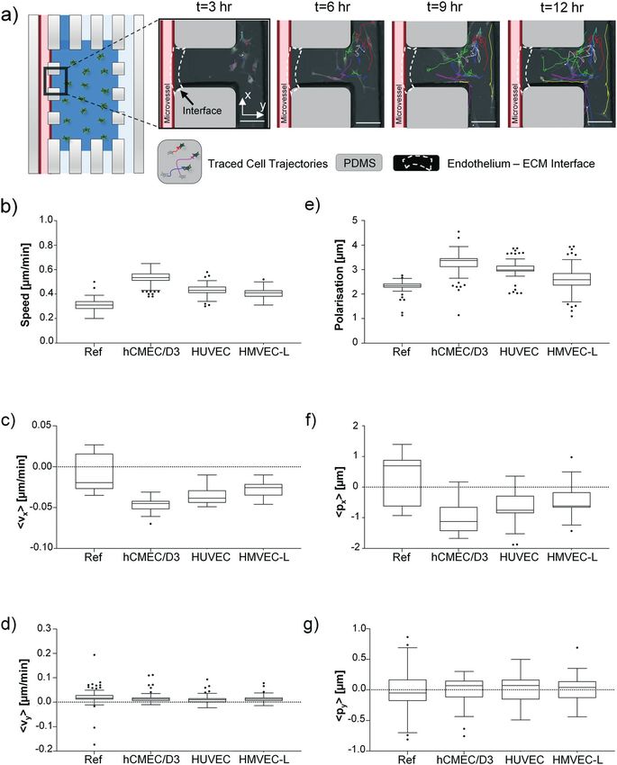
View Article Online
Paper Lab on a Chip
subsequent steps. PicoPure™ RNA isolation kit was used for marker-positive cells, the plugin cell counter was used after
RNA isolation, as per manufacturer's recommendations. gating out weakly stained cells with the threshold. To
Then, RNA concentration and purity were assessed by quantify fluorescence intensity single cells were outlined and
measuring 260/280 absorbance ratio. Pure RNA produces measured with the measure tool, which displayed the values
This article is licensed under a Creative Commons Attribution-NonCommercial 3.0 Unported Licence.
260/280 ratios around a value of 2, and lower values are for area, mean, internal density and raw internal density. As
indicative of high protein content in the sample. For an a control, sections of the background were outlined as well.
example experiment, the average ± SEM 260/280 ratio values To calculate fluorescence, the following formula was utilised:
for 2D and microfluidic systems was 1.952 ± 0.025 and 1.949
± 0.037, respectively. In each experiment, results obtained Integrated Density (sample) − (Mean (ctrl) × Area (ctrl)) =
from samples cultured in 3D ECM, as compared to the 2D Fluorescence Intensity
setup, passed Shapiro–Wilk normality tests with showing no
Open Access Article. Published on 05 May 2021. Downloaded on 7/1/2024 11:47:27 PM.
significant differences in quality of obtained RNA. For fluorescence intensity and quantification, images were
Reverse transcription PCR. The RNA samples were reverse taken with the same settings (exposure, saturation, intensity)
transcribed to obtain cDNA from isolated RNA. PrimeScript™ for each label and magnification in order to compare them
RT-PCR kit was used for cDNA synthesis. The reaction was accurately. Moreover, different cell size was taken into
set in a 0.2 ml PCR tube by using 4 μl 5x cDNA synthesis kit account for the analysis.
buffer, 1 μl PrimeScript™ enzyme mixture, x μl nuclease-free
water, and y μl 500 ng RNA sample. Next, the following Live-cell imaging and image analysis
thermal cycle was run: 5 min at 25 °C, 30 min at 42 °C, 5
An in-house image analysis programme to pre-process the
min at 85 °C, hold at 4 °C. Afterwards, 30 μl of RNase-free
time-lapse images and create a database to automatically
water was added to all samples. The aliquots were frozen at
classify cell interactions with the regional microenvironments
−20 °C for short, and −80 °C for long-term storage.
was used. This novel program utilizes several open-source
Quantitative PCR. SYBR® Green was used as a
applications, such as FIJI and CellProfiler (FIJI: https://
fluorophore. The reactions were set up in 96-well PCR plates
imagej.net/Fiji; Cell Profiler: https://cellprofiler.org), and
under semi-sterile conditions (laminar flow) to avoid
merges them with focus stack (z-stack) image analysis to
contamination. The reaction was set up by adding 5 μl
enable cell tracking and segmentation. The experiments used
SYBR® Select Master Mix, 0.75/075 μl fw/rv primer (10 μM),
brightfield (or transmission) images and green fluorescence
2.5 μl ddH2O, and 1 μl cDNA. Obtained cycle over the
protein (GFP) images. The intensity of GFP expression of the
threshold (Ct)-values were analysed by normalizing them
z-stack images acquired during the time-lapse experiments
against a housekeeping gene (GAPDH) and then comparing
was projected onto one x–y plane at each time point; thus,
the differences with a control, which was either set or chosen
the projected cell area, that is, the 2D projection of the 3D
based on the lowest relative expression (ΔΔCt-method).
cell shape, was obtained at each time point. Cell speed,
velocity and polarization were measured and analysed using
Protein analysis the image-assisted microfluidic platform. The parameters
were calculated for 3D cancer cell culture in the presence of a
Immunofluorescence. For analysis of protein expression via
microvessel or in a reference system – without endothelial
immunofluorescence, cells were fixed and stained with
lining. Cancer cell migration velocity (measured between two
fluorescent antibodies. We used immunofluorescence for
consecutive time points) was quantified. The direction of the
staining cells grown in: i) 6-well plates, ii) ibidi μ-slides, or iii)
cancer cell migration was quantified by velocity components
microfluidic devices. In all cases, cells were fixed in 4%
in the x-direction (perpendicular to the endothelium–collagen
paraformaldehyde for 20 min at room temperature, washed 3
matrix barrier) and y-direction (along the channel direction).
times in PBS and then permeabilized with 0.2% Triton™ X-100
The presence of asymmetric shape features in cancer cells
(Sigma) in PBS for 3 min. Cells were washed 3 more times in
with respect to the position of the cell nucleus was described
PBS, and then blocked by incubating the cells with 4% BSA in
by using the cell polarization parameter. The position of the
PBS for 1 h. Next, cells were incubated with primary antibodies,
cell centre of mass was calculated by the CellProfiler software,
as listed in Table S2b),† at the appropriate dilution overnight at
weighting the x and y coordinates of the pixels within the
4 °C. The next day, cells were washed 3 times with PBS and
projected cell shape with their GFP intensity; a
then incubated with a secondary antibody of choice, as listed
computational error of about 20% was made due to
in Table S2c),† as well as nuclear and/or F-actin counterstain
misrecognition of the projected cell shape profile during
for 1 h at room temperature in the dark. Cells were washed 3
automatic segmentation.
times in PBS before imaging with a Leica SP5 confocal
microscope was performed.
For experimental sets where live/dead cells were being Vessel permeability measurement by dextran diffusion
studies, the images were analysed by quantifying labelled Upon the formation of a microvessel on-chip, the diffusive
cells and by measuring the intensity of fluorescence. The permeability was measured with fluorescently labelled dextran
analysis was carried out via FIJI. To quantify the number of (fluorescein isothiocyanate-dextran, FITC-dextran) in the culture
2348 | Lab Chip, 2021, 21, 2343–2358 This journal is © The Royal Society of Chemistry 2021View Article Online
Lab on a Chip Paper
medium. The permeability measurements and calculations were gliomas.29,30 We developed a quick and straightforward
adapted from Funamoto et al.27 Briefly, an intact endothelial method to recover endothelial cells from our on-chip vascular
monolayer gave rise to an intensity drop between the channel niche coupled to microfluidic assay, to perform qPCR
and the gel region once the fluorescently labelled dextran was analysis. After cell isolation, we used a commercial RNA
This article is licensed under a Creative Commons Attribution-NonCommercial 3.0 Unported Licence.
introduced and persisted over time as dextran slowly diffused isolation kit, and the RNA concentration and purity were
across the monolayer into the gel. Hence, the microvessel assessed by absorbance measurement (260/280 ratio). RNA
permeability was assessed by observing the diffusion of 70 kDa from cells recovered from microfluidic devices and 2D
FITC-dextran (Sigma, 46945; 7 mg ml−1 in PBS, diluted further control samples was checked to ensure pure RNA from cells
1 : 9 in cell culture medium) from the microvessel channel into was obtained. The results passed Shapiro–Wilk normality test
the collagen gel. The diffusion experiments were performed in and were then subjected to a t-test. Our analysis between the
the environmentally controlled chamber (set at 37 °C, 5% CO2) 2D and 3D endothelial cell RNA quality test shows no
Open Access Article. Published on 05 May 2021. Downloaded on 7/1/2024 11:47:27 PM.
of a fluorescent microscope (Leica, CTR6500). The dextran significant difference, indicating that our RNA extraction and
solution was perfused inside the microvessel channel, and purification protocol is comparable to 2D assay routinely
images were acquired every 30 s over 30 min. Beyond 30 min, used in research. Using our optimized method, it was
the geometry of the device (e.g., pillars and the limited size of possible to extract up to 75 ng μl−1 of RNA per device. If the
the gel chamber) broke down the perfect sink condition assumed concentration of RNA in one sample was lower than 50 ng
in the calculation. Obtained fluorescent images were analysed μl−1, RNA from 2 or more samples from the same condition
using open-source software (FIJI). and the same run of a given experiment were pooled. The
extracted cells from the 3D microfluidic device are pure
Statistical methodology endothelial cell populations, as confirmed by qPCR analysis.
Data were analysed using GraphPad Prism software and its It has shown that they did not contain any GFAP, a particular
built-in tests, or RStudio. Data were tested for normality with marker for the GBM cells.
Shapiro–Wilk test and subsequently analysed with suitable
parametric or non-parametric tests. In order to assess Brain cancer cells in 3D ECM
significance, unpaired or paired t-test or ANOVA with Tukey's
post hoc test was used, depending on the experiment in U87 cells, a commonly used GBM model cell line, was used
question. The following asterisk rating system for p-value was in our study for initial protocol optimisation, and also as a
used: *p ≤ 0.05, **p ≤ 0.01. reference. In previous in vivo studies, it has been reported
that U87 cell type failed to accurately model human GBM
Results and discussion compared to patient-derived tumour stem cells.15 U87 cells,
cultured in 2D in vitro cell culture with FBS-enriched
Protocol optimization to study gene and protein expression medium, exhibit reduction in neural stem cell marker levels,
in cells cultured in 3D microfluidics such as nestin, SOX2, and CD133.31,32 Here, we performed
On-chip vascular niche (Fig. 1) was fabricated to investigate studies to culture U87 cells in 3D-collagen gel with laminin,
the interactions between patient-derived glioma cell lines and in three comparative media conditions: FBS-supplemented
different human organ-specific endothelium. With this DMEM (standard U87 culture media), FBS-supplemented
device configuration, GSCs can be cultured in the 3D ECM EGM-2MV (standard endothelial cell media), and serum-free
gel in proximity to a quasi-circular, perfusable microvessel. EGM-2MV. ESI† Fig. S2 and S3 show that switching FBS-
Despite the advantages of microfluidic cell culture, which supplemented DMEM medium with FBS-supplemented EGM-
would better emulate in vivo conditions than conventional 2D 2MV medium had negligible impacts on the U87 viability,
cell cultures, the integration of these techniques as a and the selected glial markers (e.g. EGFR, GFAP), in either 2D
research tool in mainstream cancer research has been slow.28 or 3D culture formats.33 However, 3D culture significantly
Lack of in-depth biological characterisation accompanied by increased the stemness markers of U87 cells (CD44, CD133,
limited biological readouts of the assay has been central nestin, olig2 and sox2 measured at day 3), which is consistent
issues. Genomic or proteomic analysis post microfluidic cell previous reports on GBM-derived cell lines in 3D collagen
culture requires protocol optimisation to retrieve cells in 3D culture.34–36 In a 3D collagen culture, maintaining the U87
culture from the microfluidic device. Here, we optimized cells in serum-free EGM-2MV further enhances the
several protocols for biological analysis on cells post-3D expression of sox2 and nestin, compared to the serum-
culture in a microfluid device, as summarized in Fig. 1. This supplemented media, which echoes with prior reports that
makes our platform flexible and adaptable to commonly used serum-free cultures enhanced stemness related genes.31,32
biological analysis, such as protein/gene expression analysis For all the culture format and media conditions studied,
coupled with functional analysis such as vessel permeability cellular viability of over 85% was seen for day 3, confirming
and live-cell imaging of cancer cell migration. the suitability of these culture conditions for assay purposes.
The study of the glioma-associated neovascularization 3D collagen-I gel culture preserves glioma stem cell-like
mechanisms is important as microvascular proliferation was phenotype in patient-derived cell lines. Considering the
declared a pathological hallmark of malignant high-grade complexity of both inter- and intra-tumour cellular, genetic
This journal is © The Royal Society of Chemistry 2021 Lab Chip, 2021, 21, 2343–2358 | 2349View Article Online
Paper Lab on a Chip
and molecular heterogeneity of GBMs,37,38 it is of interest to (G166, GCGR-E13, GCGR-E17, GCGR-E28 and GCGR-E35).
design an in vitro model with human cells that can be easily The FNS cell line GCGR-NS6FB_A was used as a healthy brain
used for culturing different patient-derived cancer cell lines control for stemness markers, and the U87 cell line were
and to dissect whether glioblastoma sub-types influence the used as technical control for glial markers. Since GSCs are
This article is licensed under a Creative Commons Attribution-NonCommercial 3.0 Unported Licence.
process of neovascularisation. We used five patient-derived standardly cultured in serum-free conditions in 2D, it was
cell lines, containing glioma stem-like cell (GSC) population crucial to test whether our 3D ECM and serum-free condition
Open Access Article. Published on 05 May 2021. Downloaded on 7/1/2024 11:47:27 PM.
Fig. 2 Patient-derived stem cell lines are characterised by stemness-related genes whilst cultured in 3D collagen I-based ECM for three days. a)
Representative bright-field images of each GBM cell type cultured in 3D ECM acquired by light microscopy. mRNA expression of stemness-related
gene in GBM cells normalised to 2D samples. Results for the 3D samples obtained from three independent experimental runs are presented by
mean ± SEM. Two-tailed t-test was used for significance. Note that for FNS, G166 and GCGR-E35, GFAP was not detected by qPCR; b) example
immunostaining images of GCGR-E13 cells grown in 3D for stemness-related markers: CD44, CD133, nestin, olig2, sox2. All were counter-stained
for nucleus (CyTRAK); c) the viability of GBM cells in 3D culture. i) Example images of calcein AM/EthD-1 staining of GCGR-E13 cells at day-1 and
day-7 culture, inside a microfluidic device; ii) graph presenting the cellular viability over seven days in cell culture. Data presented mean ± SD
(percentage of live cells).
2350 | Lab Chip, 2021, 21, 2343–2358 This journal is © The Royal Society of Chemistry 2021View Article Online
Lab on a Chip Paper
would influence the cellular characteristics of these five GBM coating is the prime factor controlling the expression of the
patient-derived cell lines. endothelial junction and tightness-related genes, while the
All the tested cell populations were successfully cultured in a effect of the endothelial layer format (e.g. 2D vs. 3D
serum-free condition within a 3D collagen gel/laminin for 3 days microvessel) is secondary. Tight junction ZO-1 and actin
This article is licensed under a Creative Commons Attribution-NonCommercial 3.0 Unported Licence.
with the expected cell morphology (Fig. 2a)). Relative gene (phalloidin) staining revealed that all three cell types were
expression (mRNA level) analysis was performed to study the able to form a microvessel structure with a quasi-circular
differences between 2D and 3D culture in proliferation, cross-section under 3D culture condition by
stemness and differentiation, in a manner similar to the U87 immunofluorescence, shown in Fig. 3b). Using our 3D ECM
data shown above. First, we did not detect the glial fibrillary device, HUVEC and hCMEC/D3 cells formed a more defined
acidic protein (GFAP, a marker of astrocytic differentiation) in circular microvessel structure compared to HMVEC-L cells.
the G166, GCGR-E35 and FNS cell lines in either 2D and 3D qPCR revealed that hCMEC/D3 is characterized by
Open Access Article. Published on 05 May 2021. Downloaded on 7/1/2024 11:47:27 PM.
cultures,39,40 by qPCR. The fact that GFAP was not detected in significantly higher levels of ZO-1 expression than the other
FNS (as a negative control) but in U87 (as a positive control), two cell types (Fig. 3b) ii)), where ZO-1 is required for tight
confirms the fidelity of our cell extraction and analysis process. junction formation44–46 in brain endothelial cells forming the
In all cell lines, proliferation (Ki67 marker41) in 3D ECM culture blood brain barrier. Other tight-junctions-related molecules,
was slightly decreased compared to 2D. Similar to the U87 claudin-5 and occludin47 were also shown to be higher in
results demonstrated above, 3D collagen culture enhances the hCMEC/D3 compared to HUVEC or HMVEC-L, shown in Fig.
expression of stemness-related markers (CD44, CD133, nestin, S5a).† As the process of neovascularization is important in
olig2, sox2) for all patient-derived GSC lines, compared to 2D the context of GBM, we further compared the RNA levels of
culture, while inducing insignificant changes of GFAP expression neovascularisation-related genes across the three cell types
for the selected cell lines with GFAP expression (Fig. 2a)). forming circular microvessels, and no significant differences
The stemness-related markers that we selected have been in mRNA relative levels were detected, as shown in Fig. S5b).†
indicated for their role in the maintenance of cell renewal The barrier function of the three microvessel types was
and multi-lineage differentiation, contributing to phenotypic measured by the permeability of fluorescent dextran. In
plasticity of GBM cells.42,43 Furthermore, epidermal growth particular, Fig. 3c) and Table S2† showed that in a serum-free
factor receptor (EGFR) expression was upregulated in selected condition, which is important for the subsequent co-culture for
patient-derived cell lines (i.e. GCGR-E13, GCGR-E28). Overall, maintaining GSC stemness, induces insignificant changes in
our mRNA level data are consistent with previously published vessel permeability compared to the serum-supplemented
data in GBM-derived cell lines derived from other sources, condition. The permeability values showed that the brain
cultured in collagen gels.34–36 endothelial cell line, hCMEC/D3, forms the tightest barrier
Then, we confirm the localization and protein expression compared to HUVECs or HMVECs, and the permeability on the
of CD44, CD133, nestin, olig2 and sox2 in GCGR-E13 cells microvessel decreases after 24 h of culture in the 3D device.
cultured in 3D ECM by immunostaining (Fig. 2b)). The multi- These findings further confirmed that the hCMEC/D3 cells
level analysis of factors contributing to GBM phenotype has form a more favourable microvessel than HUVECs or HMVECs-
shown comparable trends in the detection of mRNA L to study brain vasculature and GBM cells as they are specific
transcripts and protein between 2D and 3D ECM cell culture. to the brain, and that they are adequate to be used in the
Based on the above results, we suggest the 3D collagen-I specific setup we described here. Nonetheless, all three cell
culture condition created for GSCs is suitable for maintaining types were able to create vessels of acceptable permeability, as
their phenotype for 3 days. Finally, cell viability assays were compared to other microvessel-on-a-chip models where
performed in order to ensure the system is robust enough to hCMEC/D3, HUVECs or HMVECs were used.48–51
support prolonged cell culture of various cell types. Each cell
type was cultured within microfluidic devices for up to 7
days. Cells were cultured in a serum-free medium and in the Perivascular niche with GSCs on a chip
presence of gravity-driven flow. Example images of calcein Viability of GSC and microvessel co-culture: static versus
AM/EthD-1 staining of GCGR-E13 cells were shown in flow. To optimise the co-culture conditions, we evaluated the
Fig. 2c) i). As shown in Fig. 2c) ii) and S4,† it was found that cellular viability on-chip, in mono-culture, and co-culture
the cells have over 80% viability at day-3 in 3D culture, conditions with the serum-free condition, under a static
suitable for follow-on experiments. condition or in the presence of gravity-driven flow. Fig. 4a)
shows that the application of gravity-driven flow had little
impact on the viability of the microvessel in mono-culture
Microvessel-on-a-chip formed by different organotypic (∼93% at day 3), but significantly improved the co-culture
endothelial cell types cellular viability (∼77% for static, versus ∼87% for flow at
Next, we investigated endothelial cells from the human brain day 3). Therefore, in the subsequent experiments, we
(hCMEC/D3), umbilical cord (HUVECs) or the lung (HMVEC- implemented gravity-driven flow in the on-chip setup to
L) in terms of microvessel formation in the microfluidic ensure adequate cellular viability for the study of organotypic
device. Fig. 3a) shows that the application of collagen IV microvessel interaction with GSCs.
This journal is © The Royal Society of Chemistry 2021 Lab Chip, 2021, 21, 2343–2358 | 2351View Article Online
Paper Lab on a Chip
This article is licensed under a Creative Commons Attribution-NonCommercial 3.0 Unported Licence.
Open Access Article. Published on 05 May 2021. Downloaded on 7/1/2024 11:47:27 PM.
Fig. 3 Endothelial cell 3D culture with ECM is important for tightness-related gene expression and barrier function, while serum reduction has
negligible effects on permeability. a) A comparison of tightness-related gene expression in each endothelial cell type in 2D and 3D: i) hCMEC/D3; ii)
HMVEC-L; and iii) HUVEC. mRNA expression normalised to samples from 2D without collagen IV coating; results obtained from three independent
experimental runs are presented by mean ± SEM. One-way ANOVA with Tukey's post hoc test was used for significance; b) i) microvessel lumen
formed by various types of microvessels in contact with ECM. Staining of junction protein ZO-1, cytoskeleton by phalloidin, and merged image. In all
panels, on the right, a 3D reconstruction of the microvessel cross-section at a location along the vessel lumen (y-axis); ii) hCMEC/D3 express higher
levels of endothelial tightness-related genes than HUVECs and HMVECs-L in the microfluidic setup. mRNA expression normalised to HMVEC-L
(results obtained from three independent experimental runs are presented by mean ± SEM. One-way ANOVA with Tukey's post hoc test used for
significance); c) diffusion measurements showed appropriate level of permeability for the hCMEC/D3 vessel; i) typical images from an permeability
experiment showing 70 kDa FITC-dextran diffusion after 30 min; endothelial permeability measurements for cells ii) in serum-containing medium 12
and 24 h after seeding; and iii) in serum-free medium 12 and 24 h after seeding. The graphs show results from three independent experimental runs
as mean ± SEM. t-Test shows insignificant difference for permeability in serum-containing versus serum-free cultures.
Effects of GSCs on organotypic microvessels. Although hCMEC/D3, HUVEC or HMVEC-L, in serum-free medium and
angiogenesis has been regarded as essential for tumour gravity-driven flow.
growth and progression, recent studies suggest that a variety To determine how GSCs influence the microvessels,
of tumours can advance without angiogenesis, and their endothelial cells were recovered from microfluidic devices
microcirculation may be provided by nonsprouting vessels.5 after 24 h of co-culture to establish an expression pattern of
Establishing an in vitro model that allows the study of neovascularization-related genes at the mRNA level (heatmap
interactions between disperse cancer cells and nonsprouting in Fig. 4c) and unprocessed data in Fig. S6†).
microvessels can be of value towards understanding Neovascularization-related genes mRNA levels were
mechanisms contributing to brain tumour progression. comparable for FNS either in a mono-culture, or in co-culture
Therefore, three separate co-culture setups were established with a microvessel (hCMEC/D3, HUVECs or HMVECs-L cells),
to study the interactions between U87 (glial control), FNS that we defined as the reference conditions. In contrast, most
cells (stem cell control), GSCs with vessels formed by of the neovascularization-related gene mRNA levels increased
2352 | Lab Chip, 2021, 21, 2343–2358 This journal is © The Royal Society of Chemistry 2021View Article Online
Lab on a Chip Paper
This article is licensed under a Creative Commons Attribution-NonCommercial 3.0 Unported Licence.
Open Access Article. Published on 05 May 2021. Downloaded on 7/1/2024 11:47:27 PM.
Fig. 4 Viability of 3D GSCs culture with or without a microvessel, in the absence or presence of flow (∼1 dyne per cm2). a) i) Example image of
calcein AM/EthD-1 staining of GCGR-E13 cells at day three in co-culture inside a microfluidic device with gravity-driven flow; ii) three-day GSC cell
viability for the four different configurations. Data obtained from three independent experimental runs and presented as mean ± SD (percentage of
live cells); b) graphs presenting viability of six different GBM cell lines, a FNS line, and the U87 cell line at day-3 culture with different microvessel
types under flow. Data obtained from three independent experimental runs and presented as mean ± SD (percentage of live cells). c) GBM and
FNS cells induce different response patterns in different endothelial cells under flow. Heatmaps were plotted in GraphPad Prism software after the
analysis of mRNA expression in relation to GAPDH. Reference pattern was established from RNA isolated from endothelial cells cultured without
the presence of brain cancer cells in microfluidic devices (MC = microvessel monoculture). Results obtained from three independent experimental
runs. Green indicates upregulated expression, and red indicates downregulated expression.
when endothelial cells are co-cultured with all glioma cells It is interesting that MMP-2 and MMP-9 were noticeably
studied. Ang1 and Ang2 are modulators of endothelial upregulated in HUVEC, but not in the hCMEC/D3 and
permeability and barrier function via Tie2.52–54 In all cases, HMVECs-L co-cultures. Cross-comparing the heat map
Ang1 was downregulated, suggesting disruption of vascular patterns for the three endothelial cells, we found that the
stability. VEGFR1 and VEGFR2 upregulation were detectable response of endothelial cells to the co-cultures was GSC-
in selected samples, consistent with the previous report for specific and endothelial cell-specific. In particular, HUVECs
endothelial cells of the tumour neovasculature and in normal seem to show more neovascularization susceptibility
brain vessels adjacent to tumours.55 On the other hand, the compared to hCMEC/D3 and HMVEC-L. These findings
production of matrix-degrading proteases, particularly MMPs, might reflect the nature of the endothelial cell types
by endothelial cells is a critical event during angiogenesis.56 described by Uwamori et al., where the authors reported that
This journal is © The Royal Society of Chemistry 2021 Lab Chip, 2021, 21, 2343–2358 | 2353View Article Online
Paper Lab on a Chip
HUVECs had greater potential than brain microvessel niche. In every 3D device, we monitored the behaviour of 5–
endothelial cells in terms of vascular formation.57 15 cancer cells, in a field of view localized at the interface
between the outermost side channel and collagen I-based
hydrogel chamber. The cancer cell speed (micron/min) in the
This article is licensed under a Creative Commons Attribution-NonCommercial 3.0 Unported Licence.
Cancer cell migration dynamics studied by live-cell imaging 3D device with a microvessel is higher compared to a
We coupled the 3D microfluidic co-culture system with live- microvessel-free device (Fig. 5b)). Between the three organ-
cell imaging to track cell behaviours in specific microvessel specific endothelial cells, a hCMEC/D3 microvessel induced
types (Fig. 5a)). GFP-tagged patient-derived GBM cells (G166) the fastest movement of cancer cells. We also measured to
were used to observe their behaviour in an artificial vascular see if the entire cancer cell population (in the FOV) had a
Open Access Article. Published on 05 May 2021. Downloaded on 7/1/2024 11:47:27 PM.
Fig. 5 G166 cells cultured in the presence of a hCMEC/D3 microvessel with flow are characterised by higher speed, velocity, and preferential
polarisation in the direction towards the microvessel. a) Sample images showing tracking of the cell movement in 3 h time intervals. b) Speed of
the cancer cells for the reference and microvascular systems; c) cancer cell velocity component along the x-axis; d) cancer cell velocity
component along the y-axis; e) polarisation magnitude of the cancer cells in the collagen matrix; f) cellular polarisation component along the
x-axis; g) cellular polarisation component along the y-axis. x-Axis is situated perpendicular to the barrier, pointing away from the barrier, and
y-axis is along the ECM gel chamber. Scale bar = 100 μm. Note that ‘Ref’ samples are measured in microvessel-free device and all data are
obtained from three independent experimental runs.
2354 | Lab Chip, 2021, 21, 2343–2358 This journal is © The Royal Society of Chemistry 2021View Article Online
Lab on a Chip Paper
preferred direction of migration towards the endothelial cells microvessel types, a slight enhancement in directed
(right to left). The velocity components perpendicular to the migration was observed for the cancer cells facing the
ECM–endothelium barrier (x-axis as shown in Fig. 5a)) and hCMEC/D3 vessels.
along the ECM chamber were analysed (Fig. 5c)). Cancer cells Cancer cell shape axial asymmetries were also investigated
This article is licensed under a Creative Commons Attribution-NonCommercial 3.0 Unported Licence.
in the device with a microvessel were shown to move towards during cellular migration inside the collagen I matrix. Brain
the channel interface direction more (i.e. with more negative tumour cells in the presence of hCMEC/D3 microvessel were
x-velocity component) compared to the microvessel-free characterized by the highest median of polarization
control (Fig. 5c)). But there was no difference detected in the parameter, as a scalar quantity (Fig. 5e)). As illustrated in
y-direction (Fig. 5d)). Comparing the data among the three Fig. 5f) and g), investigation of cell polarization in x and y
Open Access Article. Published on 05 May 2021. Downloaded on 7/1/2024 11:47:27 PM.
Fig. 6 Increased endothelial permeability and the downregulation in mRNA expressions for junction proteins VE-cad, CD31, and ZO-1 in co-
cultures with a) G166 and b) GCGR-E13 cancer cells. i) There is an increase in endothelial permeability in co-cultures with G166 and GCGR-E13
cancer cells. The graph shows results from three independent experimental runs as mean ± SEM. Two-tailed t-test was used for significance. The
permeability increase correlates with downregulation of tightness-related genes in vessels formed by different vessel types; ii) mRNA expression
normalised to samples of endothelial cells cultured without brain cancer cells; results obtained from three independent experimental runs are
presented as mean ± SEM. Two-tailed t-test was used for significance.
This journal is © The Royal Society of Chemistry 2021 Lab Chip, 2021, 21, 2343–2358 | 2355You can also read