A Smart Multifunctional Nanoparticle for Enhanced Near-Infrared Image-Guided Photothermal Therapy Against Gastric Cancer
←
→
Page content transcription
If your browser does not render page correctly, please read the page content below
International Journal of Nanomedicine Dovepress
open access to scientific and medical research
Open Access Full Text Article
ORIGINAL RESEARCH
A Smart Multifunctional Nanoparticle for
Enhanced Near-Infrared Image-Guided
Photothermal Therapy Against Gastric Cancer
Jun Shao 1, * Background: Surgery is considered to be a potentially curative approach for gastric cancer.
Rongpu Liang 1, * However, most cases are diagnosed at a very advanced stage for the lack of typical symptoms in the
Dongbing Ding 1, * initial stage, which makes it difficult to completely surgical resect of tumors. Early diagnosis and
Xiaoming Zheng 1 precise personalized intervention are urgent issues to be solved for improving the prognosis of
gastric cancer. Herein, we developed an RGD-modified ROS-responsive multifunctional nanosys
Xudong Zhu 1
tem for near-infrared (NIR) imaging and photothermal therapy (PTT) against gastric cancer.
Shengxue Hu 2
Methods: Firstly, the amphiphilic polymer was synthesized by bromination reaction and nucleo
Hongbo Wei 1
philic substitution reaction of carboxymethyl chitosan (CMCh) and 4-hydroxymethyl-pinacol phe
Bo Wei 1 nylborate (BAPE). Then, it was used to encapsulate indocyanine green (ICG) and modified with
1
Department of Gastrointestinal Surgery, RGD to form a smart multifunctional nanoparticle targeted to gastric cancer (CMCh-BAPE-
The Third Affiliated Hospital of Sun Yat-
RGD@ICG). The characteristics were determined, and the targeting capacity and biosafety were
sen University, Guangzhou, 510630,
People’s Republic of China; 2College of evaluated both in vitro and in vivo. Furthermore, CMCh-BAPE-RGD@ICG mediated photothermal
Biological Science and Engineering, therapy (PTT) effect was studied using gastric cancer cells (SGC7901) and SGC7901 tumor model.
Fuzhou University, Fujian, 350108,
People’s Republic of China
Results: The nanoparticle exhibited suitable size (≈ 120 nm), improved aqueous stability, ROS-
responsive drug release, excellent photothermal conversion efficiency, enhanced cellular uptake, and
*These authors contributed equally to targeting capacity to tumors. Remarkably, in vivo studies suggested that CMCh-BAPE-RGD@ICG
this work
could accurately illustrate the location and margin of the SGC7901 tumor through NIR imaging in
comparison with non-targeted nanoparticles. Moreover, the antitumor activity of CMCh-BAPE-
RGD@ICG-mediated PTT could effectively suppress tumor growth by inducing necrosis and
apoptosis in cancer cells. Additionally, CMCh-BAPE-RGD@ICG demonstrated excellent biosafety
both in vitro and in vivo.
Conclusion: Overall, our study provides a biocompatible theranostic nanoparticle with
enhanced tumor-targeting ability and accumulation to realize NIR image-guided PTT in
gastric cancer.
Keywords: gastric cancer, nanoparticle, indocyanine green, near-infrared imaging,
photothermal therapy
Introduction
Gastric cancer is the second leading cause of cancer-related death and approximately
Correspondence: Hongbo Wei; Bo Wei 1,033,000 new cases are diagnosed and 783,000 deaths are estimated worldwide in
Department of Gastrointestinal Surgery,
The Third Affiliated Hospital of Sun Yat- 2018 according to GLOBACAN statistics.1 Surgery, radiotherapy, and chemotherapy
sen University, Guangzhou, 510630,
People’s Republic of China are the conventional treatments for gastric cancer.2–4 Although these therapeutic
Tel +86-20-85252228 methods have improved the survival rate to some extent, the great trauma of surgery,
Email weihb@mail.sysu.edu.cn;
weibo3@mail.sysu.edu.cn the non-targeted ability and toxic side effects of radiotherapy and chemotherapy have
International Journal of Nanomedicine 2021:16 2897–2915 2897
© 2021 Shao et al. This work is published and licensed by Dove Medical Press Limited. The full terms of this license are available at https://www.dovepress.com/terms.php
and incorporate the Creative Commons Attribution – Non Commercial (unported, v3.0) License (http://creativecommons.org/licenses/by-nc/3.0/). By accessing the work
you hereby accept the Terms. Non-commercial uses of the work are permitted without any further permission from Dove Medical Press Limited, provided the work is properly attributed. For
permission for commercial use of this work, please see paragraphs 4.2 and 5 of our Terms (https://www.dovepress.com/terms.php).
Shao et al Dovepress
seriously affected the quality of life of patients. Furthermore, attempts have been made to overcome these challenges, such
since there is no or little typical symptoms in the initial as modification of ICG with ligands to improve targeting
stage,5 the majority of patients with gastric cancer are diag ability,25 encapsulation of ICG into nanocarriers to provide
nosed middle and advanced tumors (at stage II or III). increased efficacy.14,19,26 Compared with the former which
Complete surgical resection of tumors in these cases remains usually cannot enhance the stability of ICG, the latter
extremely challenging. And for the lack of haptic feedback, (NDDS) seems to be a more hopeful strategy to enhance its
stability, prolong circulation and improve targeting ability.
surgeons may also have misjudgments about small lesions
NDDS has several advantages over the traditional pattern
during laparoscopic surgery of gastric cancer. Therefore,
of drug administration.27–29 First, the encapsulated ICG by
early diagnosis and precise personalized intervention are
nanomaterials could be rescued from nonspecific binding to
urgent issues to be solved for improving the prognosis of
plasma proteins, thus increasing its half-life in vivo. Second,
patients with gastric cancer.
the passive targeting delivery, which is associated with
Photothermal therapy, which destroys tumor cells
enhanced permeability and retention (EPR) effect and trans
through heat produced by photothermal agents, has become
cytosis (perhaps with other unknown mechanisms), makes it
a promising alternative strategy over the past few decades.6–9
possible for nanomaterials within a specific size range to
Meanwhile, nanoscale drug delivery systems (NDDS) com
selectively pass through and retain in the tumor.30–32 Third,
bining imaging and therapy is increasingly favored by the surface of nanomaterials could be modified with specific
researchers.10–13 The method of administration combining ligands to achieve active targeting delivery. For instance,
photothermal agents and nanomaterials could not only specific cell surface receptors, such as transferrin or folate
demonstrate the specific location of the lesion but also receptors, are overexpressed in tumor cells such as malignant
achieve more precise personalized intervention of the glioma and breast cancer.33 Nanomaterials modified with
tumor. For a satisfied image-guided therapy, the prerequisites peptides, aptamers, and monoclonal antibodies30,33 can
is to design a biocompatible NDDS with specific tumor- increase specific cellular uptake through receptor-mediated
targeting ability and a multifunctional theranostic agent that endocytosis (RME) effect, thus resulting in the accumulation
demonstrates imaging and therapeutic functions.14 of drugs in the target cells and increasing the payload of drug
To date, many theranostic agents including metal delivery. Integrins are a family of transmembrane receptors
nanostructures,15 near-infrared (NIR) dyes,16 carbon capable of mediating bidirectional signaling through the
nanomaterials,17 polymeric nanoparticles,13 and many others membrane, including invasion, migration, survival, and
have been widely explored for imaging-guided treatment for proliferation.34,35 Integrin αvβ3, which is overexpressed in
cancer, among which indocyanine green (ICG) is the only gastric cancer, may be helpful in the routine classification of
approved NIR dye for clinical application. Since tissues are gastric cancer subtypes and services as a novel putative
relatively transparent under NIR illumination, ICG displays prognostic biomarker.36 Arg-Gly-Asp (RGD) is a tripeptide
several obvious advantages, including minimal background sequence that can specifically recognize and bind integrin
fluorescence interference, deeper tissue penetration, and real- αvβ3. In the past few years, the RGD family has been devel
time monitoring during the operation.18,19 Additionally, ICG is oped into a variety of subtypes, such as RGD straight-chain,
also a photosensitizer with excellent photothermal conversion internalizing-RGD, and cyclic RGD.14,37 Shan et al38 pre
efficiency when exposed to single wavelength NIR light.14,20 pared an RGD-modified hepatitis B core protein virus-like
These attractive features indicate that ICG could be particle (HBc VLP) and it displayed high-efficiency endocy
a promising dye for tumor imaging and PTT.11,21 So far, tosis in vitro and improved tumor-targeted accumulation
ICG has been used for the determination of liver function in vivo compared with non-targeted particles. Therefore, the
and liver blood flow,22 cardiac output,23 and ophthalmic functionalization of RGD polypeptides can greatly improve
angiography.24 Nevertheless, several intrinsic drawbacks the tumor-targeting ability of nanomaterials.
limit its ideal application in clinical practice. Self-aggregation In practical application, the poor uptake of nanomaterials
and instability in solution lead to fluorescence quenching. by targeted cells and incomplete intracellular release limit its
Nonspecific binding to plasma proteins and rapid elimination curative effect.39 To further improve the bioavailability of
through hepatobiliary excretion result in a half-life of only 2~4 drugs, the unique microenvironment of tumor tissue, such as
min. Moreover, the nontargeting ability to tumor limits its low pH, high concentration of glutathione (GSH), high con
application in tumor diagnosis and treatment.19 Several centration of reactive oxygen species (ROS), etc., has been
2898 http://doi.org/10.2147/IJN.S289310 International Journal of Nanomedicine 2021:16
DovePress
Dovepress Shao et al
used to design environmentally responsive nano-drug delivery systematically analyzed in vitro. Their NIR imaging and
systems, which can target to the tumor site and release the anti-tumor efficiency on tumor-bearing mice (SGC7901
loaded drugs in the targeted tumor cells.10,18,26,29,39,40 In this cells) were studied. The outcome of our work may demon
paper, we synthesize a novel ICG-loaded ROS-responsive strate a promising way to integrate NIR imaging into PTT of
RGD-modified nanoparticle for simultaneous gastric cancer gastric cancer.
NIR imaging and PTT (Scheme 1). The amphiphilic block
nanoparticle is mainly composed of four parts: carboxymethyl Materials and Methods
chitosan (CMCh) acts as a hydrophilic shell to realize the Materials
water solubility of the complex; benzeneboronic acid pinacol Benzeneboronic acid pinacol ester (BAPE), carboxymethyl
ester (BAPE) forms the hydrophobic end and hydrolyzes chitosan (CMCh), phosphorus tribromide (PBr3), Na2CO3,
under the stimulation of excessive ROS in the tumor site to acetonitrile, dichloromethane, 1-ethyl-3-(3-dimethylamino
achieve targeted release of ICG; RGD polypeptides are con propyl) carbodiimide hydrochloride (EDC), and
jugated on the surface to achieve active targeting ability of the N-hydroxysuccinimide (NHS) were purchased from
nanosystem; ICG is used as a bifunctional dye to realize NIR Shanghai Macklin Biochemical Technology Co., Ltd
imaging and PTT. We investigated the preparation and char (Shanghai, China). Indocyanine green (ICG) was obtained
acterization of nanoparticles. The cellular uptake, cytotoxicity, from Shanghai Ronghe Pharmaceutical Technology Co. Ltd.
and photothermal conversion performance were (Shanghai, China). RGD polypeptide was bought from
Scheme 1 Schematic illustration of CMCh-BAPE-RGD@ICG for NIR imaging and photothermal therapy of gastric cancer.
International Journal of Nanomedicine 2021:16 http://doi.org/10.2147/IJN.S289310 2899
DovePress
Shao et al Dovepress
Shanghai Jier Biochemical Co., Ltd. Cell culture reagents Preparation of CMCh-BAPE-RGD@ICG
including Dulbecco’s modified Eagle’s medium (DMEM), Nanoparticles
RPMI 1640, fetal bovine serum (FBS), trypsin, and antibiotics CMCh-BAPE@ICG (50 mg) was first dissolved in an
were all bought from Gibco (Gaithersburg, MD, USA). ROS appropriate amount of pure water, followed by added
detection assay kit was bought from Beyotime Biotechnology with 5.6 mg EDC and 8.4 mg NHS. After stirred for 4
in China. Cell counting kit-8 (CCK-8) and Live/Dead cell h for reaction, the system was added with 10 mg RGD
staining kits were obtained from Kaiji Bio-tech Co., Ltd. polypeptide. Next, the mixture was further stirred over
(Jiangsu, China). Annexin-V/PE Apoptosis Detection Kit night for complete reaction. Then, the obtained product
was purchased from the BD in America. Mouse fibroblastic was transferred into a dialysis sack (MWCO: 1000 Da) for
cells L929 and human gastric cancer SGC7901 cells were purification. The targeted nanoparticles (CMCh-BAPE-
purchased from Beogene Biotechnology Co., Ltd. RGD@ICG) were obtained after lyophilized. Light was
(Guangzhou, China). All other reagents were of analytical avoided during the whole process. Doxorubicin-loaded
grade unless otherwise noted. (DOX) nanoparticles were prepared according to the
same procedure.
Preparation of CMCh-BAPE-RGD@ICG
Nanoparticles Characteristics of CMCh-BAPE-
Preparation of CMCh-BAPE Nanoparticles RGD@ICG Nanoparticles
CMCh-BAPE nanoparticles were prepared as previously The chemical structure of CMCh, CMCh-BAPE and
reported.41 Briefly, 0.46 g (2 mmol) 4-hydroxymethyl- CMCh-BAPE-RGD were analyzed by nuclear magnetic
pinacol phenylborate (BAPE) was weighed and dissolved resonance (1H NMR) spectrometer (AVANCE III 500M,
in 10 mL dichloromethane, followed by the addition of Bruker, Germany). Fourier transform infra-red (FTIR)
0.1 mL phosphorus tribromide (PBr3). The solution spectrometer (VERTEX 70, Bruker, Germany) was
reacted at room temperature for 2 h under nitrogen atmo applied to determine its functional groups. The morphol
sphere. Then, a rotary evaporator was applied to remove ogy images of the nanoparticles were obtained by trans
the organic solvent. Next, the resulting product was dis mission electron microscopy (TEM, JEM-2010F, JEOL
solved by an appropriate amount of acetonitrile. A 5% Ltd., Tokyo, Japan). The mean hydrodynamic diameter
Na2CO3 solution was used to adjust the PH. and size distribution were recorded by dynamic light scat
Subsequently, 0.52 g (1 mmol) carboxymethyl chitosan tering (Zetasizer Nano-ZS90, Malvern Instruments Ltd.,
(CMCh) dissolved in pure water was added to the solution. Worcestershire, UK) at 25°C. The changes in size distribu
After 48 h of reaction under the protection of nitrogen gas, tion after treated by H2O2 (1mM) were also recorded.
the mixed sample was dialyzed for 3 days against pure
water in a dialysis sack (MWCO: 3500 Da) to remove all
Drug Loading Efficiency
the impurities. The CMCh-BAPE was lyophilized and The ICG loading efficiency (LE) was calculated via UV-
stored at −20°C. vis spectrophotometric method. In brief, a series of ICG
solutions with different concentrations were prepared and
Preparation of CMCh-BAPE@ICG Nanoparticles the standard absorption curve of pure ICG in ultra-pure
The loading of ICG to the nanoparticles was based on the water was obtained by a UV-vis spectrometer (UV-
self-assembly method. Generally, 10 mg ICG was dissolved 3100PC, Mapada Instruments, Shanghai, China). Set the
in 5 mL pure water, followed by dropwise added to an λmax at 790 nm. The loaded ICG dose in CMCh-BAPE-
aqueous solution containing 50 mg CMCh-BAPE. The mix RGD@ICG was determined by the absorption value. The
ture solution was further stirred for 4 h at room temperature. formulas used were as follows:
A dialysis bag (MWCO: 1000 Da) was used for purifying
the nanoparticles for 24 h. The final production (CMCh- ICGloaded
LEð%Þ ¼ � 100%
BAPE@ICG) was lyophilized and stored at −20°C. weightnanoparticles
2900 http://doi.org/10.2147/IJN.S289310 International Journal of Nanomedicine 2021:16
DovePress
Dovepress Shao et al
In vitro ROS-Responsive Leakage control was pure water without samples. Then, samples
were irradiated and the temperature was recorded by an
Assessment
infrared thermal imager (Fotric 222, Shanghai, China).
The fluorescence-based doxorubicin (DOX) leakage
experiment was conducted to simulate the release and
ROS-responsive release of ICG from nanoparticles. The
Cell Culture and Mice
L929 cells (a mouse fibroblastic cell line, ATCC) were cul
DOX nanoparticles were prepared as mentioned above.
tured in RPMI 1640 medium while NIH/3T3 cells (a mouse
Then, 1mL pure water containing 6 mg samples in
fibroblastic cell line, ATCC) and SGC7901 cells (a human
a pretreated dialysis bag (MWCO: 1000 Da) was dialyzed
gastric cancer cell line, ATCC) were incubated in Dulbecco’s
against H2O2 solutions (0mM or 1mM) in a 15 mL cen
modified Eagle’s medium (DMEM) at 37°C in a humidified
trifuge tube. At different time intervals, 1 mL dialysate
atmosphere containing 5% CO2. All of the media was supple
sample was taken for obtaining the concentration of DOX
mented with 10% FBS, 1% penicillin, and 1% streptomycin.
by measuring the maximum DOX absorbance (λ=493 nm).
The experimental female BALB/c nude mice were obtained
The dialysate sample was returned to the centrifuge tube
from Beijing Virton Li Hua Experimental Animal Technology
immediately after detection. The following formula was
Co., Ltd (Beijing, China) and raised in the specific pathogen-
applied for cumulative release (%) calculation:
free (SPF) animal room. Before experimentation, all mice
Ai At
cumulative DOX releaseð%Þ ¼ � 100% were raised for one week to acclimatize to the environment.
Ai
All mice experiments were conducted following the guideline
Where Ai was the initial absorbance value, and At was of the Ethics Committee of the Institutional Animal Care and
the absorbance value detected at different time intervals. Use Subcommittee of the Third Hospital of Sun Yat-sen
University and the committee approved this study.
In vitro Photostability Evaluation
A UV-vis spectrometer and a fluorescence spectrometer
Intracellular ROS Level Measurement
The intracellular ROS levels of SGC7901 cells and NIH/3T3
(HR2000+, Ocean Optics) were employed to determine the
cells (negative control) were determined by ROS detection
absorption and emission spectra of ICG and CMCh-BAPE-
assay kit. Briefly, cells were harvested and incubated with 10
RGD@ICG (equivalent ICG concentration: 10 µg/mL),
µM DCFH-DA for 20 min at 37°C in the darkness, shaking
respectively. The photostability was evaluated by detecting
slightly every 5 min. Then, the cells were washed thrice with
the changes in absorption and emission spectra at selected
serum-free medium to remove unreacted DCFH-DA probe.
time intervals. All of the samples were dissolved in pure
The fluorescence was visualized immediately under
water and stored in the dark at 25°C.
a fluorescence microscope (Nikon, Tokyo, Japan) and quan
tified by flow cytometry (BD Accuri, USA).
In vitro Photothermal Efficiency
Measurement Cellular Uptake
The photothermal properties of different materials were eval The internalization of CMCh-BAPE@ICG and CMCh-
uated. One hundred microliters of PBS containing free ICG, BAPE-RGD@ICG was investigated by performing the con
CMCh-BAPE-RGD, or CMCh-BAPE-RGD@ICG (the focal laser scanning microscopy (CLSM, Zeiss LSM 710,
equivalent ICG concentration was 20 µg/mL) were placed Germany) and flow cytometry. In detail, SGC7901 cells
into Eppendorf tubes, respectively. The blank control was were harvested in the logarithmic growth phase and seeded
PBS without samples. An 808 nm laser with 1 W/cm2 energy into the 35-mm cell petri dishes (Nest, 801002) (5×10⁴ cells/
was applied for irradiation for 5 min. The temperature was dish) at 37°C and cultured for 24 h prior to the experiment.
recorded every 10 seconds to compare the photothermal Then, the medium was replaced and supplemented with
effects of different samples in vitro. CMCh-BAPE@ICG or CMCh-BAPE-RGD@ICG (ICG con
In addition, the effect of concentration on the photo centration was 10 µg/mL), respectively. After incubated for 6
thermal effect of the material was explored. Generally, h, cells were washed with PBS and fixed with paraformalde
different concentrations of CMCh-BAPE-RGD@ICG hyde (4%), followed by added with 1 μL DAPI for staining the
(10, 20, 30, and 40 µg/mL) were dissolved in pure water nuclei. Next, cells were gently washed with PBS and the
and placed into Eppendorf tubes, respectively. The blank cellular internalization was imaged under CLSM. Red and
International Journal of Nanomedicine 2021:16 http://doi.org/10.2147/IJN.S289310 2901
DovePressShao et al Dovepress
blue fluorescence emissions from ICG and DAPI were The live/dead staining assay was conducted for further
acquired at λex = 633nm for ICG and λex = 405nm for analysis. Briefly, SGC7901 cells were treated with PBS,
DAPI, respectively. ICG, or CMCh-BAPE-RGD@ICG (an equivalent ICG con
Flow cytometry was performed for further quantitative centration: 20 μg/mL), followed by laser illumination as
analysis. 5×10⁴ SGC7901 cells were seeded into 24-well mentioned above. Twenty-four hours later, calcein AM
plates per well. Twenty-four hours later, the medium was and PI were added for staining. Then, the cells were gently
replaced by fresh medium, respectively, supplemented rinsed by PBS three times and observed under the fluores
with CMCh-BAPE@ICG or CMCh-BAPE-RGD@ICG cence microscope. The images were acquired at λex = 545
and cultured for different time intervals (the equivalent nm and λex = 490 nm for PI and calcein AM, respectively.
ICG concentration was 10 µg/mL). At each time interval
(3 h and 6 h), cells were gently washed three times, In vitro Blood Compatibility Assay
followed by trypsin digestion, centrifugation, and re-sus First, hemolysis assay of erythrocytes was performed to assess
pended in PBS. Finally, flow cytometry was applied to the safety of CMCh-BAPE-RGD@ICG. Generally, red blood
determine the fluorescence intensity of cells in each cells (RBCs) from a 6-week-old nude mouse were collected
group and the corresponding fluorescence intensity was from fresh blood samples after centrifuged to remove the
quantified by FlowJoV10 software. serum (5000 rpm, 5 min). The RBCs were washed, resus
pended, and diluted with PBS to adjust the final concentration
at 16% (v: v). Next, 50 μL RBC solutions were added to 1 mL
In vitro Cytotoxicity and Anticancer PBS containing CMCh-BAPE-RGD@ICG with different
Activity Assessment concentrations (0.01, 0.1, 0.5, and 1 mg/mL), 1 mL pure
CCK-8 assay was applied for determining the toxicity of water (positive control) or 1 mL PBS (negative control),
CMCh-BAPE-RGD@ICG on L929 cells. Generally, 5000 respectively. After being incubated for a period of time (3 h,
L929 cells in the logarithmic phase were harvested, resus 5 h, 8 h, 18 h, and 24 h), the mixtures were centrifuged
pended, and seeded into 96-well plates for overnight incu (1000 rpm, 5 min) and the supernatants containing lysed
bation. Then, the medium was replaced by fresh medium RBCs were collected for absorbance values determination at
supplemented with CMCh-BAPE-RGD@ICG at different 540 nm. The hemolysis was calculated as follows:
concentrations (10, 20, 40, 80, 160, 320, and 500 μg/mL). At An
Hemolysis ð%Þ ¼ � 100%
After 24 h incubation, the cell viabilities were measured Ap An
based on the product protocol. For the photocytotoxicity
Note that At was the absorbance of samples incubated
effect assessment, SGC7901 cells seeded into 96-well
with nanoparticles measured at different time intervals. Ap
plates, and the medium was added with CMCh-BAPE-
was the absorbance of samples incubated with pure water
RGD@ICG at various concentrations (ICG equivalent
and An was the absorbance of samples incubated
concentration: 0, 5, 10, 20, and 40 μg/mL). After 6
with PBS.
h incubating, the medium was replaced and irradiated for To further observe the effect of nanoparticles on RBCs,
5 min by an 808 nm laser with 1.0 W/cm2 energy. Cells the erythrocyte morphology was determined. Briefly, the
without laser irradiation were set as the control group. RBCs were obtained as mentioned before and added to
Twenty-four hours later, the cell survival ratios were cal 1 mL PBS containing CMCh-BAPE-RGD@ICG with dif
culated. Meanwhile, for the comparison of photocytotoxi ferent concentrations (0.01, 0.1, 0.5, and 1 mg/mL). After
city effects of different materials, SGC7901 cells were incubated at 37°C for 15 min, the mixture was centrifuged
treated with PBS, ICG, or CMCh-BAPE-RGD@ICG for for 5 min to collect the precipitation. The precipitation was
6 h (ICG equivalent concentration: 20 μg/mL). Then, cells fixed by paraformaldehyde, followed by resuspension,
were treated with laser and the illuminated condition was dehydration, airing and observed under an Ultra-55 scan
consistent with the mentioned above. Twenty-four hours ning electron microscope (Zeiss, Germany).
later, the photocytotoxicity was determined by CCK-8
assay. Flow cytometry with FITC-labeled Annexin V/pro In vivo NIR Fluorescence Imaging
pidium iodide (PI) double-staining assay was further per 2×10⁶ SGC7901 cells (0.2 mL in PBS) were injected into the
formed for analysis of the apoptosis. left subcutaneous armpit areas to develop a tumor-bearing
2902 http://doi.org/10.2147/IJN.S289310 International Journal of Nanomedicine 2021:16
DovePressDovepress Shao et al
mice model. The tumor volume was calculated (V = 0.5 × A × immunostaining was also conducted to observe the prolif
B2, A stands for longest diameter and B stands for the shortest eration of tumor tissues.
diameter) and the size was recorded three times a week. When
the volume reached ≈100 mm3, mice were randomly divided
Statistical Analysis
into three groups (n = 3), followed by intravenous administra
All values were displayed as mean ± SD, and SPSS 21.0
tion with CMCh-BAPE@ICG or CMCh-BAPE-RGD@ICG
software (Chicago, IL, USA) was used for further statistical
via tail vein (an equivalent ICG dose: 2 mg/kg), respectively.
analysis. T-test was applied for comparisons between two
The experimental mice were anesthetized with 2% isoflurane
groups whereas one-way analysis of variance (ANOVA) and
prior to the imaging. At 0.5, 1, 3, 5, 8 h post-injection, the
post hoc Tukey analysis were performed for comparing
fluorescent images were acquired by the Bruker In vivo FX
multiple groups. The differences were considered significant
PRO system (Bruker company, Germany). The parameters
when (*) P < 0.05, (**) P < 0.01 and (***) P < 0.001.
were set at λex = 760 nm and λem = 830 nm. All of the
experimental mice were sacrificed and the tumors and major
organs were isolated and visualized under the same conditions Results and Discussion
as described above. And Bruker molecular imaging software Preparation and Characterization of
was used to quantify the fluorescence intensity at selected
Nanoparticles
ROIs.
In this work, the amphiphilic nanoparticles with co-encap
sulated ICG for NIR imaging and PTT were obtained by
In vivo Photothermal Therapy Evaluation bromination reaction and nucleophilic substitution reaction
When the tumor volumes developed to approximately of CMCh and BAPE. Meanwhile, to enhance their target
100 mm3, mice were randomly divided into 8 groups (n ing and penetration in solid tumors, RGD polypeptides,
= 3). Then, the experimental mice were injected with which can specifically target integrin αvβ3, were further
(2 mg/kg equivalent ICG dose for each mouse) (a) PBS, conjugated onto the surface of CMCh-BAPE. The resul
(b) CMCh-BAPE@ICG, (c) CMCh-BAPE-RGD@ICG, (d) tant products were characterized by 1H NMR as shown in
PBS+laser, (e) CMCh-BAPE@ICG +laser, (f) CMCh- Figure 1C. Resonance peak at 3.90~4.20 ppm attributed to
BAPE-RGD@ICG+laser, (g) ICG, (h) ICG+laser, respec the (O-CH2-C=O) of CMCh, and 7.2~7.3 ppm and 7.6
tively. An 808 nm laser with 1.0 W/cm2 energy was applied ppm corresponded to the benzene ring on the phenylboro
for irradiation for 10 min. The materials were intravenous nic acid pinacol ester and the pinacol ester, which indi
injected every 2 days and the treatment lasted for 21 days. cated the successful synthesis of CMCh-BAPE.41
Meanwhile, the bodyweight of mice and the volume of Meanwhile, 7.4 ppm was the characteristic peak of hydro
tumors were detected every other day. After the experimental gen atoms on the benzene ring of RGD, and 1.4~2.0 ppm
period, all tumor-bearing mice were killed, followed by was a series of proton peaks on RGD, which evidenced
blood collection, resection of the tumors and major organs RGD successful functionalization.
for further analysis. Meanwhile, FTIR spectra of CMCh, BAPE, and
CMCh-BAPE were demonstrated in Figure S1. The FTIR
Histological Analysis spectrum of CMCh-BAPE presented peak at
The isolated tissues were immersed in paraformaldehyde 3600~3200 cm−1 was assigned to the amide bond; peak
solution (4%) at least 24 h for tissue fixation, followed by at 3100~3000 cm−1 was related to the stretching vibration
dehydration with ethanol and dimethyl benzene and paraf of aromatic (V=CH); peak at 3000~2850 cm−1 was the main
fin embedding. The paraffin-embedded tissues were cut characteristic absorption band of alkane; peak at
into sections, followed by H&E staining. 1650~1500 cm−1 was corresponding to the benzene ring
TUNEL assay was performed to assess the cellular skeleton vibration (VC=C); peak at 1250–1000 cm−1
apoptosis. In all, tissue sections were incubated with pro belonged to the in-plane bending vibration of aromatic
teinase K and washed with PBS. Then, the TUNEL reac hydrogen (β=C-H). It can be noted that these characteristic
tion mixture was used according to the production peaks were obviously enhanced compared with CMCh,
protocol. Finally, the cellular apoptosis was visualized indicating that CMCh has been successfully coupled with
under a fluorescence microscope. Furthermore, Ki-67 BAPE by amide bond.41
International Journal of Nanomedicine 2021:16 http://doi.org/10.2147/IJN.S289310 2903
DovePressShao et al Dovepress
Figure 1 (A) TEM images of CMCh-BAPE-RGD@ICG. (B) Diameter distribution of CMCh-BAPE-RGD@ICG. (C) 1H-NMR of CMCh-BAPE-RGD@ICG. (D) UV-vis
absorption spectra and (G) emission spectra of ICG and CMCh-BAPE-RGD@ICG. Changes of absorption and emission spectrum of ICG (E and H) and CMCh-BAPE-
RGD@ICG (F and I) in aqueous solution within 48 h.
To determine the formation of the resultant products, Besides, due to the saturation of the double bonds in the
TEM was employed to observe their morphology. CMCh- conjugated chain, ICG tends to degrade in aqueous
BAPE-RGD@ICG appeared individual spherical core- solution.38 Meanwhile, once the concentration exceeds
shell structure and homogeneously distributed as expected 3.9 mg/l, ICG will self-quenched.14,44 Considering all the
(Figure 1A). The size was about 120 nm. DLS was further above factors, the spectra of ICG and CMCh-BAPE-
applied to confirm the particle size. As shown in Figure RGD@ICG were further characterized after reserving in the
1B, the average hydrodynamic diameter of CMCh-BAPE- dark at 4°C within 48 h. The results showed that the absorp
RGD@ICG was 151 nm with a polydispersity index (PDI) tion and fluorescence intensity of CMCh-BAPE-RGD@ICG
of 0.278. Previous studies revealed that particles less than remained 37.2% and 39.9% of the initial value (Figure 1F and
200 nm tended to accumulate at tumor sites.30,42 The ≈150 I), while ≈ 90% absorption and fluorescence intensity of free
nm size indicated that obtained nanoparticles were suitable ICG lost after 48 h (Figure 1E and H). Obviously, the encap
for drug delivery.43 Furthermore, the LE of ICG was 7.5%. sulation of ICG improved its stability for potential clinical
The spectral properties were further determined. As applications.
shown in Figure 1D and G, similar absorption and emission Due to the high level of ROS environment in cancer cells,
spectra were observed in both ICG and CMCh-BAPE- boronic ester, a ROS-responsive linkage, would be hydro
RGD@ICG group, indicating that the nanoparticles had lyzed, resulting in the decomposition of BAPE and the release
a slight effect on the spectral properties of free ICG. of ICG in the tumor site.45 To investigate the ROS response
2904 http://doi.org/10.2147/IJN.S289310 International Journal of Nanomedicine 2021:16
DovePressDovepress Shao et al
Figure 2 (A) Diameter distribution of CMCh-BAPE-RGD@ICG in the present of H2O2. (B) ROS-responsive leakage assessment of DOX in vitro. (C) Flow cytometry
analysis of ROS level. (D) Mean fluorescence intensity from flow cytometry analysis (n=3). (***) P < 0.001.
behavior of CMCh-BAPE-RGD@ICG, 1 mM H2O2 was negligible (Figure 3A and C). This result suggested that the
used to simulate the high ROS environment in vivo. As original photothermal response of ICG was not affected by
shown in Figure 2A, the diameter increased to 210 nm CMCh-BAPE-RGD particles. Intriguingly, the temperature
(PDI: 0.43) in the presence of H2O2, indicating the fracture gradually dropped after 3 minutes because of intermolecular
of the nanoparticles. Meanwhile, we further performed a drug quenching of ICG.44 Besides, the photothermal performance
release experiment. Considering the instability of ICG, DOX of CMCh-BAPE-RGD@ICG was positively correlated with
was used to simulate the ROS-triggered leakage behavior of the concentration within a certain range (Figure 3B).
ICG (Figure 2B). Under the condition of PH=7.4, only 28%
DOX release was observed. However, ≈55% DOX released Enhanced Cellular Uptake of
from the nanoparticles in the presence of H2O2 over a period
RGD-Modified Nanoparticles
of 72 h. Additionally, the intracellular ROS level was deter The cellular internalization process of free ICG and nanopar
mined. As shown in Figure 2C and D, the ROS level in ticles was evaluated in SGC7901 gastric cancer cells by
SGC7901 cells was about thrice higher than that in 3T3 CLSM and flow cytometry. For CLSM, the red fluorescence
cells. CLSM also demonstrated similar results (Figure S2). signal emitted by ICG from SGC7901 cells showed signifi
These data indicated that the obtained nanoparticles possessed cantly stronger than that of free ICG or CMCh-BAPE@ICG
the character of ROS-responsive leakage for further investi groups after incubating with CMCh-BAPE-RGD@ICG for 6
gation of SGC7901 cells. h (Figure 4D), which indicated that CMCh-BAPE-
Next, the photothermal conversion capability of these ICG RGD@ICG was largely internalized by SGC7901 cells. To
complex was assessed by being exposed to NIR laser. Under further verify the enhanced cellular uptake of RGD-modified
the 1 W/cm2 energy irradiation, the temperature increased nanoparticles, flow cytometry was performed. As shown in
rapidly and reached the maximum at 3 min in ICG and Figure 4A and B, a time-dependent internalization process of
CMCh-BAPE-RGD@ICG group, which was 47.6°C and CMCh-BAPE-RGD@ICG was demonstrated. Quantitatively,
48.5°C, respectively, whereas the temperature increases of compared with other groups, the mean fluorescence intensity
phosphate buffer saline and CMCh-BAPE-RGD were of the CMCh-BAPE-RGD@ICG group was significantly
International Journal of Nanomedicine 2021:16 http://doi.org/10.2147/IJN.S289310 2905
DovePressShao et al Dovepress
Figure 3 (A) Optothermal heating curve of PBS, ICG, CMCh-BAPE@ICG and CMCh-BAPE-RGD@ICG. (B) Temperature rise profile of CMCh-BAPE-RGD@ICG with
different concentrations. (C) Corresponding infrared thermographic images.
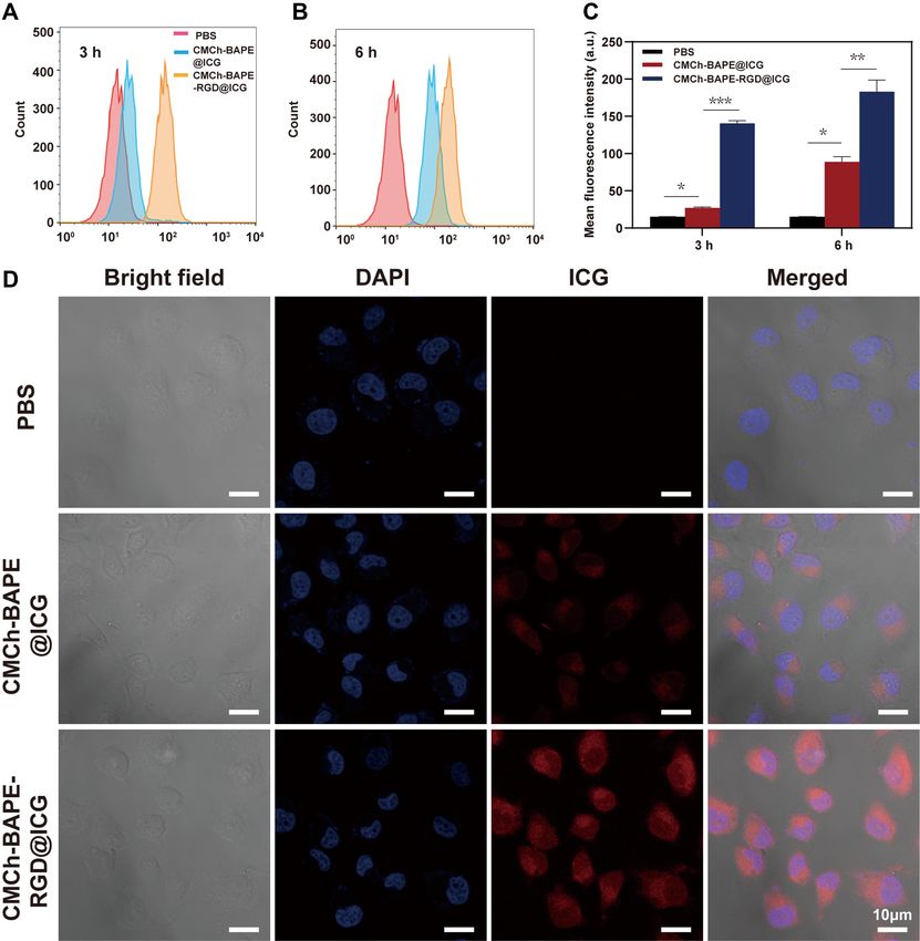
stronger at selected time intervals (P < 0.05). After 6 BAPE-RGD@ICG. As expected, CMCh-BAPE-
h incubation, the fluorescence intensity of the CMCh-BAPE- RGD@ICG had negligible toxicity on SGC7901 cells
RGD@ICG group was nearly twice than that of the CMCh- without NIR irradiation. Nevertheless, upon 5 min NIR
BAPE@ICG group (Figure 4C). Previous studies revealed irradiation, the survival rates of SGC7901 cells in this
that integrin αvβ3 is widely expressed and may be a putative group decreased remarkably (P < 0.05) when compared
prognostic biomarker in gastric cancer tissues.36 Given the fact with PBS or free ICG group (Figure 5D).
that RGD polypeptides have a strong affinity to integrin αvβ3 Simultaneously, the relative growth rates (RGR) of
which is overexpressed on the surface of SGC7901 cells, it is SGC7901 cells incubated with CMCh-BAPE-
reasonable for CMCh-BAPE-RGD@ICG to demonstrate RGD@ICG declined gradually as the concentration
enhanced cellular internalization. These results confirmed increased (Figure 5C). When the amount of ICG reached
that this targeting strategy was suitable for improving the 40 ug/mL (the equivalent ICG concentration), only 20%
delivery of ICG to SGC7901 cells, and the improved cellular of the cells were alive (IC50 = 18.37 ug/mL). As
endocytosis was considered to enhance the eventual perfor a control, NIR laser exposure alone did not result in
mance in tumor NIR imaging and photothermal therapy. inhibition in SGC7901 cells viability. Besides, the repre
sentative results through live/dead staining assay were
demonstrated in Figure 5B, indicating higher cytotoxi
In vitro Cytotoxicity and Anticancer city when treated with CMCh-BAPE-RGD@ICG com
Activity Assessment bined with NIR irradiation. Flow cytometry with
We assessed the cytotoxicity of CMCh-BAPE-RGD@ICG Annexin V-FITC/PI staining assay was conducted to
nanoparticles on L929 cells by CCK-8 assay. As demon further detect the apoptosis and necrosis induced by
strated in Figure 5A, as the concentration of CMCh- CMCh-BAPE-RGD@ICG in SGC7901 cells (Figure
BAPE-RGD@ICG increased, there was a negligible influ 5E). Notably, the proportion of early and late apoptotic
ence on the cell viabilities (P > 0.05). Even if the con cells in the CMCh-BAPE-RGD@ICG + NIR irradiation
centration was increased to 500 ug/mL, the cell viability group (45.21%) was significantly higher in comparison
remained over 80%, indicating good biocompatibility. with that of other groups (2.84% and 28.76%). Thus,
Next, CCK-8 assay based on SGC7901 cells was CMCh-BAPE-RGD@ICG nanoparticles demonstrated
performed to assess the anticancer activity of CMCh- good performance in killing the SGC7901 cells by
2906 http://doi.org/10.2147/IJN.S289310 International Journal of Nanomedicine 2021:16
DovePressDovepress Shao et al
Figure 4 Flow cytometry analysis of ICG and CMCh-BAPE-RGD@ICG by SGC7901 cells at 3 h (A) and 6 h (B). (C) Mean fluorescence intensity from flow cytometry
analysis (n=3). (D) Confocal laser scanning microscope images of SGC7901 cells after treated with ICG and CMCh-BAPE-RGD@ICG for 6 h. (*) P < 0.05, (**) P < 0.01 and
(***) P < 0.001.
PTT, which was attributed to the high cellular properties of materials. An advising from The American
internalization. Society for Testing and Materials (ASTM F756), materials
could be divided into three categories according to the hemo
In vitro Hemocompatibility Evaluation lytic ratio: non-hemolytic (hemolysis: 0–2%), slightly hemo
Hemolytic activity of erythrocytes is an alternative and reliable lytic (hemolysis: 2–5%), and hemolytic (hemolysis: >5%).
acute toxicity assay to evaluate the hemocompatibility of Once the hemolysis rate exceeds 20%, it will lead to
drugs for intravenous administration.46 The rupture of erythro a serious rupture of erythrocytes.47 As shown in Figure 6A,
cytes is highly associated with the physical and chemical the hemolysis rate was less than 5% within a certain
International Journal of Nanomedicine 2021:16 http://doi.org/10.2147/IJN.S289310 2907
DovePressShao et al Dovepress
Figure 5 (A) CCK-8 assay of CMCh-BAPE-RGD@ICG on L929 cells with different concentrations. (B) Live/Dead staining imaging of SGC7901 cells after treated with ICG
or CMCh-BAPE-RGD@ICG with or without laser irradiation. (C) The viability of SGC7901 cells incubated with CMCh-BAPE-RGD@ICG at different concentrations with
or without laser irradiation. (D) SGC7901 cell viability treated with PBS, ICG or CMCh-BAPE-RGD@ICG with or without laser irradiation. (E) Apoptosis analysis of
SGC7901 cells received the same treatment as (D) by flow cytometry with Annexin V-FITC/PI double-staining assay. (NS) P ≥ 0.05. (*) P < 0.05, (**) P < 0.01 and (***) P <
0.001.
concentration range. Meanwhile, the effects of CMCh-BAPE- enhanced delivery and accumulation in gastric cancers
RGD@ICG in different concentrations on the aggregation and in vivo. SGC7901 tumor-bearing mice were treated with
morphology of erythrocytes were observed by SEM (Figure nanoparticles with or without RGD, respectively, and the
6B). Even when the concentration reached 1 mg/mL, the whole-body fluorescence signal was captured by an in vivo
majority of erythrocytes could still maintain its normal disc imaging system (IVIS). The results are illustrated in
shape with depression in the middle. Therefore, the hemolytic Figure 7A. Apparently, the fluorescence signals of the
toxicity of CMCh-BAPE-RGD@ICG was permissible within tumor site could be detected as early as 0.5 h post-injec
its normal concentration range. tion of CMCh-BAPE-RGD@ICG. With time increasing,
the fluorescence signal increased and reached a peak at 5
In vivo NIR Fluorescence Imaging h. In contrast, CMCh-BAPE@ICG was mainly distributed
In view of the promising in vitro performance, we assessed in the liver and gastrointestinal tract, and only a weak
whether the RGD-modified nanoparticle also demonstrated fluorescence signal could be observed at the tumor site 3
2908 http://doi.org/10.2147/IJN.S289310 International Journal of Nanomedicine 2021:16
DovePressDovepress Shao et al
Figure 6 Hemolysis assay (A) and SEM (B) of erythrocytes of CMCh-BAPE-RGD@ICG with different concentrations.
h after administration. At indicated time points, an evi CMCh-BAPE-RGD@ICG plus laser irradiation group
dently stronger fluorescence signal intensity was observed (P < 0.001). More specifically, in the first 8 days of
in mice treated with the CMCh-BAPE-RGD@ICG than treatment, moderately restricted tumor growth was
other groups. Herein, the conjunction of RGD to the nano observed. After 9 days, the tumor growth of mice was
particles indeed efficiently improved the targeting ability significantly inhibited, eventually achieving nearly com
to the tumors. Additionally, the aforementioned improved plete elimination. Meanwhile, the CMCh-BAPE@ICG
tumor accumulation was also verified by ex vivo fluores plus laser irradiation demonstrated the second strongest
cent signals of the dissected tissues (Figure 7B). anti-tumor effect. We ascribed it to the passive targeting
Intriguingly, the liver and kidney in the two groups also ability of nanoparticles. After a period of 21-day experi
displayed relatively strong fluorescence signals, which was mentation, mice were killed and tumors were removed,
related to the metabolism of nanoparticles mainly through weighed, and photographed (Figures 8C, D and S4),
these organs.48 The semi-quantitative results of in vivo which further validated that CMCh-BAPE-RGD@ICG
distribution were demonstrated in Figure S3. plus laser irradiation displayed the best tumor suppres
Collectively, CMCh-BAPE-RGD@ICG has improved sion effect. Additionally, a thermal infrared imaging
tumor-specific accumulation and conducts efficient NIR camera was employed to monitor the changes of tem
imaging. perature in the tumor site upon laser irradiation. As
shown in Figure 8E, the tumor temperature reached the
In vivo Anti-Tumor Efficiency maximum after 3 minutes of laser irradiation, 46.9°C in
The above results demonstrated the potential of CMCh- CMCh-BAPE-RGD@ICG group and 44.1°C in CMCh-
BAPE-RGD@ICG as an efficient and NIR imaging BAPE@ICG group, respectively.
nanoparticles with promising photothermal therapy. Subsequently, we further performed the histological
Therefore, the anti-tumor efficiency of RGD-modified analysis to assess the therapeutic effect of different treat
nanoparticles was investigated on SGC7901 subcuta ments (Figures 8F and S4E). H&E tumor sections sug
neous tumor-bearing mice. Briefly, the experimental gested that CMCh-BAPE-RGD@ICG plus laser
mice were treated with PBS, ICG, CMCh-BAPE@ICG irradiation treatment led to the most severe structural
(non-targeted), or CMCh-BAPE-RGD@ICG (targeted), damage and tissue necrosis. Similarly, the TUNEL assay
followed with or without laser irradiation. During the indicated the most apoptotic tumor cells in this group after
21-day treatment, there were no significant changes in photothermal therapy. Besides, immunohistochemical
the mouse body weight from each group, indicating staining for Ki67 antigen on tumor sections was performed
minimal systematic toxicity of all treatments (Figures to evaluate the proliferation extent of tumor cells.
8A and S4A). Next, the volume of tumors was measured Apparently, a substantial decrease in Ki67 expression
at regular intervals to monitor the anti-tumor effect of was observed in this group, indicating that the tumor
different therapies. As shown in Figures 8B and S4B, proliferation activity was also markedly suppressed after
the strongest tumor-inhibitory effect was obtained in treatment.
International Journal of Nanomedicine 2021:16 http://doi.org/10.2147/IJN.S289310 2909
DovePressShao et al Dovepress
Figure 7 (A) In vivo fluorescent imaging of tumor-bearing mice after intravenous injection of CMCh-BAPE@ICG and CMCh-BAPE-RGD@ICG at indicated time points. (B)
Ex vivo fluorescence signal of excised tumors and organs at 8 h after injection.
The issue about the entry of nanoparticles into solid several different tumor types mainly through transcy
tumors has been argued for several decades, the central tosis (97%), and not via inter-endothelial gaps, while
concept of which is the EPR effect. However, the latest EPR effect contributed only a small part.31 How the
research reported that nanoparticles penetrate into tumor was instilled with nano-drugs may have one or
2910 http://doi.org/10.2147/IJN.S289310 International Journal of Nanomedicine 2021:16
DovePressDovepress Shao et al
Figure 8 (A) Weight changes curve within 21-day treatments of each group. (B) Tumor growth curve within 21-day treatments of each group. (C) Images of tumor in each
group collected on day 21. (D) Average tumor weight in each group after 21-day treatment. (E) Infrared thermographic images of mice received different treatments. (F)
H&E, TUNEL and Ki67 staining of tumor sections in each group. Severely damaged tumor tissue are indicated by a black circle. Note that a-f represent different treatments:
a.PBS, b.CMCh-BAPE@ICG, c.CMCh-BAPE-RGD@ICG, c.PBS+laser, d. CMCh-BAPE@ICG+laser, e. c.CMCh-BAPE-RGD@ICG +laser. (NS) P ≥ 0.05. (*) P < 0.05, (**) P <
0.01 and (***) P < 0.001.
more co-exist mechanisms, such as EPR effect, trans treatment. In this study, we integrated NIR fluorescence
cytosis, and perhaps other unknown mechanisms. There imaging with photothermal therapy, where ICG served
is no doubt that nano-drugs are beneficial in cancer as both a fluorescent agent and a photosensitizer. When
International Journal of Nanomedicine 2021:16 http://doi.org/10.2147/IJN.S289310 2911
DovePressShao et al Dovepress
Figure 9 (A) H&E staining images of mice organs after 21-day treatment. (B) Blood parameters for evaluating effects on bone marrow hematopoietic system (RBC, WBC
and platelet) and liver (ALT, AST) and kidney (Cr) function. Note that a–f represent the same treatments as Figure 8.
irradiated upon laser, the temperature could rise to underlying mechanism of hyperthermia-induced cell
46.9°C rapidly and maintained relatively stable death may be that the rise in temperature will up-
in vivo. It is reported that temperature over 42~45°C regulate the apoptosis genes, activate caspase-3, sup
will lead to apoptotic and/or necrotic death.6–8,13 The press TNF-α resistance, and damage mitochondrial.49
2912 http://doi.org/10.2147/IJN.S289310 International Journal of Nanomedicine 2021:16
DovePressDovepress Shao et al
As expected, CMCh-BAPE-RGD@ICG nanoparticles Abbreviations
exhibited an excellent tumor ablation effect in vivo. NIR, near-infrared; PTT, photothermal therapy; NDDS, nanos
cale drug delivery systems; ICG, indocyanine green; EPR,
In vivo Biosafety Evaluation enhanced permeability and retention; RME, receptor-mediated
Biosafety is an essential prerequisite for the clinical endocytosis; RGD, Arg-Gly-Asp peptide; ROS, reactive oxy
application of nanomaterials. Herein, the effects of dif gen species; CMCH, carboxymethyl chitosan; BAPE, benze
ferent treatments on mice main organs and blood para neboronic acid pinacol ester; EDC, 1-ethyl-
meters were determined. The H&E staining images in 3-(3-dimethylaminopropyl) carbodiimide hydrochloride;
NHS, N-hydroxysuccinimide; DOX, Doxorubicin; LE, loading
Figure 9A revealed no obvious injury on the tissue
efficiency; RBC, red blood cells; WBC, white blood cells; PLT,
structure and morphology of the major organs in all
platelet; AST, aspartate aminotransferase; ALT, alanine amino
groups. As demonstrated in Figure 9B, all groups dis
transferase; Cr, creatinine; 1H NMR, nuclear magnetic reso
played within the normal range in the levels of the
nance spectroscopy; CLSM, confocal laser scanning
blood indicators, including red blood cells (RBC),
microscope; TEM, transmission electron microscopy; DLS,
white blood cells (WBC), platelet (PLT), aspartate ami
dynamic light scattering; HE, hematoxylin-eosin CCK-8, cell
notransferase (AST), alanine aminotransferase (ALT)
counting kit-8.
and creatinine (Cr). These results confirmed the little
influence on the hematopoietic (WBC, RBC, and PLT),
liver (ALT and AST), and kidney (Cr) functions of each Ethical Approval and Consent to
group. Therefore, CMCh-BAPE-RGD@ICG are promis Participate
ing nanoparticles with good biosafety for potential clin All mice experiments were conducted following the guide
ical applications. line of the Ethics Committee of the Institutional Animal
Care and Use Subcommittee of the Third Hospital of Sun
Yat-sen University and the committee approved this study.
Conclusion
In summary, we successfully synthesized a promising ROS-
responsive RGD-modified nanoparticle, which could deliver Acknowledgments
ICG to gastric cancer for NIR imaging and PTT. ICG was This work was supported in part by grants from Science
coated in the core of nanoparticles as a near-infrared imaging and Technology Planning Project of Guangdong Province
agent and photosensitizer. RGD polypeptide was conjugated (2017B020227009 and 2019B030316011), by the National
to the nanoparticles for promoting targeted accumulation and Natural Science Foundation of China (81472825), by the
penetration in the tumor tissue. Boronic ester in BAPE made Outstanding Young Talents Support Program of the Third
the particles hydrolyze in tumor the high ROS environment, Affiliated Hospital of Sun Yat-sen University.
resulting in more ICG release. These nanoparticles exhibited
suitable size, improved aqueous stability, and excellent photo Disclosure
thermal conversion efficiency. Moreover, flow cytometry and The authors report no conflicts of interest in this work.
confocal microscopy confirmed the high-efficiency endocyto
sis in SGC7901 cells. Remarkably, the NIR fluorescence
imaging could accurately demonstrate the location and margin References
of the tumor in tumor-bearing mice. The photothermal therapy 1. Bray F, Ferlay J, Soerjomataram I, Siegel RL, Torre LA, Jemal A.
Global cancer statistics 2018: GLOBOCAN estimates of incidence and
of the targeted nanoparticle could efficiently ablate tumors by mortality worldwide for 36 cancers in 185 countries. CA Cancer
inducing necrosis and apoptosis in cancer cells. Additionally, J Clin. 2018;68(6):394–424. doi:10.3322/caac.21492
hemocompatibility assessment in vitro and systematic toxicity 2. Japanese Gastric Cancer A. Japanese gastric cancer treatment guide
lines 2018 (5th edition). Gastric Cancer. 2020. doi:10.1007/s10120-
evaluation in vivo displayed its good biocompatibility for 020-01042-y
potential clinical application. Collectively, the smart multi 3. Wang FH, Shen L, Li J, et al. The Chinese Society of Clinical
Oncology (CSCO): clinical guidelines for the diagnosis and treatment
functional nanoparticle provides a promising strategy for of gastric cancer. Cancer Commun (Lond). 2019;39(1):10.
tumor-targeting NIR imaging and photothermal therapy. doi:10.1186/s40880-019-0349-9
International Journal of Nanomedicine 2021:16 http://doi.org/10.2147/IJN.S289310 2913
DovePressShao et al Dovepress
4. Ajani JA, D’Amico TA, Almhanna K, et al. Gastric Cancer, Version 21. Liu R, Hu C, Yang Y, Zhang J, Gao H. Theranostic nanoparticles
3.2016, NCCN Clinical Practice Guidelines in Oncology. J Natl with tumor-specific enzyme-triggered size reduction and drug release
Compr Canc Netw. 2016;14(10):1286. doi:10.6004/jnccn.2016.0137 to perform photothermal therapy for breast cancer treatment. Acta
5. Zhang P, Zheng Z, Ling L, et al. w09, a novel autophagy enhancer, Pharm Sin B. 2019;9(2):410–420. doi:10.1016/j.apsb.2018.09.001
induces autophagy-dependent cell apoptosis via activation of the 22. Schwarz C, Plass I, Fitschek F, et al. The value of indocyanine green
EGFR-mediated RAS-RAF1-MAP2K-MAPK1/3 pathway. Autophagy. clearance assessment to predict postoperative liver dysfunction in
2017;13(7):1093–1112. doi:10.1080/15548627.2017.1319039 patients undergoing liver resection. Sci Rep. 2019;9(1):8421.
6. Peng J, Xiao Y, Li W, et al. Photosensitizer micelles together with doi:10.1038/s41598-019-44815-x
IDO inhibitor enhance cancer photothermal therapy and 23. Hwang Y, Yoon H, Choe K, et al. In vivo cellular-level real-time
immunotherapy. Adv Sci (Weinh). 2018;5(5):1700891. doi:10.1002/ pharmacokinetic imaging of free-form and liposomal indocyanine
advs.201700891 green in liver. Biomed Opt Express. 2017;8(10):4706–4716.
7. Wei W, Zhang X, Zhang S, Wei G, Su Z. Biomedical and bioactive doi:10.1364/BOE.8.004706
engineered nanomaterials for targeted tumor photothermal therapy: a 24. Lee KM, Kim JM, Lee EJ, Kim TW. Anterior optic nerve head
review. Mater Sci Eng C Mater Biol Appl. 2019;104:109891. perfusion is dependent on adjacent parapapillary choroidal
doi:10.1016/j.msec.2019.109891 perfusion. Sci Rep. 2019;9(1):10999. doi:10.1038/s41598-019-
8. Chen J, Ning C, Zhou Z, et al. Nanomaterials as photothermal 47534-5
therapeutic agents. Prog Mater Sci. 2019;99:1–26. doi:10.1016/j. 25. Asanuma D, Kobayashi H, Nagano T, Urano Y. Fluorescence ima
pmatsci.2018.07.005 ging of tumors with “smart” pH-activatable targeted probes. Methods
9. Zhang F, Liu S, Zhang N, et al. X-ray-triggered NO-released Bi-SNO Mol Biol. 2009;574:47–62. doi:10.1007/978-1-60327-321-3_5
nanoparticles: all-in-one nano-radiosensitizer with photothermal/gas 26. Wang Z, Ju Y, Ali Z, et al. Near-infrared light and tumor microenvir
therapy for enhanced radiotherapy. Nanoscale. 2020. doi:10.1039/ onment dual responsive size-switchable nanocapsules for multimodal
d0nr04634e tumor theranostics. Nat Commun. 2019;10(1):4418. doi:10.1038/
10. Yang G, Xu L, Chao Y, et al. Hollow MnO2 as a s41467-019-12142-4
tumor-microenvironment-responsive biodegradable nano-platform for 27. Kim T, Park J, Kim TI. Cholic acid-conjugated
combination therapy favoring antitumor immune responses. Nat methylcellulose-polyethylenimine nano-aggregates for drug delivery
Commun. 2017;8(1):902. doi:10.1038/s41467-017-01050-0
systems. Nanomaterials (Basel). 2019;9(3):459. doi:10.3390/
11. Wu J, Williams GR, Niu S, Gao F, Tang R, Zhu LM.
nano9030459
A multifunctional biodegradable nanocomposite for cancer
28. Xu M, Zhang C, Wu J, et al. PEG-detachable polymeric micelles
theranostics. Adv Sci (Weinh). 2019;6(14):1802001. doi:10.1002/
self-assembled from amphiphilic copolymers for tumor-acidity-trig
advs.201802001
gered drug delivery and controlled release. ACS Appl Mater
12. Yu B, Choi B, Li W, Kim DH. Magnetic field boosted ferroptosis-like
Interfaces. 2019;11(6):5701–5713. doi:10.1021/acsami.8b13059
cell death and responsive MRI using hybrid vesicles for cancer
29. Li Y, Zhai Y, Liu W, et al. Ultrasmall nanostructured drug based
immunotherapy. Nat Commun. 2020;11(1):3637. doi:10.1038/
pH-sensitive liposome for effective treatment of drug-resistant tumor.
s41467-020-17380-5
J Nanobiotechnology. 2019;17(1):117. doi:10.1186/s12951-019-0550-7
13. Guo L, Niu G, Zheng X, et al. Single near-infrared emissive polymer
30. Oh JY, Kim HS, Palanikumar L, et al. Cloaking nanoparticles with
nanoparticles as versatile phototheranostics. Adv Sci (Weinh). 2017;4
protein corona shield for targeted drug delivery. Nat Commun. 2018;9
(10):1700085. doi:10.1002/advs.201700085
(1):4548. doi:10.1038/s41467-018-06979-4
14. Yan F, Wu H, Liu H, et al. Molecular imaging-guided photothermal/
31. Sindhwani S, Syed AM, Ngai J, et al. The entry of nanoparticles into
photodynamic therapy against tumor by iRGD-modified indocyanine
solid tumours. Nat Mater. 2020;19(5):566–575. doi:10.1038/s41563-
green nanoparticles. J Control Release. 2016;224:217–228.
doi:10.1016/j.jconrel.2015.12.050 019-0566-2
15. Kim M, Lee JH, Nam JM. Plasmonic photothermal nanoparticles for 32. Liu R, Xiao W, Hu C, Xie R, Gao H. Theranostic size-reducible and
biomedical applications. Adv Sci (Weinh). 2019;6(17):1900471. no donor conjugated gold nanocluster fabricated hyaluronic acid
doi:10.1002/advs.201900471 nanoparticle with optimal size for combinational treatment of breast
16. Lu L, Li B, Ding S, et al. NIR-II bioluminescence for in vivo high cancer and lung metastasis. J Control Release. 2018;278:127–139.
contrast imaging and in situ ATP-mediated metastases tracing. Nat doi:10.1016/j.jconrel.2018.04.005
Commun. 2020;11(1):4192. doi:10.1038/s41467-020-18051-1 33. Sun Y, Zhao Y, Teng S, et al. Folic acid receptor-targeted human
17. Savchuk OA, Carvajal JJ, Cesteros Y, et al. Mapping temperature serum albumin nanoparticle formulation of cabazitaxel for tumor
distribution generated by photothermal conversion in graphene film therapy. Int J Nanomedicine. 2019;14:135–148. doi:10.2147/IJN.
using Er, Yb: naYF4Nanoparticles prepared by microwave-assisted S181296
solvothermal method. Front Chem. 2019;7:88. doi:10.3389/ 34. Nieberler M, Reuning U, Reichart F, et al. Exploring the role of
fchem.2019.00088 RGD-recognizing integrins in cancer. Cancers (Basel). 2017;9(9).
18. Yang Z, Cheng R, Zhao C, et al. Thermo- and pH-dual responsive doi:10.3390/cancers9090116
polymeric micelles with upper critical solution temperature behavior 35. Raab-Westphal S, Marshall JF, Goodman SL. Integrins as therapeutic
for photoacoustic imaging-guided synergistic chemo-photothermal targets: successes and cancers. Cancers (Basel). 2017;9(9).
therapy against subcutaneous and metastatic breast tumors. doi:10.3390/cancers9090110
Theranostics. 2018;8(15):4097–4115. doi:10.7150/thno.26195 36. Boger C, Warneke VS, Behrens HM, et al. Integrins alphavbeta3 and
19. Yan L, Qiu L. Indocyanine green targeted micelles with improved alphavbeta5 as prognostic, diagnostic, and therapeutic targets in
stability for near-infrared image-guided photothermal tumor therapy. gastric cancer. Gastric Cancer. 2015;18(4):784–795. doi:10.1007/
Nanomedicine. 2015;10(3):361–373. doi:10.2217/nnm.14.118 s10120-014-0435-2
20. Qin L, Cao J, Shao K, Tong F, Yang Z. A tumor-to-lymph procedure 37. Garcia Ribeiro RS, Belderbos S, Danhier P, et al. Targeting tumor
navigated versatile gel system for combinatorial therapy against cells and neovascularization using RGD-functionalized
tumor recurrence and metastasis. Sci Adv. 2020;6(36):eabb3116. magnetoliposomes. Int J Nanomedicine. 2019;14:5911–5924.
doi:10.1126/sciadv.abb3116 doi:10.2147/IJN.S214041
2914 http://doi.org/10.2147/IJN.S289310 International Journal of Nanomedicine 2021:16
DovePressYou can also read