Liver cell hydration and integrin signaling - De Gruyter
←
→
Page content transcription
If your browser does not render page correctly, please read the page content below
Biol. Chem. 2021; 402(9): 1033–1045

Review

Michele Bonus, Dieter Häussinger* and Holger Gohlke*

Liver cell hydration and integrin signaling
https://doi.org/10.1515/hsz-2021-0193                                    reviews, see Graf and Häussinger 1996; Häussinger 1996a,
Received March 12, 2021; accepted April 12, 2021;                        b). Although hepatocytes, like virtually every cell type,
published online April 29, 2021
                                                                         possess powerful volume-regulatory mechanisms, these
                                                                         mechanisms only prevent excessive volume changes but
Abstract: Liver cell hydration (cell volume) is dynamic and
                                                                         allow variations of cell volume within narrow limits. Most
can change within minutes under the influence of hor-
mones, nutrients, and oxidative stress. Such volume                      importantly, such small changes in liver cell volume have
changes were identified as a novel and important modu-                   been identified as an important and until then not recog-
lator of cell function. It provides an early example for the             nized regulator of diverse hepatocyte functions, including
interaction between a physical parameter (cell volume) on                metabolism, membrane transport, cell fate, and gene
the one hand and metabolism, transport, and gene                         expression (Figure 1; for reviews, see Häussinger and Lang
expression on the other. Such events involve mechano-                    1991; Häussinger 1996a, b). Many effects of amino acids
transduction (osmosensing) which triggers signaling cas-                 and hormones can, at least in part, be attributed to cell
cades towards liver function (osmosignaling). This article               hydration changes, which correspond on a short-term time
reviews our own work on this topic with emphasis on the                  scale to cell volume changes.
role of β1 integrins as (osmo-)mechanosensors in the liver,                  Indeed, cell volume changes were identified as
but also on their role in bile acid signaling.                           important mediators of proteolysis control by amino acids
                                                                         and hormones in the liver (Hallbrucker et al. 1991; Häus-
Keywords: bile acids; cell swelling; functional selectivity;
                                                                         singer et al. 1991; vom Dahl et al. 1991), and the cellular
mechanotransduction; molecular dynamics simulations;
                                                                         hydration state, in general, was suggested to be an
tauroursodeoxycholate.
                                                                         important determinant of protein catabolism in health and
                                                                         disease (Häussinger et al. 1993).
                                                                             The effectors depicted in Figure 1 can change within
Introduction and general                                                 minutes the hepatocyte water content by up to ± 15%. Such
                                                                         volume changes can also be experimentally induced by
considerations                                                           changing the extracellular osmolarity by ± 80 mosmol/L.
                                                                         Thus, hyper- or hypoosmotic exposure of isolated hepato-
Liver cell volume can change within minutes under the
                                                                         cytes or perfused rat liver was frequently used to mimic
influence of nutrients, hormones, and toxins due to the
                                                                         hepatocyte hydration changes and investigate the func-
creation or dissipation of osmotic gradients and corre-
                                                                         tional consequences of hepatocyte swelling and shrinkage.
sponding water fluxes across the plasma membrane (for
                                                                         Several long-known, but mechanistically unclear effects of
                                                                         amino acids, which could not be related to their metabolism,
*Corresponding authors: Dieter Häussinger, Clinic for                    include the stimulation of glycogen synthesis (Katz et al.
Gastroenterology, Hepatology, and Infectious Diseases, Heinrich          1976; Lavoinne et al. 1987) or inhibition of proteolysis
Heine University Düsseldorf, Moorenstr. 5, D-40225 Düsseldorf,           (Mortimore and Pösö 1987). Such effects can quantitatively
Germany, E-mail: haeussin@uni-duesseldorf.de; and Holger Gohlke,         be mimicked by hypoosmotic hepatocyte swelling to an
Institute for Pharmaceutical and Medicinal Chemistry, Heinrich Heine
                                                                         extent as these amino acids do (Baquet et al. 1990; Hall-
University Düsseldorf, Universitätsstr. 1, D-40225 Düsseldorf,
Germany; and John von Neumann Institute for Computing (NIC), Jülich      brucker et al. 1991; Häussinger et al. 1991). Against this
Supercomputing Centre (JSC), Institute of Biological Information         background, it has been suggested that Na+-dependent
Processing (IBI-7: Structural Biochemistry), and Institute of Bio- and   amino acid transport systems in the plasma membrane
Geosciences (IBG-4: Bioinformatics) Forschungszentrum Jülich GmbH,       should not merely be viewed as simple amino acid trans-
Wilhelm-Johnen-Str., D-52428 Jülich, Germany, E-mail: gohlke@uni-
                                                                         locators but also as transmembrane signaling systems,
duesseldorf.de. https://orcid.org/0000-0001-8613-1447
Michele Bonus, Institute for Pharmaceutical and Medicinal Chemistry,
                                                                         which alter cell hydration and, accordingly, cell function in
Heinrich Heine University Düsseldorf, Universitätsstr. 1, D-40225        response to amino acid supply (Häussinger 1996b). A cell
Düsseldorf, Germany. https://orcid.org/0000-0003-4411-7342               type- and development-specific expression of concentrative
  Open Access. © 2021 Michele Bonus et al., published by De Gruyter.       This work is licensed under the Creative Commons Attribution 4.0
International License.
1034         M. Bonus et al.: Liver cell hydration and integrin signaling

                                                                                            Figure 1: Liver cell volume and hepatocyte
                                                                                            function.
                                                                                            Liver cell volume, i.e., hepatocyte
                                                                                            hydration, is dynamic and under the
                                                                                            influence of hormones, nutrients, nerves,
                                                                                            ambient osmolarity either due to
                                                                                            cumulative substrate transport or
                                                                                            generation or dissipation of ion gradients
                                                                                            across the plasma membrane.
                                                                                            Osmosensing and osmosignaling triggered
                                                                                            by such cell volume changes are potent
                                                                                            regulators of hepatocyte function. SN1:
                                                                                            system N isoform 1 glutamine transporter
                                                                                            (Na+- and H+-dependent).

versus simply equilibrating amino acid transporters may                 aspects and topics on other mammalian cell types, yeast,
add to the complexity of such signaling processes. Roughly              bacteria, and plants, the reader is referred to (Häussinger
spoken, hepatocyte swelling provides a protein anabolic,                and Sies 2007).
choleretic, proliferative, and anti-apoptotic signal, whereas               In addition to a volume-regulatory uptake and syn-
hepatocyte shrinkage is catabolic, cholestatic, and                     thesis of organic osmolytes in response to cell shrinkage
proapoptotic.                                                           and their release in response to cell swelling, an early
     Much effort has been devoted to the question of how                evolutionary mechanism for the maintenance of cell
hepatocyte volume changes are sensed („osmosensing“)                    volume homeostasis is the polymerization of small
and how this information signals towards hepatocyte                     osmotically active molecules. This polymerization oc-
function („osmosignaling“). Several candidates for                      curs in response to cell swelling in hypoosmotic envi-
osmosensing have been discussed in the past, including                  ronments and, conversely, the depolymerization of such
macromolecular crowding (Minton et al. 1992; Parker and                 macromolecules in response to hyperosmotic cell
Colclasure 1992), stretch-activated ion channels (for re-               shrinkage. Such a polymerization/depolymerization
view see Naruse 2018), and in yeast, histidine kinases                  strategy is also found in some algae and primitive or-
were identified that might act as putative osmosensors                   ganisms (Chamberlin and Strange 1989): here, the
(Maeda et al. 1994). In the liver, however, we identified                cellular metabolism is at the service of cell volume
α5β1-integrins as volume(osmo)sensors in response to                    maintenance in order to compensate for osmolarity
hepatocyte swelling (Häussinger et al. 2003; Schliess et al.            changes in the surrounding watery environment. It
2004; vom Dahl et al. 2003), whereas early endosomes                    seems that this archaic mechanism is still present in
were identified as chloride-governed osmosensors that                    higher organisms, such as mammals, whose cells, how-
are activated in response to hyperosmotic hepatocyte                    ever, are mostly in an osmotically stable environment. In
shrinkage (Reinehr et al. 2006) (for review see Reinehr                 line with this, experimental hepatocyte swelling favors
et al. 2013).                                                           the “polymerization” of amino acids and glucose to
     This review summarizes some aspects of our work on                 proteins and glycogen, respectively, whereas “depoly-
osmosensing and osmosignaling in the liver. For further                 merization” (increased proteolysis and glycogenolysis
M. Bonus et al.: Liver cell hydration and integrin signaling   1035

and inhibition of glycogen and protein synthesis) occurs        noted that the hyperosmolarity-induced signaling pathway
in response to hyperosmotic hepatocyte shrinkage. A cell        is also activated by the hydrophobic bile acid glyco-
volume-stabilizing effect of such maneuvres, however, is        chenodeoxycholic acid (GCDC) (Becker et al. 2007a, b;
doubtful in view of the osmotically stable environment          Mayer et al. 2019; Reinehr et al. 2004b).
in higher organisms. Instead, it seems that these or-
ganisms use the archaic polymerization/depolymeriza-
tion strategy in an opposite way: changes in cell volume
are artificially created by hormones or cumulative
                                                                Osmosensing and osmosignaling
substrate uptake in order to regulate metabolism                in response to hypoosmotic
(Häussinger 1996a).
                                                                hepatocyte swelling
                                                                Hypoosmotic hepatocyte swelling inhibits autophagic
Osmosensing and osmosignaling                                   proteolysis and glycogenolysis but stimulates protein and
                                                                glycogen synthesis and bile acid excretion and triggers a
in response to hyperosmotic                                     volume-regulatory decrease (RVD) (for review see Häus-
hepatocyte shrinkage                                            singer 1996b). Hepatocyte swelling may also inhibit viral
                                                                replication, as shown for the duck hepatitis B virus repli-
Hyperosmotic exposure of hepatocytes leads to hepatocyte        cation (Offensperger et al. 1994), although the underlying
shrinkage and an accompanying increase of the intracel-         mechanisms remained unclear. Concerning the stimula-
lular chloride concentration due to water efflux and a          tion of glycogen synthesis and acetyl-CoA carboxylase by
volume-regulatory uptake of K+, Na+, and Cl− into the he-       hepatocyte swelling, a decrease of the intracellular chlo-
patocyte (Graf and Häussinger 1996; Häussinger et al.           ride concentration was suggested to be an important trigger
1990). Early endosomes were identified as chloride-              (Baquet et al. 1993; Meijer et al. 1992; for review, see Hue
governed osmosensors, which are activated in response           1995). However, concerning hypoosmotic proteolysis inhi-
to hyperosmotic hepatocyte shrinkage (Reinehr et al. 2006;      bition, RVD, and stimulation of bile acid excretion, α5β1-
for review, see Reinehr et al. 2013). The increase of the       integrins were identified as osmosensors (Häussinger et al.
intracellular chloride concentration directly activates the     2003; Schliess et al. 2004; vom Dahl et al. 2003), and
vacuolar H+-ATPase (for review, see Faundez and Hartzell        the downstream osmosignaling events involve activation
2004) and triggers a DIDS- and bafilomycin-sensitive             of focal adhesion kinase, c-Src, the epidermal growth fac-
endosomal acidification following hyperosmotic expo-             tor receptor (EGFR), and the mitogen-activated protein
sure, as shown in studies using endocytosed FITC-dextran        kinases Erks and p38 (Figure 2). The latter mediates RVD and
(Schreiber et al. 1994). Endosomal acidification activates       proteolysis inhibition, whereas dual MAP kinase activation
acidic sphingomyelinase, and the accompanying ceramide          is required for the insertion of Bsep and Mrp2 into the
formation leads to an activation of NADPH oxidase and the       canalicular membrane and choleresis (Häussinger et al.
formation of reactive oxygen species (ROS) (Reinehr et al.      2003; Kubitz et al. 1997; Noé et al. 1996; Schmitt et al. 2001).
2006; for review, see Reinehr et al. 2013). The hyperosmotic         Swelling-induced activation of α5β1-integrins is also an
ROS response was shown to trigger the activation not only       integral part of insulin action in the liver (Reinehr et al.
of c-Jun N-terminal kinase (JNK) but also of the Src family     2010b; Schliess et al. 2004; for review see Schliess and
kinase members Fyn and Yes (Cantore et al. 2011; Reinehr        Häussinger 2003). Activation of the insulin receptor trig-
et al. 2004a; Sommerfeld et al. 2015a) and to induce pro-       gers, among many other events, also the activation of PI3
apoptotic miRNA 15/107 (Santosa et al. 2015). Hyper-            kinase, which activates the Na+/K+/2Cl− cotransporter
osmotic Fyn activation triggers cholestasis due to retrieval    (NKCC1) leading to hepatocyte swelling (Häussinger and
of Bsep and Mrp2 from the canalicular membrane (Cantore         Lang 1992), subsequent integrin-dependent osmosensing,
et al. 2011) and Ntcp from the sinusoidal membrane              and signaling towards proteolysis and EGFR activation
(Sommerfeld et al. 2015a; Sommerfeld et al. 2015a) of the       (Reinehr et al. 2010b; Schliess et al. 2004), as schematically
hepatocyte. The activation of Yes and JNK triggers a pro-       depicted in Figure 3. The involvement of osmosensing and
apoptotic state of the hepatocyte (Reinehr et al. 2002, 2003,   osmosignaling events can well explain the prodiabetic
2004a; for review, see Reinehr et al. 2013). It should be       action of loop diuretics, hypernatremia, oxidative stress,
1036          M. Bonus et al.: Liver cell hydration and integrin signaling

                                                                         dehydration, glucagon, and gain of function mutations of
                                                                         KCNQ1 (Boini et al. 2009; Schliess and Häussinger 2003;
                                                                         Schliess et al. 2001; Unoki et al. 2008; Yasuda et al. 2008).
                                                                              The α5β1-integrins can also be activated non-
                                                                         osmotically/non-mechanically by urea in high concentra-
                                                                         tions and tauroursodeoxycholate (TUDC), and signaling
                                                                         events similar to those found after hypoosmotic exposure
                                                                         are initiated (Bonus et al. 2020; Gohlke et al. 2013; Reinehr
                                                                         et al. 2010a), which are mechanistically discussed below.
                                                                         The signaling events triggered by TUDC can explain the
                                                                         choleretic and hepatoprotective action of this bile acid
                                                                         (Häussinger et al. 2003; Kurz et al. 2001), which is also a
                                                                         mainstay in the treatment of cholestatic diseases. Inter-
                                                                         estingly, TUDC also triggers the formation of cyclic AMP
                                                                         (cAMP) in an integrin-dependent manner (Sommerfeld
                                                                         et al. 2015b). Whether cAMP formation also occurs in
                                                                         response to hypoosmotic hepatocyte swelling remains to
                                                                         be investigated.
                                                                              The TUDC-triggered cAMP formation strongly coun-
                                                                         teracts the proapoptotic action of the hydrophobic bile
                                                                         acid GCDC by preventing Yes, Fyn, and JNK activation
                                                                         (Sommerfeld et al. 2015b) and triggering an inactivating
                                                                         Ser/Thr-phosphorylation of CD95 (Reinehr and Häus-
Figure 2: Osmosensing and osmosignaling in response to
hypoosmotic hepatocyte swelling and hyperosmotic hepatocyte              singer 2004). In addition, TUDC prevented GCDC-induced
shrinkage. For details, see text.                                        changes in gene transcription (Paluschinski et al. 2019).

                                                                         Early indications of TUDC-mediated
                                                                         integrin activation
                                                                         Early studies demonstrated that TUDC activates the
                                                                         mitogen-activated protein kinases (MAPK) extracellular
                                                                         signal-regulated kinase (Erk)-1 and Erk-2 in isolated,
                                                                         cultured hepatocytes and perfused rat liver (Schliess et al.
                                                                         1997). These TUDC-mediated signal transduction events
                                                                         were similar to those triggered by cell swelling (Noé et al.
                                                                         1996; Schliess et al. 1995) but, conversely, independent
                                                                         from G protein-mediated pathways and amenable to inhi-
                                                                         bition by high concentrations of glucagon and CPT-cAMP,
                                                                         i.e., conditions that allow for a rapid induction of MAP
Figure 3: Hepatocyte hydration, insulin action, and resistance.          kinase phosphatase 1 (MKP 1). For reasons not yet fully
Insulin triggers a PI3 kinase-driven phosphorylation and activation      understood, this is in contrast to the low levels of
of NKCC1, which triggers cell swelling and activates integrin-           TUDC-induced cAMP formation that mediate the hep-
dependent osmosensing and -signaling. This results in proteolysis
                                                                         atoprotective effects of TUDC (Sommerfeld et al. 2015a; see
inhibition and EGFR activation. Factors preventing or counteracting
insulin-induced hepatocyte swelling include furosemide, oxidative
                                                                         Figure 4). Since Erks can phosphorylate proteins that
stress, hypernatremia, or KCNQ1 gain of function mutations and are       control microtubule dynamics and in this way regulate
known to be prodiabetic (Boini et al. 2009; Schliess et al. 2001;        vesicular trafficking of canalicular transporters, they were
Unoki et al. 2008; Yasuda et al. 2008).                                  considered early on as a central node for TUDC- and
M. Bonus et al.: Liver cell hydration and integrin signaling           1037

hypoosmolarity-mediated choleresis. Soon after PI3-K and
Ras activation were confirmed as upstream events of the
TUDC-mediated, choleretic Erk signal (Kurz et al. 2000),
p38MAPK was identified as the second major downstream
component to convert a hypoosmolarity- (Häussinger et al.
1999) or TUDC-induced (Kurz et al. 2001) signal into a
cellular response. While hypoosmotic cell swelling
induced inhibition of autophagic proteolysis via p38MAPK
without any involvement of Erks (Häussinger et al. 1999),
swelling and liver perfusion with TUDC triggered choler-
esis only when Erk-1/2 and p38MAPK were both activated.
Such dual activation was found essential for vesicular
trafficking of the bile salt export pump (BSEP) and multi-
drug resistance-associated protein 2 (MRP2) (Kubitz et al.
1997; Kurz et al. 2001) to the canalicular membrane.
     Considering that the described TUDC-induced signal
transduction pathways occurred without any involve-
ment of G proteins (Schliess et al. 1997) and the potency of
TUDC toward the farnesoid X receptor (FXR) is only
modest (Liu et al. 2003), GPCR and nuclear receptors were
ruled out as the primary TUDC sensors (Häussinger and
Kordes 2017). A subsequent study (Häussinger et al. 2003)
revealed an altered phosphorylation pattern of other ki-
nases associated with the Erk pathway (c-Src and FAK),
which turned the focus to integrin α5β1, the most abun-
dant integrin in hepatocytes (Volpes et al. 1993). The same
study (Häussinger et al. 2003) demonstrated abrogation of                   Figure 5: Integrin subunit assemblies and structural aspects of
the complete TUDC-induced signal transduction in the                        integrin activation.
                                                                            (A) Representation of known integrin heterodimer pairs in vertebrates
presence of the hexapeptide GRGDSP, which inhibits
                                                                            and their grouping by ECM ligand. Integrin α subunits are colored
RGD-integrins such as α5β1 (Figure 5A). Swelling-induced                    according to their evolutionary relationship; α subunits with an αI
signal transduction was likewise abolished through in-                      domain carry a gray attachment (figure modified from Hynes 2002,
hibition of integrin activation (vom Dahl et al. 2003),                     and Barczyk et al. 2010). (B) Domain architecture of an integrin with an
                                                                            αI domain. The MIDAS is depicted as a yellow sphere, the ADMIDAS
                                                                            (distal) and SyMBS (proximal) are depicted as red spheres. Membrane
                                                                            lipids indicate the membrane region in the background of the
                                                                            transmembrane regions. β-P: β-propeller domain; Hyb: hybrid
                                                                            domain; TM: transmembrane domain. (C) Integrins undergo a large
                                                                            conformational transition from a “bent-closed” (left) to an “extended-
                                                                            closed” conformation (center) by extending their ectodomain
                                                                            headpiece via the “joints” between thigh and calf-1 domains and
                                                                            EGF-1 and EGF-2 domains. The transition between “extended-closed”
                                                                            and “extended-open” conformation (right) is achieved by a swing-out
                                                                            of the hybrid domain and dissociation of the transmembrane helices.
                                                                            The latter has a contributing role in the subunit-specific sensitivity of
                                                                            integrin activation (Pagani and Gohlke 2018). The conformational
                                                                            equilibrium is depicted for an integrin lacking an αI domain (figure
                                                                            modified from Kadry and Calderwood 2020).

                                                                            leading to the hypothesis that α5β1 is a sensor for TUDC,
Figure 4: TUDC and GCDC signaling.
                                                                            besides it mediating changes in the hepatocellular
GCDC triggers signaling events such as hyperosmolarity, which
results in cholestasis and a proapoptotic state. This is counteracted
                                                                            hydration state. Likewise, TUDC-induced cAMP formation
by TUDC, which triggers via cAMP an inhibition of GCDC signaling            was inhibited by the integrin inhibitory hexapeptide GRGDSP
towards cholestasis and apoptosis.                                          (Sommerfeld et al. 2015b), and β1-integrin-dependent cAMP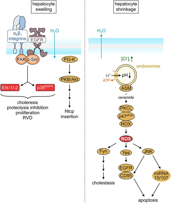
1038         M. Bonus et al.: Liver cell hydration and integrin signaling

formation has also been described in tumor cell lines                   ectodomain (Xiong et al. 2001, 2002) and electron micro-
(O’Connor and Mercurio 2001; Whittard and Akiyama                       scopy studies (Nishida et al. 2006; Takagi et al. 2002),
2001).                                                                  integrins adopt at least three stable conformational states
                                                                        during their activation cycle (Figure 5C). The “bent-closed”
                                                                        (Figure 5C, left) conformation with low affinity for extra-
                                                                        cellular matrix (ECM) ligands represents a resting state of
Structural aspects of integrin                                          integrins. In contrast, the “extended-closed” conformation
activation                                                              (Figure 5C, center) with medium affinity and the
                                                                        “extended-open” conformation (Figure 5C, right) with high
Structures of integrin ectodomains (Xiong et al. 2001, 2002)            affinity are binding-competent (Arnaout et al. 2005; Kadry
and an understanding of how conformational changes are                  and Calderwood 2020). Integrin-dependent ligand binding
associated with integrin activation opened up the oppor-                was thought to always follow the same basic multi-step
tunity to complement the in vitro and in vivo findings with a            procedure (Bachmann et al. 2019; Kadry and Calderwood
mechanistic understanding on the structural level.                      2020; Kechagia et al. 2019; Sun et al. 2019). First, intracel-
Humans express 18 α and 8 β subunits, which can arrange                 lular adaptor proteins such as talin and kindlin are
into 24 distinct integrin heterodimers (Figure 5A). The                 recruited. The binding of these adaptor proteins to the
global architecture of these heterodimers is conserved                  integrin cytoplasmic tails induces the long-range confor-
across all multicellular organisms (Kadry and Calderwood                mational changes described in Figure 5C, which increase
2020). A large extracellular region (ectodomain) that con-              the affinity for ECM ligands. Now the integrins engage their
sists of several domains is connected to two generally short            respective ECM components with high affinity and simul-
cytoplasmic tails via two single-span transmembrane re-                 taneously bind to the actin cytoskeleton. This bilateral
gions (Figure 5B). α and β subunits are unrelated with                  connection results in a clustering of integrins and the for-
respect to their sequence, but the subunits themselves are              mation of anchoring cell-matrix connections known as
globally conserved across almost all species. An exception              focal adhesions. Numerous intracellular binding partners
to this is a group of vertebrate α subunits with an additional          can subsequently be recruited, which may initiate various
inserted domain. This insertion, the αI or αA domain, forms             downstream signaling pathways (Kim et al. 2011; Morse
the binding site in collagen-binding integrins and some                 et al. 2014).
laminin-binding integrins. A divalent cation (usually Mg2+)                  Note that this model neither includes nor excludes the
within the “metal ion-dependent adhesion site” (MIDAS) of               possibility of ligand binding to the low-affinity “bent-
this binding site mediates binding of extracellular ligands             closed” conformation (Figure 5C, left). Omitting this sce-
either via an aspartate or glutamate residue. Regardless of             nario is reasonable in the context of binding to voluminous
whether an αI domain is present or absent, the ectodo-                  ECM ligands, which are unlikely to reach the binding pocket
mains of all integrin α subunits carry a β-propeller domain,            in the “bent-closed” state for steric reasons. However, the
two calf domains, and a thigh domain with an IgG-like                   model may imply that the modulation of integrin activity
structure. Ectodomains of β subunits all bear a βI (or βA)              and signaling by extracellular binding partners of lower
domain homologous to the αI domain, a hybrid domain, a                  molecular weight is not possible either. Moreover, the model
plexin–semaphorin–integrin (PSI) domain, four epidermal                 limits the concept of bidirectionality and reciprocity (Hynes
growth factor (EGF)-like domains, and a β-tail domain                   2002) of integrin signaling to the focal adhesion phase. By
(Xiong et al. 2001). In integrins without an αI domain, the βI          contrast, a large body of experimental evidence suggests
domain takes over ligand engagement; however, the βI                    that the “bent-closed” conformation may very well bind
domain carries three divalent cations instead of one. While             small- and macromolecular ligands (Adair et al. 2005; Zhu
the βI MIDAS carries a Mg2+ ion, as it does in αI domains,              et al. 2013). Furthermore, ligand binding to an isolated
the structural information available to this date suggests              headpiece induces a conformational change that exactly
that the site “adjacent to MIDAS” (ADMIDAS) and the                     matches the conformational change following adaptor
synergistic metal ion-binding site (SyMBS, formerly                     protein-induced integrin activation (Springer et al. 2008;
referred to as LIMBS for “ligand-associated metal-binding               Zhu et al. 2013). Therefore, ligand binding to the “bent-
site”) both carry a Ca2+ ion.                                           closed” ectodomain likely influences the integrin confor-
     According to the currently accepted model, derived                 mational equilibrium similar to the binding of intracellular
from the first crystal structures of a complete integrin                adaptor proteins to the cytoplasmic tail.
M. Bonus et al.: Liver cell hydration and integrin signaling   1039

Activation of integrin α5β1 by urea                             contrast to integrin activation by cell swelling, predomi-
                                                                nantly involving membrane-bound integrins (Gohlke et al.
Early studies revealed that high urea concentrations            2013; Reinehr et al. 2010a). Intriguingly, TUDC-mediated
induce a paradoxical regulatory volume decrease with            integrin activation was also suppressed in the presence of
an opening of K+ channels followed by hepatocyte                higher TC concentrations, suggesting that both bile acids
shrinkage, although the hepatocyte membrane is ex-              bind to the RGD binding site but with opposite effects
pected to be freely permeable to urea (Hallbrucker et al.       (Gohlke et al. 2013). In line with this data, MD simulations
1994). Cryosections of urea-perfused rat liver immuno-          of α5β1 integrin in complex with TUDC, TC, or GRGDSP,
stained for the active β1 integrin subunit showed that high     docked to the RGD binding site, respectively, demonstrated
urea concentrations (100 μM) cause activation of the he-        that only TUDC induces conformational changes between
patocyte β1 integrin system and trigger the same signal         the propeller and the βA domains (Figure 6A) and in helices
transduction mechanisms as hypoosmolarity and TUDC              α1 and α7 (Figure 6B) that are consistent with integrin
(Reinehr et al. 2010a). Molecular dynamics (MD) simula-         activation (Bonus et al. 2020; Gohlke et al. 2013). Hence, we
tions of a model of α5β1 integrin in aqueous urea solution      concluded that TUDC can directly activate intrahepatocytic
revealed that urea disrupts stabilizing hydrogen bonds          β1 integrins, which trigger signal transduction pathways
between the leg regions of both integrin subunits and           toward choleresis.
between the hybrid domain and the leg region of the
α-subunit. Breaking these interactions led to a pro-
nounced integrin unbending, which is part of the                Functional selectivity of TUDC and
conformational transition towards the “extended-closed”         norUDCA for integrin-mediated
state, corresponding to the first step in the activation cycle
of integrins (Figure 5C). Hence, urea-mediated α5β1             signaling pathways
integrin activation in hepatocytes is suggested to occur
through direct urea-integrin interactions, although not at      Chemical modifications to the ursodeoxycholane scaf-
the extracellular ligand binding site of α5β1.                  fold of TUDC led either to a complete loss of activity or, in
                                                                the case of taurocholic acid (TC), to an antagonist of
                                                                TUDC-induced α5β1 integrin activation (Gohlke et al.

Integrin α5β1 acts as a sensor for                              2013). Whether chemical modifications of the bile acid’s
                                                                side chain – preserving the ursodeoxycholane scaffold –
TUDC in hepatocytes                                             retain or even increase activity towards α5β1 integrin was
                                                                investigated in a subsequent study (Bonus et al. 2020).
Abrogation of the complete TUDC-induced signal trans-           Here, MD simulations were conducted first to compare
duction in the presence of the hexapeptide GRGDSP               the ability of norUDCA, TnorUDCA, GUDC, and UDCA to
(Häussinger et al. 2003), the lack of TUDC-induced kinase       evoke activation-related conformational changes in α5β1
activation after β1 integrin knockdown in isolated rat he-      integrin (Figure 6A, B) with that of TUDC and TC. The
patocytes (Gohlke et al. 2013), and the pharmacophoric          extent of these conformational changes, described using
similarity of TUDC with tirofiban, a small molecule inhib-       three geometric descriptors, was subsequently corre-
itor binding to the extracellular binding site of integrin      lated with an activity ranking of these bile acids
αIIbβ3, led to the suggestion that TUDC can activate integrin   (Figure 6C) derived from the amount of immunostained,
α5β1 by specifically binding to its extracellular site.          active β1 integrin determined at fixed time points
Immunofluorescence staining experiments revealed that            (Figure 6D). Based on this data, TUDC and norUDCA were
only TUDC, but neither taurocholic acid (TC), glyco-            classified as highly active, TnorUDCA and GUDC as
chenodeoxycholic acid (GCDC), taurochenodeoxycholic             weakly active, and UDCA and TC as inactive or inhibitory
acid (TCDC), nor taurolithocholic acid 3-sulfate (TLCS),        ligands of α5β1 integrin, respectively. A competitive
activated β1 integrins (Gohlke et al. 2013). These bile acids   ELISA-based solid-phase assay (Bochen et al. 2013)
primarily differ from TUDC with respect to substitutions at     confirmed direct binding of TUDC and norUDCA to the
or configurations of substituents of the cholane scaffold.       MIDAS in α5β1 integrins and revealed similar binding
Furthermore, integrin β1 was activated predominantly in-        affinities of both compounds (Bonus et al. 2020). In view
side hepatocytes and required TUDC uptake through the           of this, the different extent of α5β1 integrin activation by
Na+/taurocholate cotransporting polypeptide (NTCP)              TUDC and norUDCA could not have been a consequence
(Gohlke et al. 2013). This mode of activation was in stark      of different binding site occupancy.
1040           M. Bonus et al.: Liver cell hydration and integrin signaling

Figure 6: Geometric descriptors and activation of α5β1 integrin in MD simulations and immunofluorescence staining experiments.
(A) Part of the α5β1 integrin headpiece in cartoon representation. Helices α1 and α7 are highlighted in orange and blue, respectively. The
propeller-βA distance was measured between the respective centers of mass (violet circles). The propeller domain is colored in light blue, the
βA domain in pale green, and the hybrid domain in violet. (B) Close-up view of the βA domain with the docked TUDC structure (stick
representation) (Gohlke et al. 2013). This complex structure was used to generate the other starting structures by modifying the bile acid side
chain. Angles measured during the MD simulations: orange: α1 kink angle; blue: α7 tilt angle. Mg2+ ions are depicted as yellow spheres; the
metal ion at the MIDAS is labeled M, the metal ion at the ADMIDAS is labeled A. (C) Average ± SEM values of the α1 kink angle (orange), α7 tilt
angle (blue), and propeller-βA distance (violet) over three replicate MD simulations versus the rank of the bile acids according to their
agonistic activity towards α5β1 integrin as observed in immunofluorescence staining experiments (panel D). Dashed lines depict linear
regressions; fit parameters are given in the figures. Vertical lines separate the dataset into inactive (left), weakly active (middle), and highly
active (right) bile acids. (D) Effect of norUDCA, TnorUDCA, GUDC, and UDCA on β1 integrin activation. Rat livers were perfused with the
respective bile acids for up to 60 min with the concentrations indicated. Liver samples were immunostained for the active conformation of β1
integrin (blue, false-color images; for original images cf. Figure 3 in Bonus et al. 2020). Figure and caption were modified from Bonus et al.
(2020).

    As described before (Kurz et al. 2001; Schliess et al.                have been a consequence of weaker α5β1 integrin activa-
1997), TUDC (20 μM)-induced α5β1 integrin activation led to               tion. Since TUDC, but not norUDCA, significantly enhanced
sustained activation of Erk-1/-2 and p38MAPK (Figure 7A–C),               the EGFR/c-Src association, a c-Src-dependent trans-
the critical downstream signal towards choleresis. Perfu-                 activation of EGFR is likely a determining factor for pro-
sion with lower (10 μM) and higher (50 μM) TUDC con-                      longed MAPK activation (Bonus et al. 2020). Inhibiting
centrations did not change this kinetic profile, ruling out a              PI3-K abolished norUDCA-induced phosphorylation of
concentration effect (Bonus et al. 2020). In contrast,                    only Erk-1/-2, but inhibiting c-Src abolished phosphoryla-
norUDCA-induced α5β1 integrin activation only led to                      tion of both Erk-1/-2 and p38MAPK, indicating that c-Src
transient activation of these MAPKs (Figure 7C), which may                activation occurs upstream of PI3-K activation. In an earlier
M. Bonus et al.: Liver cell hydration and integrin signaling            1041

Figure 7: Comparison between TUDC- and norUDCA-induced α5β1 integrin activation and MAPK signaling.
(A) Immunofluorescence staining of active β1 integrin after liver perfusion with either 20 μM norUDCA (left) or TUDC (right) at timepoints t = 0, 1, 5,
and 15 min. The scale bar corresponds to 20 μm. Representative images of three independent experiments are depicted. (B) Quantification of β1
integrin fluorescence. (C) Densitometric quantification of Erk-1/2 (left) and p38MAPK (right) phosphorylation after liver perfusion with either 20 μM
norUDCA (violet) or TUDC (green). Total Erk-1/2 and total p38MAPK served as respective loading controls. Phosphorylation at t = 0 min was set to 1.
Data represents the mean ± SEM of at least three independent experiments. (D) Activation of α5β1 integrin by TUDC leads to high FAKY397−P levels,
which trigger both a slow, c-Src and EGFR-independent PI3-K activation (red arrow) and a fast, c-Src and EGFR-dependent PI3-K activation (green
arrows). The subsequent PI3-K-mediated Erk-1/2 activation is sustained. (E) Activation of α5β1 integrin by norUDCA leads to lower FAKY397−P levels,
which trigger the fast, c-Src and EGFR-dependent pathway of PI3-K activation (green arrows), but abolish the slower FAK-mediated PI3-K
activation. The subsequent PI3-K mediated Erk-1/2 activation is only transient. Figure and caption were modified from Bonus et al. (2020).

study (Häussinger et al. 2003), which investigated                            delayed it by ∼8 min. Hence, inhibition of c-Src activity only
TUDC-induced signaling towards choleresis, inhibition of                      prevents Erk-1/-2 phosphorylation after norUDCA-induced
c-Src did not prevent Erk-1/-2 phosphorylation but only                       but not after TUDC-induced α5β1 integrin activation.

1042           M. Bonus et al.: Liver cell hydration and integrin signaling

     Together with further experiments on FAK phosphor-                   Barczyk, M., Carracedo, S., and Gullberg, D. (2010). Integrins. Cell
ylation patterns (Bonus et al. 2020), these observations                       Tissue Res. 339: 269–280.
                                                                          Becker, S., Reinehr, R., Graf, D., vom Dahl, S., and Häussinger, D.
suggested a ligand-dependent selectivity for signaling
                                                                               (2007a). Hydrophobic bile salts induce hepatocyte shrinkage
pathways induced by α5β1-integrin (Figure 7D and E), a                         via NADPH oxidase activation. Cell. Physiol. Biochem. 19:
property widely known for G-protein coupled receptors                          89–98.
(Violin and Lefkowitz 2007), but that had not yet been                    Becker, S., Reinehr, R., Grether-Beck, S., Eberle, A., and
described for integrins without an αI domain (Simon 2011).                     Häussinger, D. (2007b). Hydrophobic bile salts trigger
                                                                               ceramide formation through endosomal acidification. Biol.
The insights gained from this study also provide a rationale
                                                                               Chem. 388: 185–196.
for the different therapeutic applications of UDCA and
                                                                          Bochen, A., Marelli, U.K., Otto, E., Pallarola, D., Mas-Moruno, C.,
norUDCA in primary biliary cholangitis and primary scle-                       Di Leva, F.S., Boehm, H., Spatz, J.P., Novellino, E., Kessler, H.,
rosing cholangitis, respectively (Trauner et al. 2015).                        et al. (2013). Biselectivity of isoDGR peptides for fibronectin
                                                                               binding integrin subtypes alpha5beta1 and alphavbeta6:
                                                                               conformational control through flanking amino acids. J. Med.
                                                                               Chem. 56: 1509–1519.
Concluding remarks                                                        Boini, K.M., Graf, D., Hennige, A.M., Koka, S., Kempe, D.S., Wang, K.,
                                                                               Ackermann, T.F., Föller, M., Vallon, V., Pfeifer, K., et al. (2009).
                                                                               Enhanced insulin sensitivity of gene-targeted mice lacking
Hormones, nutrients, and oxidative stress can lead
                                                                               functional KCNQ1. Am. J. Physiol. 296: R1695–1701.
to changes of liver cell hydration (cell volume) within
                                                                          Bonus, M., Sommerfeld, A., Qvartskhava, N., Görg, B., Ludwig, B.S.,
minutes. The changes act as important modulators of cell                       Kessler, H., Gohlke, H., and Häussinger, D. (2020). Evidence for
function and couple the physical parameter cell volume to                      functional selectivity in TUDC- and norUDCA-induced signal
metabolism, transport, and gene expression. For this to                        transduction via alpha5beta1 integrin towards choleresis. Sci.
occur, mechanotransduction (osmosensing) is required,                          Rep. 10: 5795.
                                                                          Cantore, M., Reinehr, R., Sommerfeld, A., Becker, M., and Häussinger,
which triggers signaling cascades towards liver function
                                                                               D. (2011). The Src family kinase Fyn mediates hyperosmolarity-
(osmosignaling). β1 integrins have a central role as (osmo-                    induced Mrp2 and Bsep retrieval from canalicular membrane.
)mechanosensors in the liver, but also are involved in bile                    J. Biol. Chem. 286: 45014–45029.
acid signaling.                                                           Chamberlin, M.E. and Strange, K. (1989). Anisosmotic cell volume
                                                                               regulation: a comparative view. Am. J. Physiol. 257: C159–173.
                                                                          Faundez, V. and Hartzell, H.C. (2004). Intracellular chloride channels:
Author contributions: All the authors have accepted
                                                                               determinants of function in the endosomal pathway. Sci. STKE
responsibility for the entire content of this submitted
                                                                               2004: re8.
manuscript and approved submission.                                       Gohlke, H., Schmitz, B., Sommerfeld, A., Reinehr, R., and Häussinger,
Research funding: The studies reported herein were                             D. (2013). α5β1-integrins are sensors for tauroursodeoxycholic
supported by Deutsche Forschungsgemeinschaft through                           acid in hepatocytes. Hepatology 57: 1117–1129.
Sonderforschungsbereiche SFB 575 and SFB 974.                             Graf, J. and Häussinger, D. (1996). Ion transport in hepatocytes:
                                                                               mechanisms and correlations to cell volume, hormone actions
Conflict of interest statement: The authors declare no
                                                                               and metabolism. J. Hepatol. 24: 53–77.
conflicts of interest regarding this article.                              Hallbrucker, C., vom Dahl, S., Lang, F., and Häussinger, D. (1991).
                                                                               Control of hepatic proteolysis by amino acids. The role of cell
                                                                               volume. Eur. J. Biochem. 197: 717–724.
References                                                                Hallbrucker, C., vom Dahl, S., Ritter, M., Lang, F., and Häussinger, D.
                                                                               (1994). Effects of urea on K+ fluxes and cell volume in perfused
Adair, B.D., Xiong, J.-P., Maddock, C., Goodman, S.L., Arnaout, M.A.,          rat liver. Pflüger’s Arch. 428: 552–560.
     and Yeager, M. (2005). Three-dimensional EM structure of the         Häussinger, D. (1996a). Regulation der Zellfunktion durch den
     ectodomain of integrin αVβ3 in a complex with fibronectin. J. Cell         Hydratationszustand. Naturwissenschaften 83: 264–271.
     Biol. 168: 1109–1118.                                                Häussinger, D. (1996b). The role of cellular hydration in the regulation
Arnaout, M.A., Mahalingam, B., and Xiong, J.-P. (2005). Integrin               of cell function. Biochem. J. 313: 697–710.
     structure, allostery, and bidirectional signaling. Annu. Rev. Cell   Häussinger, D., Hallbrucker, C., vom Dahl, S., Decker, S., Schweizer,
     Dev. Biol. 21: 381–410.                                                   U., Lang, F., and Gerok, W. (1991). Cell volume is a major
Bachmann, M., Kukkurainen, S., Hytönen, V.P., and Wehrle-Haller, B.            determinant of proteolysis control in liver. FEBS Lett. 283: 70–72.
     (2019). Cell adhesion by integrins. Physiol. Rev. 99: 1655–1699.     Häussinger, D. and Kordes, C. (2017). Mechanisms of
Baquet, A., Gaussin, V., Bollen, M., Stalmans, W., and Hue, L. (1993).         tauroursodeoxycholate-mediated hepatoprotection. Dig. Dis. 35:
     Mechanism of activation of liver acetyl-CoA carboxylase by cell           224–231.
     swelling. Eur. J. Biochem. 217: 1083–1089.                           Häussinger, D., Kurz, A.K., Wettstein, M., Graf, D., vom Dahl, S., and
Baquet, A., Hue, L., Meijer, A.J., van Woerkom, G.M., and Plomp,               Schliess, F. (2003). Involvement of integrins and Src in
     P.J.A.M. (1990). Swelling of rat hepatocytes stimulates glycogen          tauroursodeoxycholate-induced and swelling-induced
     synthesis. J. Biol. Chem. 265: 955–959.                                   choleresis. Gastroenterology 124: 1476–1487.
M. Bonus et al.: Liver cell hydration and integrin signaling 1043
Häussinger, D. and Lang, F. (1991). Cell volume in the regulation of by glycochenodeoxycholate and tauroursodeoxycholate. Cell.
hepatic function: a mechanism for metabolic control. Biochim. Physiol. Biochem. 52: 1427–1445.
Biophys. Acta Rev. Biomembr. 1071: 331–350. Meijer, A.J., Baquet, A., Gustafson, L., van Woerkom, G.M., and Hue, L.
Häussinger, D. and Lang, F. (1992). Cell volume and hormone action. (1992). Mechanism of activation of liver glycogen synthase by
Trends Pharmacol. Sci. 13: 371–373. swelling. J. Biol. Chem. 267: 5823–5828.
Häussinger, D., Roth, E., Lang, F., and Gerok, W. (1993). Cellular Minton, A.P., Colclasure, G.C., and Parker, J.C. (1992). Model for the
hydration state: an important determinant of protein catabolism role of macromolecular crowding in regulation of cellular volume.
in health and disease. Lancet 341: 1330–1332. Proc. Natl. Acad. Sci. USA 89: 10504–10506.
Häussinger, D., Schliess, F., Dombrowski, F., and Vom Dahl, S. (1999). Morse, E.M., Brahme, N.N., and Calderwood, D.A. (2014). Integrin
Involvement of p38MAPK in the regulation of proteolysis by liver cytoplasmic tail interactions. Biochem 53: 810–820.
cell hydration. Gastroenterology 116: 921–935. Mortimore, G.E. and Pösö, A.R. (1987). Intracellular protein catabolism
Häussinger, D. and Sies, H. (Eds.) (2007). Osmosensing and and its control during nutrient deprivation and supply. Annu.
osmosignaling. In: Methods in enzymology, Vol. 428. Academic Rev. Nutr. 7: 539–564.
Press, San Diego, CA. Naruse, K. (2018). Mechanomedicine. Biophys. Rev. 10: 1257–1262.
Häussinger, D., Stehle, T., and Lang, F. (1990). Volume regulation in Nishida, N., Xie, C., Shimaoka, M., Cheng, Y., Walz, T., and
liver: further characterization by inhibitors and ionic Springer, T.A. (2006). Activation of leukocyte beta2 integrins
substitutions. Hepatology 11: 243–254. by conversion from bent to extended conformations. Immunity
Hue, L. (1995). Regulation of liver carbohydrate and lipid metabolism 25: 583–594.
by cell volume. Ann. Endocrinol. (Paris) 56: 599–601. Noé, B., Schliess, F., Wettstein, M., Heinrich, S., and Häussinger, D.
Hynes, R.O. (2002). Integrins: bidirectional, allosteric signaling (1996). Regulation of taurocholate excretion by a hypo-
machines. Cell 110: 673–687. osmolarity-activated signal transduction pathway in rat liver.
Kadry, Y.A. and Calderwood, D.A. (2020). Chapter 22: Structural and Gastroenterology 110: 858–865.
signaling functions of integrins. Biochim. Biophys. Acta 1862: O’Connor, K.L. and Mercurio, A.M. (2001). Protein kinase A
183206. regulates Rac and is required for the growth factor-stimulated
Katz, J., Golden, S., and Wals, P.A. (1976). Stimulation of hepatic migration of carcinoma cells. J. Biol. Chem. 276:
glycogen synthesis by amino acids. Proc. Natl. Acad. Sci. USA 73: 47895–47900.
3433–3437. Offensperger, W.-B., Offensperger, S., Stoll, B., Gerok, W., and
Kechagia, J.Z., Ivaska, J., and Roca-Cusachs, P. (2019). Integrins as Häussinger, D. (1994). Effects of anisotonic exposure on duck
biomechanical sensors of the microenvironment. Nat. Rev. Mol. hepatitis B virus replication. Hepatology 20: 1–7.
Cell Biol. 20: 457–473. Pagani, G. and Gohlke, H. (2018). On the contributing role of the
Kim, C., Ye, F., and Ginsberg, M.H. (2011). Regulation of integrin transmembrane domain for subunit-specific sensitivity of
activation. Annu. Rev. Cell Dev. Biol. 27: 321–345. integrin activation. Sci. Rep. 8: 5733.
Kubitz, R., D’Urso, D., Keppler, D., and Häussinger, D. (1997). Paluschinski, M., Castoldi, M., Schöler, D., Bardeck, N., Oenarto, J.,
Osmodependent dynamic localization of the multidrug Görg, B., and Häussinger, D. (2019). Tauroursodeoxycholate
resistance protein 2 in the rat hepatocyte canalicular membrane. protects from glycochenodeoxycholate-induced gene
Gastroenterology 113: 1438–1442. expression changes in perfused rat liver. Biol. Chem. 400:
Kurz, A.K., Block, C., Graf, D., vom Dahl, S., Schliess, F., and 1551–1565.
Häussinger, D. (2000). Phosphoinositide 3-kinase-dependent Parker, J.C. and Colclasure, G.C. (1992). Macromolecular crowding and
Ras activation by tauroursodesoxycholate in rat liver. Biochem. J. volume perception in dog red cells. Mol. Cell. Biochem. 114: 9–11.
350 Pt 1: 207–213. Reinehr, R., Becker, S., Braun, J., Eberle, A., Grether-Beck, S., and
Kurz, A.K., Graf, D., Schmitt, M., vom Dahl, S., and Häussinger, D. Häussinger, D. (2006). Endosomal acidification and activation of
(2001). Tauroursodesoxycholate-induced choleresis involves NADPH oxidase isoforms are upstream events in
p38(MAPK) activation and translocation of the bile salt export hyperosmolarity-induced hepatocyte apoptosis. J. Biol. Chem.
pump in rats. Gastroenterology 121: 407–419. 281: 23150–23166.
Lavoinne, A., Baquet, A., and Hue, L. (1987). Stimulation of glycogen Reinehr, R., Becker, S., Höngen, A., and Häussinger, D. (2004a). The
synthesis and lipogenesis by glutamine in isolated rat Src family kinase yes triggers hyperosmotic activation of the
hepatocytes. Biochem. J. 248: 429–437. epidermal growth factor receptor and CD95. J. Biol. Chem. 279:
Liu, Y., Binz, J., Numerick, M.J., Dennis, S., Luo, G., Desai, B., 23977–23987.
MacKenzie, K.I., Mansfield, T.A., Kliewer, S.A., Goodwin, B., et al. Reinehr, R., Becker, S., Wettstein, M., and Häussinger, D. (2004b).
(2003). Hepatoprotection by the farnesoid X receptor agonist Involvement of the Src family kinase yes in bile salt-induced
GW4064 in rat models of intra- and extrahepatic cholestasis. apoptosis. Gastroenterology 127: 1540–1557.
J. Clin. Invest. 112: 1678–1687. Reinehr, R., Gohlke, H., Sommerfeld, A., vom Dahl, S., and Häussinger,
Maeda, T., Wurgler-Murphy, S.M., and Saito, H. (1994). A two- D. (2010a). Activation of integrins by urea in perfused rat liver.
component system that regulates an osmosensing MAP kinase J. Biol. Chem. 285: 29348–29356.
cascade in yeast. Nature 369: 242–245. Reinehr, R., Graf, D., Fischer, R., Schliess, F., and Häussinger, D.
Mayer, P.G.K., Qvartskhava, N., Sommerfeld, A., Görg, B., and (2002). Hyperosmolarity triggers CD95 membrane trafficking and
Häussinger, D. (2019). Regulation of plasma membrane sensitizes rat hepatocytes toward CD95L-induced apoptosis.
localization of the Na+-taurocholate co-transporting polypeptide Hepatology 36: 602–614.1044 M. Bonus et al.: Liver cell hydration and integrin signaling
Reinehr, R. and Häussinger, D. (2004). Inhibition of bile salt-induced Takagi, J., Petre, B.M., Walz, T., and Springer, T.A. (2002). Global
apoptosis by cyclic AMP involves serine/threonine conformational rearrangements in integrin extracellular domains in
phosphorylation of CD95. Gastroenterology 126: 249–262. outside-in and inside-out signaling. Cell 110: 599–511.
Reinehr, R., Schliess, F., and Häussinger, D. (2003). Hyperosmolarity Trauner, M., Halilbasic, E., Claudel, T., Steinacher, D., Fuchs, C.D.,
and CD95L trigger CD95/EGF receptor association and tyrosine Moustafa, T., Pollheimer, M.J., Krones, E., Kienbacher, C.,
phosphorylation of CD95 as prerequisites for CD95 membrane Traussnigg, S., et al. (2015). Potential of nor-ursodeoxycholic acid
trafficking and DISC formation. FASEB J 17: 731–733. in cholestatic and metabolic disorders. Dig. Dis. 33: 433–439.
Reinehr, R., Sommerfeld, A., and Häussinger, D. (2010b). Insulin Unoki, H., Takahashi, A., Kawaguchi, T., Hara, K., Horikoshi, M.,
induces swelling-dependent activation of the epidermal growth Andersen, G., Ng, D.P.K., Holmkvist, J., Borch-Johnsen, K.,
factor receptor in rat liver. J. Biol. Chem. 285: 25904–25912. Jørgensen, T., et al. (2008). SNPs in KCNQ1 are associated with
Reinehr, R., Sommerfeld, A., and Häussinger, D. (2013). The Src family susceptibility to type 2 diabetes in East Asian and European
kinases: distinct functions of c-Src, Yes, and Fyn in the liver. populations. Nat. Genet. 40: 1098–1102.
Biomol. Concepts 4: 129–142. Violin, J.D. and Lefkowitz, R.J. (2007). Beta-arrestin-biased ligands at
Santosa, D., Castoldi, M., Paluschinski, M., Sommerfeld, A., and seven-transmembrane receptors. Trends Pharmacol. Sci. 28:
Häussinger, D. (2015). Hyperosmotic stress activates the 416–422.
expression of members of the miR-15/107 family and induces Volpes, R., van den Oord, J.J., and Desmet, V.J. (1993). Integrins as
downregulation of anti-apoptotic genes in rat liver. Sci. Rep. 5: differential cell lineage markers of primary liver tumors. Am.
12292. J. Pathol. 142: 1483–1492.
Schliess, F. and Häussinger, D. (2003). Cell volume and insulin vom Dahl, S., Hallbrucker, C., Lang, F., Gerok, W., and Häussinger, D.
signaling. Int. Rev. Cytol. 225: 187–228. (1991). Regulation of liver cell volume and proteolysis by
Schliess, F., Kurz, A.K., vom Dahl, S., and Häussinger, D. (1997). glucagon and insulin. Biochem. J. 278: 771–777.
Mitogen-activated protein kinases mediate the stimulation of vom Dahl, S., Schliess, F., Reissmann, R., Görg, B., Weiergräber, O.,
bile acid secretion by tauroursodeoxycholate in rat liver. Kocalkova, M., Dombrowski, F., and Häussinger, D. (2003).
Gastroenterology 113: 1306–1314. Involvement of integrins in osmosensing and signaling toward
Schliess, F., Reissmann, R., Reinehr, R., vom Dahl, S., and Häussinger, autophagic proteolysis in rat liver. J. Biol. Chem. 278:
D. (2004). Involvement of integrins and src in insulin signaling 27088–27095.
toward autophagic proteolysis in rat liver. J. Biol. Chem. 279: Whittard, J.D. and Akiyama, S.K. (2001). Positive regulation of cell-cell
21294–21301. and cell-substrate adhesion by protein kinase A. J. Cell Sci. 114:
Schliess, F., Schreiber, R., and Häussinger, D. (1995). Activation of 3265–3272.
extracellular signal-regulated kinases Erk-1 and Erk-2 by cell Xiong, J.-P., Stehle, T., Diefenbach, B., Zhang, R., Dunker, R., Scott,
swelling in H4IIE hepatoma cells. Biochem. J. 309: 13–17. D.L., Joachimiak, A., Goodman, S.L., and Arnaout, M.A. (2001).
Schliess, F., vom Dahl, S., and Häussinger, D. (2001). Insulin Crystal structure of the extracellular segment of integrin αVβ3.
resistance induced by loop diuretics and hyperosmolarity in Science 294: 339–345.
perfused rat liver. Biol. Chem. 382: 1063–1069. Xiong, J.-P., Stehle, T., Zhang, R., Joachimiak, A., Frech, M., Goodman,
Schmitt, M., Kubitz, R., Lizun, S., Wettstein, M., and Häussinger, D. S.L., and Arnaout, M.A. (2002). Crystal structure of the
(2001). Regulation of the dynamic localization of the rat Bsep extracellular segment of integrin αVβ3 in complex with an Arg-
gene-encoded bile salt export pump by anisoosmolarity. Gly-Asp ligand. Science 296: 151–155.
Hepatology 33: 509–518. Yasuda, K., Miyake, K., Horikawa, Y., Hara, K., Osawa, H., Furuta, H.,
Schreiber, R., Stoll, B., Lang, F., and Häussinger, D. (1994). Effects of Hirota, Y., Mori, H., Jonsson, A., Sato, Y., et al. (2008). Variants in
aniso-osmolarity and hydroperoxides on intracellular pH in KCNQ1 are associated with susceptibility to type 2 diabetes
isolated rat hepatocytes as assessed by (2’,7’)- mellitus. Nat. Genet. 40: 1092–1097.
bis(carboxyethyl)-5(6)-carboxyfluorescein and fluorescein Zhu, J., Zhu, J., and Springer, T.A. (2013). Complete integrin headpiece
isothiocyanate-dextran fluorescence. Biochem. J. 303: 113–120. opening in eight steps. J. Cell Biol. 201: 1053–1068.
Simon, D.I. (2011). Opening the field of integrin biology to “biased
agonism”. Circ. Res. 109: 1199–1201.
Sommerfeld, A., Mayer, P.G.K., Cantore, M., and Häussinger, D.
(2015a). Regulation of plasma membrane localization of the Na+-
Bionotes
taurocholate cotransporting polypeptide (Ntcp) by
hyperosmolarity and tauroursodeoxycholate. J. Biol. Chem. 290: Michele Bonus
24237–24254. Institute for Pharmaceutical and Medicinal
Sommerfeld, A., Reinehr, R., and Häussinger, D. (2015b). Chemistry, Heinrich Heine University
Tauroursodeoxycholate protects rat hepatocytes from bile acid- Düsseldorf, Universitätsstr. 1, D-40225
induced apoptosis via β1-integrin- and protein kinase Düsseldorf, Germany
A-dependent mechanisms. Cell. Physiol. Biochem. 36: 866–883. https://orcid.org/0000-0003-4411-7342
Springer, T.A., Zhu, J., and Xiao, T. (2008). Structural basis for
distinctive recognition of fibrinogen gamma C peptide by the
platelet integrin alphaIIbbeta3. J. Cell Biol. 182: 791–800.
Sun, Z., Costell, M., and Fässler, R. (2019). Integrin activation by talin, Michele Bonus graduated in pharmacy from Heinrich Heine University
kindlin and mechanical forces. Nat. Cell Biol. 21: 25–31. Düsseldorf in 2012 and has been working in Holger Gohlke’s researchM. Bonus et al.: Liver cell hydration and integrin signaling 1045
group since 2013. His research focuses on the computational Wilhelm-Leibniz-Prize in 1991, the Dr. Robert Pfleger Prize in 2002, the
modeling of dynamic protein-ligand interactions in membrane Order of Merit 1st class of the German Federal Republic in 2011, and
proteins and the computer-aided identification and design of novel the Order of Merit of North-Rhine Westfalia (2020).
small-molecule protein ligands. From 2014 onwards, he studied the
activation of α5β1 integrins by bile acids and the translocation Holger Gohlke
mechanism of the lipid floppase ABCB4 as a Research Associate in the Institute for Pharmaceutical and Medicinal
Collaborative Research Centre 974 SFB (CRC 974) – “Communication Chemistry, Heinrich Heine University
and System Relevance in Liver Injury and Regeneration”. During his Düsseldorf, Universitätsstr. 1, D-40225
research work for SFB CRC 974, he was head of the organizing Düsseldorf, Germany
committee of the international PhD symposium “deLIVER – John von Neumann Institute for Computing
Technology in Hepatology”. Since 2017, as a Research Associate in the (NIC), Jülich Supercomputing Centre (JSC)
Research Unit 2518 – “Functional dynamics of ion channels and Institute of Biological Information Processing
transporters – DynIon”, he has been studying the dynamics of (IBI-7: Structural Biochemistry), and Institute
Hyperpolarization-activated cyclic nucleotide-gated (HCN) channels in of Bio- and Geosciences (IBG-
the presence and absence of cyclic nucleotides. He received several 4: Bioinformatics), Forschungszentrum Jülich
computing time grants from the Jülich Supercomputing Centre (JSC). GmbH, Wilhelm-Johnen-Str., D-52428 Jülich,
Germany
Dieter Häussinger gohlke@uni-duesseldorf.de
Clinic for Gastroenterology, Hepatology, and https://orcid.org/0000-0001-8613-1447
Infectious Diseases, Heinrich Heine University Holger Gohlke is Professor of Pharmaceutical and Medicinal
Düsseldorf, Moorenstr. 5, D-40225 Düsseldorf, Chemistry at the Heinrich-Heine-University Düsseldorf and head of the
Germany NIC Research Group “Computational Biophysical Chemistry” at
haeussin@uni-duesseldorf.de Forschungszentrum Jülich. He obtained his diploma in chemistry from
the Technical University of Darmstadt and his PhD from Philipps-
University, Marburg. He then did postdoctoral research at The Scripps
Research Institute, La Jolla, USA. After appointments as Assistant
Dieter Häussinger was born on June 22, 1951, in Nördlingen/Bavaria. Professor in Frankfurt and Professor in Kiel, he moved to Düsseldorf in
From 1994 until 2020, he was a full professor in internal medicine at 2009 and started in Jülich in 2017. He was awarded the
the Heinrich-Heine-University Düsseldorf and the director of the Clinic “Innovationspreis in Medizinischer und Pharmazeutischer Chemie”
for Gastroenterology, Hepatology and Infectious Diseases, the Center from the GDCh and the DPhG, the Hansch Award of the
for Liver and Infectious Disease and of the Hirsch Institute for Tropical Cheminformatics and QSAR Society, and the Novartis Chemistry
Medicine at Arsi University, Ethiopia. He was the coordinator of the Lecturship, and he is the speaker of the DFG-funded Research Training
Colloborative Research Centers SFB 575 (Experimental Hepatology) Group GRK 2158 (Natural products and natural product analogs
and SFB 974 (Liver Injury and Regeneration), and the Clinical Research against therapy-resistant tumors and microorganisms). His current
Group Klinische Forschergruppe 217 (Hepatobiliary Transport). He is research focuses on the understanding, prediction, and modulation of
also a member of the National Academy of Sciences Leopoldina and interactions involving biological macromolecules from a
the North Rhine-Westphalian Academy for Science and Arts. From computational perspective. His group applies and develops
2010 to 2018 he was also a senator of the Scientific Society Leibniz techniques grounded in molecular bioinformatics, computational
(WGL). He received the Thannhauser-Prize in 1989, the Gottfried biology, and computational biophysics.You can also read