Outer Membrane Vesicles (OMVs) of Pseudomonas aeruginosa Provide Passive Resistance but Not Sensitization to LPS-Specific Phages - MDPI
←
→
Page content transcription
If your browser does not render page correctly, please read the page content below
viruses
Article
Outer Membrane Vesicles (OMVs) of Pseudomonas aeruginosa
Provide Passive Resistance but Not Sensitization to
LPS-Specific Phages
Daria Augustyniak                , Tomasz Olszak        and Zuzanna Drulis-Kawa *

                                          Department of Pathogen Biology and Immunology, University of Wroclaw, Przybyszewskiego 63/77,
                                          51-148 Wroclaw, Poland; daria.augustyniak@uwr.edu.pl (D.A.); tomasz.olszak@uwr.edu.pl (T.O.)
                                          * Correspondence: zuzanna.drulis-kawa@uwr.edu.pl; Tel.: +48-71-375-6290

                                          Abstract: Outer membrane vesicles (OMVs) released from gram-negative bacteria are key elements in
                                          bacterial physiology, pathogenesis, and defence. In this study, we investigated the role of Pseudomonas
                                          aeruginosa OMVs in the anti-phage defence as well as in the potential sensitization to LPS-specific
                                          phages. Using transmission electron microscopy, virion infectivity, and neutralization assays, we
                                          have shown that both phages efficiently absorb on free vesicles and are unable to infect P. aeruginosa
                                          host. Nevertheless, the accompanying decrease in PFU titre (neutralization) was only observed for
                                          myovirus KT28 but not podovirus LUZ7. Next, we verified whether OMVs derived from wild-type
                                          PAO1 strain can sensitize the LPS-deficient mutant (∆wbpl PAO1) resistant to tested phages. The
                                          flow cytometry experiments proved a quite effective and comparable association of OMVs to ∆wbpl
                                          PAO1 and wild-type PAO1; however, the growth kinetic curves and one-step growth assay revealed
                                          no sensitization event of the OMV-associated phage-resistant P. aeruginosa deletant to LPS-specific
                                          phages. Our findings for the first time identify naturally formed OMVs as important players in

                                          passive resistance (protection) of P. aeruginosa population to phages, but we disproved the hypothesis
                                   of transferring phage receptors to make resistant strains susceptible to LPS-dependent phages.
Citation: Augustyniak, D.; Olszak, T.;
Drulis-Kawa, Z. Outer Membrane            Keywords: Pseudomonas aeruginosa; outer membrane vesicles (OMV); lytic phages; resistance to
Vesicles (OMVs) of Pseudomonas            phages; sensitization to phages; passive protection
aeruginosa Provide Passive Resistance
but Not Sensitization to LPS-Specific
Phages. Viruses 2022, 14, 121.
https://doi.org/10.3390/v14010121
                                          1. Introduction
Academic Editor: Mikael Skurnik                 Outer membrane vesicles (OMVs) released by gram-negative bacteria are proteoli-
Received: 1 December 2021
                                          posomal nanoparticles that play an important role in bacterial physiology, pathogenesis,
Accepted: 6 January 2022
                                          intraspecies or interspecies interactions, as well as mammals interactions [1,2]. In host-
Published: 11 January 2022
                                          pathogen interactions during infection, OMVs may serve as enhancers of microbial adher-
                                          ence and biofilm formation [3–5], inductors of inflammation and apoptosis [6–9], and as
Publisher’s Note: MDPI stays neutral
                                          inhibitors of host immune response [10–12].
with regard to jurisdictional claims in
                                                In microbial communities, OMVs may be used by bacteria as an offensive or defensive
published maps and institutional affil-
                                          weapon. Concerning the former, OMVs can deliver bactericidal toxins or enzymes to
iations.
                                          other bacteria [13,14] as well as enzymes, DNA, and small RNA to mammalian cells,
                                          causing their injury [15]. In the defensive aspect, Moraxella catarrhalis OMVs carrying
                                          β-lactamases confer protection to the producer and accompanying Streptococcus pneumoniae
Copyright: © 2022 by the authors.
                                          against β-lactam antibiotics [16]. It has been also shown that OMVs are involved in the
Licensee MDPI, Basel, Switzerland.        trapping of antibiotics, cationic peptides, or serum complement, thus providing cross-
This article is an open access article    resistance and increasing virulence among bacteria or pathogenic yeasts [17,18]. Recent
distributed under the terms and           studies show that OMVs can participate in bacteria–phage interplay, by being a phage
conditions of the Creative Commons        decoy and sequestering phage particles as first documented for OMVs of E. coli and phage
Attribution (CC BY) license (https://     T4 [19]. Furthermore, membrane vesicles (MV) derived from gram-positive Bacillus genera
creativecommons.org/licenses/by/          can mediate the exchange of cell surface components, including phage receptors, thus
4.0/).                                    facilitating sensitization to phages [20].

Viruses 2022, 14, 121. https://doi.org/10.3390/v14010121                                                  https://www.mdpi.com/journal/viruses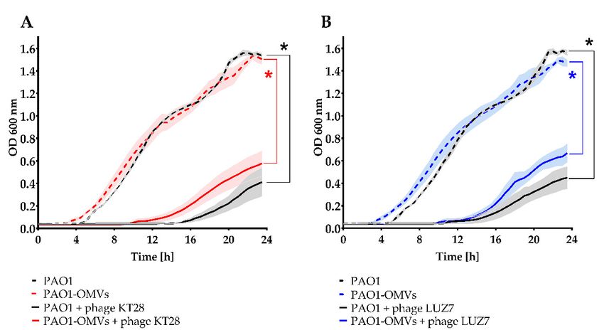
Viruses 2022, 14, 121                                                                                             2 of 18

                              Pseudomonas aeruginosa is one of the most life-threatening pathogens due to its high
                        intrinsic drug resistance and genome plasticity that condition high adaptation to adverse
                        environmental changes and promote survival and persistence at different stages of patho-
                        genesis [21,22]. These bacteria cause opportunistic infections, including burn wound
                        infections, urinary tract infections, keratitis, otitis externa, and respiratory tract infection.
                        P. aeruginosa strains possess a huge arsenal of virulence properties/factors, including pro-
                        duction of alginate, biofilm formation, pyoverdine siderophore, lipopolysaccharide (LPS),
                        quorum sensing (QS), type 4 pili (T4P), the type II and III and VI secretion systems, lipases,
                        proteases (AprA and PIV), elastases (LasA and LasB), urease, and exotoxin A [22–24].
                        Many of these virulence factors have been identified as OMV elements [25]. OMVs can
                        be released by planktonic or sessile cells by PQS (Pseudomonas quinolone signal)-induced
                        mechanism or cell lysis, significantly increasing the virulence capacity or contributing to
                        virulence-associated processes [26]. P. aeruginosa OMVs can (i) prime host tissue surfaces for
                        bacterial adhesion [5], (ii) facilitate the removal of competing bacteria from the environment
                        during infection, as well as (iii) reduce CFTR (cystic fibrosis transmembrane conductance
                        regulator) Cl− secretion from cystic fibrosis bronchial epithelial cells, thus reducing the
                        bacterial clearance from the lungs [13,27].
                              The use of phages for the treatment of bacterial infections has been extensively studied
                        as an alternative therapeutic strategy [28–30]. Since P. aeruginosa is one of the leading
                        opportunistic pathogens involved in hospital-acquired infections, large fractions of the
                        phage application studies and genome-driven phage–bacteria interplay projects are focused
                        on this bacterium [31–34]. To our knowledge, there are no reports published yet deliberating
                        the role of P. aeruginosa OMVs in fighting phages. In general, two groups of defence
                        strategies against phage infection can be recognized.
                              The first one is triggered in the presence of a phage genome inside the cell (cellu-
                        lar interior). These mechanisms are exemplified by bacterial expression of anti-phage
                        defences cutting phage DNA, including innate systems (restriction-modification, BREX,
                        DND, DISARM, etc.) as well as adaptive ones, such as CRISPR–Cas (clustered regularly
                        interspaced short palindromic repeats-CRISPR associated proteins), destroying the invader
                        and remembering it [35]. The phage life cycle can be inhibited also by the chemical defence,
                        viperins, retrons, and cell signalling coupled with abortive infection leading to cell death
                        before completion of phage reproduction and many other mechanisms that have not yet
                        been fully elucidated [35–38].
                              The second strategy is the primary mechanism to avoid phage infection, and it is
                        based on blocking phage adsorption to the bacterial cell. That purpose may be achieved by
                        the modification or alteration of surface receptors by (i) mutation or deletion of genomic
                        DNA encoding receptor that leads to receptor alteration/loss, (ii) masking its binding
                        site, (iii) and regulation of receptor expression by lysogenic phages as well as (iv) global
                        regulatory pathways [34,39,40]. Since OMVs released from bacterial cells are equipped with
                        numerous surface macromolecules, such as LPS, type IV fimbriae, outer membrane proteins
                        (OMPs), and capsular polysaccharides (CPS) [37,41], their contribution to potential phage
                        receptor transfer and alteration is also very likely. Such phenomenon of phage protection
                        by OMVs has already been shown in Salmonella and Vibrio cholerae models [41,42].
                              That enormous (extraordinary) adaptive capability that enables P. aeruginosa to survive
                        various hostile conditions, such as inhabiting various environmental niches, host invasion,
                        or phage exposure, constitute an axis of interconnection, the understanding of which
                        is of great importance. The goal of the study was to extend the OMV-mediated phage-
                        protection paradigm to an important human opportunistic pathogen and characterize
                        the role of P. aeruginosa OMVs in bacteria–phage interplay and anti-phage defence in the
                        PAO1 strain model. We verified two main hypotheses of OMVs phage-bacteria interactions
                        first as a protective barrier and secondly as a sensitization element to LPS-dependent
                        phage infection.
Viruses 2022, 14, 121 3 of 18
2. Materials and Methods
2.1. Reagents
TSB (Tryptone Soya Broth, OXOID, Hampshire, UK); TSA (Tryptone Soya Agar, OX-
OID, Hampshire, UK); Bradford reagent (Protein Assay Dye Reagent Concentrate, Bio-Rad,
München, Germany); Gelcode Blue stain reagent (Thermo Scientific, Rockford, IL, USA),
and fluorescein isothiocyanate (FITC, ThermoScientific, Rockford, IL, USA) were used.
2.2. Bacterial Strain and Phages
The P. aeruginosa PAO1 (ATCC 15692) reference strain and its ∆wbpl knock-out mutant
deficient in the A-band and B-band O-antigens biosynthesis (provided by Andrew M.
Kropinski from the Laboratory of Foodborne Zoonoses, Guelph, ON, Canada) were used
in this study. Bacteria were stored at −70 ◦ C in TSB supplemented with 20% glycerol.
Pseudomonas phages KT28 and LUZ7 were propagated as previously described [43]. The
phage titre was assessed using the double-agar layer technique. Purified phage samples
were stored at 4 ◦ C. Phage characteristics are presented in Table 1.
Table 1. Characteristics of phages used in this work.
Phage Taxonomy (Family, Genus) Genome Size GenBank Recognized Bacterial Receptor Reference
KT28 ** Myoviridae, Pbunavirus 66,381 bp KP340287 LPS [43]
LUZ7 * Schitoviridae, Luzseptimavirus 74,901 bp NC_013691 LPS [44]
* Laboratory of Gene Technology, KU Leuven, Leuven, Belgium. ** Department of Pathogen Biology and
Immunology, Institute of Genetics and Microbiology, University of Wroclaw, Wroclaw, Poland.
2.3. Outer Membrane Vesicles Isolation
Outer membrane vesicles (OMVs) isolation was adapted according to our previous
protocol with few modifications [12]. Briefly, overnight culture of P. aeruginosa was diluted
50-fold in 500 mL of TSB and incubated at 37 ◦ C for 16–18 h, with agitation (150 rpm). The
culture was harvested by centrifugation (8000× g for 15 min at 4 ◦ C). The supernatant was
collected and passed through a 0.22-µm-pore size filter vacuum pump (Merck, Millipore,
Billerica, MA, USA). The filtrates were concentrated using 100 kDa Vivaspin centrifugal
concentrators (Amicon Ultra, Merck Millipore, Cork, Ireland) at 5000× g for 30 min at
4 ◦ C. The concentrated supernatants were subsequently pelleted overnight (100,000× g, at
4 ◦ C) in an ultracentrifuge (Beckman Coulter Optima L-90K, USA). The pellets containing
OMVs were re-suspended in 500 µL of sterile PBS buffer (pH 7.4), aliquoted, and stored
at −20 ◦ C. For some experiments, OMVs were subjected to additional centrifugation at
16,000× g for 30 min at 4 ◦ C to clear most contaminating flagella [5]. The sterility of the
OMV preparations was confirmed on TSA. The protein concentrations in OMV preparations
were measured using Bradford assay, and the quality of the OMV samples was confirmed
in 12% SDS-PAGE.
2.4. OMV Association Assay by Flow Cytometry
OMV association assay was performed as described previously [18] with some modifi-
cations.
(i) OMVs’ labelling: Initially, 500 µL OMVs (100 µg/mL) in PBS were concentrated
using 30 kDa Vivaspin centrifugal concentrators (Amicon Ultra, Merck Millipore,
Cork, Ireland) at 14,000× g for 10 min at 4 ◦ C to remove PBS. The collected OMVs
were reconstituted with 500 µL of 0.05 M carbonate/bicarbonate buffer (pH 9.5) and
washed by centrifugation on Vivaspin as described before. The collected OMVs
(~50 µL) were labelled with 500 µL of 1 mg/mL FITC at carbonate/bicarbonate buffer
for 30 min at 37 ◦ C with gentle mixing in the dark. The remaining fluorochrome was
rinsed 3 times with a 500 µL of cold carbonate/bicarbonate buffer each time, using
30 kDa Vivaspin. The final FITC-labelled OMVs were resuspended in PBS containing
at the concentration of 500 µg/mL.Viruses 2022, 14, 121 4 of 18
(ii) OMV association with bacteria: 0.5 mL of fresh bacterial culture corresponding to
OD600 = 0.23–0.25 was centrifuged and subsequently washed with 1 mL of PBS
(8000× g, 10 min, 4 ◦ C). The pellet was resuspended in 100 µL of OMV-FITC conjugate
(from 20 to 320 µg/mL OMVs) supplemented with 5 mM CaCl2 and incubated for
3 h at 37 ◦ C with gentle mixing in the dark. Afterwards, samples were washed twice
with PBS by centrifugation (8000× g, 10 min, 4 ◦ C) to remove free OMVs particles and
finally resuspended in 500 µL of PBS.
(iii) Flow cytometric analysis: To detect bacterial cells associated with FITC-labelled OMVs,
flow cytometry analysis was performed using GUAVA® EasyCyte flow cytometer
(Millipore, Seattle, WA, USA). Before analysis, the samples were diluted at 1:10
to obtain approximately 1–5 × 10⁶ CFU/mL (colony-forming units/mL) in PBS.
Fluorescence intensity of bacterial cells associated with OMVs was analysed for green
fluorescence in the FL1 channel by collecting 5000 events. Data were expressed as
mean fluorescence intensity (MFI). Data analysis was performed using InCyte Merck
Guava software (Millipore, Hayward, CA, USA).
2.5. Bacterial Growth Assay with Free OMVs and Phages
The overnight TSB culture of P. aeruginosa PAO1 was refreshed to early log phase
(OD = 0.2, A600 nm) by incubation at 37 ◦ C with agitation. The bacteria were centrifuged
(8000× g, 10 min, 4 ◦ C), washed with saline, adjusted to OD 0.2 and diluted 100×. For
testing the lytic activity of phages in the presence of OMVs, the log-phase bacterial sus-
pension (105 CFU/mL) was treated with phages (multiplicity of infection, MOI = 1) and
with or without 20 µg/mL of OMVs. Each option was incubated in a water bath at 37 ◦ C
in 1% TSB medium (w/v) in the final volume of 200 µL for 120 min. Every 30 min, the
10 µL aliquots of 10-times diluted bacterial suspensions were plated in triplicate on TSA
agar plates. After 18 h of incubation at 37 ◦ C, the colony counts and the CFU/mL were
calculated. The lytic activity of phages was expressed in each time point as a decrease
in CFU/mL in the reference to uninfected control (time 0). All microbicidal assays were
performed at least two times in triplicate.
2.6. Phage Neutralization Assays
Then, an 18-h TSB culture of P. aeruginosa PAO1 was refreshed to early log phase
(OD600 = 0.2) by incubation at 37 ◦ C with agitation. The bacteria were centrifuged (8000× g,
10 min, 4 ◦ C), washed with saline, adjusted to OD600 = 0.2, and diluted 100×. The following
test options were prepared: (i) phage inoculum of 105 PFU/mL (plaque-forming units/mL)
and 80 µg/mL OMVs and (ii) phage inoculum of 105 PFU/mL. The contents were mixed
and incubated in a water bath at 37 ◦ C for 3 h. Every 30 min, the PFU/mL was determined
by the double-layer TSA plates method.
2.7. Growth Kinetics Measurements after OMV Association
To fuse OMVs with bacteria, 0.5 mL of fresh bacterial culture corresponding to
OD600 = 0.2 was transferred to a 2-mL Eppendorf tube, centrifuged, and subsequently
washed with 1 mL of PBS, pH 7.4 (8000× g, 10 min, 4 ◦ C). The pellet was resuspended in
100 µL of OMVs at the final concentration of 320 µg/mL in PBS containing 5 mM CaCl2
to facilitate fusion [45] and incubated for 3 h at 37 ◦ C with mixing (200 rpm) in the dark.
Afterwards, samples were washed twice with 1 mL of PBS by centrifugation (8000× g,
10 min, 4 ◦ C) to remove free OMVs particles and finally resuspended in 0.5 mL of PBS.
The bacteria were diluted 50× in TSB to obtain 106 CFU/mL. A hundred µL of bacterial
suspension and 100 µL of phages (MOI = 1) were added to each microplate well. All growth
kinetics experiments were performed on the flat-bottomed 96-well microplates (Nunclon
Delta Surface 167008, Thermo Scientific, Roskilde, Denmark) at 37 ◦ C in the final volume
of 200 µL. The growth rate was measured using a Varioskan LUX multimode microplate
reader (Thermo Scientific, Vantaa, Finland) with OD600 measurements for 24 h at 30 minViruses 2022, 14, 121 5 of 18
intervals with agitation (60 rpm). The data for statistical analyses were expressed as AUC
(area under the curve).
2.8. Lytic Phage Cycle after OMV Association (One-Step Growth Assay)
The bacteria-OMVs-associated population was prepared as described in Section 2.7,
and then, 100 µL of bacterial suspension in TSB (108 CFU/mL) was mixed with 100 µL
of phages (MOI = 1) and incubated at 37 ◦ C for 90 min. The phage titre (PFU/mL) was
monitored every 15 min by the double-layer TSA plates method.
2.9. Transmission Electron Microscopy Analyses (TEM)
The OMVs preparation for TEM was adapted from our previous protocol [9]. Briefly,
OMVs were visualized by standard negative staining using a formvar-coated copper grid
(Christine Gröpl Electronenmikroskopie, Tulln, Austria) and 2% (w/v) aqueous solution
of uranyl acetate. The OMVs were imaged with a TEM operating at an acceleration
voltage of 150 kV (Hitachi H-800, Tokyo, Japan) or (Tesla BS 540, Brno, Czech Republic
operated at 80 kV. During the purification of phages, the sterile and filtered phage lysate
was centrifuged at 25,000× g, 60 min, 4 ◦ C. The pellets were washed twice with 500 µL
of ammonium acetate (0.1 M, pH 7.0) by centrifugation (parameters as above), and the
quality of phage particles was checked using TEM. To visualize the interaction of phages
with OMVs, the phage particles in PBS supplemented with 5 mM CaCl2 (108 PFU/mL)
were mixed with OMVs (100 µg/mL) and incubated for 30 min at 37 ◦ C. Formed OMV-
phage complexes were next deposited on carbon-coated 200-mesh Formvar copper grids
(Christine Gröpl Electronenmikroskopie, Tulln, Austria), stained with uranyl acetate (2%,
pH 4.5), and examined using TEM.
2.10. Statistical Analysis
The data were expressed as the mean ± SEM (standard error of the mean). Normality
and homogeneity of variance assumptions were checked by Shapiro–Wilk and Levene’s test,
respectively. For comparisons between two groups, the t-test for independent variables was
used. For multiple comparisons, data were analysed using non-parametric Kruskal–Wallis
ANOVA rang test. Differences were considered statistically significant if p < 0.05 using the
Statistica (version 13.1) software (StatSoft, Krakow, Poland).
3. Results
3.1. Charateristics of P. aeruginosa OMVs
The P. aeruginosa PAO1 strain was used as an OMV source because of its comprehensive
genotypic and phenotypic characterization in the literature and as the host of lytic phages
selected for this study. As shown in the transmission electron microscopy (TEM) images,
the diameters of OMVs from PAO1 planktonic cells varied between 25–75 nm (Figure 1A,B).
The zeta potential of vesicles in miliQ (20 µg/mL) was −26.8 ± 0.71 mV as assessed by
the Zeta-sizer Nano-ZS 90 (Malvern, UK). The protein components of OMVs are shown in
Figure 1C.Viruses 2022, 14, x FOR PEER REVIEW 6 of 18
Viruses 2022, 14, 121 6 of 18
Figure 1. Physical characterization
Figure 1. membrane vesicles
characterization of outer membrane vesicles (OMVs)
(OMVs) produced
produced by P. aeruginosa
by P. aeruginosa
PAO1 cells: (A)
(A)TEM micrographofofP.P.aeruginosa
TEMmicrograph aeruginosacells
cells releasing
releasing OMVs
OMVs (magnification,
(magnification, ×12,000);
×12,000); (B)
TEM
(B) micrograph
TEM micrographof isolated P. aeruginosa
of isolated OMVs,
P. aeruginosa OMVs, with
withvesicles
vesiclesindicated
indicatedby
byarrows
arrows(magnification,
(magnification,
×30,000);
×30,000);(C)
(C) the
the representative proteinogramofof12%
representative proteinogram 12%SDS-PAGE
SDS-PAGE electrophoresis
electrophoresis of of OMVs;
OMVs; thethe pro-
protein
tein profiles
profiles werewere visualized
visualized usingusing Coomassie
Coomassie staining.
staining.
3.2. P. aeruginosa Free
Free OMVs
OMVs Passively Protect
Protect against
against Infection
Infection with
with Phages
Phages Recognizing
Recognizing LPS
LPS
To evaluate whether OMVs can inhibit phage infection, OMVs obtained
To evaluate whether OMVs can inhibit phage infection, OMVs obtained from P. ae- from P. aerugi-
nosa PAO1
ruginosa wild-type
PAO1 strain
wild-type werewere
strain combined withwith
combined the PAO1 population
the PAO1 treated
population with with
treated LPS-
specific phages.
LPS-specific Two Two
phages. different lytic phages,
different a myovirus
lytic phages, KT28 KT28
a myovirus (Pbunavirus) and podovirus
(Pbunavirus) and pod-
LUZ7, were used
ovirus LUZ7, wereasused
models (Table (Table
as models 1). The1).
selection of phages
The selection for thefor
of phages vesicle experiments
the vesicle exper-
was
imentsnotwas
random; both recognise
not random; LPS as their
both recognise LPS receptor, and bothand
as their receptor, are capable
both areofcapable
infecting
of
the same host, P. aeruginosa PAO1, but not its LPS-deficient ∆wbpl mutant.
infecting the same host, P. aeruginosa PAO1, but not its LPS-deficient Δwbpl mutant. SinceSince phageViruses 2022, 14, 121 7 of 18
adsorption to target receptors on the host cell surface is sometimes hindered, most of-
ten by envelopes, mucus, and polymeric molecules anchored to the cell wall/membrane,
the tail fibres of some phages are equipped with specific virion-associated enzymes (e.g.,
depolymerases, deacetylases), which pave the way to the final receptor. Analysis of the
annotated genomes of phages KT28 and LUZ7 did not unequivocally show the presence
of genes encoding depolymerases in either virus. Although the ORF56 of phage LUZ7
contains a hypothetical domain of the GDSL-like lipase/acylhydrolase family, no visible
halo zone on the bacterial lawn was detected as a characteristic plaque morphology for de-
polymerase producing phages. Therefore, we concluded that the taxonomic affiliations and
the virion types (LUZ7-podovirus, KT28-myovirus) are the main differences between these
phages. The differences in virion structure imply different infection strategies. Podoviruses
equipped with a short, noncontractile tail eject their DNA following the rearrangement
of structural tail proteins, which create a specific tail extension that serves as a channel
for the transfer of genetic material [46]. Myoviruses have a tail composed of a rigid tube
surrounded by a contractile sheath. As a result of conformational changes within the base-
plate, the sheath contracts, and the rigid tube pierces through the cell envelope, enabling
the DNA transfer [47].
The phage lytic activity in the presence or absence of OMVs was measured by assessing
two independent parameters: (i) PFU count after neutralization of infective virions by
free OMVs (Figure 2A) and (ii) bacterial counts of phage-infected culture (Figure 2B,C). In
preliminary experiments, the titration of optimal concentrations of OMVs was performed
using from 10 µg/mL to 160 µg/mL of vesicles in neutralization assays and from 10 µg/mL
to 80 µg/mL of vesicles in passive protection assays. Based on calibration results, the lowest
concentration of OMVs that produced the clear effect, i.e., 80 µg/mL (neutralization) and
20 µg/mL (protection), was selected and used in remaining experiments (Supplementary
Figure S1).
Evaluating virion neutralization of myovirus KT28 over a one-log reduction (>90%) in
PFU/mL was obtained after 3 h of incubation with OMVs. In contrast, phage LUZ7 particles
infectivity seemed to be not counteracted by bacterial vesicles, as in the presence of OMVs,
the PFU titre remained at the initial level throughout the experiment (Figure 2A). The incon-
sistency in the neutralization efficacy by LPS deposited on OMV surface might be correlated
with the differences in the adsorption mechanisms between myo- and podoviruses.
The passive protection with OMVs was confirmed in the second set of experiments
for both cases, showing almost 100% of bacterial survival in the presence of phages and
OMVs, whereas phage infected population in the absence of externally applied vesicles
was reduced by over 3 logs for KT28 and LUZ7 phages (Figure 2B,C).
To confirm that the ability of vesicles to interact with LPS-dependent phages is respon-
sible for the mentioned biological effects, we performed an OMV-phage binding assay and
visualization by electron microscopy (Figure 3). It turned out that free OMVs effectively
bind KT28 particles followed by active injection of capsid content into the vesicle interior
(empty capsids and contractile tails), which explains the neutralization event. On the con-
trary, phage LUZ7 virions were adsorbed on OMVs surface, but it did not result in genetic
material transfer into vesicles (lack of neutralization). We may thus conclude that even
without a direct neutralization of phage particles, the protective shield made by externally
delivered OMVs is provided probably both by sequestrating infective particles and/or by
the physical barrier of vesicles surrounding bacterial cells.Viruses 2022, 14, 121
x FOR PEER REVIEW 8 8ofof 18
18
Figure 2.
Figure The inhibition
2. The inhibition of
of lytic
lytic phage
phage activity
activity by
by OMVs:
OMVs: (A)(A) phage
phage particles
particles neutralization
neutralization assay
assay
by OMVs
by OMVs at atthe
theconcentration
concentrationofof8080μg/mL;
µg/mL; thethe comparisons
comparisons of AUC
of AUC were
were performed
performed by t-test
by t-test for
independent
for independent variables; * p < *0.0001.
variables; (B,C) The
p < 0.0001. (B,C)passive protection
The passive of P. aeruginosa
protection PAO1 cells
of P. aeruginosa PAO1against
cells
lytic
againstphage
lyticinfection provided
phage infection by free OMVs
provided by freeatOMVs
20 μg/mL. The infection
at 20 µg/mL. was monitored
The infection by the col-
was monitored by
ony count of
the colony the surviving
count population
of the surviving treatedtreated
population with myovirus KT28 (B)
with myovirus KT28and(B)
podovirus LUZ7 (C)
and podovirus LUZ7at
MOI = 1. The phage-uninfected PAO1 culture without the addition of OMVs was
(C) at MOI = 1. The phage-uninfected PAO1 culture without the addition of OMVs was considered considered as the
control. The curves were established on the average of at least two independent experiments per-
as the control. The curves were established on the average of at least two independent experiments
formed in at least two working replicates, PFU/mL or CFU/mL measurement ± SEM bars.
performed in at least two working replicates, PFU/mL or CFU/mL measurement ± SEM bars.
The passive protection with OMVs was confirmed in the second set of experiments
for both cases, showing almost 100% of bacterial survival in the presence of phages and
OMVs, whereas phage infected population in the absence of externally applied vesicles
was reduced by over 3 logs for KT28 and LUZ7 phages (Figure 2B,C).fectively bind KT28 particles followed by active injection of capsid content into the vesicle
interior (empty capsids and contractile tails), which explains the neutralization event. On
the contrary, phage LUZ7 virions were adsorbed on OMVs surface, but it did not result
in genetic material transfer into vesicles (lack of neutralization). We may thus conclude
that even without a direct neutralization of phage particles, the protective shield made by
Viruses 2022, 14, 121 9 of 18
externally delivered OMVs is provided probably both by sequestrating infective particles
and/or by the physical barrier of vesicles surrounding bacterial cells.
Figure 3. Interactions between OMVs and LPS-dependent phages visualized in TEM technique.
Figure 3. Interactions
(A) Adsorption and (B)between OMVs and
capsid content LPS-dependent
injection of myovirusphages visualized
KT28; (C,D) in TEM
podovirus technique.
LUZ7 (A)
adsorption
Adsorption andcontent
lacking capsid (B) capsid content
injection injection
into OMVsofinterior.
myovirus KT28; (C,D) podovirus
Magnifications: LUZ7
(A) 50,000 adsorption
×, (B) 30,000×,
lacking capsid
(C,D) 16,000 ×. content injection into OMVs interior. Magnifications: (A) 50,000×, (B) 30,000×, (C,D)
16,000×.
3.3. OMVs Associated with Resistant Bacterial Cell Do Not Sensitize to Phage Infection
3.3. OMVs Associated
The next part ofwith
the Resistant
study was Bacterial Cell Do
dedicated Notquestion
to the Sensitize to
of Phage Infection
whether OMVs being
The next
associated withpart
the of the study
bacterial wasprotect
cell can dedicated to the
against the question of whether
LPS-specific phages or,OMVs being
conversely,
associated with the
sensitize resistant bacterial
bacteria cell infection.
to phage can protect against that,
To answer the the
LPS-specific phagesstrain
PAO1 wild-type or, con-
was
versely, sensitize
used as the resistant bacteria
phage-sensitive host and its ∆wbpl
to phage infection. To answer
knock-out mutant that, the PAO1
deficient wild-type
in the A-band
and B-band
strain O-antigens
was used as the phage-resistant
as the phage-sensitive host andstrain. First,
its Δwbpl the association
knock-out mutantcapability
deficient in of
OMVs
the was and
A-band determined using flow as
B-band O-antigens cytometry (Figure 4). strain.
the phage-resistant In thisFirst,
assaythe
initially, various
association ca-
concentrations
pability of OMVs of OMVs were applied
was determined to flow
using find out the efficiency
cytometry (Figureof4).
cell-vesicle interactions
In this assay initially,
and to check whether the entire population could be covered with FITC-labelled OMVs.
As shown in Figure 4, there were no notable differences in association efficacy between
PAO1 and the ∆wbpl mutant after 3 h of incubation. Although the intensity of association
of OMVs was generally correlated with the number of vesicles available, even at their
maximum tested amount (corresponding to a concentration of 320 µg/mL), an association
of the entire bacterial population was not achieved. Both strains similarly interacted with
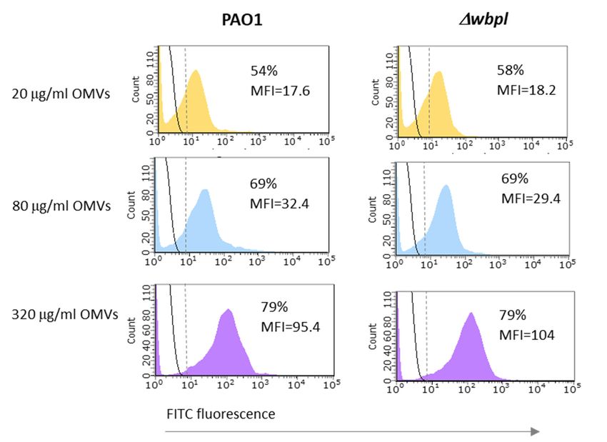
OMVs at a level close to 80%.OMVs. As shown in Figure 4, there were no notable differences in association ef
between PAO1 and the Δwbpl mutant after 3 h of incubation. Although the inten
association of OMVs was generally correlated with the number of vesicles available
at their maximum tested amount (corresponding to a concentration of 320 μg/mL),
Viruses 2022, 14, 121 sociation of the entire bacterial population was not achieved. Both strains
10 of 18similarly
acted with OMVs at a level close to 80%.
Figure 4. The association
Figure 4. of
TheOMVs to P. aeruginosa
association of OMVscells
to P.determined by the
aeruginosa cells flow cytometry
determined technique.
by the flow cytometry tech
The experiment The
was experiment
carried out was carried
on OMVs out onfrom
derived OMVs derived
PAO1. from were
Bacteria PAO1. Bacteria were
incubated incubated with
with FITC-
37 ◦ C with
OMVs for 3 h atOMVs for 3 agitation
h at 37 °C(200
with agitation
rpm). (200 rpm). The
The fluorescence fluorescence
intensities intensities of OMVs-asso
of OMVs-associated
bacteria
bacteria are shown are shown
as coloured as coloured
histograms, histograms,
whereas control whereas control
bacteria by blackbacteria by black
solid lines. solid lines. Calc
Calculated
percentages of OMV-associated cells include cells separated by a vertical dashed line. Data
percentages of OMV-associated cells include cells separated by a vertical dashed line. Data are
pressed as mean fluorescent intensity (MFI) from a representative experiment.
expressed as mean fluorescent intensity (MFI) from a representative experiment.
To the
To verify whether verify whether the
cell-associated cell-associated
vesicles vesiclesoraffect
affect (positively (positively
negatively) or negativel
the phage
adsorption andphage adsorption
finally and finally
the propagation thea propagation
cycle, cycle, agrowth
modified one-step modifiedwasone-step
assessedgrowth w
(Figure 5). As calculated by PFU counting, we could not detect the propagation of KT28the
sessed (Figure 5). As calculated by PFU counting, we could not detect norpropagat
KT28 nor LUZ7 phages on the Δwbpl mutant lacking O-antigens
LUZ7 phages on the ∆wbpl mutant lacking O-antigens when associated with OMVs since it when associated
remained resistant to both phages, indicating a complete lack of sensitization (Figure 5A,C). of sen
OMVs since it remained resistant to both phages, indicating a complete lack
tion (Figure
On the other hand, 5A,C).
verifying On the
a potential other hand,
adverse verifying
influence of OMVs a potential adverse
association, influence of O
we found
association,
that the wild-type we found
PAO1 strain thatsensitive
was still the wild-type PAO1
to KT28 andstrain
LUZ7was still sensitive
phages regardlesstoof
KT28 and
phages
vesicle presence. regardless
The eclipse of vesicle
period presence.
was similar The eclipse period
for OMV-associated was
and similar for OMV-asso
non-associated
bacteria (Figureand non-associated bacteria (Figure 5B,D).
5B,D).
The protection/sensitization potential of OMVs associated with bacteria was further
investigated in the bacteria culture growth kinetics during 24 h of incubation, and indi-
vidual curves were analysed based on the area under the curve (AUC) ± SEM (Figure 6).
Growth curves for PAO1 and separately for the ∆wbpl mutant, after or without association
showed similar characteristics, indicate that association does not interfere with cell mul-
tiplication. Analogous to the previous (one-step growth) experiment, the ∆wbpl mutant
culture was not sensitized to lytic phage infection when associated with OMVs; thus, the
bacteria could propagate freely regardless of KT28 or LUZ7 phages’ presence. On the
contrary, the significant KT28-dependent growth inhibition of PAO1 (AUC = 2.48 ± 0.55 vs.
AUC = 17.79 ± 0.16; p < 0.0005) and PAO1-OMVs (AUC = 4.91 ± 0.56 vs. AUC = 17.93 ± 1.06;
p < 0.01) were demonstrated compared to bacteria cultured without phages. The analogous
results were obtained for LUZ7-dependent growth inhibition of PAO1 (AUC = 2.64 ± 0.32
vs. AUC = 17.14 ± 0.32; p < 0.001) and PAO1-OMVs (AUC = 4.80 ± 0.53 vs. AUC = 17.85
± 1.07; p < 0.005) when bacteria were not treated with phages. These results indicate that
OMVs associated with phage-sensitive bacteria do not constitute a barrier against phage
adsorption and propagation. However, although the PAO1-OMVs culture proved to beViruses 2022, 14, 121 11 of 18
slightly less sensitive to both phages (starting MOI = 1) as compared to the sensitivity of
non-associated PAO1, these differences were not statistically significant. The tendency
of the faster emergence of phage resistant forms in the PAO1-OMVs growth curve might
suggest that associated vesicles even gradually dissolved within dividing cells somehow
affect the whole population, inducing phenotypic resistance to phages, thus indirectly
Viruses 2022, 14, x FOR PEER REVIEW 11 of 18
protecting bacteria from infection.
Figure5.5.The
Figure The OMVs association to P.
OMVs association P. aeruginosa
aeruginosa cells as a potential
potential sensitizer/disturbance
sensitizer/disturbanceagent
agentto
tophage
phageadsorption
adsorption and
andpropagation
propagation evaluated in in
evaluated a modified
a modifiedone-step
one-step growth cycle
growth (MOI
cycle (MOI= 1).= The
1).
The PAO1 wild-type strain was used as the phage-sensitive host, whereas ∆wbpl knock-out mutant
PAO1 wild-type strain was used as the phage-sensitive host, whereas Δwbpl knock-out mutant de-
ficient ininO-antigens
deficient O-antigens served
servedasas
thethe
phage-resistant
phage-resistantstrain. Phage
strain. titretitre
Phage (PFU/mL) of myovirus
(PFU/mL) KT28
of myovirus
(A,B) and podovirus LUZ7 (C,D) were determined every 15 min by the double-agar
KT28 (A,B) and podovirus LUZ7 (C,D) were determined every 15 min by the double-agar layer layer technique.
The curves were established on the average of triplicate PFU/mL measurement from two independ-
technique. The curves were established on the average of triplicate PFU/mL measurement from two
ent experiments ±SEM bars.
independent experiments ± SEM bars.
The protection/sensitization potential of OMVs associated with bacteria was further
investigated in the bacteria culture growth kinetics during 24 h of incubation, and indi-
vidual curves were analysed based on the area under the curve (AUC) ± SEM (Figure 6).
Growth curves for PAO1 and separately for the Δwbpl mutant, after or without association
showed similar characteristics, indicate that association does not interfere with cell multi-
plication. Analogous to the previous (one-step growth) experiment, the Δwbpl mutant cul-
ture was not sensitized to lytic phage infection when associated with OMVs; thus, the
bacteria could propagate freely regardless of KT28 or LUZ7 phages’ presence. On the con-
trary, the significant KT28-dependent growth inhibition of PAO1 (AUC = 2.48 ± 0.55 vs.
AUC = 17.79 ± 0.16; p < 0.0005) and PAO1-OMVs (AUC = 4.91 ± 0.56 vs. AUC = 17.93 ± 1.06;
p < 0.01) were demonstrated compared to bacteria cultured without phages. The analo-
gous results were obtained for LUZ7-dependent growth inhibition of PAO1 (AUC = 2.64
± 0.32 vs. AUC = 17.14 ± 0.32; p < 0.001) and PAO1-OMVs (AUC= 4.80 ± 0.53 vs. AUC =
17.85 ± 1.07; p < 0.005) when bacteria were not treated with phages. These results indicate
that OMVs associated with phage-sensitive bacteria do not constitute a barrier againstof the faster emergence of phage resistant forms in the PAO1-OMVs growth curve
suggest that associated vesicles even gradually dissolved within dividing cells som
Viruses 2022, 14, 121 affect the whole population, inducing phenotypic resistance to phages,
12 of 18thus ind
protecting bacteria from infection.
Figure 6. The OMVs association to P. aeruginosa cells as a potential sensitizer/disturbance agent to
phage adsorption and propagation evaluated in bacterial culture growth kinetics. The PAO1 wild-type
Figure
strain was used as 6. The OMVs association
the phage-sensitive to P.
host, whereas aeruginosa
∆wbpl cellsmutant
knock-out as a potential
deficientsensitizer/disturbance
in O-antigens a
phage adsorption and propagation evaluated in bacterial culture growth kinetics. The PAO1
served as the phage-resistant strain. Bacterial growth was monitored by OD600 measurement using a
type strain was used as the phage-sensitive host, whereas Δwbpl knock-out mutant deficien
microplate reader for 24 h at 30 min intervals. The populations were treated with myovirus KT28
antigens served as the phage-resistant strain. Bacterial growth was monitored by OD600 me
(A,C) or podovirus
ment LUZ7
using(B,D) at MOI =reader
a microplate 1. Thefor
phage-uninfected culture without
24 h at 30 min intervals. the addition
The populations wereoftreated wi
OMVs was considered
ovirusas the control.
KT28 (A,C) orThe curves were
podovirus LUZ7 established on the
(B,D) at MOI = 1.average of two independent
The phage-uninfected culture witho
experiments measured OMVs was±considered
additioninoftriplicates SEM bars.asStatistics were
the control. Theperformed
curves were byestablished
Kruskal–Wallis
on the average
ANOVA rang with independent
pairwise AUCexperiments measured
comparisons acrossingroups:
triplicates
* p ±SEM
< 0.05.bars. Statistics were performed by Kr
Wallis ANOVA rang with pairwise AUC comparisons across groups: * p < 0.05.
4. Discussion
Membrane 4. vesicles
Discussion
(MVs) are an extremely sophisticated bacterial secretion system,
allowing the transport of a widevesicles
Membrane variety(MVs)
of compounds or structures
are an extremely and therefore
sophisticated facilitat-
bacterial secretion sy
ing rapid adaptation
allowing the transport of a wide variety of compounds or structures andortherefore
of the microbial population to changing environmental conditions
protection against adverse
tating factors. Depending
rapid adaptation on theirpopulation
of the microbial content (orto
composition of surface
changing environmental cond
structures), OMVs can associate with the same or another cell of the same population of a
competing species or even with eukaryotic host cells. OMVs contain both surface virulence
factors (e.g., LPS, OMPs, iron and zinc acquisition systems) and specific cargoes, such as
signalling molecules, enzymes, toxins, genetic elements, harmful metabolites, or otherViruses 2022, 14, 121 13 of 18
substances toxic to the bacterial cell. In summary, OMVs can have signalling, nutritional,
offensive, or defensive functions [48].
In this study, we attempted to investigate the overall effect of OMVs on the interaction
dynamics between two LPS-dependent bacteriophages (KT28 and LUZ7) and the P. aerugi-
nosa population in the model of PAO1 strain. Several aspects have been analysed, such as
the OMVs-bacteria interactions, the phage particles neutralization by OMVs, the passive
protection against phage infection, and lastly a possible sensitization of phage resistant
population. Confronting these complex issues, we performed a series of experiments using
both the wild-type PAO1 strain and the ∆wbpl mutant incapable of O-antigen production.
During all research procedures, only naturally produced OMVs by PAO1 (not acquired
by mechanical or enzymatic cell damage) were used to mimic as closely as possible the
common mechanisms occurring in bacterial culture. First, the adsorption ability of KT28
and LUZ7 phages on the surface of OMVs was tested using transmission electron mi-
croscopy and the virion infectivity and neutralization by the incubation of phages with
the vesicles suspension. Next, the ability of P. aeruginosa cells to associate with OMVs was
assessed using flow cytometry. The propagation of LPS-dependent phages on bacteria
associated with OMVs and bacterial growth dynamics in the presence or absence of the
phage were investigated. Finally, to dot the i’s, we tested whether the OMVs derived from
PAO1 wild-type and associated with ∆wbpl PAO1 O-chain-deficient cells would transfer the
receptors for KT28 and LUZ7 phages, enabling virus propagation on the sensitized deletant.
At the outset of designing the experiments for this study, we deliberately abandoned the
dual negative control of using OMVs isolated from the ∆wbpl strain. We considered it
logical that vesicles produced by a strain lacking CPA/OSA could not sequester phage
particles recognising the O-antigen. We found confirmation of our supposition in the work
of Tzipilevich et al. [20], where this type of control was performed.
The results of the OMVs passive protection against phage infection left no illusions
since vesicles protected the PAO1 population against both phages. However, we noted an
interesting effect testing the neutralization of phage particles by OMVs. As a result, we
observed that incubation with vesicles decreases the titre of myovirus KT28 only but did
not affect the podovirus LUZ7 concentration.
The cloud of vesicles surrounding the bacteria is not just a physical veil because,
as a piece of the outer bacterial membrane, it carries the receptors for phages (LPS, pili,
OMPs). This allows phage particles to bind to OMVs surface, which might be followed
by the ejection of viral genetic material into the vesicle interior, thus classified as an
abortive infection (Abi) [19,41,42]. It should be emphasised that the vesicles containing
the components of the bacterial periplasmic space might later be effectively fused with
bacterial cells and potentially transfer phage genes into the new host [48].
The process of binding phages to bacterial surface structures is much more complicated
and can provide permanent or reversible adsorption. Moreover, even if a given phage
recognises and binds to a particular structure (e.g., the O-antigen of LPS), it does not
mean the successful capsid content release or permanent neutralization of the phage.
Firstly, there are different recognition capacities of phage tail spikes/fibres, and secondly,
phages may require additional receptors (e.g., OMPs) for effective infection. In the case
of P. aeruginosa, the O-antigen is produced in two variants as a common polysaccharide
antigen (CPA, A-band), which is formed with repeated rhamnose residues, and as an
O-specific antigen (OSA, B-band), which is composed of a wide variety of sugars [49,50].
These two types of LPS are produced by P. aeruginosa at different intensities depending
on the population lifestyle (planktonic or sessile) [51]. Undoubtedly, it is difficult to say
what ratio of CPA/OSA is present on the surface of the currently produced vesicle, but it
would be relevant to LPS-dependent phages adsorption and eventually their inactivation.
The previous report done on naturally produced OMVs by P. aeruginosa detected only the
anionic OSA but not the neutral CPA presence [52]; thus, the receptor for both phages tested
in our study was available in abundance in OMVs samples. A possibility that podovirus
may be equipped with virion-associated O-chain depolymerase has been also taken intoViruses 2022, 14, 121 14 of 18
consideration as one of the factors differentiating the adsorption mechanism. Although
SGNH hydrolase domain-containing tail fibre protein has been found in the phage LUZ7
genome, the lack of a characteristic halo zone surrounding phage LUZ7 plaques suggested
no active depolymerase presence. The above aspects explain the discrepancies in the
efficiency of phage particles interaction with purified LPS vs. OMVs [42] as well as the
differences in KT28 and LUZ7 phages neutralization by P. aeruginosa OMVs in our studies.
The biogenesis of OMVs has not yet been thoroughly elucidated. Intensive research re-
veals new groups of molecules involved in the formation and release of vesicles, extending
the three main models (related to lipoproteins, peptidoglycan, and LPS) that have been in
existence for some time [53]. So far, we know that the main stimulator of OMVs production
in P. aeruginosa is PQS (Pseudomonas quinolone signal), one of the key molecules of the com-
plex quorum-sensing system of this bacterium. PQS, as a highly hydrophobic molecule, is
transported to the outer membrane, where it changes the conformation of lipid A, resulting
in membrane convexity [48,49,53–58]. In addition to lipid A, O-antigen is also involved in
the formation of OMVs, and its electrical charge is crucial for the physical size and protein
composition of OMVs [52,59]. Murphy et al. demonstrated that in the case of P. aeruginosa,
proteomes of the OMVs produced by CPA-deficient mutants contain high amounts of
proteins related to the transport of small-molecule substances. In contrast, the mutants
possessing only uncharged A-band LPS produce OMVs accumulating proteins related to
transcriptional regulation, adaptation, cell protection, and phage/plasmid/transposon
handling [51,53]. It is therefore clear that the surface of OMVs produced by P. aeruginosa is
heterogeneous in sugar, lipid, and protein content. The complex process of phage adsorp-
tion usually depends on more than one receptor, which may be some OM proteins; thus,
once missing on the vesicle, the phage will not bind to it permanently.
Although we could not experimentally confirm the phenomenon of permanent neu-
tralization of phage LUZ7 by OMVs, nevertheless, microscopic analysis confirmed that
both the LPS-dependent phages tested in this study were adsorbed to vesicles, justifying
the positive role of OMVs in passive protection against phage infection. The differences
in the neutralization process might also depend on the dissimilarity of the DNA ejection
mechanism present in podoviruses (phage LUZ7) vs. myoviruses (phage KT28).
MVs produced by bacteria (both gram-positive and gram-negative) usually contain
receptors for phages. The possibility of transferring these receptors between different
bacterial strains is therefore of great interest, as this could enable the sensitization of strains
originally resistant to phages [60]. Encouraged by the work of Tzipilevich et al. [20], who
demonstrated the existence of such a phenomenon in Bacillus subtilis, we tried to verify if it
is possible to sensitize a CPA/OSA-deficient mutant (∆wbpl PAO1) resistant to tested LPS-
dependent phages by the association with OMVs isolated from a wild-type PAO1 strain.
Documenting a quite effective association of OMVs to ∆wbpl mutant, firstly, we checked the
growth curve of the culture with phage addition, and secondly, we measured the phage titre
during a 90-min incubation with the bacterial cells associated with OMVs. Unfortunately,
none of these experiments was successful since no sensitization was observed in contrast to
previous reports published by other researchers [20]. Apart from the obvious difference in
cell wall structure between gram-negative and gram-positive bacteria as well as completely
different mechanisms of phage infection, the reason for our failure can be attributed to
two main issues. First, unlike in the latter study, we did not co-culture a susceptible
strain with a resistant strain but attempted to sensitise the O-antigen-deficient mutant with
OMVs released from uninfected wild-type PAO1. This is important insofar as naturally
produced vesicles differ in composition from those formed by lysis of phage-sensitive
cells [42,61] or obtained by the French press procedure, as was done by other teams [42].
It is therefore quite possible that commonly formed OMVs did not contain the complete
set of receptors for LPS-dependent phages (LUZ7 and KT28) and might not be able to
transfer the susceptibility patterns to resistant cells. Finally, we cannot exclude the lack of
potential fusion between the donor (OMVs) and the recipient (bacteria) cell membranes.
Interestingly, to our knowledge, none of the published papers documenting a vesicle-Viruses 2022, 14, 121 15 of 18
dependent sensitization has experimentally proven the fusion event between OMVs and
bacterial membranes [19,20,41,42].
The second problem arises from the association potential of vesicles, which can vary
depending on the species of bacteria and the growth phase of the culture [62]. For example,
during the incubation with OMVs (80 µg/mL), Moraxella catarrhalis can associate more than
90% of the OMVs [18], whereas our measurements in P. aeruginosa revealed an association
at the level of ~70%. Thus, even if the transfer of receptors was effective, only a part of the
population received them, and likely, the number of vesicles associated with the OM of
individual bacteria was limited.
Although the idea of OMVs role as the phage receptor vehicle to sensitize resistant
bacteria is very tempting, we should remember that from the co-evolutionary point of
view both bacteria and phages have developed specific strategies/mechanisms to avoid the
susceptibility overload to viral infection in order to protect the resources of both parties.
Considering the future directions, a very interesting issue seems to be the influence
of the prophages on the intensity of secretion and the composition of released OMVs. So
far, we used only the prophage-free P. aeruginosa PAO1 strain. In future studies, we will
certainly use prophage-carrying strains to determine whether a lysogenic strain producing
vesicles can protect strains lacking prophages against phage infection in the superinfection
exclusion (Sie) phenomenon [63].
Supplementary Materials: The following supporting information can be downloaded at:
https://www.mdpi.com/article/10.3390/v14010121/s1, Figure S1: The inhibition of the lytic activity
of myovirus KT28 against P. aeruginosa PAO1 (MOI = 1) in the presence of various concentrations
of OMVs from PAO1: (A) An assay for phage particles neutralization on OMVs surface deter-
mined by PFU/mL counts. The phage-uninfected PAO1 culture without the addition of OMVs was
considered as the control. (B) The OMV-mediated passive protection assay of PAO1 cells against
phages determined by CFU/mL counts. The representative experiments show mean CFU/mL or
PFU/mL ± SEM bars.
Author Contributions: Conceptualization, D.A. and Z.D.-K.; methodology, D.A. and T.O.; formal
analysis, D.A., T.O. and Z.D.-K.; investigation, D.A. and T.O.; resources, Z.D.-K.; data curation, D.A.
and T.O.; writing—original draft preparation, D.A., T.O. and Z.D.-K.; writing—review and editing,
D.A., T.O. and Z.D.-K.; visualization, D.A. and T.O.; supervision, Z.D.-K.; project administration,
Z.D.-K.; funding acquisition, Z.D.-K. All authors have read and agreed to the published version of
the manuscript.
Funding: This study was partially supported by the National Science Centre, Poland (Narodowe
Centrum Nauki), grant number 2016/21/B/NZ6/01157.
Institutional Review Board Statement: Not applicable.
Informed Consent Statement: Not applicable.
Data Availability Statement: Not applicable.
Acknowledgments: The authors thank Roger Sitek, for his assistance with neutralization and pas-
sive protection assays. We thank Sylwia Nowak, Ryszard Adamski, and Marek Chmielewski for
performing TEM analyses and photos. We acknowledge Rob Lavigne from the Department of Biosys-
tems, Laboratory of Gene Technology, KU Leuven, Belgium, for providing the phage LUZ7 for the
experiments. We acknowledge Andrew M. Kropinski for providing the PAO1 ∆wbpl mutant for
the experiments.
Conflicts of Interest: The authors declare no conflict of interest.
References
1. Yu, Y.; Wang, X.; Fan, G.-C. Versatile effects of bacterium-released membrane vesicles on mammalian cells and infec-
tious/inflammatory diseases. Acta Pharmacol. Sin. 2018, 39, 514–533. [CrossRef] [PubMed]
2. Cecil, J.D.; Sirisaengtaksin, N.; O’Brien-Simpson, N.M.; Krachler, A.M. Outer membrane vesicle-host cell interactions. Microbiol.
Spectr. 2019, 7. [CrossRef] [PubMed]Viruses 2022, 14, 121 16 of 18
3. Yonezawa, H.; Osaki, T.; Kurata, S.; Fukuda, M.; Kawakami, H.; Ochiai, K.; Hanawa, T.; Kamiya, S. Outer membrane vesicles of
Helicobacter pylori TK1402 are involved in biofilm formation. BMC Microbiol. 2009, 9, 197. [CrossRef] [PubMed]
4. Baumgarten, T.; Sperling, S.; Seifert, J.; von Bergen, M.; Steiniger, F.; Wick, L.Y.; Heipieper, H.J. Membrane vesicle formation as a
multiple-stress response mechanism enhances Pseudomonas putida DOT-T1E cell surface hydrophobicity and biofilm formation.
Appl. Environ. Microbiol. 2012, 78, 6217–6224. [CrossRef]
5. Metruccio, M.M.E.; Evans, D.J.; Gabriel, M.M.; Kadurugamuwa, J.L.; Fleiszig, S.M.J. Pseudomonas aeruginosa outer membrane
vesicles triggered by human mucosal fluid and lysozyme can prime host tissue surfaces for bacterial adhesion. Front. Microbiol.
2016, 7, 871. [CrossRef] [PubMed]
6. Zhao, K.; Deng, X.; He, C.; Yue, B.; Wu, M. Pseudomonas aeruginosa outer membrane vesicles modulate host immune responses by
targeting the Toll-like receptor 4 signaling pathway. Infect. Immun. 2013, 81, 4509–4518. [CrossRef]
7. Winter, J.; Letley, D.; Rhead, J.; Atherton, J.; Robinson, K. Helicobacter pylori membrane vesicles stimulate innate pro- and
anti-inflammatory responses and induce apoptosis in Jurkat T cells. Infect. Immun. 2014, 82, 1372–1381. [CrossRef]
8. Cecil, J.D.; O’Brien-Simpson, N.M.; Lenzo, J.C.; Holden, J.A.; Chen, Y.-Y.; Singleton, W.; Gause, K.T.; Yan, Y.; Caruso, F.; Reynolds,
E.C. Differential responses of pattern recognition receptors to outer membrane vesicles of three periodontal pathogens. PLoS ONE
2016, 11, e0151967. [CrossRef]
9. Augustyniak, D.; Roszkowiak, J.; Wiśniewska, I.; Skała, J.; Gorczyca, D.; Drulis-Kawa, Z. Neuropeptides SP and CGRP diminish
the Moraxella catarrhalis outer membrane vesicle- (OMV-) triggered inflammatory response of human A549 epithelial cells and
neutrophils. Mediat. Inflamm. 2018, 2018, 4847205. [CrossRef]
10. Perez Vidakovics, M.L.A.; Jendholm, J.; Mörgelin, M.; Månsson, A.; Larsson, C.; Cardell, L.-O.; Riesbeck, K. B Cell activation by
outer membrane vesicles—a novel virulence mechanism. PLoS Pathog. 2010, 6, e1000724. [CrossRef]
11. Koeppen, K.; Hampton, T.H.; Jarek, M.; Scharfe, M.; Gerber, S.A.; Mielcarz, D.W.; Demers, E.G.; Dolben, E.L.; Hammond, J.H.;
Hogan, D.A.; et al. A novel mechanism of host-pathogen interaction through sRNA in bacterial outer membrane vesicles. PLoS
Pathog. 2016, 12, e1005672. [CrossRef] [PubMed]
12. Augustyniak, D.; Seredyński, R.; McClean, S.; Roszkowiak, J.; Roszniowski, B.; Smith, D.L.; Drulis-Kawa, Z.; Mackiewicz, P.
Virulence factors of Moraxella catarrhalis outer membrane vesicles are major targets for cross-reactive antibodies and have adapted
during evolution. Sci. Rep. 2018, 8, 4955. [CrossRef] [PubMed]
13. Kadurugamuwa, J.L.; Beveridge, T.J. Bacteriolytic effect of membrane vesicles from Pseudomonas aeruginosa on other bacteria
including pathogens: Conceptually new antibiotics. J. Bacteriol. 1996, 178, 2767–2774. [CrossRef] [PubMed]
14. Li, Z.; Clarke, A.J.; Beveridge, T.J. Gram-negative bacteria produce membrane vesicles which are capable of killing other bacteria.
J. Bacteriol. 1998, 180, 5478–5483. [CrossRef]
15. Rompikuntal, P.K.; Thay, B.; Khan, M.K.; Alanko, J.; Penttinen, A.-M.; Asikainen, S.; Wai, S.N.; Oscarsson, J. Perinuclear localization
of internalized outer membrane vesicles carrying active cytolethal distending toxin from Aggregatibacter actinomycetemcomitans.
Infect. Immun. 2012, 80, 31–42. [CrossRef]
16. Schaar, V.; Nordström, T.; Mörgelin, M.; Riesbeck, K. Moraxella catarrhalis outer membrane vesicles carry β-lactamase and promote
survival of Streptococcus pneumoniae and Haemophilus influenzae by inactivating amoxicillin. Antimicrob. Agents Chemother. 2011, 55,
3845–3853. [CrossRef]
17. Duperthuy, M.; Sjöström, A.E.; Sabharwal, D.; Damghani, F.; Uhlin, B.E.; Wai, S.N. Role of the Vibrio cholerae matrix protein Bap1
in cross-resistance to antimicrobial peptides. PLoS Pathog. 2013, 9, e1003620. [CrossRef]
18. Roszkowiak, J.; Jajor, P.; Guła, G.; Gubernator, J.; Żak, A.; Drulis-Kawa, Z.; Augustyniak, D. Interspecies outer membrane vesicles
(OMVs) modulate the sensitivity of pathogenic bacteria and pathogenic yeasts to cationic peptides and serum complement. Int. J.
Mol. Sci. 2019, 20, 5577. [CrossRef]
19. Manning, A.J.; Kuehn, M.J. Contribution of bacterial outer membrane vesicles to innate bacterial defense. BMC Microbiol. 2011,
11, 258. [CrossRef]
20. Tzipilevich, E.; Habusha, M.; Ben-Yehuda, S. Acquisition of phage sensitivity by bacteria through exchange of phage receptors.
Cell 2017, 168, 186–199.e12. [CrossRef]
21. Dorotkiewicz-Jach, A.; Augustyniak, D.; Olszak, T.; Drulis-Kawa, Z. Modern therapeutic approaches against Pseudomonas
aeruginosa infections. Curr. Med. Chem. 2015, 22, 1642–1664. [CrossRef]
22. Moradali, M.F.; Ghods, S.; Rehm, B.H.A. Pseudomonas aeruginosa lifestyle: A paradigm for adaptation, survival, and persistence.
Front. Cell. Infect. Microbiol. 2017, 7, 39. [CrossRef]
23. Cullen, L.; Weiser, R.; Olszak, T.; Maldonado, R.; Moreira, A.; Slachmuylders, L.; Brackman, G.; Paunova-Krasteva, T.; Zarnowiec,
P.; Czerwonka, G.; et al. Phenotypic characterization of an international Pseudomonas aeruginosa reference panel: Strains of cystic
fibrosis (CF) origin show less in vivo virulence than non-CF strains. Microbiology 2015, 161, 1961–1977. [CrossRef]
24. Jurado-Martín, I.; Sainz-Mejías, M.; McClean, S. Pseudomonas aeruginosa: An audacious pathogen with an adaptable arsenal of
virulence factors. Int. J. Mol. Sci. 2021, 22, 3128. [CrossRef] [PubMed]
25. Choi, D.-S.; Kim, D.-K.; Choi, S.J.; Lee, J.; Choi, J.-P.; Rho, S.; Park, S.-H.; Kim, Y.-K.; Hwang, D.; Gho, Y.S. Proteomic analysis of
outer membrane vesicles derived from Pseudomonas aeruginosa. Proteomics 2011, 11, 3424–3429. [CrossRef] [PubMed]
26. Cooke, A.C.; Nello, A.V.; Ernst, R.K.; Schertzer, J.W. Analysis of Pseudomonas aeruginosa biofilm membrane vesicles supports
multiple mechanisms of biogenesis. PLoS ONE 2019, 14, e0212275. [CrossRef] [PubMed]You can also read