Perspectives in solubility measurement and interpretation
←
→
Page content transcription
If your browser does not render page correctly, please read the page content below
ADMET & DMPK 7(2) (2019) 88-105; doi: http://dx.doi.org/10.5599/admet.686

                                                                                        Open Access : ISSN : 1848-7718
                                                                      http://www.pub.iapchem.org/ojs/index.php/admet/index
Review

Perspectives in solubility measurement and interpretation
Christel A.S. Bergström1 and Alex Avdeef 2,*
1
    Department of Pharmacy, Uppsala University, BMC P.O. Box 580, SE-751 23 Uppsala, Sweden
2
    in-ADME Research, 1732 First Avenue, #102, New York, NY 10128, USA
*Corresponding Author: E-mail: Alex@in-ADME.com; Tel.: +1-646-678-5713
ORCID ID: Christel A.S. Bergström: 0000-0002-8917-2612; Alex Avdeef: 0000-0002-3139-5442
Received: March 12, 2019; Revised: March 26, 2019; Published: April 05,2019.

       Abstract
        Several key topics in solubility measurement and interpretation are briefly summarized and illustrated with
        case studies drawing on published solubility determinations as a function of pH. Featured are examples of
        ionizable molecules that exhibit solubility-pH curve distortion from that predicted by the traditionally used
        Henderson-Hasselbalch equation and possible interpretations for these distortions are provided. The scope
        is not exhaustive; rather it is focused on detailed descriptions of a few cases. Topics discussed are
        limitations of kinetic solubility, ‘brick-dust and grease-balls,’ applications of simulated and human intestinal
        fluids, supersaturation and the relevance of pre-nucleation clusters and sub-micellar aggregates in the
        formation of solids, drug-buffer/excipient complexation, hydrotropic solubilization, acid-base
        ‘supersolubilization,’ cocrystal route to supersaturation, as well as data quality assessment and solubility
        prediction. The goal is to highlight principles of solution equilibria – graphically more than mathematically
        – that could invite better assay design, to result in improved quality of measurements, and to impart a
        deeper understanding of the underlying solution chemistry in suspensions of drug solids. The value of solid
        state characterizations is stressed but not covered explicitly in this mini-review.

         Keywords
        Solubility-pH; shake-flask solubility; intrinsic solubility; thermodynamic solubility; Henderson-Hasselbalch
        equation; supersaturation; pre-nucleation clusters, drug aggregates; drug salts; pharmaceutical cocrystals

Introduction
   For many oral drugs, the rate-limiting step in intestinal absorption is dictated by the kinetics of
dissolution of the active pharmaceutical ingredient from the solid form. The dissolution rate is a function of
a number of factors, foremost being the aqueous solubility of the drug, which, for ionizable molecules, can
be affected by the pH of the medium [1]. Also, depending on the drug that has been introduced, the
dissolution may overshoot the saturation concentration and produce a supersaturated solution. The
supersaturation is typically achieved when the API is introduced in its amorphous form or in a charged form
as a salt. In such a supersaturated solution, drug molecules may self-associate as sub-micellar
aggregates/clusters, particularly if they are surface-active or polarizable. For bases introduced as drug salts
into an alkaline-pH solution, the charged drug in the supersaturated solution can disproportionate into oil
or undergo precipitation into a new amorphous solid, with which charged water-soluble aggregates may co-
exist. Given enough time, the oil or amorphous solid (and any other type of aggregates) are expected to
undergo phase transformation into a thermodynamically more stable crystalline solid, the steps governed

doi: 10.5599/admet.686                                                                                                     88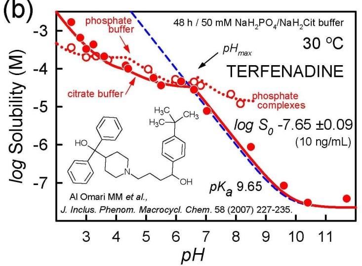
ADMET & DMPK 7(2) (2019) 88-105                         Perspectives in solubility measurements and interpretation

by kinetics of nucleation and crystal growth. For drugs that are practically insoluble in water, dissolution
and re-crystallization can be particularly complicated kinetic processes occurring at different interfaces of,
e.g., undissolved API or colloidal structures composed of excipients. Aggregation and recrystallization are to
date not fully understood and hence may be difficult to predict. About two-thirds of the compounds in the
drug discovery pipeline are very poorly soluble in water, and therefore show low and/or erratic intestinal
absorption. To increase solubility (and thus absorption), oral drugs are often formulated as salts. However,
there are numerous other strategies which have been successfully implemented [2,3].
   Several aspects of the above complexity are summarized in this mini-review. It is intended to serve as
prologue or accompaniment to an upcoming session on solubility at the IAPC-8 meeting in Split, Croatia, 9-
11 September 2019, to be co-chaired by the authors. Since 2009, the International Association of Physical
Chemists (IAPC, www.iapchem.org) series of symposia maintained extensive coverage of the topic of
solubility measurement, both from solid state and solution perspectives. At the 2015 IAPC-4 meeting, the
“Thermodynamic Solubility Measurement of Practically Insoluble Ionizable Drugs - Case Studies & Suggested
Method Improvements” special session resulted in a ‘white paper’ publication drawing on expert consensus
thoughts of researchers from six countries (Hungary, Russia, Serbia, Spain, Sweden, United States) [4]. At
the 2017 IAPC-6 meeting, the “Pharmaceutical Cocrystals -Physicochemical Properties and Formulations”
special session included presentations from internationally-recognized experts in cocrystal solubility-pH
measurement. Several publications on the topic emerged out of the meeting. It is anticipated that future
sessions will continue to cover solubility methods and strategies, to critically address the different needs in
pharmaceutical research, spanning from drug discovery to drug development.

Brief survey of topics in solubility
   Several key topics are briefly summarized below and are illustrated with case studies drawing on
solubility determinations as a function of pH. Significant effort was made to find useful examples from the
published literature which illustrated principles of solution equilibria. The coverage is not exhaustive, but is
considerable in some cases, to address issues of measurement and interpretation of pH dependency in
solubility profiles. The value of solid state characterizations is stressed but not covered explicitly in this
mini-review.

Kinetic or thermodynamic?
   It’s been more than two decades since Lipinski’s Rule of 5 was published [5], addressing the problems of
poorly soluble compounds. High-throughput microtitre plate methods for determining ‘kinetic’ solubility
based on turbidity detection became very popular. Usually, a DMSO solution of the drug is added gradually
to an aqueous buffer, until the first signs of solid formation are detected. The resultant precipitate is most
often amorphous, up to a hundred times more soluble than that measured by conventional
‘thermodynamic’ (slow-to-medium throughput) methods. Pharmaceutical company databases mostly
contain kinetic values. Gradually, it became evident that placing heavy focus on using kinetic data could
drive the lead-to-candidate optimization process in the wrong direction: towards making more amorphous-
tending compounds, generating problems later on in development [6-8]. Limitations of these early methods
can lead to misconceptions and promote the lack of understanding of solubility data [7]. One way to
mitigate some of the development-stage risks is to implement ‘miniaturized’ solubility methods (which can
produce values closer to those of shake-flask thermodynamic measurements), brought forward alongside
potency assessments [9]. This is not necessarily easy to do due to the inherently higher costs of
measurement, particularly when directed at large compound libraries [10]. Also, such implementation
requires some understanding of solubility-pH relationships.

doi: 10.5599/admet.686                                                                                         89
Bergström and Avdeef                                                   ADMET & DMPK 7(2) (2019) 88-105

Brick dust and grease balls
   Low-soluble compounds are often colloquially classified as ‘brick dust’ or ‘grease balls’ [11]. The
solubility of the former type is mainly limited by a strong crystal lattice, making it difficult for the compound
to dissociate from the solid form. Such ‘brick dusts’ are high melting point crystalline solids (Tm > 200 °C) in
which strong intermolecular bonds within the crystal lattice overcome the competing pull of solvation by
water. In contrast, the ‘grease ball’ molecules are limited in their solubility by poor hydration. They typically
have high octanol-water partition coefficients (log P > 3), and are hydrophobic compounds that form
relatively low melting point solids. These compounds are unable to form strong bonds with the water
molecules, and hence exhibit high log P [12]. Contemporary medicinal chemistry synthesizes compounds as
insoluble grease balls rather than brick dust [8,12,13]. Traditional formulation strategies (excipients,
solubilizing agents, cyclodextrins, amorphous dispersions, etc.) are likely to improve oral absorption of
grease balls, more so than that of brick dust [12]. One may also speculate that many of the ‘brick dust’
molecules never reach the development stage because they are too problematic to work with in assays
(e.g., pharmacological potency, toxicity and ADME properties). Hence, they are screened out early during
the discovery stage.

Solubility in simulated and human intestinal fluids
   A drug’s solubility and dissolution behavior can be investigated in human intestinal fluid (HIF), but such
media are difficult to obtain since they require in vivo sampling of the fluid. HIF composition is also highly
variable [14], both intra- and inter-individually, and it is relatively difficult to work with on the lab bench
due to its low buffer capacity. While the variability is important to capture to understand and predict
potential population effects, this complexity also makes it more suitable for later stage studies. The
development of multiple simulated intestinal fluid (SIF) recipes (FaSSIF, FeSSIF, others) applicable to the
early drug development stage has therefore evolved [15]. A recent study by Khadra et al. [16] applied a
mathematically-rigorous design-of-experiment (DOE) statistical technique to investigate how equilibrium
drug solubility can be influenced by seven typical SIF components (sodium taurocholate, lecithin, sodium
phosphate, sodium chloride, pH, pancreatin and sodium oleate) within concentration ranges found in HIF.
Poorly-soluble acids (naproxen, indomethacin, phenytoin, and piroxicam), bases (aprepitant, carvedilol,
zafirlukast, tadalafil), and uncharged drugs (fenofibrate, griseofulvin, felodipine, probucol) were tested.
With the exception of pancreatin, all of the factors individually had a significant influence on equilibrium
solubility. Differences between acids, bases, and neutral drugs were evident; however, the study also
pointed towards (as many other studies also have done) that responses to changes in a solvent, e.g.,
micellar fraction and composition, salt and pH, are often compound specific and there are no generalizing
rules or one-size-fits-all solutions.

Supersaturation, liquid-liquid phase separation, amorphous solid, oil-to-crystal transformations
   Buffers can affect the formation of drug aggregates (i.e., molecular clusters). As an example: citrate,
succinate, phthalate increase surface activity of phenothiazines; acetate decreases it [17]. Certain surface
active bases produce oil when the drug hydrochloride is converted to the free base in situ at pH > pHmax. For
example, a 30 μM chlorpromazine hydrochloride solution at pH 9 becomes supersaturated, which then
partly separates as oil, which is more soluble than the crystalline free base. In contrast, a 2-5 μM alkaline
solution (pH 9-12) prepared from chlorpromazine free base showed no surface activity [17].
    Figure 1 shows log solubility-pH profiles of haloperidol free base at different temperatures and in
different buffer media reported by several groups [18-21]. The dashed curves refer to the profiles
calculated from the simple Henderson-Hasselbalch (HH) equation (not corrected for ionic strength or

90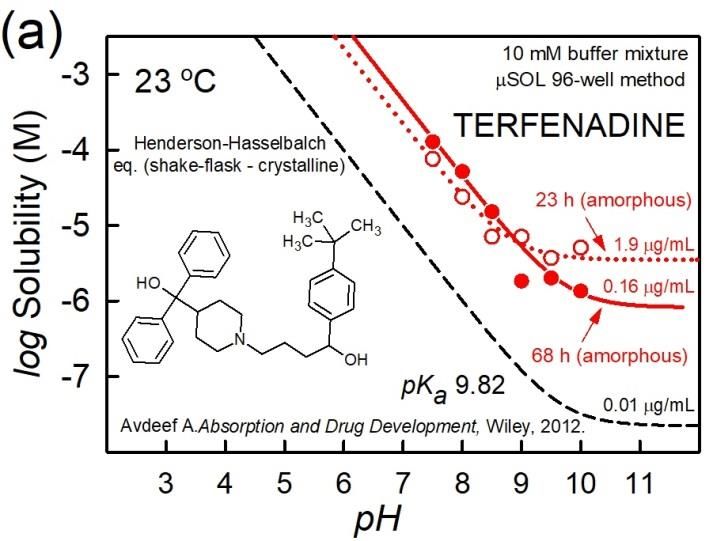
ADMET & DMPK 7(2) (2019) 88-105                             Perspectives in solubility measurements and interpretation

dilution effects), using the independently-determined pKa values appropriate for the temperature. The solid
curves are calculated using the refined constants from fitting the measured log S-pH data (filled circles) to
the proposed equilibrium model, using the pDISOL-X program (in-ADME Research; www.in-
adme.com/pdisol_x.html). S0 refers to the thermodynamic intrinsic solubility in pure water of the
uncharged form of the dissolved substance in equilibrium with the crystalline solid. Its value is indicated by
the lowest value in the dashed curve, in the highly alkaline region. The supersaturation in the pHmax region
noted in the case of chlorpromazine hydrochloride also occurs for the haloperidol mesylate salt (as
illustrated in Figs. 3c,d in Ref. [4]). However, it does not occur for the haloperidol free base. Figures 1a,d do
show deviations from the theoretical dashed curve (HH) in the alkaline region (pH > 7), where the
uncharged species appears to possess elevated solubility.

      Figure 1. Haloperidol (free base) log S-pH profiles, illustrating alkaline-region distortions from the shape
    expected from the Henderson-Hasselbalch equation (dashed blue line). The solid red lines are the best fit to
     the measured data, using the regression analysis program pDISOL-X [22]. See text for further elaboration.
    Several phenomena can account for such a behavior: (i) the neutral (but not the charged) species forms
a supersaturated solution; (ii) stable water-soluble uncharged aggregates of haloperidol form; (iii) the
added crystalline free base, soon after dissolution, re-precipitate into an amorphous solid phase; (iv) liquid-
liquid phase separation (LLPS) takes place, where oil forms as the second liquid phase, in addition to the
aqueous phase; and (v) the free-base (but not the charged-form) drug forms a complex with a component
of the buffer, thus elevating solubility. Overlapping contributions from the above effects may also play a
role.

   Without a specific molecular model for the phenomenon of ‘supersaturation’ (i), it is not clear why the
extent of distortions is different in each of the frames of Figure 1 (and also in Figs. 3b,c,e in Ref. [4]). Case

doi: 10.5599/admet.686                                                                                               91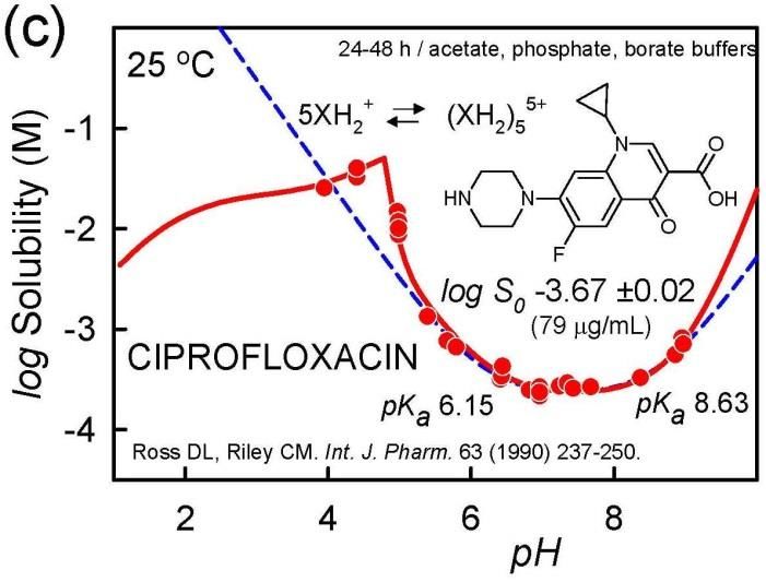
Bergström and Avdeef                                                         ADMET & DMPK 7(2) (2019) 88-105

(ii) – thermodynamically-stable aggregation reaction – is not supported by the data, since the distortion is
different in each of the frames. Cases (iii) and (iv) – amorphous solid and oil, resp. – are possible
explanations for the distortion. Figure 1c, based on a 48-h equilibration time, shows no distortion in the
alkaline region. It could be that the amorphous solid or oil have essentially converted into the
thermodynamically most stable crystalline form by then, but not entirely during the 24-h equilibration in
Figures 1a,d. The extent of distortion in Figure 1a, compared to Figure 1d, could be indicative of a possible
role for drug-phosphate complex formation (v), since 0.15 M phosphate constituted the medium in Figure
1a, but no buffers were in added in Figure 1d.

   It is evident from the above examples that solid state characterization would be needed to help decide
on the most likely mechanism behind the observed distortions in Figure 1. Changing buffer types in the
medium and selecting different equilibration-time approaches [4] would add further insight into the
mechanistic basis for the shape distortions in Figure 1.

    It is worth noting that a comprehensive analysis of each of the log S-pH sets in Figure 1 (e.g., mass action
model using all relevant chemical equilibria [22]) yields very similar log S0 values, showing systematic
temperature dependence. Based on van’t Hoff analysis (e.g., Eq. (3) in Ref. [23]), the intercept value of log
S025 at 25 °C is -5.78 ±0.02 and the slope value (enthalpy of solution), ΔHsolo = 29 ± 6 kJ/mol. Based on the
analysis of ten different published studies, the interlaboratory mean value for haloperidol is log S025= -5.71
± 0.17 (0.73 μg/mL) for haloperidol [24]. The above 0.17 log unit standard deviation is precisely the value
obtained in the averaging of 870 molecules whose solubility was reported in two or more studies, as
described below.

   A common practice in the literature is to report the measured solubility near pH = pKa +/- 2 (for
bases/acids, resp.) as the intrinsic value. The above examples suggests that caution is warranted for such a
procedure: from Figures 1a,c,d, the mean value would be -5.07 ±0.82 (3.2 ±16 μg/mL), which would not
represent the realistic accuracy of the underlying data. A common perception is that reproducibility in
solubility measurements is typically not better than 0.7 log unit [25]. The haloperidol example suggests that
better reproducibility can be achieved if published data are critically analyzed. The diclofenac example
below will further elaborate this.

     Figure 2. Time sequence of liquid-liquid phase boundary transformation to crystalline phase at 0, 12, and 22 h
          [26]. Reprinted with permission from S. Veesler, L. Lafferrère, E. Garcia, C. Hoff, Phase transitions in
        supersaturated drug solution. Org. Proc. Res. Dev. 7 (2003) 983-989. Copyright 2003 American Chemical
                                                        Society.
   The haloperidol discussion suggests that oil or amorphous solid forms in alkaline solutions. Generally, a
supersaturated solution forms on initial dissolution. Such a solution can then spontaneously form two liquid
phases (aqueous and oil), followed soon after by the appearance of crystals, as shown in the time sequence
in Figure 2. Solute is transported from oil drops to crystal via the continuous phase in a solvent-mediated
process in which the drops disappear as the crystal grows. After heating and cooling the crystals, LLPS is not
regenerated, which further confirms that the LLPS is not a thermodynamically stable phase [26,27].

92
ADMET & DMPK 7(2) (2019) 88-105 Perspectives in solubility measurements and interpretation
Amorphous and crystalline solids
High-throughput solubility (HTS) methods, where DMSO solutions of the sample are added to buffer
media, generally indicate a measure of the solubility of amorphous solids, with values greater than that
indicated by crystalline solids. Amorphous phases are not thermodynamically stable and will undergo
crystallization over time. Two amorphous phases may co-exist, or a transient oil phase may appear. An
example of this is shown in Figure 3a for the case of terfenadine measurement using the μSOL method
(Pion Inc.), with 1 %v/v DMSO in each of the 96-well buffer solutions [28]. The unfilled circles represent
solubility values collected at the end of 23-h equilibration, and may suggest the presence of oil plus
amorphous solid. (Alternatively, the ‘oil’ can be a more energetic amorphous solid co-precipitated with a
more stable amorphous form.) The filled circles represent data collected at 68-h equilibration time, and
may suggest the sole presence of the more stable amorphous solid. The apparent solubility at pH 11 is
1.9 μg/mL for the shorter time. This value appears to be an order of magnitude greater than the analyzed
value of 0.16 μg/mL, corresponding to the apparent intrinsic solubility of the more stable amorphous solid.
Solid state characterizations were not reported for the above HTS measurements.
Figure 3. Terfenadine examples of oil/amorphous vs. crystalline phases solubility-pH profiles. (a) High-
throughput measurement [29]. Dotted curve is best-fit of 23-h equilibration data; solid curve is best-fit of 68-
h data. As a visual guide, the dashed curve is the HH curve calculated for crystalline terfenadine, based on the
analysis of the data from Al Omari et al. (2007) [29], shown in the frame on the right. (b) Shake-flask
thermodynamic measurements [29]. See text.
The dashed curve in Figure 3a represents the thermodynamic solubility of the crystalline solid, 0.01
μg/mL, based on the reported citrate-buffer data of Al Omari et al. [29]. The detailed analysis of the latter is
shown in Figure 3b. The authors studied the solubility characteristics in 50 mM phosphate (unfilled circles)
as well as citrate (filled circles) buffer. The 30 °C shake-flask data at 48-h equilibration appears to be of
good quality. The phosphate-containing medium seems to indicate drug-buffer complexation for pH > pHmax
(not seen in the citrate medium), as evidenced by the elevation in solubility above the dashed HH curve (pH
7-8). The analysis suggests the equilibrium constant K = [Tf.TfH+.HPO42-]/[Tf][TfH+][HPO42-] = 10+8.96 M-2 for
the monoanionic complex [24]. Between pHmax 6.7 and pH 8.3, the complex appears to shed one proton
with increasing pH. In both the citrate and phosphate media, at pH below pHmax, two salts form between
the drug and each buffer, consistent with the solid and dotted curves being below the HH dashed curve.
The salt solubilities are quite similar for the citrate and phosphate systems.
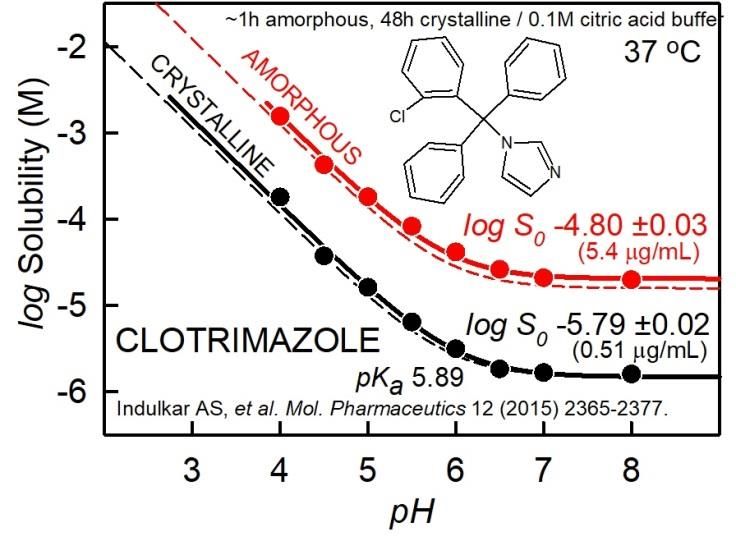
Figure 4 shows a simpler example of a single amorphous-phase log S-pH profile of clotrimazole,
measured at about 1-h equilibration time, compared to the corresponding crystalline phase profile,
measured at 48-h equilibration [30]. In both cases, the curves nearly follow the pH dependence calculated
by the Henderson-Hasselbalch equation (with a constant vertical displacement between the two), although
doi: 10.5599/admet.686 93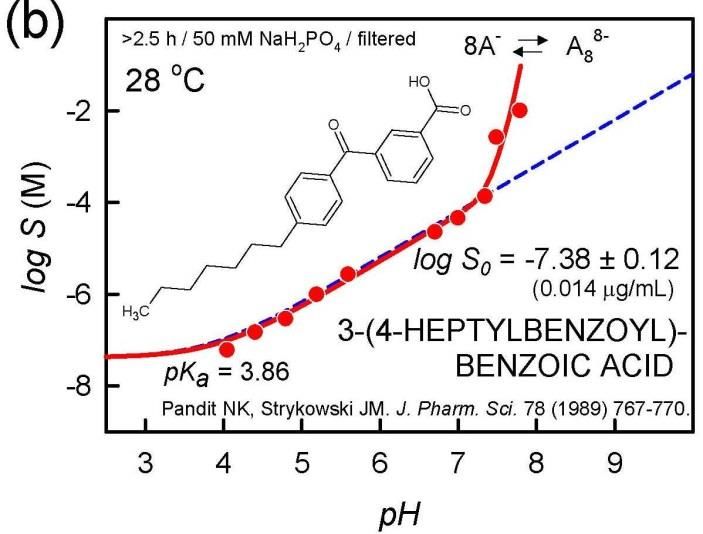
Bergström and Avdeef                                                     ADMET & DMPK 7(2) (2019) 88-105

the amorphous curve shows a slight amount liquid-liquid separation above pH 7, as indicated by a small
difference between the solid and dashed upper curves in neutral solution (which is absent in the lower
crystalline curves).

                                                                          Figure 4. Clotrimazole example of
                                                                           amorphous vs. crystalline phase
                                                                        solubility-pH profiles [30]. See text for
                                                                                        details.

Pre-nucleation clusters (i.e. self-aggregates, sub-micelles, drug-buffer complexes)
Although classical nucleation theory describes drug crystallization from a supersaturated aqueous solution,
the understanding of the molecular basis underlying the nucleation steps is still incomplete [31]. The role of
pre-nucleation clusters as solute precursors to nucleation and the link between the chemical speciation of
homogeneous solutions and the process of phase separation of such clusters was reviewed by Gebauer et
al. [31]. Sub-micellar pre-nucleation clusters form in many supersaturated solutions: calcium phosphate,
amino acids – particularly glycine (see below), and many surface-active drug molecules, such as in
phosphate-buffered solutions of chlorpromazine, acetylpromazine, verapamil, orphenadrine, hydralazine,
pramoxine, and amiodarone, at pH slightly above that of pHmax [21,32]. The buffers in solution critically
influence the stability of such clusters. The link between the clusters and the kinetics of crystallization is still
work in progress, and could turn out to be a vibrant area for future investigations. It should also be noted
that the loss of supersaturation may potentially start at the surface of the dissolving material that is
provided in excess and not always from the supersaturated solution [33].
   Solubility-pH profiles can indicate self-aggregation/cluster formations. In the pH region where the drug
is charged and self-aggregation takes place, the slope in the log S-pH plot can reveal the order of
aggregation [28]. Figure 5 shows three examples indicating formation of charged aggregates. In Figure 5a,
brequinar appears to form a negatively-charged dimer at pH above 7, as indicated by a slope of +2 in the
log S-pH plot; the corresponding sodium-drug salt precipitates above pH 9 [34]. Figure 5b shows the surface
active molecule, 3-(4-heptylbenzoyl)-benzoic acid, apparently forming a negatively-charged octamer above
pH 7, as indicated by the slope of +8 in the plot [35]. The authors proposed that anionic micelles formed,
with a critical micelle concentration of about 0.3 mM. Figure 5c shows ciprofloxacin cation appearing to
form a pentameric aggregate below pH 5 (slope = -5), overlapping with the formation of a phosphate salt at
lower pH [36]. (The possibility of the formation of a soluble drug-phosphate complex near pH 5 has not
been explored.) Most often, when such sub-micellar charged aggregates form, the distortion in the log S-pH
curve is most prominent right below the pHmax for acids and right above the pHmax for bases, as indicated by
the examples in Figure 5.

94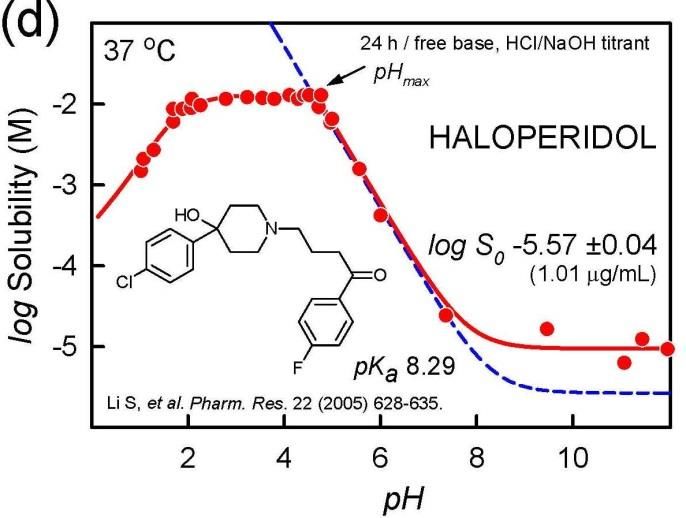
ADMET & DMPK 7(2) (2019) 88-105                        Perspectives in solubility measurements and interpretation

                                                         Figure 5. Examples of charged-species aggregation.
                                                         (a) Brequinar appears to form a negatively-charged
                                                       dimer above pH 7, as indicated by a slope of +2 in the
                                                       log S-pH plot. Sodium-drug salt precipitates above pH
                                                        9 [34]. (b) 3-(4-Heptyl-benzoyl)-benzoic acid appears
                                                          to form an 8-monomer aggregate above pH 7, as
                                                             indicated by the slope of +8 in the plot [35].
                                                       (c) Ciprofloxacin cation appears to form a pentameric
                                                       aggregate below pH 6, as well as a phosphate salt for
                                                          pH < pHmax [36]. The possibility of drug-phosphate
                                                                 complexation has not been ruled out.

    As discussed by Gebauer et al. [31], amino acids, particularly glycine, form sub-micellar pre-nucleation
clusters. Figure 6 displays log S-pH profiles of six amino acids in buffer-free solutions, where pH was
modified with HCl/NaOH [37-39]. Amino acids are highly soluble in water. Consequently, the ionic strength
reaches nearly 5 M at the low-pH end of the profile in three of the cases. The pH < 3 and pH > 9 electrode
readings may be significantly affected. The symmetrically U-shaped dashed curves in the figures were
calculated using the simple HH equation, which does not factor in the effects of ionic strength. The solid
curves, which are less symmetrical than the HH curves, are the results of regression analysis where effects
of ionic strength were factored in [22]. The apparent log S0 values listed in the figures were calculated using
only the data in the pH 4-8 interval, with the rest of the points assigned zero weights during the regression
analysis. It can be seen that for pH < 3 and pH > 9, the measured points are notably below the solid curves.
This is extremely so in the case of glycine in Figure 6b. Ordinarily, such distortion would suggest that the
zwitterionic form of the molecules self-associate, thus raising the solubility [28]. However, it was not
possible to fit the glycine data with an aggregation model that contained a single zwitterionic
stoichiometry. At least several overlapping oligomers appear to be necessary to rationalize the nearly flat
log S-pH profile of polymeric glycine. It may be that glycine forms a high-molecular weight polymer strands
in saturated solutions, which persist across a broad swathe of pH.

Hydrotropic solubilization via cluster formation
   In 1916, Neuberg [40] coined the term ‘hydrotrope’ for compounds that increase the solubility of
sparingly-soluble organic substances, by interacting as ‘co-solubilizers.’ Usually, high concentrations of
hydrotropes are necessary to increase solubility of the sparingly-soluble substance. Hydrotropes are
thought to form aggregates in solution. Typical examples of hydrotropes include urea, nicotinamide,

doi: 10.5599/admet.686                                                                                          95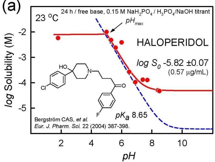
Bergström and Avdeef                                                         ADMET & DMPK 7(2) (2019) 88-105

sodium salicylate, and benzoates with a short alkyl chains, as these molecules can self-associate into small,
sub-micellar clusters of 3-4 molecules [41]. A poorly-soluble molecule can be associated with these clusters
and become solubilized in the process. Figure 7 shows an example of the hydrotropic solubilization of
medazepam by salicylate anions [42]. It would be interesting to explore whether phosphate at high
concentrations acts as a hydrotrope.

     Figure 6. The solubility-pH profiles of amino acids showing departures from the ideal Henderson-Hasselbalch
     shape. The distortion is particularly extreme in the case of glycine. Since the data for pH < 4 and pH > 8 were
      not used in the regression analysis, the listed log S0 values are ‘apparent.’ The actual values may be lower,
                                                   especially for glycine.

                                                                      Figure 7. Hydrotropic solubilization. Plot of
                                                                      the total concentration of medazepam in a
                                                                       saturated solution as a function of added
                                                                         sodium salicylate. The solubility data of
                                                                      Badwan et al. [42] were fit (using pDISOL-X)
                                                                       to an equilibrium model consisting of the
                                                                       drug forming a complex with four units of
                                                                     salicylate. ‘CAC’ is the abbreviation for critical
                                                                                 aggregate concentration.

96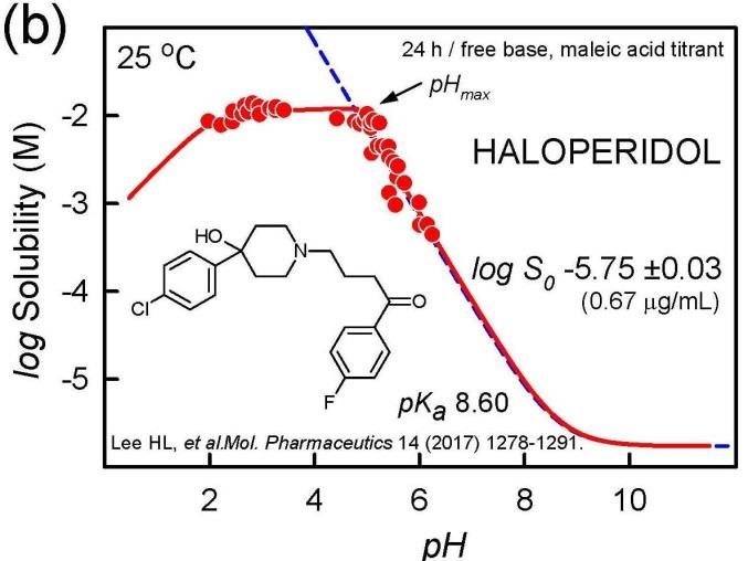
ADMET & DMPK 7(2) (2019) 88-105 Perspectives in solubility measurements and interpretation
Supersolubilization and amorphous dispersions
Parikh and Serajuddin [43] have developed a novel method of preparing solid dispersion by using
acid-base interaction in water. Haloperidol was dissolved in concentrated aqueous solutions of a weak acid
(e.g., malic, tartaric, or citric acid). Each weak acid was selected such that it would not form salts with the
drug. The solubility of haloperidol exceeded 300 mg/g aqueous solution of malic acid. This was an
extremely high aqueous solubility for haloperidol, considering that its intrinsic aqueous solubility in water
as the free base is only 0.7 μg/mL, and the solubility of its salt forms (e.g., HCl, phosphate, maleate) is only
about 1-5 mg/mL. Similar solubility enhancements were observed with itraconazole by the investigators.
The extraordinary enhancement by acid-base interaction has been called ‘supersolubilization.’ On drying,
the drug-containing material is a semisolid and sticky amorphous solid dispersion. Free-flowing powders
that can be compressed into tablets are obtained by e.g., adsorbing drug solid dispersions with acids onto
porous silica [44].
Cocrystals enhancement of solubility
Xanthines (e.g., caffeine, theophylline, theobromine) are known to form complexes with many
pharmaceutical compounds. In a series of papers (1952-1954) Higuchi, Zuck and Lach [45] studied the
xanthine molecular associations formed with simple organic molecules in saturated solutions. Weak
caffeine-containing complexes were determined to form, in the decreasing order of stability: p-
hydroxybenzoic acid > benzocaine > butylparaben > p-aminobenzoic acid > salicylic acid > benzoic acid >
aspirin > o-phthalic acid > benzoate > sulfathiazole > picric acid > sulfadiazine > suberic acid. Such
complexes raised the solubility of the xanthine. These classic studies may bear relevance to the selection of
‘coformers’ in cocrystal formation.
A recent and still evolving formulation strategy to increase intestinal absorption is based on using
cocrystals [46]. Cocrystals are materials composed of two or more different uncharged molecules (e.g.,
drug and ‘coformer’) within the same crystal lattice, which are associated by nonionic and noncovalent
bonds. The altered solid state properties in cocrystals are generally expected to elevate the drug
concentration in a similar way that drug salts do, as the drug is released in a dissolution process. The
initially elevated concentration of the drug could lead to increased oral absorption of the drug. The analyses
of solubility of salts and cocrystals are similar, since both are multi-component solids. Drug salts are
characterized by a solubility product, KspSALT= [drug][counterion], where the drug is charged. Cocrystals are
similarly characterized by an equilibrium solubility product, KspCC = [drug][coformer], but in contrast to salts,
both the drug and the coformer are uncharged in the cocrystal. A method to predict KspCC and the solubility
enhancement of cocrystals, using an approach based on measured drug and coformer intrinsic solubility
(S0drug, S0cof), combined with in silico H-bond descriptors, has been recently described [47,48]. The latter
publications arose out of the IAPC-6 special session on cocrystal solubility.
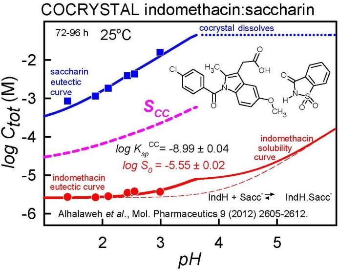
Figure 8 shows an example of a solubility profile as a function of pH for the indomethacin:saccharin
cocrystal [49]. The points represent the measured total concentrations of the coformer saccharin (blue
squares) and the drug indomethacin (red circles) under equilibrium conditions poised at 72-96 h, where
both the cocrystal and the free-acid crystalline drug are indefinitely stable co-precipitates below pH 3.7. As
the cocrystal partially dissolves below pH 3.7, it releases the solvated free drug at the concentration
indicated by the SCC thick dashed line. This is the initial supersaturated concentration of the drug. It may not
persist long, but while the drug is in the elevated concentration, its intestinal absorption may be
significantly enhanced. The Henderson-Hasselbalch curve for indomethacin is indicated by the dashed red
curve in the bottom of the drawing. The equilibrium concentration of indomethacin (solid red curve) is
doi: 10.5599/admet.686 97Bergström and Avdeef ADMET & DMPK 7(2) (2019) 88-105
above the dashed curve, with the difference peaking at about pH 3.7. This can be explained by the
complexation of uncharged drug by the anionic saccharin. The effect diminishes above pH 5, since the
concentration of the free drug diminishes due to ionization [47].
Figure 8. Plot of the total equilibrium
concentrations of saccharin (upper solid line,
blue), indomethacin (lower solid line, red), and
of the maximum possible concentration of the
solubilized indomethacin released from the
cocrystal (thick dashed curve, SCC), as a function
of pH. SCC is termed the ‘cocrystal solubility.’
The elevation of the SCC dashed curve above
that of the indomethacin equilibrium solid red
curve is viewed as the cocrystal ‘solubility
advantage.’ The solubility product is no longer
satisfied above pH 3.7, so the cocrystal no
longer forms above that pH.
Data quality
A comprehensive study of equilibrium solubility data quality based on the review of over 800
publications was reported in 2015 [50]. Since then, more studies have been examined and a more robust
estimate of the true interlaboratory reproducibility can be offered here. Improved estimates [4,50] of
interlaboratory reproducibility are better revealed when solubility data are normalized for pH (to produce
intrinsic solubility, S0, derived from water solubility, Sw, or multiple-pH measurements) and temperature (by
transforming measurements performed in the range 10-50 °C to the standard value of 25 °C [23]).
By way of an addendum to the 2015 study, the data-quality study now has 6355 intrinsic solubility
entries in the Wiki-pS0 database (in-ADME Research), for 3014 different pharmaceutically-relevant
molecules (solids at room temperature), drawing on the review of 1325 publications. Of all the entries,
2144 are singletons, generally reported as Sw, but transformed to S0, and temperature adjusted (if needed)
to 25 °C. These single-source results offer no direct indication of interlaboratory reproducibility. That
indication comes from the 870 molecules for which solubility was reported from at least two different
sources. Some molecules have been studied in surprisingly large number of different laboratories. For
example, there were 34 different reports of the solubility of diclofenac found to date. Seventeen of these
were measured at several different pH values, and are displayed in Figure 9. The next most-frequently
studied molecules appear to be phenytoin, barbital, and ketoprofen with 30, 26, and 24 interlaboratory
determinations, respectively. The average interlaboratory reproducibility, based on the curated 870
replicated studies, has been determined to be 0.17 log unit, significantly lower than the interlaboratory
reproducibility (~0.7 log unit) suggested in past studies [25]. Many factors can lead to the perception of
poor quality of data. Comparing “raw” water solubilities (Sw) determined in unbuffered solutions can be
problematic. For example, using inaccurate values of pKa to interpret the log S-pH data can lead to
erroneous intrinsic solubility values. Ambient levels of carbon dioxide can significantly affect measured Sw
values in the cases of sparingly soluble bases. So can unsuspected buffer-drug interactions,
aggregate/micelle formations, presence of amorphous solids, supersaturated solutions, differences in the
measurement temperature, and other experimental conditions. When an effort is made to critically factor
in the possible sources of systematic error, the interlaboratory reproducibility is actually quite good. Still,
98ADMET & DMPK 7(2) (2019) 88-105 Perspectives in solubility measurements and interpretation
for some drug molecules, the intrinsic solubility is very uncertain, with standard deviation (SD) values
exceeding 0.5 log unit.
Table 1. Averaged intrinsic solubility of 32 drugs at 25 °C with the poorest interlaboratory reproducibility
25 25
log S0 log S0
a b
Compound (M) SD n Compound (M) SD n
Clofazimine -9.05 0.93 5 Amiodarone -10.41 0.59 4
Telmisartan -6.73 0.84 5 Saquinavir -5.92 0.58 3
Buprenorphine -6.07 0.83 3 Quinine -3.06 0.57 7
Mifepristone -5.22 0.75 4 Diflunisal -4.99 0.56 11
Tamoxifen -7.52 0.72 7 Raloxifene -6.82 0.56 6
Terfenadine -7.74 0.71 11 Diphenhydramine -3.21 0.55 4
Sulfadimethoxine -3.74 0.70 3 Etoxadrol -1.96 0.55 3
Curcumin -5.36 0.68 3 Didanosine -1.24 0.54 3
Rifabutin -4.09 0.66 3 Danazol -6.10 0.52 10
Iopanoic_Acid -5.49 0.66 3 Ezetimibe -4.94 0.51 4
Pioglitazone -6.20 0.66 4 Chlorprothixene -5.99 0.51 6
Amodiaquine -5.49 0.65 3 Bromocriptine -5.50 0.51 5
Fentiazac -5.84 0.65 4 Miconazole -5.82 0.50 6
Itraconazole -8.98 0.61 3 Omeprazole -3.70 0.50 3
Procaine -2.30 0.60 3 Amantadine -2.19 0.50 3
Bisoprolol -2.09 0.59 3 Thiabendazole -3.97 0.50 4
a b
Calculated interlaboratory standard deviation. Number of reported studies from different laboratories.
In addressing data quality, it needs to be emphasized that standard conditions are essential: intrinsic
solubility values are to be compared at the designated reference temperature of 25 °C. Mixing different
types of solubility and not normalizing for temperature increase the apparent variance between different
laboratory results, as noted in the haloperidol example (cf., Fig. 1). Although the realistic average
interlaboratory reproducibility is relatively good, some low-soluble molecules are still problematic. Table 1
lists the 32 compounds with the poorest reproducibility, drawing on sources where at least three different
studies were reported. At the top of the list is clofazimine. Based on five reported values, the standard
deviation is 0.93 log unit. Similarly, even though terfenadine solubility has been reported in eleven studies,
its interlaboratory reproducibility at 0.71 is still high. Still, 32 out of 3014 molecules is a small fraction. The
rest of the molecules are much better determined. Absent from the list in Table 1 is diclofenac, because its
intrinsic solubility has a tight consensus value. Its averaged log S025 = -5.34 ±0.18, based on 34 published
studies. The standard deviation is almost identical to the estimated interlaboratory reproducibility of 0.17.
Thus, the diclofenac data from different laboratories offer an opportunity to examine issues that affect the
quality of solubility measurements as a whole. That is, many of the possible challenges in the interpretation
of data in the assessment of interlaboratory reproducibility can be revealed in the examination of
diclofenac.
It is evident that several different patterns of distortion occur in the diclofenac log S-pH profiles in Figure
9 which cannot be predicted by the Henderson-Hasselbalch equation.
One case, in Figure 9g, shows distortion similar to those shown in Figure 5, suggestive of charge-species
aggregation reaction. The data can be rationalized by the formation of the hexameric diclofenac anion
complex associated with three protonated 1-(2-hydroxyethyl)pyrrolidine cations. The points with the
highest solubility (above the solid curve) appear to be indicative of supersaturation, and could not be
explained by a simple equilibrium model.
Distortions involving the free acid are evident in Figures 9f-o, which show elevated solubility in low-pH
region, above that indicated by the dashed HH-calculated curve. These may indicate amorphous phase
solubility (cf., Figure 1a,d). In some cases, the effect may be due to the limit of detection in the analytical
technique used to measure the concentration of the practically-insoluble drug.
doi: 10.5599/admet.686 99Bergström and Avdeef ADMET & DMPK 7(2) (2019) 88-105
Different solubility methods were used to collect the data in the figures. Figure 9a is based on HTS data
(μSOL method) – there are many points in the plot, but they show considerable scatter. The rest of the
examples are based on shake-flask measurement. Also, the temperature is not the same in the figure. Two
of the studies were done at 22 °C, four at 23 °C (‘room temperature’), ten at 25 °C, and one at 37 °C.
Figure 9. The diclofenac log S-pH measurements from 17 publications [28,51-64] reveal a plethora of effects
that might lead to poor reproducibility in the determination of the intrinsic solubility. See text for details.
100ADMET & DMPK 7(2) (2019) 88-105 Perspectives in solubility measurements and interpretation
The rigorous regression analysis of the data in the figures, taking into account all relevant reactions in
the equilibrium model [22], produced the intrinsic solubility values indicated in each frame of Figure 9.
When these values are further transformed to a common reference temperature of 25 °C [23], the
resultant mean value of the 17 cases turns out to be log S025 = -5.30 ±0.17 (1.6 ± 0.6 μg/mL). This mean is
essentially the same as the one obtained from the averaging all 34 cited values, as noted above. The above
standard deviation of the mean is identical to the interlaboratory reproducibility obtained from the analysis
of 870 different molecules, and is quite a bit lower than the frequently cited uncertainty of ~0.7 log unit in
solubility measurement, when comparing results from different laboratories [25].
How does such an impression of poor reproducibility in solubility measurement arise? As mentioned
earlier, it is not uncommon to assume that the solubility 2 pH units below the pKa (for acids) or above the
pKa (for bases) can be equated with the intrinsic solubility. Clearly, such an approximation would not be
accurate in cases of the distortions found in Figures 9f-o (and those in Fig. 1a,d). The consequences of the
above approximation can be illustrated by selecting the lowest solubility in each of the 17 cases and
calculating the average: log S025 (approx.) = -5.01 ±0.59 (6.5 ± 8.5 μg/mL). The increased apparent standard
deviation, 0.59, seems to be consistent with the ~0.7 value.
Prediction of intrinsic solubility
Many computational methods for predicting solubility have been described. These serve the important
need to predict solubility risks during structural design in the early stages of drug discovery. The earlier
methods used relatively small training sets (Bergström and Avdeef ADMET & DMPK 7(2) (2019) 88-105
References
[1] D. Hörter, J.B. Dressman. Influence of physicochemical properties on dissolution of drugs in the
gastrointestinal tract. Adv. Drug Deliv. Rev. 25 (1997) 3-14.
[2] N.G. Solankin, K. Lam, M. Tahsin, S.G. Gumaste, A.V. Shah, A.T.M. Serajuddin. Effects of surfactants
on itraconazole-HPMCAS solid dispersion prepared by hot melt extrusion. I: Miscibility and drug
release. J. Pharm. Sci. (2018) in press. doi: 10.1016/j.xphs.2018.10.058.
[3] C.A.S. Bergström, R. Holm, S.A. Jørgensen, S.B.E. Andersson, P. Artursson, S. Beato, A. Borde, K. Box,
M. Brewster, J. Dressman, K.-I. Feng, G. Halbert, E. Kostewicz, M. McAllister, U. Muenster, J. Thinnes,
R. Taylor, A. Mullertz. Early pharmaceutical profiling to predict oral drug absorption: Current status
and unmet needs. Eur. J. Pharm. Sci. 57 (2014) 173-199.
[4] A. Avdeef, E. Fuguet, A. Llinàs, C. Ràfols, E. Bosch, G. Völgyi, T. Verbić, E. Boldyreva, K. Takács-Novák.
Equilibrium solubility measurement of ionizable drugs – consensus recommendations for improving
data quality. ADMET & DMPK 4 (2016) 117-178.
[5] C.A. Lipinski, F. Lombardo, B.W. Dominy, P.J. Feeney. Experimental and computational approaches to
estimate solubility and permeability in drug discovery and development settings. Adv. Drug Delivery
Rev. 23 (1997) 3-25.
[6] C. Saal, A.C. Petereit. Optimizing solubility: Kinetic versus thermodynamic solubility temptations and
risks. Eur. J. Pharm. Sci. 47 (2012) 589-595.
[7] N.M. Ahmad. Solubility-driven lead optimization: Recent examples and personal perspectives. Bioorg.
Med. Chem. Lett. 26 (2016) 2975-2979.
[8] L. Di, P.V. Fish, T. Mano. Bridging solubility between drug discovery and development. Drug Disc.
Today 17 (2012) 486-495.
[9] T. Sou, C.A.S. Bergström. Automated assays for thermodynamic (equilibrium) solubility
determination. Drug Discov. Today: Technol. 2 (2018) 11-19.
[10] Á. Könczöl, G. Dargó. Brief overview of solubility methods: Recent trends in equilibrium solubility
measurement and predictive models. Drug Discov. Today: Technol. 27 (2018) 3-10.
[11] C.A.S Bergström, W.N. Charman, C.J.H. Porter. Computational prediction of formulation strategies for
beyond-rule-of-5 compounds. Adv. Drug Deliv. Rev. 101 (2016) 6-21.
[12] C.A. Bergström, C.M. Wassvik, K. Johansson, I. Hubatsch. Poorly soluble marketed drugs display
solvation limited solubility. J. Med. Chem. 50 (2007) 5858-5862.
[13] A.P. Hill, R.J. Young. Getting physical in drug discovery: a contemporary perspective on solubility and
hydrophobicity. Drug Disc. Today 15 (2010) 648-655.
[14] D. Riethorst, J. Brouwers, J. Motmans, P. Augustijns. Human intestinal fluid factors affecting intestinal
drug permeation in vitro. Eur. J. Pharm. Sci. 121 (2018) 338-346.
[15] E. Galia, E. Nicolaides, D. Horter, R. Lobenberg, C. Reppas, J.B. Dressman. Evaluation of various
dissolution media for predicting in vivo performance of class I and II drugs. Pharm. Res. 15 (1998)
698-705.
[16] I. Khadra, Z. Zhou, C. Dunn, C.G. Wilson, G. Halbert. Statistical investigation of simulated intestinal
fluid composition on the equilibrium solubility of biopharmaceutics classification system class II
drugs. Eur. J. Pharm. Sci. 67 (2015) 65-75.
[17] G. Zografi, I. Zarenda. The surface activity of phenothiazine derivatives at the air-solution interface.
Biochem. Pharmacol. 15 (1966) 591-598.
[18] H.L. Lee, J.M. Vasoya, M de L. Cirqueira, K.L. Yeh, T. Lee, A.T.M. Serajuddin. Continuous preparation
of 1:1 haloperidol-maleic acid salt by a novel solvent-free method using a twin screw melt extruder.
Mol. Pharmaceutics 14 (2017) 1278-1291.
[19] S. Li, S. Wong, S. Sethia, H. Almoazen, Y.M. Joshi, A.T.M. Serajuddin. Investigation of solubility and
dissolution of a free base and two different salt forms as a function of pH. Pharm. Res. 22 (2005) 628-
635.
102ADMET & DMPK 7(2) (2019) 88-105 Perspectives in solubility measurements and interpretation
[20] M.M. Al Omari, M.B. Zughul, J.E.D. Davies, A.A. Badwan. A Study of haloperidol inclusion complexes
with β-cyclodextrin using phase solubility, NMR spectroscopy and molecular modeling techniques. J.
Solution. Chem. 38 (2009) 669-683.
[21] C.A.S. Bergström, K. Luthman, P. Artursson. Accuracy of calculated pH-dependent aqueous drug
solubility. Eur. J. Pharm. Sci. 22 (2004) 387-398.
[22] G. Völgyi, A. Marosi, K. Takács-Novák, A. Avdeef. Salt solubility products of diprenorphine
hydrochloride, codeine and lidocaine hydrochlorides and phosphates – Novel method of data
analysis not dependent on explicit solubility equations. ADMET & DMPK 1 (2013) 48-62.
[23] A. Avdeef. Solubility temperature dependence predicted from 2D structure. ADMET & DMPK 3 (2015)
298-344.
[24] A. Avdeef. unpublished results (2018).
[25] D.S. Palmer, J.B.O. Mitchell. Is experimental data quality the limiting factor in predicting the aqueous
solubility of druglike molecules?. Mol. Pharmaceutics 11 (2014) 2962-2972.
[26] S. Veesler, L. Lafferrère, E. Garcia, C. Hoff. Phase transitions in supersaturated drug solution. Org.
Proc. Res. Dev. 7 (2003) 983-989.
[27] P.E. Bonnett, K.J. Carpenter, S. Dawson, R.J. Davey. Solution crystallization via a submerged liquid–
liquid phase boundary: oiling out. Chem. Commun. (2003) 698–699.
[28] A. Avdeef. Absorption and Drug Development Second Edition, Wiley-Interscience, Hoboken NJ, 2012.
[29] M.M. Al Omari, M.B. Zughul, J.E.D. Davies, A.A. Badwan. Effect of buffer species on the complexation
of basic drug terfenadine with β-cyclodextrin. J. Inclus. Phenom. Macrocycl. Chem. 58 (2007) 227-235.
[30] A.S. Indulkar, K.J. Box, R. Taylor, R. Ruiz, L.S. Taylor. pH-Dependent liquid−liquid phase separation of
highly supersaturated solutions of weakly basic drugs. Mol. Pharmaceutics 12 (2015) 2365-2377.
[31] D. Gebauer, M. Kellermeier, J.D. Gale, L. Bergström, H. Cölfen. Pre-nucleation clusters as solute
precursors in crystallization. Chem. Soc. Rev., 43 (2014) 2348-2371.
[32] A. Pobudkowska, C. Ràfols, X. Subirats, E. Bosch, A. Avdeef. Phenothiazines solution complexity –
determination of pKa and solubility-pH profiles exhibiting sub-micellar aggregation at 25 and 37 °C.
Eur. J. Pharm. Sci. 93 (2016) 163-176.
[33] K. Edueng, D. Mahlin, P. Larsson, C.A.S. Bergström. Mechanism-based selection of stabilisation
strategy for amorphous formulations: Insights into crystallization pathways. J. Control. Rel. 256
(2017) 193-202.
[34] S.-Y. P. King, A.M. Basista, G. Oros. Self-association and solubility behaviors of a novel anticancer
agent, brequinar sodium. J. Pharm. Sci. 78 (1989) 95-100.
[35] N.K. Pandit, J.M. Strykowski. Effect of temperature on the solubility of a surface active carboxylic
acid. J. Pharm. Sci. 78 (1989) 767-770.
[36] D.L. Ross, C.M. Riley. Aqueous solubilities of some variously substituted quinolone antimicrobials. Int.
J. Pharm. 63 (1990) 237-250.
[37] H.-C. Tseng, C.-Y. Lee, W.-L. Weng, I.-M. Shiah. Solubilities of amino acids in water at various pH
values under 298.15 K. Fluid Phase Equilib. 285 (2009) 90-95.
[38] X. Yang, X. Wang, C.B. Ching. Solubility of form alpha and form gamma of glycine in aqueous
solutions. J. Chem. Eng. Data 53 (2008) 1133-1137.
[39] T.E. Jr. Needham, A.N. Paruta, R.J. Gerraughty. Solubility of amino acids in pure solvent systems. J.
Pharm. Sci. 60 (1971) 565-567.
[40] C. Neuberg. Hydrotropic phenomena. Biochem. Z. 76 (1916) 107-176.
[41] W. Kunz, K. Holmberg, T. Zemb. Hydrotropes. Curr. Opin. Coll. Interf. Sci. 22 (2016) 99-107.
[42] A.A. Badwan, L.K. El-Khordagui, A.M. Saleh, S.A. Khalil. The solubility of benzodiazepines in sodium
salicylate solution and a proposed mechanism for hydrotropic solubilization. Int. J. Pharm. 13 (1983)
67-74.
doi: 10.5599/admet.686 103Bergström and Avdeef ADMET & DMPK 7(2) (2019) 88-105
[43] T. Parikh, A.T.M. Serajuddin. Development of fast-dissolving amorphous solid dispersion of
itraconazole by melt extrusion of its mixture with weak organic carboxylic acid and polymer. Pharm.
Res. 35 (2018) 127. In press. doi: 10.1007/s11095-018-2407-4.
[44] A. Shah, A.T.M. Serajuddin. Conversion of solid dispersion prepared by acid–base interaction into
free-flowing and tabletable powder by using Neusilin® US2. Int. J. Pharm. 484 (2015) 172-180.
[45] T. Higuchi, J.L. Lach. Investigation of some complexes formed in solution by caffeine VI. Comparison
of complexing behaviors of methylated xanthines with p-aminobenzoic acid, salicylic acid,
acetylsalicylic acid, and p-hydroxybenzoic acid. J. Amer. Pharm. Assoc. 43 (1954) 527-530.
[46] S.L. Childs, N. Rodríguez-Hornedo, L.S. Reddy, A. Jayasankar, C. Maheshwari, L. McCausland, R.
Shipplett, B.C. Stahly. Screening strategies based on solubility and solution composition generate
pharmaceutically acceptable cocrystals of carbamazepine. Cryst. Eng. Comm. 10 (2008) 856-864.
[47] A. Avdeef. Cocrystal solubility product analysis – Dual concentration-pH mass action model not
dependent on explicit solubility equations. Eur. J. Pharm. Sci. 110 (2017) 2-18.
[48] A. Avdeef. Cocrystal solubility product prediction using an in combo model and simulations to
improve design of experiments. Pharm. Res. (2018) 35:40. https://doi.org/10.1007/s11095-018-2343-
3.
[49] A. Alhalaweh, L. Roy, N. Rodríguez-Hornedo, S.P. Velaga. pH-dependent solubility of indomethacin-
saccharin and carbamazepine-saccharin cocrystals in aqueous media. Mol. Pharmaceutics 9 (2012)
2605-2612.
[50] A. Avdeef. Suggested improvements for measurement of equilibrium solubility-pH of ionizable drugs.
ADMET & DMPK 3 (2015) 84-109.
[51] B. Bard, S. Martel, P.-A. Carrupt. High throughput UV method for the estimation of thermodynamic
solubility and the determination of the solubility in biorelevant media. Eur. J. Pharm. Sci. 33 (2008)
230-240.
[52] A. Chiarini, A. Tartarini, A. Fini. pH-Solubility relationship and partition coefficients for some
antiinflammatory arylaliphatic acids. Arch. Pharm. (Weinheim) 317 (1984) 268-273.
[53] A. Avdeef, C.M. Berger, C. Brownell. pH-metric solubility. 2. Correlation between the acid-base
titration and the saturation shake-flask solubility-pH methods. Pharm. Res. 17 (2000) 85-89.
[54] A. Fini, G. Fazio, G. Feroci, Solubility and solubilization properties of non-steroidal antiinflammatory
drugs. Int. J. Pharm. 126 (1995) 95-102.
[55] M.T. Ledwidge, O.I. Corrigan. Effects of surface active characteristics and solid state forms on the pH
solubility profiles of drug-salt systems. Int. J. Pharm. 174 (1998) 187-200.
[56] P. Luger, K. Daneck, W. Engel, G. Trummlitz. K. Wagner. Structure and physicochemical properties of
meloxicam, a new NSAID. Eur. J. Pharm. Sci. 4 (1996) 175-187.
[57] L. Zhou, L. Yang, S. Tilton, J. Wang. Development of a high throughput equilibrium solubility assay
using miniaturized shake-flask method in early drug discovery. J. Pharm. Sci. 96 (2007) 3052-3071.
[58] M.L. Manca, M. Zaru, G. Ennas, D. Valenti, C. Sinico, G. Loy, A.M. Fadda. Diclofenac-beta-cyclodextrin
binary systems: physicochemical characterization and in vitro dissolution and diffusion studies. AAPS
PharmSciTech 6 (2005) E464-E472.
[59] B. Chuasuwan, V. Binjesoh, J.E. Polli, H. Zhang, G.L. Amidon, H.E. Junginger, K.K. Midha, V.P. Shah, S.
Stavchansky, J.B. Dressman, D.M. Barends. Biowaiver monographs for immediate release solid oral
dosage forms: diclofenac sodium and diclofenac potassium. J. Pharm. Sci. 98 (2009) 1206-1219.
[60] C.D. Herzfeldt, R. Kuemmel. Dissociation constants, solubilities and dissolution rates of some selected
nonsteroidal antiinflammatories. Drug Dev. Ind. Pharm. 9 (1983) 767-793.
[61] Y. Maitani, M. Nakagaki, T. Nagai. Determination of the acid dissociation constants in ethanol-water
mixtures and partition coefficients for diclofenac. Int. J. Pharm. 74 (1991) 105-116.
[62] A. Pobudkowska, U. Domańska, Study of pH-dependent drugs solubility in water. Chem. Ind. Chem.
Eng. Quart. 20 (2014) 115-126.
104ADMET & DMPK 7(2) (2019) 88-105 Perspectives in solubility measurements and interpretation
[63] W. Riess, H. Stierlin, P. Degen, J.W. Faigle, A. Gerardin, J. Moppert, W. Theobald, J. Wagner.
Pharmacokinetics and metabolism of the anti-inflammatory agent Voltaren. Scand. J. Rheumato.
Suppl. 22 (1978) 17-29.
[64] G. Völgyi, D. Csicsák, K. Takács-Novák. Right filter-selection for phase separation in equilibrium
solubility measurement. Eur. J. Pharm. Sci. 123 (2018) 98-105.
[65] A. Llinàs, R.C. Glen, J.M. Goodman, Solubility challenge: can you predict solubilities of 32 molecules
using a database of 100 reliable measurements?. J. Chem. Inf. Model. 48 (2008) 1289-1303.
[66] A.J. Hopfinger, E.X. Esposito, A. Llinàs, R.C. Glen, J.M. Goodman. Findings of the challenge to predict
aqueous solubility. J. Chem. Inf. Model. 49 (2009) 1-5.
[67] J.H. Fagerberg, E. Karlsson, J. Ulander, G. Hanisch, C.A.S. Bergström. Computational prediction of
drug solubility in fasted simulated and aspirated human intestinal fluid. Pharm. Res. 32 (2015) 578-
589.
©2019 by the authors; licensee IAPC, Zagreb, Croatia. This article is an open-access article distributed under the terms and
conditions of the Creative Commons Attribution license (http://creativecommons.org/licenses/by/3.0/)
doi: 10.5599/admet.686 105You can also read