Review Article Hypomagnesemia in the Cancer Patient - Kidney360
←
→
Page content transcription
If your browser does not render page correctly, please read the page content below
Review Article

  Hypomagnesemia in the Cancer Patient
  Biruh T. Workeneh,1 Nupur N. Uppal,2 Kenar D. Jhaveri              ,2 and Helbert Rondon-Berrios3

  Abstract
  Hypomagnesemia is a common medical problem that contributes to the morbidity and mortality of patients with
  cancer. This review summarizes magnesium physiology and highlights the mechanisms underlying magnesium
  disturbances due to cancer and cancer treatment. The causes of hypomagnesemia can be categorized according to
  the pathophysiologic mechanism: decreased intake, transcellular shift, gastrointestinal losses, and kidney losses.
  Patients with cancer are at risk for opportunistic infections, frequently experience cardiovascular complications,
  and often receive classes of medications that cause or exacerbate hypomagnesemia. Also, cancer-specific
  therapies are responsible for hypomagnesemia, including platinum-based chemotherapy, anti-EGF receptor mAbs,
  human EGF receptor-2 target inhibitors (HER2), and calcineurin inhibitors. Urinary indices, such as the fractional
  excretion of magnesium, can provide useful information about the etiology. The management of hypomagnesemia
  depends on the magnitude of hypomagnesemia and the underlying cause. We recommended checking serum
  magnesium at the beginning of treatment and as part of routine monitoring throughout cancer treatment.
  Opportunities exist for potential research and practice improvement, including further characterization of
  hypomagnesemia regarding the clinical effect on cancer outcomes, preventing hypomagnesemia in patients
  receiving high-risk anticancer agents, and developing effective therapeutic strategies.
                                     KIDNEY360 2: 154–166, 2021. doi: https://doi.org/10.34067/KID.0005622020

  Introduction                                                        or critically ill are at enhanced risk for hypomagnese-
  Hypomagnesemia is defined as a serum magnesium                       mia, occurring in up to 50%–60% of patients (3). Cancer
  (Mg) concentration of ,1.8 mg/dl (1). It is essential to            often results in compromised immunity, predomi-
  anticipate, identify, and treat hypomagnesemia in                   nantly liquid tumors, or those that affect hematopoi-
  patients with cancer. This review summarizes the mech-              esis. Furthermore, most cancer therapies compromise
  anisms underlying the disturbances of Mg deficiency                  the immune system, and patients are at exceptionally
  due to cancer and during cancer treatment. Although                 high risk for opportunistic infections. The administra-
  Mg is frequently referred to as the “forgotten ion,” it is          tion of antibiotics and antiviral drugs contributes to
  the second most abundant intracellular cation (after                hypomagnesemia (4). Cardiovascular and kidney re-
  potassium [K]), acts as a cofactor for hundreds of en-              lated complications resulting from cancer and cancer
  zymatic reactions, and has structural functions for both            therapy frequently require the use of cardiovascular
  proteins and nucleic acids (2). The range of symptoms               medications that can contribute to hypomagnesemia.
  associated with hypomagnesemia is consequently ex-                  Traditional chemotherapeutic agents cause hypomag-
  pansive; patients can be asymptomatic and exhibit                   nesemia that can persist for months to years after
  nonspecific symptoms (such as anorexia, nausea, and                  cessation of cancer therapy (5). Patients who survived
  fatigue) and severe symptoms (such as tetany, seizures,             childhood cancer are at a particular risk of developing
  and lethal arrhythmias) (2). Hypomagnesemia is graded               adverse effects caused by multimodal treatment for
  on the basis of serum concentration (Table 1) and the               their malignancy (6). Studies that have assessed hypo-
  degree of hypomagnesemia is generally correlated with               magnesemia in patients who have survived cancer
  adverse outcomes. However, it is important to note that             determined a prevalence ranging between 13% and
  clinically significant adverse effects and outcomes can              29% (7–10).
  occur with any degree of hypomagnesemia. This review                   Hypomagnesemia, in both acute and chronic forms,
  will summarize Mg’s physiology and review cancer-                   is associated with poor clinical outcomes. Chronic
  related causes of hypomagnesemia.                                   hypomagnesemia is implicated in developing insulin
                                                                      resistance, diabetes, more rapid progression of diabetic
                                                                      nephropathy, nephrolithiasis, fracture, and increased
  Epidemiology and Clinical Outcomes                                  risk for cancer (11–13). Chronic hypomagnesemia has
    In general, hypomagnesemia frequently develops in                 also been implicated in cancer development, possibly
  patients with cancer, and patients who are hospitalized             in relation to the induction of chronic inflammation
  1
    Section of Nephrology, The University of Texas MD Anderson Cancer Center, Houston, Texas
  2
    Division of Kidney Diseases and Hypertension, Donald and Barbara Zucker School of Medicine at Hofstra/Northwell, Northwell Health,
  Great Neck, New York
  3
    Renal-Electrolyte Division, University of Pittsburgh School of Medicine, Pittsburgh, Pennsylvania

  Correspondence: Dr. Kenar D. Jhaveri, Division of Kidney Diseases and Hypertension, Donald and Barbara Zucker School of Medicine at
  Hofstra/Northwell, 100 Community Drive, Great Neck, NY 11021, or Dr. Helbert Rondon-Berrios, Renal-Electrolyte Division, University of
  Pittsburgh School of Medicine, 3550 Terrace St., A915 Scaife Hall, Pittsburgh, PA 15261. E-mail: kjhaveri@northwell.edu or
  rondonberriosh@upmc.edu

154    Copyright © 2021 by the American Society of Nephrology                                                              www.kidney360.org Vol 2 January, 2021
KIDNEY360 2: 154–166, January, 2021 Hypomagnesemia of Malignancy, Workeneh et al. 155
Table 1. Grades of hypomagnesemia according to common terminology criteria used by cancer societies for adverse events reported,
version 4.0
Grade Serum Magnesium (mg/dl) Clinical Significance
1 1.2–1.7 Mild or no symptoms, fatigue
2 0.9–1.2 Muscle weakness, fasciculations
3 0.7–0.9 Neurologic deficits, atrial fibrillation
4 ,0.7 Psychosis, seizures, tetany, nystagmus, lethal arrhythmia
(14). Hypomagnesemia can result in higher viral titers of process and accounts for 10% of Mg reabsorption. Trans-
patients infected with Epstein–Barr virus, which raises the cellular Mg transport requires the activity of transient re-
risk for lymphomas and other malignancies (15). Preclinical ceptor potential melastatin 6 (TRPM6) and 7 (TRPM7) Mg
data support hypomagnesemia as a contributing factor transporters in the enterocyte apical membrane.
to metastatic disease (16), and studies in patients with
cancer show hypomagnesemia is associated with worse
outcomes (17). Kidney Handling of Mg
In the kidney, nonprotein-bound Mg is freely filtered
across the glomerulus. The proximal tubule (PT) reabsorbs
Physiology of Mg Homeostasis 15% of filtered Mg through a paracellular mechanism
The physiology of Mg regulation is complex, and the (23,24). Water reabsorption along the early parts of the
dysregulation of Mg homeostasis is common in patients PT increases Mg concentration in the tubular lumen, creat-
with cancer and results in frequent complications. Under- ing a favorable gradient for Mg reabsorption in the distal
standing the physiology discussed in this section will clarify section of the PT. Solvent drag also contributes to Mg
the effect of some of the cancer-specific and targeted ther- reabsorption in this segment of the nephron.
apies we will discuss in this review. Extracellular-fluid volume expansion results in decreased
Mg reabsorption along with the PT (25).
Mg Distribution Unlike most solutes, most Mg reabsorption occurs in the
Total body Mg is close to 24 g for an average adult, and cortical thick ascending limb of the loop of Henle (TAL)
99% of total body Mg is located in the intracellular fluid rather than the PT. In the TAL, approximately 70% of the
compartment (bone, muscle, and soft tissues), leaving 1% filtered Mg is reabsorbed, mainly through the paracellular
present in the extracellular fluid compartment. Nearly 30% route (Figure 1). Claudins 16 and 19 are considered the main
of the total plasma Mg is bound to proteins, mainly albumin. claudins responsible for Mg permeability through the para-
The remaining 70% is available for glomerular filtration, cellular route (26). Claudin 14 interacts with claudin 16 in
either as the Mg cation complexed to anions—including the TAL and decreases the cation selectivity of the claudin
oxalate (10%), phosphate, and citrate—or as ionized Mg 16–19 complexes (27). Claudin 10 has also been identified as
(60%). a vital constituent in cation selectivity in the TAL, as dem-
onstrated in claudin 10–knockout mice which demonstrated
hypermagnesemia, nephrocalcinosis, and impaired paracel-
Mg Balance lular sodium (Na) permeability (28). In the absence of clau-
Mg is a micronutrient mostly derived from nuts din 10, TAL tight junctions become more permeable to
(almonds), green vegetables, cereal, and milk. The intestines calcium and Mg. The driving force for paracellular Mg
absorb 120 mg/d (30%–50%) of Mg and secrete 20 mg/d in reabsorption, along with calcium and Na, is the lumen-
bile and pancreatic and intestinal juices, representing a net positive transepithelial voltage of the TAL determined by
absorption of 100 mg/d (18). Under conditions of Mg de- the activity of the Na-K–2 chloride (Cl) cotransporter
ficiency, the intestines can absorb up to 80% of dietary Mg (NKCC2) and the associated K recycling via the renal outer
(19). The kidneys filter approximately 2400 mg/d of Mg, of medullary K (ROMK) channel (29). Cl ions leave the TAL
which they reabsorb 2300 mg/d (90%–95%) for a net ex- cells via ClC-Kb channels on the basolateral membrane.
cretion of 100 mg/d. In the setting of Mg deficiency, net Mutations in the genes encoding for NKCC2, ROMK,
kidney Mg excretion is reduced to ,12 mg/d (20). ClC-Kb, and Barttin (a subunit of ClC-Kb) lead to Bartter
syndromes type 1, 2, 3, and 4, respectively, all of which are
Gastrointestinal Absorption associated with various degrees of hypomagnesemia. Com-
Mg absorption in the gut occurs via two routes: a satura- pensation for reduced TAL Mg reabsorption may occur in
ble, paracellular route and a nonsaturable, transcellular the distal convoluted tubule (DCT), which explains why
route (21). The paracellular route is a passive mechanism patients with Bartter syndrome often have normal Mg lev-
and accounts for the bulk (90%) of total Mg reabsorption. els. ClC-Kb and Barttin are also expressed in DCT, and
The paracellular route is modulated by tight junction pro- patients with mutations in these genes frequently exhibit
teins called claudins. Claudins 2, 7, and 12 are expressed in hypomagnesemia. Activation of the calcium-sensing recep-
the intestines and might facilitate Mg reabsorption (22). The tor (CaSR) in the TAL by calcium or Mg inhibits paracellular
final segment for Mg reabsorption occurs in the cecum and Mg transport via inhibition of NKCC2 and ROMK (30).
colon using the transcellular route, which is an active CaSR also regulates claudin 14 expression and calcium156 KIDNEY360
15%
Anti-EGFR monoclonal antibodies
EGFR tyrosine kinase inhibitors
HER-2 inhibitors
Thiazide diuretics
Calcineurin inhibitors
mTOR inhibitors
Loop diuretics Pro-EGF
70% Na+
NCC EGFR
EGF
Cl- Mg2+
SLC41A3
TRPM6 Na+
Mg2+ CNNM2
Mg2+
+ -
NKCC2 Kiv1.1 3Na+
Na+ 3Na+
K+ 2K+
K+ 2K+ FXYD2
PCBD1
Cl- HNF1B Kir4.1
CLC-Kb
Cl- K+
ROMK
LUMEN BLOOD
K+ Barttin
+ -
CaSR Aminoglycosides
Claudin 16 Claudin 14
Ca2+ Mg2+
Claudin 19
LUMEN BLOOD
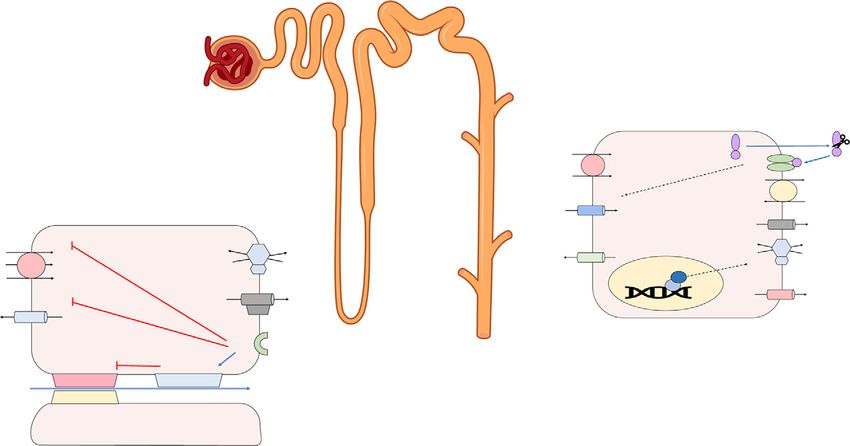
Figure 1. | Kidney handling of magnesium (Mg) and nephron site of action of magnesiuric drugs. Numbers in blue (%) refers to the percent Mg
that is reabsorbed in the specific segment of the nephron. Nonprotein-bound Mg is freely filtered across the glomerulus. The proximal tubule
(PT) reabsorbs 15% of filtered magnesium via a paracellular mechanism. The bulk of Mg reabsorption occurs in the cortical thick ascending limb
of the loop of Henle (TAL), where approximately 70% of Mg is reabsorbed via a paracellular route. Claudins 16 and 19 are considered the main
claudins responsible for the Mg permeability through the paracellular route. Claudin 14 may interact with claudin 16 in TAL and decreases the
cation selectivity of the claudin-16 and -19 complexes. Recently, claudin 10 has also been identified as an important factor in cation selectivity
in TAL. The driving force for paracellular Mg reabsorption is the lumen-positive transepithelial voltage of the TAL, which is determined by the
activity of NKCC2 and the subsequent potassium recycling back into the lumen via the ROMK channel at the apical membrane. Cl ions leave the
TAL cells via ClC-Kb channels on the basolateral membrane. Activation of the CaSR in the TAL inhibits paracellular Mg transport via inhibition of
NKCC2 and ROMK. Further, CaSR regulates claudin-14 expression and calcium and Mg reabsorption in the TAL. Aminoglycosides target the
CaSR and foscarnet can chelate the Mg molecule. The site for fine-tuning magnesium regulation is the distal convoluted tubule (DCT), which is
responsible for the reabsorption of 10% of filtered magnesium. No Mg reabsorption takes place beyond the DCT. Mg is reabsorbed in this
nephron segment via the transcellular route through the TRPM6 Mg channels. EGF regulates TRPM6 by increasing its expression. EGF is
synthesized as pro-EGF, which is then secreted by DCT cells to undergo cleavage by extracellular proteases to become EGF. EGF then binds to
the EGFR at the basolateral membrane, thereby activating a tyrosine kinase, which stimulates TRPM6. NCC seems to be involved in Mg
reabsorption in the DCT. Mg transport via TRPM6 depends almost exclusively on the negative membrane potential in the DCT cells because no
significant chemical gradient for Mg exists in this nephron segment. Kiv1.1 is primarily responsible for maintaining the necessary negative
membrane potential for Mg reabsorption in the DCT by providing an efflux of potassium, resulting in hyperpolarization of the luminal
membrane. The activity of the Na-K-ATPase in the basolateral membrane also affects the membrane potential and is the driving force for Mg
reabsorption. The FXYD2 gene encodes for the g-subunit of the Na-K-ATPase. The transcription factor hepatocyte NF 1b (HNF1B) regulates the
expression of FXYD2. The PCBD1 gene encodes for pterin-4a carbinolamine dehydratase, which is a dimerization cofactor for HNF1B. The
activity of the Na-K-ATPase is also dependent on potassium recycling via Kir4.1 channels in the basolateral membrane. The elucidation of the
mechanism for basolateral Mg extrusion in the DCT has been challenging because there is no chemical gradient for Mg and the electrical
gradient favors Mg uptake rather than extrusion. Therefore, it is likely that Mg extrusion is dependent on the sodium gradient set by the Na-K-
ATPase. Several proteins in the basolateral membrane of DCT cells have been postulated to mediate Mg transport into the bloodstream.
SCL41A3 and CNNM2 have been identified as potential Mg transporters in the basolateral membrane of DCT cells. Calcineurin and mTOR
inhibitors affect the TRPM6 channel, and EGFR monoclonal antibodies and HER-2 inhibitors inhibit the basolateral EGFR. Cisplatin and
pentamidine affect the transport of the Mg within the DCT (not shown in the figure). Ca21, calcium ion; CaSR, calcium-sensing receptor; Cl2,
chloride ion; ClC-Kb, Cl channel Kb; CNNM2, cyclin and CBS domain divalent metal cation transport mediator 2; EGFR, EGF receptor; FXYD2,
FXYD domain containing ion transport regulator 2; K1, potassium ion; Kir4.1, inwardly rectifying potassium channel subtype 4.1; Kv1.1,
voltage-gated potassium channel; Mg21, Mg ion; mTOR, mammalian target of rapamycin; Na1, sodium ion; NCC, Na1-Cl2 cotransporter;
NKCC2, Na1-K1-2Cl2 cotransporter; PCBD1, pterin-4a carbinolamine dehydratase; ROMK, renal outer medullary K1 channel; SLC41A3,
solute carrier family 41 member 3; TRPM6, transient receptor potential cation channel subfamily M member 6.
and Mg reabsorption in the TAL by downregulation of two of 10% of filtered Mg. Mg is reabsorbed in this nephron
microRNAs, miR-9 and miR-374 (27). segment via the transcellular route through the TRPM6 Mg
The site for fine-tuning Mg regulation in the nephron is channels. Insulin and EGF regulate TRPM6 by increasing its
the DCT (Figure 1), which is responsible for the reabsorption expression. EGF is synthesized as pro-EGF, which is thenKIDNEY360 2: 154–166, January, 2021 Hypomagnesemia of Malignancy, Workeneh et al. 157
secreted by DCT cells to undergo cleavage by extracellular also have other risk factors for hypomagnesemia. Addition-
proteases to become EGF. EGF then binds to the EGF ally, massive blood transfusions (typically ten or more units
receptor (EGFR) at the basolateral membrane, thereby of packed RBCs) may cause low ionized Mg due to the
activating a tyrosine kinase, which stimulates TRPM6. chelation of Mg by citrate (40). Lastly, acute pancreatitis can
Mutations in the EGF gene lead to isolated recessive hypo- cause hypomagnesemia, presumably from the saponifica-
magnesemia due to impaired basolateral sorting of pro-EGF tion of Mg in necrotic fat (41,42).
(31), therefore preventing TRPM6 activity. The Na-Cl
cotransporter (NCC) seems to be involved in Mg reabsorp-
tion in the DCT. Mutations in NCC caused Gitelman syn- GI Losses
drome, which is characterized by normotensive hypokalemic GI secretions contain a significant concentration of Mg,
metabolic alkalosis and hypomagnesemia. NCC-knockout and losses via the GI tract are frequently observed in patients
mice express reduced levels of TRPM6, possibly explaining with cancer. Although nausea and vomiting are frequently
the renal Mg wasting observed in Gitelman syndrome; how- conflated as causes, Mg depletion is primarily related to
ever, the atrophy of the DCT segment observed in NCC- diarrhea (43). This is because the Mg content of lower GI
knockout mice may partially explain this phenomenon tract secretions is significantly higher (up to 15 mEq/L
(32,33). Mg transport via TRPM6 depends almost exclusively versus approximately 1 mEq/L for the upper GI tract),
on the negative membrane potential in the DCT cells because and the loss of volume is typically greater, than in the upper
no significant chemical gradient for Mg exists in this nephron GI tract. Cancer and cancer therapies can potentiate chronic
segment. The voltage-gated K channel Kv1.1 is primarily diarrhea by one or more mechanisms that can be charac-
responsible for maintaining the necessary negative mem- terized as secretory, osmotic, inflammatory, and relating to
brane potential for Mg reabsorption in the DCT by providing dysmotility.
an efflux of K, resulting in hyperpolarization of the luminal Neuroendocrine tumors can cause diarrhea that is secre-
membrane (34). tory (no osmotic gap between serum and stool), typically
The Na-K-ATPase activity in the basolateral membrane causing large-volume stools (43). A classic example of tumor-
also affects the membrane potential and is the driving force induced secretory diarrhea is a carcinoid tumor, which is
for Mg reabsorption. For example, mutations in the FXYD2 associated with serotonin syndrome. Other paraneoplastic
gene, encoding for the g-subunit of the Na-K-ATPase (35), syndromes can cause secretory diarrhea and subsequent
cause a defective routing of the protein and results in iso- hypomagnesemia (44,45). Traditional chemotherapy drugs,
lated dominant hypomagnesemia. Na-K-ATPase activity is such as 5-fluorouracil and irinotecan, affect cells with
also dependent on K recycling via Kir4.1 K channels in the rapid turnover, like those typically found in the GI tract,
basolateral membrane. and the epithelial damage results in secretory diarrhea.
Frequently, the treatment plan for cancer involves cytor-
eductive surgery involving partial resection of the GI tract.
Etiology of Hypomagnesemia in Cancer This can result in short-gut or dumping syndrome, giving
The causes of hypomagnesemia in cancer are diverse, and rise to osmotic diarrhea caused by the premature introduc-
their pathophysiologic mechanisms can be categorized as tion of the undigested nutrients into sections of the small
follows: decreased intake, transcellular shift, gastrointesti- bowel that are not prepared to handle them. Patients after
nal (GI) losses, and kidney losses (Table 2). surgery or tumor obstruction may require total parenteral
nutrition (TPN). Balance studies in patients on TPN indicate
that about 0.5 mEq of Mg is retained for each gram of
Decreased Intake nitrogen. These values indicate that Mg requirements are
The recommended daily Mg allowance prescribed by the
substantial in such patients and, in most cases, explain the
Food and Drug Administration (FDA) is 300–400 mg/d.
development of hypomagnesemia during a course of TPN.
Unfortunately, appetite loss and involuntary weight loss are
Patients with solid tumor malignancy who are receiving
part and parcel of progressive cancer and cancer treatment,
TPN are more likely to develop hypomagnesemia, possibly
occurring in .80% of diagnosed patients (36). Micronutrient
because of the increased requirements for Mg in lympho-
deficiency is common and, early in the course of cancer,
cytolysis of tumor cells, and they must be carefully moni-
serum Mg levels may mask deficiency because they can be
tored to prevent this complication (46).
drawn from intracellular and skeletal stores (37). Therefore,
Patients with cancer may suffer from severe GI disease
this condition demands vigilance on the part of providers.
and chronic pancreatitis from complications of therapy. In
the setting of steatorrhea, Mg deficiency often develops (47).
Transcellular Shift There are several agents, mainly traditional chemotherapies,
Patients with cancer frequently have periods of low or no that cause pancreatitis, and pancreatic insufficiency can be
caloric intake and can be at risk for refeeding syndrome, a cause of chronic hypomagnesemia. A number of agents,
which, in addition to other solutes, causes the shift of Mg such as 5-fluorouracil, are associated with autonomic neu-
from plasma into red blood cells (RBCs) and platelets (38). ropathy and GI dysmotility, resulting in chronic diarrhea
Pamidronate is often used to treat hypercalcemia of malig- (48). Peripheral autonomic neuropathy is a common feature
nancy and has been found to cause significant hypomag- of bortezomib. The GI damage is time dependent and di-
nesemia, which is attributed to the transcellular shift of Mg arrhea is reported in .30% of patients who receive the drug
into cells (39). Catecholamines also shift Mg into cells due to (49,50).
the stimulation of b-adrenergic receptors, which is com- Inflammatory diarrhea has been reported with specific
monly observed in patients critically ill with cancer and who chemotherapy agents, such as pemetrexed, carboplatin, and158 KIDNEY360
Table 2. Etiology of hypomagnesemia
Etiology Causes and Consequences
Dietary deficiency of magnesium Starvation, protein-calorie malnutrition, total parenteral nutrition, enteral feeding with
inadequate magnesium
Magnesium redistribution Blood transfusions, acute pancreatitis, refeeding syndrome
Gastrointestinal losses Diarrhea, vomiting, nasogastric suction, malabsorption, gastrointestinal fistulae, bowel
resection, drug related (e.g., PPIs, laxative abuse)
Kidney losses Ketoacidosis, hypercalcemia, hypoparathyroidism, hyperaldosteronism, hypervitaminosis D,
chemotherapeutic agents (e.g., cisplatin, cetuximab), nonchemotherapy drugs (e.g.,
diuretics)
Transdermal losses Burns, excess sweating
PPI, proton-pump inhibitor.
gemcitabine (51). Novel agents, such as immune checkpoint Cancer-Specific Therapies
inhibitors, have also been implicated as a cause of diarrhea. Anti-EGF Receptor mAbs
Checkpoint inhibitors are increasingly being used for Both incidence and severity of hypomagnesemia are
a broad spectrum of cancers in clinical practice and they high in patients receiving mAbs targeting the EGFR,
have the potential to induce inflammation in the GI tract that particularly cetuximab and panitumumab. Urine Mg
is immune mediated (48). Pelvic or abdominal radiation wasting is the causative etiology of hypomagnesemia
therapy can cause acute injury and chronic enteritis, result- at the DCT (Figure 1) (46). Studies show the incidence
ing in diarrhea (52). Inflammatory diarrhea can be seen with of hypomagnesemia related to anti-EGFR mAbs was 34%
cytomegalovirus and other opportunistic infections, and the compared with 10% in controls (95% CI, 28% to 41%;
antimicrobials used for their treatment are another major P,0.001) (54). Patients with colorectal cancer had the
cause of chronic diarrhea. Lastly, the availability and rate highest risk of grade 3/4 hypomagnesemia events among
of allogeneic stem cell transplantation has expanded, and patients with cancer: compared with chemotherapy
diarrhea is a frequent complication as a result of opportu- alone, the addition of cetuximab increased the risk of
nistic infections and in cases of acute graft-versus-host grade-3/4 hypomagnesemia with RRs of 7.14 (95% CI,
disease (53). 3.13 to 16.27; P,0.001), whereas patients receiving pan-
The use of proton-pump inhibitors (PPIs) in patients with itumumab were even more vulnerable to grade-3/4 hy-
cancer is pervasive and deserves consideration as a potential pomagnesemia (RR, 18.29; 95% CI, 7.29 to 48.41; P,0.001)
cause of hypomagnesemia (41). PPIs have been demon- (54). The most important risk factor for hypomagnesemia
strated to reduce the expression of claudins 7 and 12 in in patients receiving anti-EGFR mAbs is treatment dura-
the gut (49). PPIs decrease the negative electric-field tion. Other risk factors that have been reported include
strength within the claudin-7 and -12 channels required a patient’s age (greater incidence in the elderly) and the
to strip the Mg ions’ hydration shell before passing through baseline serum Mg level (31,55–58). Hypomagnesemia is
the claudin channel. In a study of 366 patients hospitalized seen less frequently with zalutumumab, with an inci-
with hypomagnesemia and matched controls (50), current dence reported to be only 4% (59). Hypomagnesemia
PPI use was associated with a 43% higher relative risk (RR) has been reported with all three EGFR-related tyrosine
for hypomagnesemia (adjusted odds ratio, 1.43; 95% CI, 1.06 kinase inhibitors—such as afatinib, erlotinib, and gefiti-
to 1.93), and the risk was significantly increased among nib—to treat non–small cell lung cancer; the overall in-
patients receiving diuretics (odds ratio, 1.73; 95% CI, 1.11 to cidence of hypomagnesemia with these drugs seems to be
2.70). The sum of experimental and clinical investigations less than that with anti-EGFR mAbs (60).
appear to suggest that PPI use may contribute to hypomag-
nesemia, primarily in patients who have other risk factors
Platinum-Based Chemotherapy
for hypomagnesemia, and this applies to most patients with
Cisplatin and, to a much lesser extent, carboplatin ther-
cancer.
apy, is associated with hypomagnesemia, more so than any
other electrolyte deficiency (5,61,62). Hypomagnesemia
Kidney Related Losses affects 40%–90% of patients on cisplatin; in contrast, 10%
Supportive drugs commonly used in cancer cause hypo- of patients treated with carboplatin or oxaliplatin experience
magnesemia (Table 3). Thiazide and loop diuretics, which hypomagnesemia (63). Platinum-induced hypomagnesemia
are used in patients with cancer, can cause hypomagnese- can persist for up to 6 years after cessation of treatment and
mia due to reduced paracellular Mg absorption via claudins is primarily attributed to renal Mg wasting (62,64). Cisplatin
16 and 19 (44,52) and downregulation of TRPM6 in the DCT, causes direct injury to tubular cells in the TAL and DCT and
respectively (45,52). Infectious disease is also a common is the likely mechanism by which cisplatin induces hypo-
complication of cancer and cancer therapy, and several magnesemia. Also, cisplatin can lead to Mg loss from the
therapies result in hypomagnesemia (these are outlined in gut because vomiting, diarrhea, and anorexia are common
Tables 3 and 4). Other drugs causing hypomagnesemia complications of platinum therapy. Importantly, hypomag-
include pamidronate, denosumab, and—rarely—nonsteroi- nesemia can potentiate cisplatin-induced AKI (65), and pre-
dal anti-inflammatory drugs (44,47,49,53). clinical studies have shown a protective effect of normalKIDNEY360 2: 154–166, January, 2021 Hypomagnesemia of Malignancy, Workeneh et al. 159
serum Mg levels in models of cisplatin-induced nephrotox- reabsorption (86). Paradoxically, severe hypomagnesemia
icity (66–68). can also cause hypoparathyroidism and secondary hypo-
calcemia (87). This is because severe hypomagnesemia inter-
Human EGFR-2 Target Inhibitors feres with the activation of a-subunits of heterotrimeric
Human EGFR-2 (HER-2) is a member of the EGFR family G-proteins of the CaSR, mimicking its activation (86).
of transmembrane receptors and is overexpressed in ap- Chronic hypomagnesemia can cause demineralization of
proximately 20% of breast cancers. A recent review of the bone and osteoporosis (88). Chronic hypomagnesemia can
FDA adverse-events reporting for trastuzumab and pertu- also result in hypercalciuria, which can contribute to
zumab has uncovered significant hypomagnesemia rates nephrolithiasis (89).
with HER-2 inhibitors (61,69,70). It is hypothesized that It has been recognized for some time that hypomagnese-
kidney related Mg loss is due to decreased reabsorption mia has a critical influence on K homeostasis. It is estimated
from the DCT (71). that .50% of clinically significant hypokalemia has con-
comitant Mg deficiency (90). Hypomagnesemia results in
the release of inhibition of ROMK channels, increasing the
Calcineurin Inhibitors
Calcineurin inhibitors (CNIs) are also used in patients for secretion of K into the tubular lumen. Correction of K alone
several hematologic cancers and post–hematopoietic stem will not resolve hypokalemia in these cases, the correction of
cell transplantation to prevent graft-versus-host disease hypomagnesemia is also required.
(72). Hypomagnesemia is a well‐recognized and common
complication of CNI treatment (73). It has been linked with
post–kidney transplant diabetes mellitus (hazard ratio, 1.78; Diagnosis
95% CI, 1.29 to 2.45; P,0.001) (74). Treatment with two Hypomagnesemia may become evident from the medical
chemically distinct CNIs, cyclosporine or tacrolimus, was history or symptoms listed in Table 1. RBC Mg levels may
found to reduce the abundance of calbindin‐D28K, an effect better reflect total body stores than serum levels. A typical
postulated to account for calcium wasting (75,76). Tacroli- RBC Mg level ranges from 4.2 to 6.8 mg/dl (91). There are
mus treatment increases the fractional Mg and calcium other Mg measurement methods, including the ratio of
excretion and reduces the expression of TRPV5 and calbin- ionized calcium and Mg and the Mg content of hair, muscle,
din‐D28K (77). The transcription for TRPM6 is also reduced and bone. These alternative measures are not readily acces-
by tacrolimus treatment (78). These actions were suggested sible to most laboratories, and the normal values are not
to account for the hypomagnesemia and hypercalciuria firmly established.
that result from CNI treatment (78). Table 4 summarizes The distinction between GI and kidney losses can be made
the antineoplastic agents that have been associated with by measuring the 24-hour urinary Mg excretion. One can
hypomagnesemia. calculate the fractional excretion of Mg (FEMg) on a random
urine specimen using the following formula:
FEMg 5 ðU 3 PÞ=ð½0:7 3 P 3 UÞ 3 100
Clinical Manifestations of Hypomagnesemia
Mg is an essential electrolyte that plays a significant role U and P refer to urine and plasma concentrations of Mg,
as a cofactor for nearly every major biochemical pathway. respectively. If the FEMg is .2% in someone with normal
Its deficiency can cause a wide array of acute and chronic renal function, then renal Mg wasting is likely. If the FEMg is
clinical manifestations, either solely due to lack of Mg or in ,2%, it suggests GI losses.
association with other electrolyte abnormalities, including
hypocalcemia and hypokalemia (79). Neuromuscular man-
ifestations have been well characterized, but there is emerg- Management
ing evidence that Mg influences BP, specifically low Mg Mg Replacement
leading to elevated BP, although studies are mixed (80,81). The management of hypomagnesemia is guided by the
The effects of hypomagnesemia on chemotherapy-induced magnitude of hypomagnesemia and its etiology. There is
peripheral neuropathy also vary (82,83). In addition, a sys- a strong rationale for Mg replacement in symptomatic cases;
tematic review of Mg infusions to prevent oxaliplatin- however, the utility of Mg replacement in milder forms has
induced chronic peripheral neuropathy concluded that been extrapolated from associative data that we have
there was no benefit to supplemental Mg in this setting reviewed, showing adverse outcomes. Hypomagnesemia
(84). Nevertheless, there are data showing that hypomag- without acute symptomatology can be treated with oral
nesemia can contribute to atherosclerotic cardiovascular Mg replacement and by eliminating medications that may
disease and congestive heart failure, which is common in be contributing to the hypomagnesemia. Table 5 summa-
patients who have survived cancer (85). rizes the various available oral Mg supplementations and
The contribution of hypomagnesemia to endocrinopa- their advantages and disadvantages (92). In severe or symp-
thies and derangements in mineral metabolism is also in- tomatic hypomagnesemia, parenteral administration is re-
creasingly recognized. Hypomagnesemia has been linked to quired. Intramuscular replacement is also an option, but
impaired glucose homeostasis and may be a risk factor for there is delayed absorption, of a few hours, from muscle
the development of diabetes (11). Mild to moderate hypo- stores. In the absence of seizures or lethal arrhythmia, the
magnesemia can interfere with mineral metabolism, increas- parenteral replacement rate should not exceed 1 g/h (93).
ing the secretion of parathyroid hormone, which can, in Mg is cleared renally, therefore, parenteral or intramuscular
turn, inhibit the CaSR in the kidneys and promote Mg replacement should be monitored in patients with advanced160
KIDNEY360
Table 3. Drug-induced hypomagnesemia in a patient with cancer: adjunct agents used in patients with cancer
Drug Class or Name Incidence Mechanism Reference
PPIs 19% of PPI users Intestinal loss, malabsorption of magnesium. PPIs (105–107)
interfere with TRPM6 and TRPM7 genes, leading to
intestinal malabsorption and possible renal Mg loss
Thiazide diuretics Unknown TRPM6 inhibition, leading to increase in renal Mg loss, (45,52)
increase in potassium excretion causes hypokalemia,
leading to decrease in passive Mg reabsorption
Loop diuretics Unknown Decrease in paracellular reabsorption in thick ascending (44,52)
LOH, increase renal Mg loss, hypokalemia
Pamidronate Case reports Renal impairment, increased Mg excretion, and cellular (44)
shifting
No other bisphosphate has been reported to cause
hypomagnesemia
RANKL mAb (denosumab) Isolated case report Unknown (53)
Ibuprofen Isolated case report Unknown (47)
Aminoglycosides (amikacin, gentamicin, tobramycin, Unknown Positively charged antibiotics act via a polyvalent cation- (108–110)
neomycin, streptomycin) sensing extracellular receptor in DCT, leading to
inhibition of PTH-mediated cAMP formation and Mg
uptake in the DCT
Antituberculous agents (viomycin, capreomycin) Unknown Proximal tubular dysfunction, secondary (111–113)
hyperaldosteronism with consequent renal Mg loss
Amphotericin B Unknown This drug is a polyene antibiotic, and Mg participates in (114)
the polyene-sterol binding process, leading to
a functional Mg deficiency
Theophylline Unknown Renal Mg wasting (115)
Pentamidine Unknown Renal Tubular injury (116–118)
Foscarnet Up to 70% Chelates divalent ions, thereby leading to acute reduction (119,120)
in ionized magnesium
PPI, proton-pump inhibitor; TRPM, transient receptor potential melastatin; Mg, magnesium; LOH, loop of Henle; RANKL, receptor activator of NF-kΒ ligand; DCT, distal convoluted tubule;
PTH, parathyroid hormone.KIDNEY360 2: 154–166, January, 2021
Table 4. Drug-induced hypomagnesemia in a patient with cancer: antineoplastic agents
Drugs Reported to Cause
Drug Class Incidence Mechanism Reference
Hypomagnesemia
Anti-EGFR mAbs Cetuximab, panitumumab, 34% for cetuximab; 4% for Decrease stimulation of TRPM6 in DCT (31,54,59,60,69,121)
zalutumumab zalutumumab leading to renal Mg wasting (1),
inhibition of TRPM6 channels in gut,
causing decrease in Mg absorption
from gut (2)
EGFR tyrosine kinase inhibitors Afatinib, erlotinib, gefitinib None reported Postulated similar mechanism as EGFR (60)
antibodies
Platinum-based agents Cisplatin, carboplatin, oxaliplatin Cisplatin, 40%–90%;carboplatin and Downregulation of TRPM6/EGF (63,122,123)
oxaliplatin, 10% pathway, may lead to persistent distal
tubular dysfunction with a Gitelman-
like syndrome, can also cause Mg loss
from gut due to anorexia, vomiting,
diarrhea
HER-2 inhibitors Trastuzumab, pertuzumab Patients on pertuzumab: 14% with Inhibition of Mg reabsorption in DCT (71)
HypoMg ($G1), 9% with HypoMg due to EGF blockade, secretory
($G1) in neoadjuvant setting diarrhea
Calcineurin inhibitors Cyclosporine, tacrolimus Case series and reports EGF production is downregulated, (124)
which in turn inhibits TRPM6
activation. Reduce mRNA expression
of NCC, reduce transcript for TRPM6
in DCT
Immunotherapy IL-2 Case reports Unknown (125)
Hypomagnesemia of Malignancy, Workeneh et al.
mTOR inhibitors Rapamycin Case report Reduction in mRNA expression of (126)
TRPM6 at the DCT via inhibition of
EGF-induced increase in TRPM6
expression, likely by reducing the
stability of TRPM6 mRNA
Topoisomerase inhibitors Amsacrine Case reports only Unknown (127)
Anthracyclines Pegylated liposomal doxorubicin Case reports only Unknown (128)
Alkylating agents Ifosfamide 1% Unknown (129,130)
EGFR, EGF receptor; TRPM, transient receptor potential melastatin; DCT, distal convoluted tubule; Mg, magnesium; HER-2, human EGF receptor 2; HypoMg, hypomagnesemia; $G1, grade 1 or
higher; NCC, renal sodium-chloride cotransporter; mTOR, mammalian target of rapamycin.
161162 KIDNEY360
Table 5. Common oral magnesium formulations and doses
Supplement Elemental Magnesium Content Advantages/Disadvantages
Magnesium oxide 61% elemental magnesium Requires higher dosages to meet
repletion, diarrhea is limiting
complication
242 mg in 400-mg tablet
Magnesium hydroxide (milk of 42% elemental magnesium Over the counter, avoid in patients
magnesia) 167 mg in 400 mg per 5-ml oral with creatinine clearance ,30 cc/
suspension min
Magnesium citrate 16% elemental magnesium Diarrhea is a concern, used as
48 mg elemental magnesium and a laxative, avoid in patients with
13 mg potassium in 290 mg per 5-ml creatinine clearance ,30cc/min
oral solution
Magnesium gluconate 5% elemental magnesium Over the counter, excessive dosages
27 mg in 500-mg tablet lead to diarrhea
Magnesium chloride 12% elemental magnesium Slowly absorbed, less renal excretion,
64 mg in 535-mg tablet Less diarrhea
Magnesium sulfate (Epsom salts) 10% elemental magnesium Slowly absorbed, less renal excretion,
98.6 mg in 1-g salts less diarrhea
Magnesium lactate 12% elemental magnesium Sustained release
84 mg in 84-mg tablet
Magnesium aspartate hydrochloride 10% elemental magnesium Diarrhea is a side effect
122 mg in 1230-mg dietary
supplement granules
Magnesium-protein complex 133 mg elemental magnesium bound Less diarrhea, mostly used in
to 26 mg soy protein pediatric patients
This data was obtained from Lexicomp Online and Ref. 92.
CKD to avoid the risk of heart block. However, otherwise (98). This property has been used to treat patients with
normal individuals excrete .80% of the sizable parenteral renal Mg wasting of various etiologies, including nephro-
load of Mg in the urine within 48 hours, even in the setting of toxicity after amphotericin therapy and other refractory
deficiency, making adequate repletion of Mg a significant hypomagnesemia (99,100). Amiloride can be considered
challenge (94). A potential explanation for the latter phe- in patients with cancer who have refractory hypomagnese-
nomenon is that intravenous Mg administration will result mia and in whom it can be used safely.
in plasma Mg peaks that may stimulate the CaSR in the
TAL, with subsequent inhibition of paracellular Mg trans- Na-glucose cotransporter 2 Inhibitors
port and magnesiuria. Furthermore, a rapid correction may Na-glucose cotransporter 2 (SGLT2) inhibitors inhibit
contribute to other electrolyte losses, including kaliuresis; glucose reabsorption at the PT, increase urinary glucose
therefore, chemistries should be closely followed during excretion, and have been proven to be effective at control-
repletion (95). ling hyperglycemia in patients with type 2 diabetes. A meta-
The management of cetuximab-induced hypomagnese- analysis of data collected from .15,000 patients showed
mia can be challenging and deserves special mention. significantly higher serum Mg levels in patients treated with
Firstly, oral Mg supplementation is poorly tolerated in SGLT2 inhibitors than in patients who were untreated (101).
the colorectal cancer population due to diarrhea (96). In On average, serum Mg levels increased by 0.15–0.24 mg/dl,
this setting, patients with grade-2 hypomagnesemia may depending on the formulation. The authors hypothesize that
require weekly intravenous replacement (4 g of Mg sulfate). elevated serum Mg levels might result from osmotic diuresis
Mg replacement in patients with severe (grade-3/4) hypo- caused by SGLT2 inhibitors, but the exact mechanism is
magnesemia can be very challenging and requires 6–10 g of unknown. A more recent analysis investigated a similar
Mg sulfate daily. An initial strategy of intravenous replace- treatment effect with dapagliflozin on serum Mg in patients
ment and frequent (three times a week) serum Mg moni- with type 2 diabetes (102). The observation that SGLT2
toring helps guide the frequency of replacement until inhibitors can improve serum Mg might be useful in re-
a steady state is reached. An alternative strategy for patients fractory cases in those patients with cancer who meet the
requiring frequent Mg sulfate infusion and who do not have indication for, and who can safely use, SGLT2 inhibitors
an enormous tumor burden may be a “stop-and-go” ap- (103).
proach to anti-EGFR mAb therapy (96,97), which does not
usually result in recurrence of severe hypomagnesemia.
Other Therapies
The limitations of oral Mg supplementation are often
Amiloride reached without achieving goal concentrations because
Amiloride is widely used in clinical medicine as a K-spar- Mg itself can induce diarrhea. Alternatives have been pro-
ing diuretic. In addition to blocking the Na reabsorption in posed—such as Epsom salt baths (Mg sulfate), Mg oils, and
the distal tubule and collecting duct, amiloride has an creams—which the patient can absorb transdermally
additional property of enhancing renal Mg conservation (37,104), but the magnitude of their effects and applicationsKIDNEY360 2: 154–166, January, 2021 Hypomagnesemia of Malignancy, Workeneh et al. 163
at higher concentrations have not been well studied 8. Musiol K, Sobol-Milejska G, Nowotka Ł, Torba K, Kniażewska
(Table 5). M, Wos H: Renal function in children treated for central nervous
system malignancies. Childs Nerv Syst 32: 1431–1440, 2016
https://doi.org/10.1007/s00381-016-3130-2
9. Othman F, GuoC-Y, Webber C, Atkinson SA, Barr RD: Osteo-
Areas of Potential Research Opportunities penia in survivors of Wilms tumor. Int J Oncol 20: 827–833,
On a fundamental level, we need more comprehensive, 2002
descriptive analyses of patients with cancer to provide in- 10. Brock PR, Koliouskas DE, Barratt TM, Yeomans E, Pritchard J:
formation about the incidence, prevalence, and cancer- Partial reversibility of cisplatin nephrotoxicity in children.
J Pediatr 118: 531–534, 1991 https://doi.org/10.1016/S0022-
related risk factors for hypomagnesemia. Opportunities
3476(05)83372-4
exist to examine the relationship between Mg and carcino- 11. de Lordes Lima M, Cruz T, Pousada JC, Rodrigues LE, Barbosa K,
genesis, survival, and response to therapy. Preventive strat- Canguçu V: The effect of magnesium supplementation in in-
egies in patients receiving high-risk drugs, such as cisplatin creasing doses on the control of type 2 diabetes. Diabetes Care
and cetuximab, need further corroboration. Finally, the pace 21: 682–686, 1998 https://doi.org/10.2337/diacare.21.5.682
12. Sakaguchi Y, Shoji T, Hayashi T, Suzuki A, Shimizu M, Mitsu-
of drug development in oncology is unprecedented, and moto K, Kawabata H, Niihata K, Okada N, Isaka Y, Rakugi H,
onconephrologists will need to maintain vigilance to iden- Tsubakihara Y: Hypomagnesemia in type 2 diabetic nephrop-
tify therapies that induce or exacerbate hypomagnesemia athy: A novel predictor of end-stage renal disease. Diabetes Care
and develop effective preventive and therapeutic strategies. 35: 1591–1597, 2012 https://doi.org/10.2337/dc12-0226
13. Castiglioni S, Maier JAM: Magnesium and cancer: A dangerous
liason. Magnes Res 24: S92–S100, 2011 https://doi.org/
Disclosures
10.1684/mrh.2011.0285
K.D. Jhaveri is a consultant for Astex Pharmaceuticals and
14. Colotta F, Allavena P, Sica A, Garlanda C, Mantovani A: Cancer-
Natera; is a paid contributor to Uptodate.com; and reports receiving related inflammation, the seventh hallmark of cancer: Links to
honorarium from the International Society of Nephrology and the genetic instability. Carcinogenesis 30: 1073–1081, 2009 https://
American Society of Nephrology. B.T. Workeneh reports being on doi.org/10.1093/carcin/bgp127
a speakers bureau for AstraZeneca. All remaining authors have 15. Ravell J, Otim I, Nabalende H, Legason ID, Reynolds SJ,
Ogwang MD, Ndugwa CM, Marshall V, Whitby D, Goedert JJ,
nothing to disclose. Engels EA, Bhatia K, Lenardo MJ, Mbulaiteye SM: Plasma
magnesium is inversely associated with Epstein-Barr virus load
Funding in peripheral blood and Burkitt lymphoma in Uganda [pub-
H. Rondon-Berrios is funded by National Institute of Diabetes lished correction appears in Cancer Epidemiol 55: 192,
and Digestive and Kidney Diseases exploratory/developmental 2018 10.1016/j.canep.2018.05.010]. Cancer Epidemiol 52:
research grant R21DK122023. 70–74, 2018 https://doi.org/10.1016/j.canep.2017.12.004
16. Nasulewicz A, Wietrzyk J, Wolf FI, Dzimira S, Madej J, Maier
JAM, Rayssiguier Y, Mazur A, Opolski A: Magnesium deficiency
inhibits primary tumor growth but favors metastasis in mice.
Author Contributions
Biochim Biophys Acta 1739: 26–32, 2004 https://doi.org/
K.D. Jhaveri and H. Rondon-Berrios conceptualized the study; 10.1016/j.bbadis.2004.08.003
K.D. Jhaveri, N.N. Uppal, H. Rondon-Berrios, and B.T. Workeneh 17. Gile J, Ruan G, Abeykoon J, McMahon MM, Macon WR, Witzig
wrote the original draft and were responsible for visualization; K.D. TE: Hypomagnesemia is associated with an increased risk of
Jhaveri, H. Rondon-Berrios, N.N. Uppal, and B.T. Workeneh early clinical failure in patients with Burkitt lymphoma. Leuk
Lymphoma 61: 2274–2276, 2020 https://doi.org/10.1080/
reviewed and edited the manuscript; H. Rondon-Berrios was re- 10428194.2020.1759056
sponsible for methodology; and B.T. Workeneh was responsible for 18. de Baaij JHF, Hoenderop JGJ, Bindels RJM: Magnesium in man:
validation. Implications for health and disease. Physiol Rev 95: 1–46, 2015
https://doi.org/10.1152/physrev.00012.2014
19. Graham LA, Caesar JJ, Burgen AS: Gastrointestinal absorption
References and excretion of Mg 28 in man. Metabolism 9: 646–659, 1960
1. Elin RJ: Assessment of magnesium status. Clin Chem 33: 20. Swaminathan R: Magnesium metabolism and its disorders. Clin
1965–1970, 1987 https://doi.org/10.1093/clinchem/ Biochem Rev 24: 47–66, 2003
33.11.1965 21. Quamme GA: Recent developments in intestinal magnesium
2. Elin RJ: Magnesium metabolism in health and disease. Dis Mon absorption. Curr Opin Gastroenterol 24: 230–235, 2008 https://
34: 161–218, 1988 https://doi.org/10.1016/0011-5029(88) doi.org/10.1097/MOG.0b013e3282f37b59
90013-2 22. Fujita H, Chiba H, Yokozaki H, Sakai N, Sugimoto K, Wada T,
3. HansenB-A, Bruserud Ø: Hypomagnesemia in critically ill Kojima T, Yamashita T, Sawada N: Differential expression and
patients. J Intensive Care 6: 21, 2018 https://doi.org/10.1186/ subcellular localization of claudin-7, -8, -12, -13, and -15 along
s40560-018-0291-y the mouse intestine. J Histochem Cytochem 54: 933–944, 2006
4. Chernow B, Bamberger S, Stoiko M, Vadnais M, Mills S, https://doi.org/10.1369/jhc.6A6944.2006
Hoellerich V, Warshaw AL: Hypomagnesemia in patients in 23. Quamme GA, Wong NL, Dirks JH, Roinel N, De Rouffignac C,
postoperative intensive care. Chest 95: 391–397, 1989 https:// Morel F: Magnesium handling in the dog kidney: A micro-
doi.org/10.1378/chest.95.2.391 puncture study. Pflugers Arch 377: 95–99, 1978 https://doi.org/
5. Lajer H, Daugaard G: Cisplatin and hypomagnesemia. Cancer 10.1007/BF00584380
Treat Rev 25: 47–58, 1999 https://doi.org/10.1053/ 24. Le Grimellec C: Micropuncture study along the proximal
ctrv.1999.0097 convoluted tubule. Electrolyte reabsorption in first con-
6. Kooijmans EC, Bökenkamp A, Tjahjadi NS, Tettero JM, van volutions. Pflugers Arch 354: 133–150, 1975 https://doi.org/
Dulmen-den Broeder E, van der Pal HJ, Veening MA: Early and 10.1007/BF00579944
late adverse renal effects after potentially nephrotoxic treatment 25. Poujeol P, Chabardes D, Roinel N, De Rouffignac C: Influence of
for childhood cancer. Cochrane Database Syst Rev 3: extracellular fluid volume expansion on magnesium, calcium
CD008944, 2019 and phosphate handling along the rat nephron. Pflugers Arch
7. Bailey S, Roberts A, Brock C, Price L, Craft AW, Kilkarni R, Lee 365: 203–211, 1976 https://doi.org/10.1007/BF01067020
REJ, Skillen AW, Skinner R: Nephrotoxicity in survivors of 26. Konrad M, Schaller A, Seelow D, Pandey AV, Waldegger S,
Wilms’ tumours in the North of England. Br J Cancer 87: Lesslauer A, Vitzthum H, Suzuki Y, Luk JM, Becker C,
1092–1098, 2002 https://doi.org/10.1038/sj.bjc.6600608 Schlingmann KP, Schmid M, Rodriguez-Soriano J, Ariceta G,164 KIDNEY360
Cano F, Enriquez R, Juppner H, Bakkaloglu SA, Hediger MA, 43. Naraev BG, Halland M, Halperin DM, Purvis AJ, OʼDorisio TM,
Gallati S, Neuhauss SCF, Nurnberg P, Weber S: Mutations in the Halfdanarson TR: Management of diarrhea in patients with
tight-junction gene claudin 19 (CLDN19) are associated with carcinoid syndrome. Pancreas 48: 961–972, 2019 https://
renal magnesium wasting, renal failure, and severe ocular in- doi.org/10.1097/MPA.0000000000001384
volvement. Am J Hum Genet 79: 949–957, 2006 https://doi.org/ 44. Gröber U: Magnesium and drugs. Int J Mol Sci 20: 2094, 2019
10.1086/508617 https://doi.org/10.3390/ijms20092094
27. Gong Y, Renigunta V, Himmerkus N, Zhang J, Renigunta A, 45. Kieboom BCT, Zietse R, Ikram MA, Hoorn EJ, Stricker BH:
Bleich M, Hou J: Claudin-14 regulates renal Ca11 transport in Thiazide but not loop diuretics is associated with hypo-
response to CaSR signalling via a novel microRNA pathway. magnesaemia in the general population. Pharmacoepidemiol
EMBO J 31: 1999–2012, 2012 https://doi.org/10.1038/ Drug Saf 27: 1166–1173, 2018 https://doi.org/10.1002/
emboj.2012.49 pds.4636
28. Breiderhoff T, Himmerkus N, Stuiver M, Mutig K, Will C, Meij 46. Tejpar S, Piessevaux H, Claes K, Piront P, Hoenderop JGJ,
IC, Bachmann S, Bleich M, Willnow TE, Müller D: Deletion of Verslype C, Van Cutsem E: Magnesium wasting associated
claudin-10 (Cldn10) in the thick ascending limb impairs par- with epidermal-growth-factor receptor-targeting antibodies in
acellular sodium permeability and leads to hypermagnesemia colorectal cancer: A prospective study. Lancet Oncol 8:
and nephrocalcinosis [published correction appears in Proc 387–394, 2007 https://doi.org/10.1016/S1470-2045(07)
Natl Acad Sci U S A 109: 15072, 2012]. Proc Natl Acad Sci U S 70108-0
A 109: 14241–14246, 2012 https://doi.org/10.1073/ 47. al-Harbi NN, Domrongkitchaiporn S, Lirenman DS: Hypocal-
pnas.1203834109 cemia and hypomagnesemia after ibuprofen overdose. Ann
29. Mandon B, Siga E, Roinel N, de Rouffignac C: Ca21, Mg21 and Pharmacother 31: 432–434, 1997 https://doi.org/10.1177/
K1 transport in the cortical and medullary thick ascending 106002809703100408
limb of the rat nephron: Influence of transepithelial voltage. 48. Ahmed M: Checkpoint inhibitors: What gastroenterologists
Pflugers Arch 424: 558–560, 1993 https://doi.org/10.1007/ need to know. World J Gastroenterol 24: 5433–5438, 2018
BF00374924 https://doi.org/10.3748/wjg.v24.i48.5433
30. Hebert SC: Extracellular calcium-sensing receptor: Implications 49. Thongon N, Krishnamra N: Apical acidity decreases inhibitory
for calcium and magnesium handling in the kidney. Kidney Int effect of omeprazole on Mg(21) absorption and claudin-7 and
50: 2129–2139, 1996 https://doi.org/10.1038/ki.1996.539 -12 expression in Caco-2 monolayers. Exp Mol Med 44:
31. Groenestege WMT, Thébault S, van der Wijst J, van den Berg D, 684–693, 2012 https://doi.org/10.3858/emm.2012.44.11.077
Janssen R, Tejpar S, van den Heuvel LP, van Cutsem E, Hoen- 50. Zipursky J, Macdonald EM, Hollands S, Gomes T, Mamdani
derop JG, Knoers NV, Bindels RJ: Impaired basolateral sorting of MM, Paterson JM, Lathia N, Juurlink DN: Proton pump inhib-
pro-EGF causes isolated recessive renal hypomagnesemia. J Clin itors and hospitalization with hypomagnesemia: A population-
Invest 117: 2260–2267, 2007 https://doi.org/10.1172/JCI31680 based case-control study. PLoS Med 11: e1001736, 2014 https://
32. Nijenhuis T, Vallon V, van der Kemp AWCM, Loffing J, Hoen- doi.org/10.1371/journal.pmed.1001736
derop JGJ, Bindels RJM: Enhanced passive Ca21 reabsorption 51. Thongon N, Penguy J, Kulwong S, Khongmueang K, Thongma
and reduced Mg21 channel abundance explains thiazide- M: Omeprazole suppressed plasma magnesium level and du-
induced hypocalciuria and hypomagnesemia. J Clin Invest 115: odenal magnesium absorption in male Sprague-Dawley rats.
1651–1658, 2005 https://doi.org/10.1172/JCI24134 Pflugers Arch 468: 1809–1821, 2016 https://doi.org/10.1007/
33. Loffing J, Vallon V, Loffing-Cueni D, Aregger F, Richter K, Pietri s00424-016-1905-7
L, Bloch-Faure M, Hoenderop JGJ, Shull GE, Meneton P, 52. Sica DA: Diuretic-related side effects: Development and
Kaissling B: Altered renal distal tubule structure and renal Na(1) treatment. J Clin Hypertens (Greenwich) 6: 532–540, 2004
and Ca(21) handling in a mouse model for Gitelman’s syn- https://doi.org/10.1111/j.1524-6175.2004.03789.x
drome. J Am Soc Nephrol 15: 2276–2288, 2004 https://doi.org/ 53. Marlow CF, Sharma S, Babar F, Lin J: Severe hypocalcemia and
10.1097/01.ASN.0000138234.18569.63 hypomagnesemia with denosumab in advanced chronic kidney
34. Glaudemans B, van der Wijst J, Scola RH, Lorenzoni PJ, Heister disease: Case report and literature review. Case Rep Oncol Med
A, van der Kemp AW, Knoers NV, Hoenderop JG, Bindels RJ: A 2018: 2059364, 2018
missense mutation in the Kv1.1 voltage-gated potassium 54. Wang Q, Qi Y, Zhang D, Gong C, Yao A, Xiao Y, Yang J, Zhou F,
channel-encoding gene KCNA1 is linked to human autosomal Zhou Y: Electrolyte disorders assessment in solid tumor patients
dominant hypomagnesemia. J Clin Invest 119: 936–942, 2009 treated with anti-EGFR monoclonal antibodies: A pooled
https://doi.org/10.1172/JCI36948 analysis of 25 randomized clinical trials. Tumour Biol 36:
35. Meij IC, Koenderink JB, van Bokhoven H, Assink KF, Groe- 3471–3482, 2015 https://doi.org/10.1007/s13277-014-2983-9
nestege WT, de Pont JJ, Bindels RJ, Monnens LA, van den Heuvel 55. Schlingmann KP, Weber S, Peters M, Niemann Nejsum L,
LP, Knoers NV: Dominant isolated renal magnesium loss is Vitzthum H, Klingel K, Kratz M, Haddad E, Ristoff E, Dinour D,
caused by misrouting of the Na(1),K(1)-ATPase gamma- Syrrou M, Nielsen S, Sassen M, Waldegger S, Seyberth HW,
subunit. Nat Genet 26: 265–266, 2000 https://doi.org/10.1038/ Konrad M: Hypomagnesemia with secondary hypocalcemia is
81543 caused by mutations in TRPM6, a new member of the TRPM
36. Bruera E, Sweeney C: Cachexia and asthenia in cancer patients. gene family. Nat Genet 31: 166–170, 2002 https://doi.org/
Lancet Oncol 1: 138–147, 2000 https://doi.org/10.1016/S1470- 10.1038/ng889
2045(00)00033-4 56. Schrag D, Chung KY, Flombaum C, Saltz L: Cetuximab therapy
37. Razzaque MS: Magnesium: Are we consuming enough? and symptomatic hypomagnesemia. J Natl Cancer Inst 97:
Nutrients 10: 1863, 2018 https://doi.org/10.3390/nu10121863 1221–1224, 2005 https://doi.org/10.1093/jnci/dji242
38. Xu LHR, Maalouf NM: Effect of acute hyperinsulinemia on 57. Chubanov V, Gudermann T, Schlingmann KP: Essential role for
magnesium homeostasis in humans. Diabetes Metab Res Rev TRPM6 in epithelial magnesium transport and body magnesium
33: e2844, 2017 https://doi.org/10.1002/dmrr.2844 homeostasis. Pflugers Arch 451: 228–234, 2005 https://doi.org/
39. Elisaf M, Kalaitzidis R, Siamopoulos KC: Multiple electrolyte 10.1007/s00424-005-1470-y
abnormalities after pamidronate administration. Nephron 79: 58. Ikari A, Okude C, Sawada H, Yamazaki Y, Sugatani J, Miwa M:
337–339, 1998 https://doi.org/10.1159/000045059 TRPM6 expression and cell proliferation are up-regulated by
40. Li K, Xu Y: Citrate metabolism in blood transfusions and its phosphorylation of ERK1/2 in renal epithelial cells. Biochem
relationship due to metabolic alkalosis and respiratory acidosis. Biophys Res Commun 369: 1129–1133, 2008 https://doi.org/
Int J Clin Exp Med 8: 6578–6584, 2015 10.1016/j.bbrc.2008.03.002
41. Ryzen E, Rude RK: Low intracellular magnesium in patients with 59. Saloura V, Cohen EEW, Licitra L, Billan S, Dinis J, Lisby S, Gauler
acute pancreatitis and hypocalcemia. West J Med 152: TC: An open-label single-arm, phase II trial of zalutumumab,
145–148, 1990 a human monoclonal anti-EGFR antibody, in patients with
42. Agus ZS: Mechanisms and causes of hypomagnesemia. Curr platinum-refractory squamous cell carcinoma of the head and
Opin Nephrol Hypertens 25: 301–307, 2016 https://doi.org/ neck. Cancer Chemother Pharmacol 73: 1227–1239, 2014
10.1097/MNH.0000000000000238 https://doi.org/10.1007/s00280-014-2459-zYou can also read