Roots, Tissues, Cells and Fragments-How to Characterize Peat from Drained and Rewetted Fens
←
→
Page content transcription
If your browser does not render page correctly, please read the page content below
Article

Roots, Tissues, Cells and Fragments—How to
Characterize Peat from Drained and Rewetted Fens
Dierk Michaelis *, Almut Mrotzek and John Couwenberg
 Peatland Studies and Palaeoecology, Institute of Botany and Landscape Ecology, University of Greifswald,
 partner in the Greifswald Mire Centre, Soldmannstr 15, 17487 Greifswald, Germany;
 almut.mrotzek@uni‐greifswald.de (A.M.); couw@gmx.net (J.C.)
 * Correspondence: dierk.michaelis@uni‐greifswald.de

 Received: 20 January 2020; Accepted: 24 February 2020; Published: 28 February 2020

 Abstract: We present analyses of macroscopic and microscopic remains as a tool to characterise
 sedge fen peats. We use it to describe peat composition and stages of peat decomposition, to assess
 the success of rewetting of a formerly drained fen, and to understand the workings of these novel
 ecosystems. We studied two percolation fen sites, one drained and one drained and rewetted 20
 years ago. Years of deep drainage have resulted in a layer of strongly decomposed peat which lacks
 recognizable macro‐remains. We could associate micro‐remains with macro‐remains, and thus still
 characterise the peat and the plants that once formed it. We show that the strongly decomposed
 peat is of the same origin as the slightly decomposed peat below, and that is was ploughed
 We present descriptions of eight types of the main constituent of sedge peat: plant roots, including
 Carex rostrata type, C. lasiocarpa/rostrata type, C. limosa type, C. acutiformis type, C. echinata
 type, Phragmites australis type, Cladium type, Equisetum type. We describe three new non‐pollen
 palynomorph types (microscopic remains) and five new subtypes. The rewetted fen provides
 insights into plant succession after rewetting and the formation of peat that predominantly consists
 of roots. Results indicate that leaf sheaths may be a consistent component of the peat.

 Keywords: displacement peat; percolation fen; macro‐remains; micro‐remains; drainage;
 rewetting; peat degradation; non‐pollen palynomorphs

1. Introduction
     Peatlands cover large stretches of land in the boreal and nemoral zones of the Earth [1]. Large
parts of these peatlands are fens (i.e., minerotrophic mires) that receive not only rainwater, but also
water that has been in contact with the mineral soil or bedrock. Fens are commonly characterized by
a dominant graminoid vegetation [2]. Their peat deposits contain, in varying proportions, mosses,
roots and rhizomes of herbaceous plants and amorphous organic material or detritus. There is a
fundamental difference between moss peat and peat formed by herbaceous plants. Mosses grow
upward and the new material of the deposit is added at the surface. The peat of herbaceous plants
consists mainly of roots and rhizomes and the new material is added at some depth in an existing,
older matrix. This latter type of peat is called displacement peat [3,4]. Displacement peats are always
associated with differing ages of individual components at the same depth, making them inherently
more difficult to analyse and less studied than simpler moss peats.
     The continued accumulation of peat in graminoid‐dominated fens is not straightforward. If the
peat consists of dead roots and rhizomes that grow down to displace an existing matrix of dead roots
and rhizomes, how does the peat body as a whole become thicker or ‘grow upward’? If mosses are
present, they can provide an upward‐growing matrix to be displaced by roots and rhizomes. If
mosses are absent, aboveground plant litter layer could play a similar role. However, only very little
aboveground plant remains are found in these displacement peats. Degradation tests show a rapid

Soil Syst. 2020, 4, 12; doi:10.3390/soilsystems4010012                      www.mdpi.com/journal/soilsystems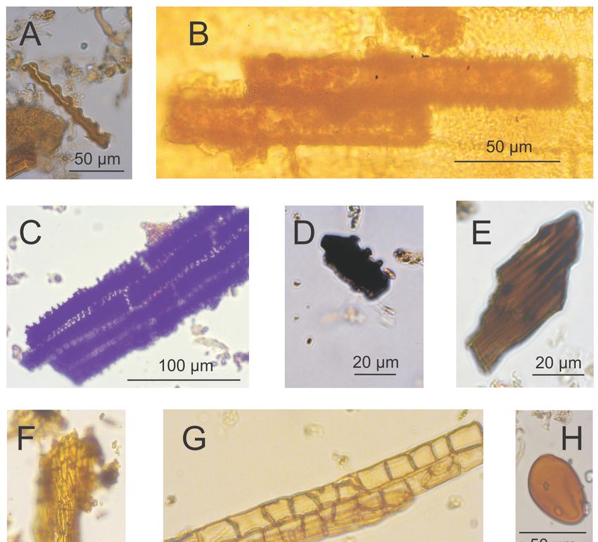
Soil Syst. 2020, 4, 12                                                                             2 of 16

decomposition of aboveground material of more than 90 % within a few years [5]. With a few
exceptions (e.g., leaves of Ericaceae, leaf tips of Scheuchzeria palustris, leaf fragments of Rhynchospora
alba and Thelypteris palustris [6]) and, apart from diaspores, no recognizable aboveground plant
remains are known from peat formed by herbaceous plants.
      If aboveground litter plays a role in peat formation it must be heavily decomposed and part of
the detritus. Detritus can constitute more than half the volume of sedge peats ([7], own experience).
Studies focussing on macro‐remains typically discard the detritus and microscopic studies rarely try
to identify material beyond pollen and spores. Yet, there is increasing interest in identification of
microscopic remains other than pollen—so‐called non‐pollen palynomorphs (NPPs) [8]. Plenty of
aboveground biomass may be part of the detritus, but it is difficult to estimate its extent. In this
paper, we will describe microscopic remains from sedge peats that may be of aboveground origin.
      With respect to the roots and rhizomes that constitute the peats we describe in this paper, the
description and identification of remains has had a long history. In the early 20th century, a detailed
characterization of botanical peat types [9,10]) was accompanied by a request for research towards a
more precise identification of vegetative herbaceous plant remains in fen peats. Matjuschenko [11]
developed a key for the identification of Carex fine roots (radicels) in peat; this key was later adapted
by Bertsch [12]. In the following decades, palaeoecological research focused on refining the rapidly
evolving field of pollen analysis and little attention was paid to macro‐remains. Only in the 1970s
did Grosse‐Brauckmann [6] formulate fundamental criticism on the keys to identifying Carex roots,
as a result of which identification to the species level was, from then on, often restricted to Carex
limosa roots alone [13,14]. Despite some available reviews [15,16] based at least partly on the use of
living plant material, recent macrofossil analyses are often limited to the identification of Carex sp.
roots [17] or Cyperaceae roots [18‒21]. Occasionally, additional root types are distinguished such as
Cladium, Equisetum and Thelypteris roots [22] or Phragmites and Scheuchzeria roots [23].
      This study aims to characterize macroscopic and microscopic remains of peat in different stages
of decomposition. We analysed peat from percolation fens that have been drained and, in one case,
rewetted again. We want i) to describe different types of macro‐ and micro‐remains that can reliably
be identified, ii) to test how analysis of micro‐remains can contribute to characterize particularly
decomposed peat, iii) to characterize the material that has been deposited since the rewetting and iv)
to fill in knowledge gaps on the possible contribution of aboveground biomass in displacement peat
and on the composition of the main component of displacement peat: the root mass.

2. Material and Methods
     Two peat monoliths were recovered from a percolation fen complex in north‐eastern Germany.
Detailed descriptions of the study sites in the Recknitz valley (PD) and the Trebel valley (PW) are
given in [24]. One site, PD, is drained and used for grass fodder production, the other site, PW, has
been drained as well, but was rewetted in 1997. The water table has remained close to the ground
surface since the rewetting [25]. The site is now under nature conservation. The first attempts at
drainage of the peatland complex date back to at least 1744, but strong degradation of the peat
probably only started in 1967 when deep drainage ditches of ~1.5 m deep were dug to allow for
high‐intensity grass cropping [26]. Peat cores of 20 cm diameter and ~50 cm length were recovered
using a “Clymo corer” [27] and frozen after extraction.
     Bricks of 7 × 13 × c. 50 cm were cut from the frozen monoliths and subsequently sampled into
contiguous 0.5 cm thick slices using DAMOCLES [28]. Respective subsamples of these slices were
analysed for macro‐ and micro‐remains at selected depths (n = 17 for site PD and n = 26 for site PW).

2.1. Analysis of Macro‐Remains
     For the analysis of macro‐remains, samples of 6 cm³ (4 × 3 × 0.5 cm) were slurried with purified
water and examined under an incident light microscope (10 to 40 times magnification). For tissue
and moss remains, volume percentages were estimated with reference to the total volume. Seeds,
fruits, and intact mollusk shells were counted. The following literature was used in te identification
Soil Syst. 2020, 4, 12 3 of 16
of plant tissue and moss remains: [6, 11‒13, 29], of fruits and seeds: [13, 30‒33], of animal remains:
[34, 35].
In addition, we collected fresh root material of 15 sedge species to establish a catalogue of
radicel morphotypes. For AMS 14C dating, 0.3 mg C (PD) and 0.7 mg C (PW) of aboveground plant
material was collected (Table 1). The radiocarbon dates were calibrated to calendar years using the
software CALIB v. 7.0.4. To arrive at a single point age estimate, we calculated the weighted average
of the probability distribution function [36].
With respect to the question of whether the root material we found was still alive or already
dead, we argued as follows: young, living or recently dead (subrecent) roots will be longer than old,
dead or subfossil roots and will possess more intact branches. However, the limited sample volume
and the small sample thickness (0.5 cm) made these criteria unsuitable for identification. Instead, all
flattened roots were considered long‐dead or (sub‐)fossil and all others, i.e., with intact aerenchyma,
were considered (sub‐)recent. It is not possible to separate roots that died 10 or 20 years ago, and
whose aerenchyma has already been completely degraded, from fossil roots of several thousand
years old.
2.2. Analysis of Micro‐Remains
For the analysis micro‐remains, samples of 2 cm3 (2 × 2 × 0.5 cm) were prepared. Sample
preparation included treatment with HCl and KOH, sieving (125 μm), HF‐treatment, acetolysis
(7 min), and mounting in silicone oil [37]. Lycopodium clavatum tablets were added for the
calculation of pollen concentrations [38]. Counting was done with a Zeiss Axioskop 40 light
microscope with 400× magnification. The micro‐remain counts are presented in concentrations of
particles per volume. The following literature was used for the identification: for pollen: [39], for
non‐pollen palynomorphs: EMA types (Department of Peatland studies and Palaeoecology at
Ernst‐Moritz‐Arndt University Greifswald) [40,41], and HdV types (Hugo de Vries‐Laboratory at
University of Amsterdam) [42‒50]. New EMA types and subtypes were identified and are first
described here. In order to differentiate clearly between plant taxa and pollen types, the latter are
displayed in small capitals [51].
2.3. Diagrams
The relative abundance or number of macro‐remains and concentrations of micro‐remains were
plotted against depth using the software C2 v. 1.7.7 [52]. Zonation of the diagrams was carried out
visually to distinguish zones that are a combination of informal acme zones and informal interval
zones [53, 54].
3. Results
3.1. Description of Macro‐ and Micro‐Remain Morphotypes
In this section we will first present descriptions of roots of eight plants commonly occurring in
percolation fens. In our descriptions of these macro‐remains we will focus on features that remain
identifiable even if the material is degraded. Descriptions of new EMA types of micro‐remains will
follow, complemented by new insights on previously described EMA types.
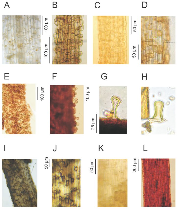
3.1.1. Radicel Types (Figure 1)
Carex acutiformis type (Figure 1I‒J): the colour of the radicels is whitish to slight greyish. The
root hairs are often broken and only the basal part, the pustule, is left. The pustules are rather
thick‐walled and, when viewed from above, rectangular to trapezoid with sharp edges. The lateral
walls of the pustules show a dark grey‐brown colour. Pustules are often arranged in small groups.
This type includes radicels of Carex acutiformis and C. riparia.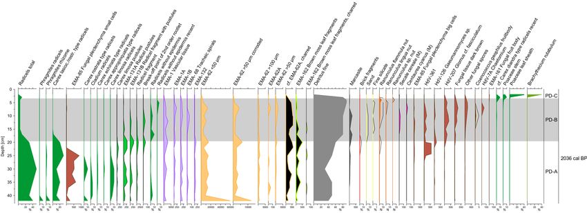
Soil Syst. 2020, 4, 12                                                                                4 of 16

      Figure 1. Radicel types: (A), (B) Carex rostrata type, (C) Carex lasiocarpa/rostrata type, (D)
      Phragmites australis type, (E) Cladium type, (F) Carex limosa type, (G) pustules of Carex limosa
      type, (H) EMA‐131C cf. Carex limosa pustules (from microscopic sample), (I), (J) Carex acutiformis
      type, (K) Carex echinata type, (L) Equisetum type.

     Carex limosa type (Figure 1F‒G): the colour of the radicels is yellowish to reddish. The root
hairs are more frequently preserved than in other sedges. The pustules are remarkably thick‐walled
and higher than they are wide. In side‐view, they often show a narrowed middle section and a wider
head. This type includes Carex limosa.
     Carex rostrata type (Figure 1A‒B): the colour of the radicels is whitish. The root hairs are often
broken and only the pustules are left. The pustules are thick‐walled and, when viewed from above,
square to rectangular with rounded edges. The lateral walls of the pustules show a whitish colour.
Soil Syst. 2020, 4, 12 5 of 16
The pustules are arranged in a chessboard pattern or in small groups. This type includes Carex
rostrata and probably its close relatives in Carex sect. Vesicariae, but not C. vesicaria itself with more
elongated pustules [11].
Carex lasiocarpa/rostrata type (Figure 1C): the radicels of this type are very similar to the Carex
rostrata type, but the pustules are less pronounced and arranged in a chessboard pattern. Single
radicels can show both the somewhat more pronounced pustules of the Carex rostrata type and the
less pronounced pustules of the Carex lasiocarpa type (in the sense of [11]), and therefore this type is
called Carex lasiocarpa/rostrata type here. It includes Carex lasiocarpa and perhaps C. rostrata.
Carex echinata type (Figure 1K): the colour of the radicels is whitish. Root hairs are mostly not
preserved; the basal part does not differentiate from the other epidermis cells, i.e., no pustules can be
seen. The cells of the epidermis are approximately five times longer than they are wide.
Cladium type (Figure 1E): the radicels of this type are very similar to the Carex rostrata type,
but the pustules show a light to clear brown colour. The pustules are arranged in a chessboard
pattern. This type probably only includes Cladium mariscus.
Equisetum type (Figure 1L): radicels of this type have a reddish, reddish‐brown to burgundy
colour. Root hairs are occasionally preserved. Small V‐shaped notches are often found at the former
place of the root hairs. This type includes Equisetum fluviatile and probably other Equisetum species as
well.
Phragmites australis type (Figure 1D): the colour of the radicels is whitish with a yellow touch.
The elongated parts of root hairs are often broken and only the basal part, the pustule, is left. The
pustules are rather thick‐walled and, when viewed from above, rectangular to trapezoid with rather
sharp edges. The pustules are arranged more or less in a chessboard pattern. The walls of the
pustules show a yellowish colour, which gives the radicels a spotty whitish/yellowish appearance.Soil Syst. 2020, 4, 12 6 of 16
Figure 2. Microscopic remains: (A) EMA‐62A Cell infills with undulating edges, (B) EMA‐62A inside
leaf sheath (from macroscopic sample), (C) EMA‐132 charred: charred cell complex of Cyperaceae
epidermis, (D) separate cells of EMA‐132 charred, (E)‒(F): EMA‐162: moss leaf fragment with
prosenchymatic cell pattern, charred (E) and not charred (F). (G), (I), (J) EMA‐85: Fungal
plectenchyma in shape of vascular plant cell, (H) EMA‐160 Fungal spore, (K) EMA‐161 Fungal fruit
body.
3.1.2. Microscopic Morphotypes (NPP types, Figure 2)
The exact origin of most of the microscopic morphotypes described here is unknown. Besides
origin, taphonomic processes further determine morphotype characteristics. The depth at which a
morphotype occurs and the accompanying assemblage of micro‐ and macro‐remains provides clues
to the age, origin and site conditions during and after deposition. In our descriptions of the
morphotypes, we therefore refer to stratigraphic information that is presented in detail in the next
section (Section 3.2, Figures 3 and 4).
EMA‐1 [41]: tissue including fragments of tracheids of vascular bundles like EMA‐1A and
EMA‐1B (see below) and EMA‐68 [40]. Abundant over the entire depth of both cores, but only inSoil Syst. 2020, 4, 12 7 of 16
very small amounts in the newly accumulated material (see Figure 3 zones PW‐C2, ‐C3, ‐D). The
highest amounts occur in both profiles in the upper parts of the A zone and in the lower parts of the
B zone, indicating that this NPP is concentrated when the peat is slightly to moderately affected by
drainage. Where drainage effects are strong, like close to the (former) soil surface, its amount is
lower (in upper part of zone PD‐B, Figure 4; in zones PW‐B2 and PW‐C1, Figure 3).
EMA‐1A: separate tracheids consisting of continuous rings, type (a) in [41]. Frequent in the
layers A and B, with old and degraded peat.
EMA‐1B: separate tracheids with pits, type (b) in [41]. Frequent in peat layers PW‐A and PW‐B,
but also in the newly accumulated material of radicels and leaf sheaths, where a lot of Carex
acutiformis remains were found.
EMA‐62 [40]: well‐preserved to highly corroded cell infills. They are elongated and rectangular
to slightly polygonal. The size of well‐preserved specimen is 17.5 (20) × 120‒180 μm, but frequently
shorter and/or narrower particles were found, and therefore counted in classes 50 μm show the highest values in zone PW‐B (Figure 3), indicating highly degraded peat.
Not‐corroded particles >50 μm are prominent in the old peat (zones PD‐A Figure 4, PW‐A Figure 3)
and the newly accumulated material (PW‐C3, PW‐D), indicating well‐preserved material.
EMA‐62A (Figure 2A): cell infill of Cyperaceae epidermis. Features are as described for
EMA‐62, but additionally characterized by evenly undulating edges. Found in‐situ in
macro‐remains of Cyperaceae leaf sheath (Figure 2B), but might occur in epidermis of other plant
parts with regularly undulating walls as well (see EMA‐132 [40]). Sporadically occurring in PD
(Figure 4), and in PW in the old peat (zone A) and the newly accumulated material (zone C3 and D,
Figure 3). In the diagram (Figure 3), the curve of EMA‐62A runs parallel to the curve of Cyperaceae
leaf sheaths. Black particles of similar shape (Figure 2D) are apparently charred EMA‐62A and were
separately counted. Charred particles are more frequent than non‐charred ones throughout PD
(Figure 4), and with low amounts but a steady occurrence characteristic for zones A and B1 in PW
(Figure 3).
EMA‐62A indicates the presence of Cyperaceae, especially when macroscopic remains are lost
due to decomposition. Based on its origin from the leaf sheath epidermis, in our study EMA‐62A
indicates that material grown above the surface is incorporated into the peat.
EMA‐85 [40] (Figure 2G‒J): fungal plectenchyma in shape of vascular plant epidermis cells,
12−18 x 43−110 (170) μm, fungal cells round to rectangular and folded, variable, occasionally look
pressed‐in, 2−4 x 5.7−11 μm in diameter, yellowish to brown.
These are probably plant cells filled with fungi that consume the cell content. The
plectenchyma was only once recorded with cell wall attached (Figure 2I).
We counted two variants. The variant with roundish, small cells (diameter 2−5.7 μm,
Figure 2I‒J) was found in the old peat in PD‐A in high amounts (Figure 4). The variant withSoil Syst. 2020, 4, 12 8 of 16
rectangular to polygonal, big cells (diameter 4−11 μm, Figure 2G) occurred in low amounts in the
upper part of PD‐A and in PD‐B (Figure 4), indicating the former presence of plant tissue.
EMA‐131 [40]: basal cells of radicel pustules, basal parts of Cyperaceae rootlets. Thick‐walled
cells. We separate two morphotypes described under this type number in [40] and add another one:
EMA‐131A: quadrangular to rectangular radicel pustules, see pl. I Figure 131.a in [40];
EMA‐131B: radicel fragment with EMA‐131A type pustules, see pl. I Figure 131.a in [40];
EMA‐131C (Figure 1H): cf. Carex limosa pustules. Single cells, thick‐walled, elongated
(23‒27 × 10−11 μm) with constriction (5.5−7.5 μm) in the middle, one end rounded, the other end
ruptured, yellow to hyaline.
Basal parts of roothairs probably of Carex limosa rootlets (see Figure 1G‒H). Indicating local
presence of Carex limosa type radicels.
EMA‐132 [40] charred (Figure 2C): epidermal tissue, probably of Cyperaceae. We found black,
apparently charred material that looks like a negative image of EMA‐132, where walls are lacking
and cell content is charred. Single charred ‘cells’ (Figure 2D) have the same shape as EMA‐62 A and
are discussed under this type (see above). Separated cells do not show the complete cell pattern of
the tissue and may also derive from Poaceae, which have comparatively short cells.
EMA‐160 (Figure 2H): fungal spore. Ellipsoid, 56 x 39 μm, at one end ca. 9 μm wide protruding
pore, one‐celled, brown. Occurring only in the newly accumulated material (zones PW‐C2, ‐C3, ‐D,
Figure 3), probably connected to the decomposition of fresh plant material.
EMA‐161 (Figure 2K): fungal fruit body. Globose to ovoid, 70−90 × 75−100 (115) μm, ruptured,
cells thick‐walled, irregularly, jigsaw puzzle‐like shaped, brown. Occurring in the uppermost
sample of zone PW‐B and in PW‐C and PW‐D (Figure 3), probably connected to the decomposition
of recent plant material.
EMA‐162 (Figure 2E‒F): brown moss leaf fragments. Various shapes and sizes, prosenchymatic
cell pattern, yellowish to light green (Figure 2F), when brownish to black (Figure 2E), supposed to
be charred.
Yellow to green specimen very abundant in zone PW‐A2 (Figure 3), together with
macro‐remains of brown mosses (Drepanocladus sp., Calliergonella cuspidata, Calliergon giganteum,
Campylium stellatum). This type indicates the presence of brown mosses in PD where no
macroscopic moss remains were found.
3.2 Diagrams
Proportions of the different macro‐ and micro‐remains are shown in Figure 3 and 4. We
distinguish four assemblage zones in PW and three in PD.
In Zone PW‐A (Figure 3), we find a radicel peat which seems only slightly influenced by
drainage. The proportion of radicels is higher than the proportion of fine detritus. Most of these
radicels are of the Carex lasiocarpa/rostrata type. Subzone A2 contains the highest amounts of moss
stems and brown moss leaf fragments of the entire profile. Carex lasiocarpa nutlets are present in
subzone A2.
Zone PW‐B is characterized by peat that is strongly modified due to agricultural drainage
followed by compaction, aerobic decay and ploughing. Ploughing is evident from the disturbed
pollen sequence with, e.g., SECALE CEREALE and CENTAUREA CYANUS pollen in the lower part of the
zone B, which is normally found only in the uppermost part of pollen diagrams in the study region
(Figure 3). The proportion of fine detritus is higher than the proportion of radicels. Clamydospores
of the soil fungus Glomus cf. fasciculatum (HdV‐207) are concentrated in this zone and microscopic
fungal types in total have the highest concentrations here. Microscopic vascular tissue (EMA‐1) is
especially prominent in subzone B1.
Zone PW‐C is somewhat heterogeneous. The line between zones B and C is drawn where the
proportion of fine detritus is below 70 % and the proportion of radicels is higher than 20 %.
Subzones 1 and 2 contain less than 5 % of Cyperaceae leaf sheaths, subzone 3 contains 10% to 30 %.
The difference between subzone 1 and 2 is the exclusive occurrence of Urtica dioica nutlets inSoil Syst. 2020, 4, 12 9 of 16
subzone 1. Fungal fruit bodies of Tetraploa (HdV‐89) and Microthyrium (HdV‐8b), as well as a big
unknown fungal spore (EMA‐160), occur only in the subzones 2‐3.
Zone PW‐D contains 60% to 90 % of Cyperaceae leaf sheaths and 2% to 20 % of radicels. This
zone comprises the only samples in which Typha angustifolia fruits and Pisidium sp. scales have been
found.
Figure 3. Selected curves of macro‐ and micro‐remain analyses of site Trebel valley (PW). Note the
scales and units of the x‐axes. Curves show percentages [%] or number [N] of macro‐remains as
indicated, or otherwise concentrations [101 N cm−3] of micro‐remains.Soil Syst. 2020, 4, 12 10 of 16
Figure 4. Selected curves of macro‐ and micro‐remain analyses of site Recknitz valley (PD). Note the
scales and units of the x‐axes. Curves show percentages [%] or number [N] of macro‐remains as
indicated, or otherwise concentrations [101 N cm‒3] of micro‐remains.Soil Syst. 2020, 4, 12 11 of 16
Zone PD‐A (Figure 4) contains radicel peat which seems only slightly influenced by drainage.
The proportion of total radicels is of the same order of magnitude as fine detritus. The major part of
these radicels belongs to the Carex lasiocarpa/rostrata type. Besides this dominant radicel type,
Carex limosa type and Carex echinata type radicels were found.
Zone PD‐B contains strongly modified peat due to agricultural drainage followed by
compaction, aerobic decay and ploughing. As in case of PW, ploughing is evident from the
disturbed pollen sequence with, e.g., SECALE CEREALE pollen in the lower part of the zone B
(Figure 4). The proportion of fine detritus is higher than of radicels. Due to the strong
decomposition, a significant proportion of the radicels no longer shows any epidermis cells and
cannot be further identified. Microscopic remains of radicels (fragments, pustules, root break‐off
points) still occur in the lower part and vascular tissue (EMA‐1, ‐1A and ‐1B) occurs throughout the
zone. The concentration of clamydospores of the soil fungus Glomus cf. fasciculatum (HdV‐207) is
high. Diaspores of Juncus sp. and Ranunculus sp. show a significantly better state of preservation
than the surrounding matrix material.
Zone PD‐C is characterized by the remains of living grasses and ruderal mosses.
3.3. Dating
The peat just below the strongly degraded agriculturally influenced layer is of considerable
age in both profiles (Table 1).
Table 1. Radiocarbon dates in PW and PD.
Sample Calibrated 14C‐AMS‐date weighted
Material Lab. no 14C date
number average (2σ range) [cal. BP]
Carex lasiocarpa 3365 ±
PW 40 cm Poz‐107576 3607 (3484‐3694)
nuts 35 BP
Cladium mariscus 2065 ±
PD 26 cm Poz‐114673 2036 (1934−2128)
fruits 35 BP
The year of the rewetting of PW in 1997 can be pinpointed as well. Rewetting was carried out
after the area had been abandoned and Urtica dioica had spread. Fruits of U. dioica must have fallen
into cracks that had developed in the strongly degraded soil [55]. After the rapid increase in the
water table upon rewetting [56], U. dioica died off and was replaced by Carex sp., as evidenced by
the presence of molluscs and by aboveground remains of various Carex species. The year 1997 is
placed at the PW‐C1 to PW‐C2 boundary (Figure 3).
4. Discussion
4.1 Site Development
In both cores the older, well preserved peat (zones PW‐A, PD‐A) shows indicators of loose,
permanently water‐saturated peat formed under mesotrophic nutrient conditions. The PW core
contains Meesia triquetra, Calliergon giganteum and Carex limosa roots, the PD core Carex limosa roots
and EMA‐162 (brown moss leaf fragments), all of which are indicative of stable high‐water tables
relative to the surface. The remains of Menyanthes trifoliata and Carex lasiocarpa that occur
simultaneously in PW support the picture of high‐water tables and mesotrophic conditions, as they
are typically found in sedge‐dominated percolation mires [57, 58]. Floating mires can have similar
vegetation and they did occur in the Recknitz valley during regression phases of the Litorina
Transgression in the Pomeranian Border Valley. However, they had transitioned into percolation
mires more than 5000 years cal. BP ago [22].
Hardly any macro‐remains that make up the original peat have remained in the layers of
highly decomposed peat (PW‐B, PD‐B). The Carex rostrata type radicels found in PW‐B may alsoSoil Syst. 2020, 4, 12 12 of 16
have grown into the matrix after rewetting, as scattered Carex rostrata plants are found in close
vicinity to the coring location. Carex acutiformis currently dominates the vegetation at the coring
location and any radicels of Carex acutiformis type observed in PW‐B2 are likely of recent origin.
The fine roots without epidermis in PD‐B are difficult to interpret. They may have originated
during the time of drainage, but could also be younger. Certainly, the clamydospores of type
HdV‐207 Glomus cf. fasciculatum, a fungus that lives in aerated soils, were deposited when the site
was drained. They occur in the layer of highly decomposed peat that corresponds with the formerly
drained and ploughed agricultural soil. In both cores, the micro‐remains EMA‐162 (moss leaf
fragments with prosenchymatic cell pattern), EMA‐1 (vascular bundles), EMA‐131 (isolated
pustules and radicel fragments with pustules), microscopic radicel fragments and EMA‐62 occur in
both zone A and B and indicate that the peat of PW‐B and PD‐B originally had a similar
composition to the well‐preserved sedge peat immediately below.
Isolated and surprisingly well‐preserved diaspores found in these layers (e.g., Ranunculus
flammula nuts, Juncus sp. seeds) were probably added to the matrix when the peat was drained.
Either they were mechanically worked into the soil during agricultural site preparation, or, like the
Urtica dioica nutlets, they fell into soil cracks in the degraded peat. Although Ranunculus flammula is
regularly found in wet peatlands and the nuts are commonly found in peat deposits [31,56], it also
grows at the drained grassland site PD.
In PW, the layer of organic material that has been deposited after rewetting does not show
uniform contents over depth, but rather a development from initial pioneer vegetation (Carex
flava‐agg.) to the establishment of potentially peat‐forming dominant stands of Carex acutiformis. In
the top 6 cm of the profile, the leaf sheaths of Cyperaceae are dominant, but they are absent or very
rare further down. As all Carex species have leaf sheaths and not only the currently dominant Carex
acutiformis, it is most likely that they are absent from the sedge peat matrix because they are easily
decomposed as part of the aboveground litter.
There are many fungi present in the upper layer, like Tetraploa (HdV‐89), a mold on plant
leaves, Coniochaeta cf. lignaria (HdV‐172), an ascomycete that can decompose lignocellulosic material
[59], Microthyrium (HdV‐8b), another ascomycete, and further fungal remains (EMA‐160, HdV‐124).
Probably, the fungi in this (periodically) oxic layer are instrumental in decomposing the
aboveground litter, including leaf sheaths.
We have found cell fillings in leaf sheaths (Figure 2B) that look exactly like NPP type EMA‐62
(> 100 μm). This NPP type is present over the entire depth of both profiles. Whether these cell
fillings are only present in leaf sheaths, or also in other aboveground material, but not in
belowground roots and rhizomes, is yet unclear. If EMA‐62 were indeed of aboveground origin, it
would indicate that aboveground litter could play an important role in the accumulation of sedge
root displacement peat.
We find micro‐remains of type EMA‐62 in various sizes; we interpret the increasingly smaller
remains as increasing stages of decomposition. Whether this interpretation is valid or whether there
may be some other origin, particularly of the smallest EMA‐62 remains, is thus far unclear.
4.2 Morphotypes
In the light of rewetting and restoration measures that have been and are carried out on the
large valley mires of Central Europe, there is an interest in understanding the mechanisms of peat
formation and the plant species involved. In other words, we would like to be able to identify plant
remains up to the species level or at least to species groups. A large problem in the identification of
fine roots is their variability depending on their state of decomposition. With very little peat
decomposition, fine roots with completely preserved root hairs can occasionally be found.
However, the elongated part of the root hairs often breaks off and only the basal part of the root
hair (pustule) remains. If the root hair base is thicker‐walled and more resistant to decay than the
surrounding epidermal cells, it becomes pronounced during decomposition, resulting in the typical
wavy silhouette of a pustular radicel (Figure 1B). As decomposition progresses, the pustules are
often lost and only the sclerenchymatic tissue remains, which we call ’radicels without epidermis‘Soil Syst. 2020, 4, 12 13 of 16
and which look the same across all species. Grosse‐Brauckmann [6] argues that the form and shape
of radicels may depend on growth conditions, and that pustules may be weak or absent in species
that would normally display them. Whether and how far this caveat applies remains unclear.
It is particularly difficult to identify radicels, whose root hairs are not clearly differentiated
from the other epidermal cells. Matjuschenko [11] and Bertsch [12] define different root types
according to the length to width ratio of the epidermal cells, but such ratios are variable depending
on growth rates. For these morphotypes of Matjuschenko [11] and Bertsch [12] (e.g., Carex echinata
type, Carex diandra type, and Carex appropinquata type) we deem identification to the species
level to be very uncertain. The exceptions are hairless roots of Equisetum (reddish) and Eriophorum
vaginatum (black‐grey) which differ in colour from the mostly whitish Carex radicels.
Even among those radicels with more or less distinct pustules there are types that may be
somewhat difficult to distinguish. Matjuschenko [11], for example, points out the great similarity of
young roots of Carex rostrata and Carex lasiocarpa. In a footnote in Matjuschenko [11], Selma Ruoff
(translator of the Russian original) mentions that also the fine roots of Carex panicea are so similar to
those of Carex rostrata that a distinction would be hardly possible.
Phragmites fine roots are easily recognisable in the field by their yellowish colour when
well‐preserved, but at the cellular level they show clear similarities to Carex fine roots in general
and to Carex vesicaria radicels in particular. This similarity raises two questions: How can Phragmites
fine roots be separated from Carex vesicaria radicels in the event of the yellowish pustules fading,
and how do palaeoecological studies, which only address Cyperaceae roots in general, exclude the
possibility of including Phragmites radicels?
5. Conclusion
High‐resolution analysis of macro‐remains, even with relatively small sample volumes, is an
excellent method for the investigation of fen peat. In contrast to ancient DNA, it allows for
separation of vegetative and generative plant remains in peat from different times and origins. The
combination of root types and diaspores can provide a detailed picture of the former vegetation of
fens, especially as it also breaks down taxa that are difficult to differentiate in pollen analysis
(Cyperaceae) or are lost during pollen sample preparation (Juncus).
In the analysis of highly decomposed peat, however, analysis of macro‐remains quickly
reaches its limits, while analysis of micro‐remains still delivers worthwhile results. The
identification of the decomposition products of sedge roots (isolated pustules, EMA‐131A, ‐131C)
and what are probably leaf sheaths (EMA‐62) helps to understand the formation of fen peat also in
an advanced state of decomposition.
The establishment of new deposits after rewetting of fens seems to be a complex process,
which in the first years is probably only an accumulation of litter and only gradually leads to a kind
of ’proto‐peat’. Analysis of macro‐ and micro‐remains shows that various degradation processes
have taken place in the deposits since rewetting, making the lower part of the new layer
structurally more similar to the original peat, with its high radicel and low macroscopic leaf sheath
content. Together with the stratification found, the presence of young layers rich in radicels
indicates that the rewetting has, thus far, successfully led to the re‐establishment of an
accumulating fen ecosystem.
Author Contributions: D.M., A.M. and J.C. conceived the study; D.M. analysed macro‐remains; A.M. analysed
and described micro‐remains; D.M. led writing of the manuscript in which A.M. and J.C. participated. All
authors have read and agreed to the published version of the manuscript.
Founding: This study was supported by the European Social Fund (ESF) and the Ministry of Education, Science
and Culture of Mecklenburg‐Western Pomerania within the scope of the project WETSCAPES
(ESF/14‐BM‐A55‐0035/16) and by the Deutsche Forschungsgemeinschaft (JO 332/1‐15) in the frame of the
ERA‐NET Cofunds BiodivERsA3 project REPEAT.
Acknowledgement: We thank Stella Gribbe and Sabine Kell for help in preparation of the core samples.
Conflicts of Interest: The authors declare no conflict of interest.Soil Syst. 2020, 4, 12 14 of 16
References:
1. Loisel, J.; van Bellen, S.; Pelletier, L.; Talbot, J.; Hugelius, G.; Karran, D.; Yu, Z.; Nichols, J.; Holmquist,
J. Insights and issues with estimating northern peatland carbon stocks and fluxes since the Last
Glacial Maximum. Earth‐Science Rev. 2017, 165, 59−80.
2. Moen, A.; Joosten, H.; Tanneberger, F. Mire diversity in Europe: mire regionality. In Mires and
peatlamds of Europe; Joosten, H.; Tanneberger, F.; Moen, A., Eds.; Schweizerbart: Stuttgart, Germany,
2017; pp. 97−149.
3. Weber, C.A. Grenzhorizont und älterer Sphagnumtorf. Abhandl. Naturwissen. Ver. Bremen 1930, 28,
57–65.
4. Grosse‐Brauckmann, G. Analysis of vegetative plant macrofossils. In Handbook of Holocene
Palaeoecology and Palaeohydrology; Berglund, B.E., Ed.; John Wiley & Sons: Chichester, UK, 1986;
pp. 591–618.
5. Pegman, A.P.M.; Ogden, J. Productivity‐decomposition dynamics of Typha orientalis at Kaitoke
Swamp, Great Barrier Island, New Zealand. New Zealand J. Bot. 2005, 43, 779−789.
6. Grosse‐Brauckmann, G. Über pflanzliche Makrofossilien mitteleuropäischer Torfe. I. Gewebereste
krautiger Pflanzen und ihre Bestimmung. Telma 1972, 2, 19−56.
7. Ronkainen, T.; McClymont, E.L.; Tuittila, E.‐S.; Väliranta, M. Plant macrofossil and biomarker
evidence of fen–bog transition and associated changes in vegetation in two Finnish peatlands.
Holocene 2014, 24, 828−841.
8. van der Linden, M.; Kooistra, L.I.; Engels, S. Non‐pollen palynomorphs as relevant indicators in
palaeoecology and archaeobotany. Rev. Palaeobot. Palynol. 2012, 186, 1−162.
9. von Post, L.; Granlund, E. Södra Sveriges Torvtillgångar. Sveriges Geol. Unders., Årsbok 1926, 19, 1−127.
10. von Bülow, K. 1929: Allgemeine Moorgeologie. Einführung in das Gesamtgebiet der Moorkunde;
Borntraeger: Berlin, Germany, 1929; p. 308.
11. Matjuschenko, W. Schlüssel zur Bestimmung der in den Mooren vorkommenden Carexarten
(translation by S. Ruoff). Geol. Archiv, Z. Gesamtgeb. d. Geol. 1924, 3, 183−188, 192−193.
12. Bertsch, K. Lehrbuch der Pollenanalyse. Ferdinand Enke: Stuttgart, Germany, 1942; p. 195.
13. Mauquoy, D.; van Geel, B. Mire and peat macros. In Encyclopedia of Quaternary Science; Elias, S.A.
Eds.; Elsevier: Amsterdam, Netherlands, 2007; Volume 3, pp. 2315–2336.
14. Drzymulska, D.; Kłosowski, S.; Pawlikowski, P.; Zieliński, P.; Jabłońska, E. The historical
development of vegetation of foreshore mires beside humic lakes: Different successional pathways
under various environmental conditions. Hydrobiologia 2013, 703, 15−31.
15. Nilsson, T. Kvartärpaleontologi och Kvartärpaleontologiska undersökningsmetoder. 4th ed.; Lunds
Universitet, Lund, Sweden, 1972; Volumes 2, p. 238, Plates 63.
16. Katz, N.J.; Katz, S.W.; Skobejewa, E.I. Atlas rastitelnich ostakow w torfach. Nedra: Moscow, USSR, 1977;
p. 371.
17. Hughes, P.D.M.; Mauquoy, D.; Barber, K.E.; Langdon, P.G. Mire‐development pathways and
palaeoclimatic records from a full Holocene peat archive at Walton Moss, Cumbria, England.
Holocene 2000, 10, 465−479.
18. Tobolski, K. Przewodnik do oznaczania torfów i osadów jeziornych. Vademecum Geobotanicum 2000, 2,
9−508.
19. Loisel, J.; Garneau, M. Late Holocene paleoecohydrology and carbon accumulation estimates from
two boreal peat bogs in eastern Canada: Potential and limits of multi‐proxy archives. Palaeogeogr.
Palaeoclimat. Palaeoecol. 2010, 291, 493−533.
20. Gałka, M.; Lamentowicz, Ł.; Lamentowicz, M. Palaeoecology of Sphagnum obtusum in NE Poland.
Bryologist 2013, 116, 238−247.
21. Jabłońska, E.; Falkowski, T.; Chormański, J.; Jarzombkowski, F.; Kłosowski, S.; Okruszko, T.;
Pawlikowski, P.; Theuerkauf, M.; Wassen, M.J.; Kotowski, W. Understanding the Long Term
Ecosystem Stability of a Fen Mire by Analyzing Subsurface Geology, Eco‐Hydrology and Nutrient
Stoichiometry Case Study of the Rospuda Valley (NE Poland). Wetlands 2014, 34, 815−828.
22. Michaelis, D.; Joosten, H. Mire development, relative sea level change, and tectonic movement along
the Northeast‐German Baltic Sea coast. Ber. Römisch‐German. Komm. 2007, 88, 101−134.Soil Syst. 2020, 4, 12 15 of 16
23. Gałka, M.; Miotk‐Szpiganowicz, G.; Goslar, T.; Jęśko, M.; van der Knaap, W.O.; Lamentowicz, M.
Palaeohydrology, fires and vegetation succession in the southern Baltic during the last 7500 years
reconstructed from a raised bog based on multi‐proxy data. Palaeogeogr. Palaeoclimatol. Palaeoecol.
2013, 370, 209−221.
24. Jurasinski, G.; Ahmad, S.; Anadon‐Rosell, A.; Berendt, J.; Beyer, F.; Bill, R.; Blume‐Werry, G.;
Couwenberg, J.; Günther, A.; Joosten, H.; et al. From understanding to sustainable use of peatlands:
The WETSCAPES approach. Agricultural Sciences & Agronomy 2020,
doi:10.20944/preprints202001.0250.v1
25. Bönsel, A.; Sonneck, A.‐G. Development of ombrotrophic raised bogs in North‐east Germany
17 years after the adoption of a protective program. Wetlands Ecol. Management 2012, 20, 503−520.
26. Succow, M. Die Vegetation nordmecklenburgischer Flußtalmoore und ihre anthropogene
Umwandlung. Doctoral Thesis, E.‐M.‐Arndt Universität, Greifswald, Germany, 1970.
27. Clymo, R.S. A high‐resolution sampler of surface peat. Functional Ecology 1988, 2, 425–431
28. Joosten, H.; de Klerk, P. DAMOCLES: A DAshing MOnolith Cutter for fine sectioning of peats and
sediments into LargE Slices. Boreas 2007, 36, 76–81.
29. Michaelis, D. Ein Schlüssel zur Bestimmung von Braunmoosen in Torfen anhand einzelner Blättchen.
Telma 2001, 31, 79−104.
30. Berggren, G. Atlas of seeds and small fruits of Northwest‐European plant species with
morphological descriptions. 2: Cyperaceae. Swedish Natural Science Research Council: Stockholm,
Sweden, 1969; pp 68.
31. Birks, H.H. Plant macrofossil introduction. In Encyclopedia of Quaternary Science; Elias, S.A. Eds.;
Elsevier: Amsterdam, Netherlands, 2007; Volume 3, pp. 2266−2288.
32. Grosse‐Brauckmann, G.; Streitz, B. Pflanzliche Makrofossilien mitteleuropäischer Torfe. III. Früchte,
Samen und einige Gewebe. Telma 1992, 22, 53−102.
33. Nilsson, Ö.; Hjelmqist, H. Studies on the nutlet structure of South Scandinavian species of Carex. –
Bot. Notiser 1967, 120, 460−485.
34. Lozek, V. Quartärmollusken der Tschechoslowakei. Rozpravy ustredniho ustavu geologickeho: Praha,
ČSSR, 1964; p. 374.
35. Jaeckel, S.H. Mollusca – Weichtiere. In Exkursionsfauna Wirbellose 1, 5th ed.; Stresemann, E. Eds.;
Volk und Wissen: Berlin, Germany, 1976, pp. 102−229.
36. Telford, R.J.; Heegaard, E.; Birks, H.J.B. The intercept is a poor estimate of a calibrated radiocarbon
age. Holocene 2004, 14, 296−298.
37. Fægri, K.; Iversen, J. Textbook of Pollen Analysis, 4th. ed.; Wiley: Chichester, UK, 1989; p. 328.
38. Stockmarr, J. Tablets with spores used in absolute pollen analysis. Pollen et. Spores 1971, 13, 615–621.
39. Moore, P.D.; Webb, J.A.; Collinson, M.E. 1991: Pollen analysis. 2. Ed.; Blackwell: Oxford, UK; p. 216
40. Barthelmes, A.; de Klerk, P.; Prager, A.; Theuerkauf, M.; Unterseher, M.; Joosten, H. Expanding NPP
analysis to eutrophic and forested sites: Significance of NPPs in a Holocene wood peat section (NE
Germany). Rev. Palaeobot. Palynol. 2012, 186, 22−37.
41. Prager, A.; Barthelmes, A.; Theuerkauf, M.; Joosten, H. Non‐pollen palynomorphs from modern
Alder carrs and their potential for interpreting microfossil data from peat. Rev. Palaeobot. Palynol.
2006, 141, 7−31.
42. van Geel, B. A Paleoecological Study of Holocene Peat Bog Sections: Based on the Analysis of Pollen,
Spores and Macro‐and Microscopic Remains of Fungi, Algae, Ph.D. Thesis, Universiteit van
Amsterdam, Amsterdam, 1976.
43. van Geel, B. A palaeoecological study of Holocene peat bog sections in Germany and the
Netherlands. Rev. Palaeobot. Palynol. 1978, 25, 1–120.
44. van Geel, B. Application of fungal and algal remains and othermicrofossils in palynological analyses.
In Handbook of Holocene Palaeoecology and Palaeohydrology, Berglund, B.E. Ed.; Blackburn Press:
Caldwell, USA; pp. 497–505.
45. van Geel, B.; Bohncke, S.J.P.; Dee, H., A palaeoecologicalstudy of an upper Late Glacial and Holocene
sequence from“De Borchert”, The Netherlands. Rev. Palaeobot. Palynol. 1980/81, 31, 367–448.
46. van Geel, B.; Hallewas, D.P.; Pals, J.P. A late Holocenedeposit under the Westfriese Zeedijk near
Enkhuizen (Prov. of Noord‐Holland, The Netherlands): palaeoecological and archae‐ological aspects.
Rev. Palaeobot. Palynol. 1982/83, 38, 269–335.Soil Syst. 2020, 4, 12 16 of 16
47. van Geel, B.; Klink, A.G.; Pals, J.P.; Wiegers, J. An upper Eemian lake deposit from Twente, Eastern
Netherlands. Rev. Palaeobot. Palynol. 1986, 47, 31–61.
48. van der Wiel, A.M. A palaeoecological study of a section from the foot of the Hazendonk
(Zuid‐Holland, The Netherlands), based on the analysis of pollen, spores and macroscopic plant
remains. Rev. Palaeobot. Palynol. 1982, 38, 35−90.
49. Pals, J.P.; van Geel, B.; Delfos, A. Paleoecological studies in the Klokkeweel bog near Hoogkarspel
(prov. of Noord‐Holland). Rev. Palaeobot. Palynol. 1980, 30, 371−418.
50. Kuhry, P. Transgressions of a raised bog across a coversand ridge originally covered with an
oak‐lime forest. Rev. Palaeobot. Palynol. 1985, 44, 313–353.
51. Joosten, H., de Klerk, P. What’s in a name?: Some thoughts on pollen classification, identification,
and nomenclature in quaternary palynology. Rev. Palaeobot. Palynol. 2002, 122, 29−45.
52. Juggins, S. C2 Version 1.7.7. Software for ecological and palaeoecological data analysis and
visualization. Available on line: https://www.staff.ncl.ac.uk/stephen.juggins/software/C2Home.htm
(accessed on 28 February 2020).
53. Hedberg, H.D. International stratigraphic guide: a guide to stratigraphic classification, terminology,
and procedure. John Wiley and Sons: New York, USA, 1976; p. 200.
54. Salvador, A. International stratigraphic guide: a guide to stratigraphic classification, terminology,
and procedure, 2nd ed.; Geological Society of America: Boulder, CO, 1994.
55. Stegmann, H.; Zeitz, J. Bodenbildende Prozesse entwässerter Moore. In Landschaftsökologische
Moorkunde, 2nd ed.; Succow, M; Joosten, H., Eds.; Schweizerbart: Stuttgart, Germany, 2001; pp.
47−57.
56. Vegelin, K. Das mittlere Trebeltal im Jahr 2008 ‐ Entwicklungen in Wasserhaushalt und Vegetation
im EU‐LIFE Projektgebiet, Mittleres Trebeltal und Wiesen um das Grenztalmoor. Unpublished work,
2009.
57. Michaelis, D. Die spät‐ und nacheiszeitliche Entwicklung der natürlichen Vegetation von
Durchströmungsmooren in Mecklenburg‐Vorpommern am Beispiel der Recknitz. J. Cramer: Berlin,
Germany, 2002; p. 188.
58. Joosten, H.; Moen, A.; Couwenberg, J.; Tanneberger, F. 2017. Mire diversity in Europe: mire and
peatland types. In Mires and Peatlands of Europe; Joosten, H.; Tanneberger, F.; Moen, A., Eds.;.
Schweizerbart: Stuttgart, Germany, 2017; pp. 5−64.
59. Jiménez, D.J.; Hector, R.E.; Riley, R.; Lipzen, A.; Kuo, R.C.; Amirebrahimi, M.; Barry, K.W.; Grigoriev,
I.V.; Van Elsas, J.D.; Nichols, N. Draft Genome Sequence of Coniochaeta ligniaria NRRL 30616, a
Lignocellulolytic Fungus for Bioabatement of Inhibitors in Plant Biomass Hydrolysates. Genome
Announc. 2017, 5, 1−2.
© 2020 by the authors. Licensee MDPI, Basel, Switzerland. This article is an open access
article distributed under the terms and conditions of the Creative Commons Attribution
(CC BY) license (http://creativecommons.org/licenses/by/4.0/).You can also read