The Combination of Gefitinib With ATRA and ATO Induces Myeloid Differentiation in Acute Promyelocytic Leukemia Resistant Cells
←
→
Page content transcription
If your browser does not render page correctly, please read the page content below
ORIGINAL RESEARCH
                                                                                                                                            published: 28 September 2021
                                                                                                                                           doi: 10.3389/fonc.2021.686445

                                              The Combination of Gefitinib
                                              With ATRA and ATO Induces
                                              Myeloid Differentiation in Acute
                                              Promyelocytic Leukemia
                                              Resistant Cells
                          Edited by:
                        Harinder Gill,        Luciana Yamamoto de Almeida 1,2†, Diego A. Pereira-Martins 1,2,3†, Isabel Weinhäuser 1,2,3,
University of Hong Kong, Hong Kong,           César Ortiz 1,2, Larissa A. Cândido 1,2, Ana Paula Lange 1, Nayara F. De Abreu 1,
                          SAR China           Sı́lvia E. S. Mendonza 1,2, Virgı́nia M. de Deus Wagatsuma 1,2, Mariane C. Do Nascimento 1,2,
                        Reviewed by:          Helder H. Paiva 1,2, Raquel M. Alves-Paiva 1,2, Camila C. O. M. Bonaldo 2,
                 Michael Diamantidis,         Daniele C. Nascimento 4, José C. Alves-Filho 3, Priscila S. Scheucher 1, Ana Sı́lvia G. Lima 1,
University Hospital of Larissa, Greece        Jan Jacob Schuringa 3, Emanuele Ammantuna 3, Tiziana Ottone 5,6,7, Nelida I. Noguera 5,
                            Dijiong Wu,       Cleide L. Araujo 2 and Eduardo M. Rego 1,2,7*
Zhejiang Chinese Medical University,
                                              1 Department of Medical Images, Hematology, and Clinical Oncology, University of Sao Paulo at Ribeirao Preto Medical
                                  China
                                              School, Ribeirao Preto, Brazil, 2 Center for Cell-Based Therapy, University of Sao Paulo, Ribeirao Preto, Brazil, 3 Department
                 *Correspondence:
                                              of Experimental Hematology, Cancer Research Center Groningen, University Medical Center Groningen, University of
                   Eduardo M. Rego
                                              Groningen, Groningen, Netherlands, 4 Department of Pharmacology, University of Sao Paulo, Ribeirao Preto Medical School,
             eduardo.rego@fm.usp.br
                                              Ribeirao Preto, Brazil, 5 Department of Biomedicine and Prevention, University of Tor Vergata, Rome, Italy, 6 Santa Lucia
     †
      These authors have contributed          Foundation, I.R.C.C.S., Neuro-Oncohematology, Rome, Italy, 7 Hematology Division, Laboratórios de Investigação Médica 31
                 equally to this work         (LIM 31), Faculdade de Medicina, University of Sao Paulo, Sao Paulo, Brazil

                   Specialty section:
         This article was submitted to
                                              In approximately 15% of patients with acute myeloid leukemia (AML), total and
           Hematologic Malignancies,          phosphorylated EGFR proteins have been reported to be increased compared to
               a section of the journal
                                              healthy CD34+ samples. However, it is unclear if this subset of patients would benefit
                 Frontiers in Oncology
                                              from EGFR signaling pharmacological inhibition. Pre-clinical studies on AML cells provided
          Received: 26 March 2021
     Accepted: 06 September 2021              evidence on the pro-differentiation benefits of EGFR inhibitors when combined with ATRA
     Published: 28 September 2021             or ATO in vitro. Despite the success of ATRA and ATO in the treatment of patients with
                               Citation:      acute promyelocytic leukemia (APL), therapy-associated resistance is observed in 5-10%
                            Almeida LY,
    Pereira-Martins DA, Weinhäuser I,
                                              of the cases, pointing to a clear need for new therapeutic strategies for those patients. In
      Ortiz C, Cândido LA, Lange AP,         this context, the functional role of EGFR tyrosine-kinase inhibitors has never been
       De Abreu NF, Mendonza SES,
                                              evaluated in APL. Here, we investigated the EGFR pathway in primary samples along
             de Deus Wagatsuma VM,
       Do Nascimento MC, Paiva HH,            with functional in vitro and in vivo studies using several APL models. We observed that
    Alves-Paiva RM, Bonaldo CCOM,             total and phosphorylated EGFR (Tyr992) was expressed in 28% and 19% of blast cells
      Nascimento DC, Alves-Filho JC,
            Scheucher PS, Lima ASG,
                                              from APL patients, respectively, but not in healthy CD34+ samples. Interestingly, the
         Schuringa JJ, Ammantuna E,           expression of the EGF was lower in APL plasma samples than in healthy controls. The
Ottone T, Noguera NI, Araujo CL and
                                              EGFR ligand AREG was detected in 29% of APL patients at diagnosis, but not in control
     Rego EM (2021) The Combination
       of Gefitinib With ATRA and ATO          samples. In vitro, treatment with the EGFR inhibitor gefitinib (ZD1839) reduced cell
 Induces Myeloid Differentiation in Acute     proliferation and survival of NB4 (ATRA-sensitive) and NB4-R2 (ATRA-resistant) cells.
Promyelocytic Leukemia Resistant Cells.
               Front. Oncol. 11:686445.
                                              Moreover, the combination of gefitinib with ATRA and ATO promoted myeloid cell
        doi: 10.3389/fonc.2021.686445         differentiation in ATRA- and ATO-resistant APL cells. In vivo, the combination of gefitinib

Frontiers in Oncology | www.frontiersin.org                                           1                                    September 2021 | Volume 11 | Article 686445
Almeida et al.                                                                                              Gefitinib Sensitizes APL-Resistant Cells to ATRA/ATO

                                              and ATRA prolonged survival compared to gefitinib- or vehicle-treated leukemic mice in a
                                              syngeneic transplantation model, while the gain in survival did not reach statistical
                                              difference compared to treatment with ATRA alone. Our results suggest that gefitinib is
                                              a potential adjuvant agent that can mitigate ATRA and ATO resistance in APL cells.
                                              Therefore, our data indicate that repurposing FDA-approved tyrosine-kinase inhibitors
                                              could provide new perspectives into combination therapy to overcome drug resistance in
                                              APL patients.
                                              Keywords: epidermal growth factor receptor (EGFR), erlotinib, gefitinib, all-trans retinoic acid (ATRA), acute
                                              promyelocytic leukemia (APL), ATRA-resistance, ATO-resistance, arsenic trioxide (ATO)

INTRODUCTION                                                                        erlotinib monotherapy discontinued treatment because of disease
                                                                                    progression. Nevertheless, combination therapies between
The clinical introduction of all-trans retinoic acid (ATRA) and                     differentiation agents with EGFR inhibitors have not been
arsenic trioxide (ATO) revolutionized the treatment of acute                        evaluated in AML patients (18).
promyelocytic leukemia (APL), leading to a disease-free survival                        Here, we evaluated the effects of EGFR pharmacological
rate of 80-90% (1). Nevertheless, 5-10% of APL patients still relapse               inhibition in distinct APL models. Gefitinib monotherapy
due to ATRA or ATO resistance (2). Despite the cytotoxic activities                 induced apoptosis and inhibited the proliferation of NB4
of ATRA and ATO in APL cells, low doses of those agents result in                   (ATRA-sensitive) and NB4-R2 (ATRA-resistant) APL cells.
induction of terminal myeloid cell differentiation (3, 4). In this                  Additionally, the combination between gefitinib with ATRA
context, previous reports demonstrated that inhibitors of the                       and ATO rewired NB4-R2 and NB4 ATOr (ATO-resistant)
epidermal growth factor receptor (EGFR) increased ATRA and                          cells into sensitivity to standard therapy for APL. In vivo, APL
ATO-induced expression of the myeloid differentiation marker                        mice treated with ATRA alone or in combination with gefitinib
CD11b in AML cells (3–7). Nonetheless, the use of EGFR inhibitors                   exhibited increased overall survival in comparison with the
in combination with standard therapy was not previously explored                    vehicle-treated group.
in APL cells resistant to ATRA and ATO.
    Non-small cell lung cancer (NSCLC) demonstrated constitutive
activation of the epidermal growth factor (EGF)/EGFR pathway,                       MATERIAL AND METHODS
due to mutations on the EGFR (8). Although EGFR mutations are
rare in AML (9–11), the level of EGF—the main EGFR ligand—was                       Chemicals
elevated in the urine of patients diagnosed with APL and decreased                  Gefitinib (#S1025) and erlotinib (#S7786) were purchased from
after ATRA-induced complete remission (12). Hence, it is                            Selleck Chemicals (Houston, TX, USA). ATRA and ATO were
conceivable that the activation of the EGF/EGFR signaling                           purchased from Sigma-Aldrich (St. Louis, MO, USA). Gefitinib,
pathway could also confer APL leukemic cells with a survival                        erlotinib, and ATRA were dissolved in dimethyl sulfoxide
advantage. However, the prevalence and clinical significance of                      (DMSO). ATO was dissolved in NaOH (1 M). All compounds
EGFR and its interactors in APL patients remains unknown.                           were stored at −20°C.
    It has been well established that the distinct dimer interfaces
formed between the extracellular domain of the EGF receptor                         Cell Culture
and its respective ligands EGF and amphiregulin (AREG)                              The human APL cell lines NB4 (ATRA-sensitive), NB4-R2
differentially activate intracellular signaling cascades to regulate                (ATRA-resistant), NB4 ATOr (ATO-resistant), and NB4 clone
cell proliferation and differentiation (13). The EGFR tyrosine                      21 (parental line of NB4 ATOr) were cultured in Roswell Park
kinase inhibitors gefitinib (ZD1839) and erlotinib (CP-358774)                       Memorial Institute 1640 medium (Gibco, Rockville, MD, USA)
are small-molecule compounds that prevent the binding of ATP                        with 2 mM L-glutamine (Invitrogen, Carlsbad, CA, USA) and
to the intracellular domain of EGFR, thus impairing                                 10% of fetal bovine serum (FBS; Vitrocell, Campinas, Brazil) at
autophosphorylation and downstream signal transduction (14).                        37°C in a humidified atmosphere of 5% CO2. Cell lines were
The efficacy and safety of gefitinib and erlotinib as first-line                       tested and authenticated by STR DNA fingerprinting analysis
therapies for NSCLC have been demonstrated in several clinical                      (Laboratory of Biochemical Genetics, Department of Genetics,
trials and retrospective studies (15). Although there is evidence of                Medical School of Ribeirao Preto – University of Sao Paulo).
patients with co-occurrence of acute myeloid leukemia (AML) and
NSCLC, which achieved complete hematological remission when                         Patient Samples
treated with erlotinib monotherapy (16, 17), subsequent studies                     Primary patient APL blasts, healthy CD34+ cells, and plasma
evaluating the response of AML patients to EGFR inhibitors alone                    samples were collected from BM aspirates. Mononuclear cells
could not corroborate these findings (18–20). In a phase II trial, 26/               were isolated by Ficoll density gradient centrifugation
29 (90%) patients with refractory or relapsed AML who received                      (Histopaque-1077; Sigma-Aldrich). CD34+ cells were isolated

Frontiers in Oncology | www.frontiersin.org                                     2                                September 2021 | Volume 11 | Article 686445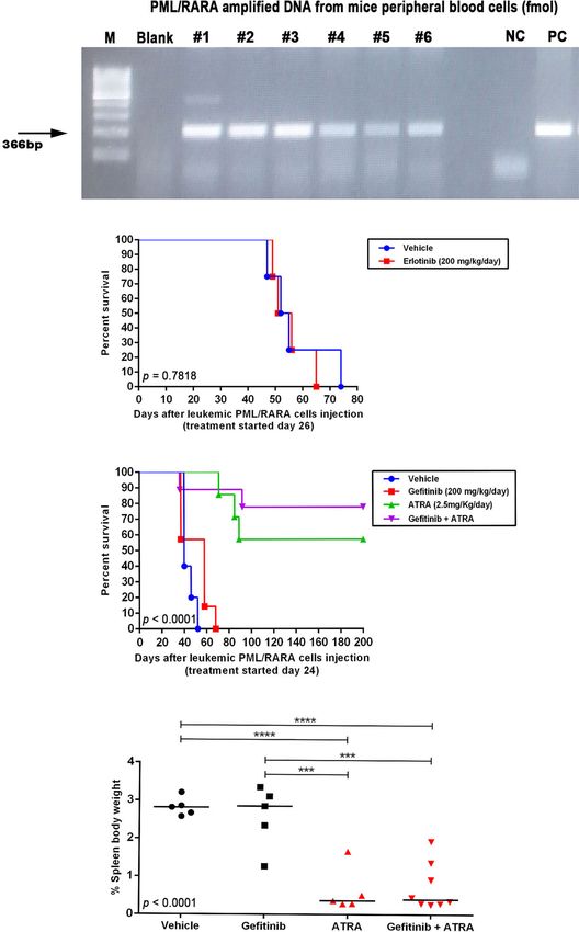
Almeida et al.                                                                                  Gefitinib Sensitizes APL-Resistant Cells to ATRA/ATO

from the BM of healthy volunteers using the CD34 Microbead Kit            CD15 (#562371, clone: 7C3.rMAb), and CD16 (#557758, clone:
(#130-046-703; Miltenyi Biotec, Auburn, CA, USA) according to             3G8) (BD Biosciences). Cells obtained from BM, or the spleen of
the manufacturer’s instructions. Plasma was obtained by                   leukemia model mice were labeled with antibodies against
centrifugation (500 g for 10 minutes) of heparinized BM aspirate          CD11b-PE (#553311, clone: M1/70), CD117-FITC (#561680,
and stored in aliquots at −80°C until use. BM CD34+ cells or plasma       clone: 2B8), Gr1-FITC (#551460, clone: 1A8; all from BD
samples from healthy donors were used as controls. The study was          Biosciences), then collected and washed and resuspended in
approved by the local Research Ethics Committee of the Medical            PBS. The percentage of positive cells and MFI were determined
School of Ribeirao Preto, University of Sao Paulo, Ribeirao Preto,        by flow cytometry.
Sao Paulo, Brazil (Reference: CAAE 05060818.9.0000.5440). All
human samples were collected after obtaining written, informed            Western Blotting
consent from patients according to the recommendations of the             Whole-cell lysates were prepared with extraction buffer (10 mM
Declaration of Helsinki.                                                  EDTA, 100 mM Tris, 10 nM Na4P2O7, 100 mM NaF, 10 mM
                                                                          Na3VO4, 2 mM phenylmethylsulfonyl fluoride, and 1% Triton X-
Apoptosis Assay, Determination                                            100) followed by centrifugation at 10 000 × g for 20 min at 4°C.
of 50% Effective Dose (ED50)                                              Protein concentration was determined with the Bradford assay and
and Combination Index                                                     50 mg of lysate was analyzed by sodium dodecyl sulfate-
To evaluate apoptosis, NB4 and NB4-R2 cells were seeded in 24-            polyacrylamide gel electrophoresis on a 10% polyacrylamide gel.
well plates at a density of 5 × 10⁵/well and treated with ATO (1–4        The proteins were transferred to a polyvinylidene difluoride
mM), gefitinib (5–40 mM, alone or in combination with 2 mM of              membrane (Amersham Hybond-P; GE Healthcare, Memphis,
ATO), erlotinib (5–120 mM), or vehicle (DMSO, 0.01%) for 24 h.            TN, USA) that was probed with antibodies against total EGFR
To detect apoptotic cells, the cells were washed and resuspended          (#2232, polyclonal, 1:1000) and phosphorylated (p-)EGFR
in 100 µL binding buffer, 3 µL Annexin V-fluorescein                       (Tyr992) (#2235, polyclonal, 1:1000) (both from Cell Signaling
isothiocyanate (BD Biosciences, San Jose, CA, USA), and 3 µL              Technology, Danvers, MA, USA); SYK (#1240, clone: 4D10, 1:1000;
propidium iodide (PI; 50 mg/ml), followed by an incubation in             Santa Cruz Biotechnology, Santa Cruz, CA, USA); and b-actin
the dark for 20 min. Fluorescence was detected by flow cytometry           (#A5441, clone: AC-15, 1:60 000; Sigma-Aldrich). Protein bands
on a FACSCalibur instrument (Becton Dickinson, San Jose, CA,              were visualized using SuperSignal West Dura Extended Duration
USA) and analyzed with FlowJo software (Treestar, Ashland,                Substrate (Thermo Fisher Scientific, Waltham, MA, USA) and the
OR, USA). A minimum of 10 000 events was acquired for each                Gel Doc XR+ system (Bio-Rad, Hercules, CA, USA).
sample. The ED50 and combination index were calculated using
CompuSyn software (CompuSyn, Paramus, NJ, USA); the latter                Enzyme-Linked Immunosorbent Assay
is a quantitative measure of drug interaction, with a value < 1 or        Plasma EGF and AREG concentrations were measured with the
> 1 indicating synergism and antagonism, respectively, and a              Human EGF Quantikine ELISA Kit (#DEGFR0) and Human
value of 1 indicating an additive effect (21).                            Amphiregulin Quantikine ELISA Kit (#DAR00; both from R&D
                                                                          Systems, Minneapolis, MN, USA), respectively, according to the
Proliferation Assay                                                       manufacturer’s instructions.
After 24 h of exposure to gefitinib, cells were washed with
phosphate-buffered saline (PBS); 4 mL cold 70% ethanol was                PCR for Genotyping
then added dropwise to the cell pellet while vortexing, followed          DNA was isolated using the QIAamp DNA Mini Kit (Qiagen,
by storage at −20°C for up to 15 days before staining. The cells          Germantown, MD, USA), according to the manufacturer’s
were resuspended and washed with staining buffer (PBS with 1%             instructions, and used as the template for PCR. The 25 mL
FBS and 0.09% NaN3), and 100 mL of cell suspension (1 × 10⁷/              reaction contained 2.5 mL of 5× reaction buffer, 2 mL of 25
ml) was transferred to a tube containing 5 mL of Ki-67-PE                 mM MgCl2, 3 mL of 10 mM dNTP mix, 2 mL of each primer
antibody (#12-5698-82, clone: SolA15; eBioscience, San Diego,             (5 mM), and 0.2 mL GoTaq DNA polymerase (Promega,
CA, USA) or PE-conjugated IgG1as an isotype control. After                Madison, WI, USA). A 3 mL volume of diluted DNA sample
incubation for 30 min, cells were washed twice, resuspended in            (300 ng) was used for conventional PCR, and amplified products
staining buffer, and analyzed by flow cytometry on a                       (20 mL) were visualized by electrophoresis with Tris–acetic acid–
FACSCalibur instrument (Becton Dickinson, San Jose, CA,                   EDTA buffer on a 1.2% (w/v) agarose gel stained with ethidium
USA) and analyzed with FlowJo software (Treestar, Ashland,                bromide under ultraviolet light. PCR amplification was
OR, USA). A minimum of 10 000 events was acquired for each                performed on a GeneAmp PCR System 9700 thermocycler
sample. Positivity is expressed as a percentage of positive cells         (Applied Biosystems, Foster City, CA, USA) under the
and mean fluorescence intensity (MFI).                                     following conditions: 94°C for 5 min; 35 cycles of 94°C for
                                                                          30 s, annealing at the melting temperature for 45 s, and 60°C for
Differentiation Assay                                                     30 s; and 60°C for 7 min. The following forward and reverse
For in vitro experiments, NB4, NB4-R2, NB4-ATOr, and NB4                  primers were used: PML, 5’-TCAAGATGGAGTCTGAGG
clone 21 cells were collected 72 h after drug treatment, washed,          AGG-3’ and 5’-CTGCTGCTCTGGGTCTCAAT-3’; and b-actin,
and resuspended in 100 mL PBS and incubated with CD11b-PE                 5’-TCTTGATAGTTCGCCATGGAT-3’ and 5’-GGTCATC
(#347557, clone: D12), CD11c-APC (#559877, clone: B-ly6),                 TTTTCACGGTTGG-3’.

Frontiers in Oncology | www.frontiersin.org                           3                              September 2021 | Volume 11 | Article 686445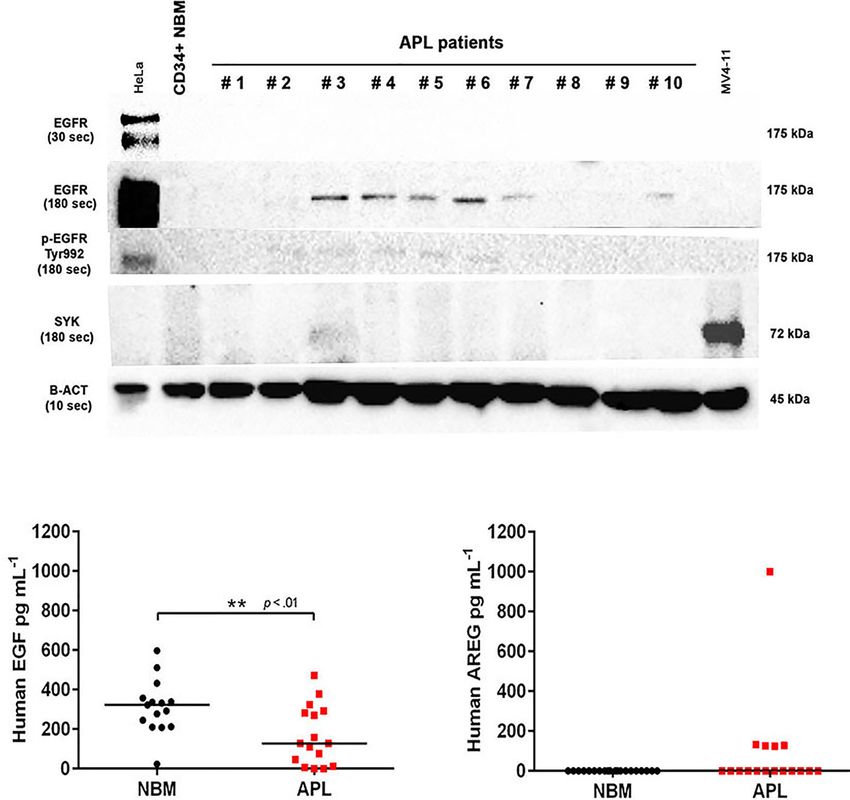
Almeida et al.                                                                                       Gefitinib Sensitizes APL-Resistant Cells to ATRA/ATO

In Vivo Experiments                                                            Statistical Analysis
To investigate the in vivo effects of the EGFR inhibitors gefitinib             Significant differences between groups were evaluated with the
or erlotinib as monotherapy or combined with ATRA, we used a                   unpaired t-test or Kruskal–Wallis test, followed by Dunn’s post
syngeneic transplantation mouse model of APL with leukemia                     hoc test. Bivariate correlation analysis with Spearman’s test was
cells from human chorionic gonadotropin (hCG)–promyelocytic                    performed to determine the correlation between BM plasma
locus–retinoic acid receptor A (PML–RARA) transgenic mice                      concentration of EGF or AREG and WBC count at the time of
(B6129 mixed background), as previously described (22, 23). The                diagnosis. The log-rank test (with Kaplan–Meier curves) was
hCG-PML-RARA mice were kindly donated by Dr. Pier Paolo                        used for overall survival analysis. Statistical analyses were
Pandolfi (Beth Israel Deaconess Medical Center, Harvard                         performed using Prism v.7.03 software (GraphPad, La Jolla,
University) and maintained at the Laboratory of Experimental                   CA, USA). The significance level was set as P ≤ 0.05.
Animal Studies (Fundaç ão Hemocentro de Ribeirão Preto–
Ribeirão Preto, SP, Brazil).
    8 to 12-week-old male wildtype (WT) littermates, weighing
approximately 30 g each, were used as transplant recipients after
                                                                               RESULTS
lethal irradiation (7 Gy split into two doses from an X-ray source i.e.,       EGFR Protein Expression Is Only Detected
two 3.5 Gy doses, 4 h apart - RS200 from Rad Source Technologies,
                                                                               in a Subset of APL Patients.
Inc., Georgia, USA). In the next day, the animals were exposed to a
                                                                               We evaluated EGFR and the non-receptor tyrosine kinase SYK [a
dose of 2% isoflurane for 5 min to induce anesthesia, and immediately
                                                                               potential off-target of EGFR inhibitors (24)] protein levels in
afterward 4 × 10⁶ viable leukemic blasts from hCG-PML-RARA mice
                                                                               bone marrow (BM) cells obtained from 21 patients diagnosed
(200 µL in PBS) were injected intravenously using a syringe with a 30-
                                                                               with APL and a pool of BM-derived CD34+ cells isolated from six
gauge disposable needle (BD Biosciences) through the retro-orbital
                                                                               healthy subjects (controls). EGFR protein was expressed in 6/21
sinus. After this time, the mice were monitored and assessed for
                                                                               (28.5%) APL patients (Figure 1A), but not in control samples.
engraftment analysis. The engraftment was confirmed by
                                                                               Notably, 4/21 (19%) APL samples also showed positivity for
conventional PCR analysis of the DNA isolated from 100 mL of
                                                                               p-EGFR (Tyr992) (Figure 1A), indicating activation of the EGF/
heparinized peripheral blood samples collected via submandibular
                                                                               EGFR signaling pathway. SYK protein expression was neither
vein by using a 5-mm lancet (Goldenrod Animal Lancet, Medipoint,
                                                                               detected in APL nor control subjects (Figures 1A and S1). In
Mineola, NY), under 2% isoflurane anesthesia.
                                                                               addition, the EGFR gene expression (by real-time quantitative
    This analysis was done once per week until the PML–RARA
                                                                               polymerase chain reaction) was not detected in any of these
fusion gene expression was detected (Figure 5A). After molecular
                                                                               specimens (Table S1).
engraftment confirmation in peripheral blood samples, mice were
randomly (by the physical method of paper sortition) assigned to               EGF and AREG Concentrations in BM
treatment groups: gefitinib (100 mg/Kg/day; n=7) and vehicle (1:10
                                                                               Plasma Samples
solution of DMSO : PBS; n=7) (Figure S1A); gefitinib (200 mg/Kg/
                                                                               Next, we sought to investigate whether the levels of EGF and AREG
day; n=5) and vehicle (n=6) (Figure S1B); erlotinib (200 mg/Kg/
                                                                               (EGFR ligands) measured in the plasma of APL patients correlates
day; n=4) and vehicle (n=4) (Figure 4B); gefitinib (200 mg/kg/day;
                                                                               with the protein expression of EGFR on APL blasts. The EGF levels
n=7), ATRA (2.5 mg/kg/day; n=7), gefitinib plus ATRA (n=9), or
                                                                               were lower in BM aspirates of APL patients at diagnosis (n=16)
vehicle (n=5) (Figure 4D). Gefitinib (100 or 200 mg/kg/day),
                                                                               compared to healthy control subjects (n=15) (median
erlotinib (200 mg/kg/day), ATRA (2.5 mg/kg/day) diluted in 200
                                                                               concentration of 127.3 ± 149 vs 322.2 ± 136 pg ml−1, P1000 pg ml−1). No correlation was observed between EGF (n=14)
until the date of spontaneous death or euthanasia. All animal
                                                                               or AREG (n=5) levels in BM plasma of APL patients and peripheral
experiments were performed at the Laboratory of Experimental
                                                                               white blood cell (WBC) counts (EGF: r2 = 0.01 – Figure S2A;
Animal Studies (Fundação Hemocentro de Ribeirão Preto –
                                                                               AREG: r2 = 0.045 – Figure S2B).
Ribeirão Preto, SP, Brazil).
    Animal experiment protocol and experimental procedures were
approved by the Animal Care and Use Committee of the Medical                   Effects of EGFR Inhibitors Alone or in
School of Ribeirao Preto of the University of Sao Paulo (Protocol no.          Combination With ATO or ATRA on APL
#016/2016) and conformed to the rules and regulations of the                   Cell Lines
National Council for Control of Animal Experimentation of Brazil               We first evaluated EGFR and SYK protein levels in NB4 and
(CONCEA). This manuscript was written following the ARRIVE                     NB4-R2 cells. As previously demonstrated, both APL cell lines
reporting guideline for reporting animal research (29). A completed            are negative for EGFR (Figure S3B) but express SYK (Figure
ARRIVE guidelines checklist is included in Table S2.                           S3A), an off-target of EGFR inhibitors. The 50% effective dose

Frontiers in Oncology | www.frontiersin.org                                4                              September 2021 | Volume 11 | Article 686445
Almeida et al. Gefitinib Sensitizes APL-Resistant Cells to ATRA/ATO
A
B C
FIGURE 1 | EGFR, p-EGFR (Tyr992), SYK, EGF, and AREG protein expression in APL patient samples. (A) Western blotting analysis of EGFR, p-EGFR (Tyr992),
SYK, and b-actin protein levels in CD34+ cells isolated from normal BM of one healthy adult volunteer and 10 representative primary APL samples collected at
diagnosis. HeLa cells served as a positive control to assess EGFR and p-EGFR (Tyr992) expression, and MV4-11 cell extracts were used as a positive control for
SYK expression. EGFR and p-EGFR (Tyr992) were detectable when exposed for 180 seconds (sec). (B) EGF levels in BM plasma samples from healthy donors
(n=15) and APL patients at diagnosis (n=16). (C) Plasma AREG levels in BM from healthy donors (n=20) and APL patients at diagnosis (n=17). (**) p < 0.01 (Mann-
Whitney U-test).
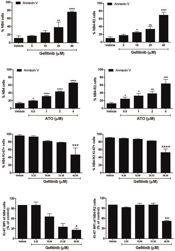
(ED50) values for the cytotoxic activities of gefitinib, erlotinib, observed at 40 mM for gefitinib treatment. Erlotinib had higher
and ATO, as well as the combination index (CI) values for ED50 values in both NB4 and NB4-R2 cells compared with
gefitinib plus ATO in the two APL cell lines, are shown in gefitinib (Table 1 and Figure S4C); therefore, the analysis of
Table 1. Since ATRA does not induce cytotoxicity at synergism with ATO was performed using only the latter
physiological concentrations after 24h in APL cell lines (30), (Table 1 and Figures 3E, F). Evaluation of Ki-67 staining
we did not perform experiments using EGFR inhibitors (MFI) revealed that gefitinib was only able to impair APL cell
combined with ATRA. Compared to the vehicle, gefitinib proliferation at concentrations > 40 µM in NB4 and NB4-R2 cells
induced increased apoptosis in NB4 cells (Figure 2A and (Figures 2E–H). In addition, treatment with gefitinib did not
Figure S4A) and NB4-R2 cells (Figure 2B and Figure S4B). alter the proliferative rate of primary human (n=3; Figure S5A)
With a dose-dependent effect, the maximum cell death was or murine (Figure S5B) APL blast cells. We next evaluated
TABLE 1 | The 50% effective dose (ED50) values of gefitinib, erlotinib, and arsenic trioxide (ATO) in NB4 and NB4-R2 cells, and combination index (CI) values of gefitinib
plus ATO at different effective levels.
Cell type ED501 CI1(Gefitinib+ATO)
Gefitinib(mM) Erlotinib ATO(mM) at ED50 at ED75 at ED90 at ED95
NB4 22.08±5.22 71.66±2.97 2.28±0.14 1.51±0.25 1.42±0.23 1.40±0.29 1.42±0.34
NB4-R2 27.0±7.35 79.95±3.36 2.91±0.94 1.35±0.19 1.10±0.41 1.00±0.56 0.99±0.65
1
ED50 and CI values were calculated by using the CompuSyn software according to the Chou and Talalay method (21).
Frontiers in Oncology | www.frontiersin.org 5 September 2021 | Volume 11 | Article 686445Almeida et al. Gefitinib Sensitizes APL-Resistant Cells to ATRA/ATO
A B
C D
E F
G H
FIGURE 2 | Effects of gefitinib or ATO monotherapy on APL cell apoptosis and proliferation. Gefitinib (5, 10, 20, and 40 mM) (A, B) and ATO (0.5, 1, 2, and 4 mM)
(C, D) treatment for 24 h decreased the fraction of apoptotic NB4 and NB4-R2 cells in a concentration-dependent manner, as determined by Annexin V/PI staining
and flow cytometry analysis. Proliferation of NB4 (E) and NB4-R2 (F) cells was reduced at gefitinib concentrations higher than twice the ED50 after 24 h of treatment.
(G, H) MFI values for NB4 and NB4-R2. Bar graphs show mean ± SD of at least three independent experiments. (*) p < 0.05, (**) p < 0.01, (***) p < 0.001, (****) p <
0.0001 (Kruskal–Wallis test, followed by Dunn’s post hoc test).
myeloid differentiation using NB4, NB4-R2, and NB4 ATOr cells differentiation (Figures 3A–D); however, the combined
(including the respective parental NB4 cells – hereafter called therapy of gefitinib plus ATRA and ATO enhanced APL cell
NB4 clone 21) when treated with ATRA (0.01, 0.1 and 1 µM) differentiation, measured by the surface expression of the
and ATO (0.5 µM), in the presence or absence of gefitinib (10 myeloid differentiation markers CD11b, CD11c, CD15, and
µM). Gefitinib monotherapy did not induce APL cell CD16 (Figures 4A–L and S6).
Frontiers in Oncology | www.frontiersin.org 6 September 2021 | Volume 11 | Article 686445Almeida et al. Gefitinib Sensitizes APL-Resistant Cells to ATRA/ATO
A B
C D
E F
FIGURE 3 | The effect of gefitinib combined with ATRA or ATO on NB4 and NB4- R2 cells. (A, B) CD11b MFI of NB4 and NB4-R2 cells treated with ATRA alone
(0.01 or 1 mM), gefitinib alone (10 mM), or ATRA plus gefitinib for 72 h. Gefitinib potentiated the myeloid differentiation-inducing effect of ATRA in NB4 (A) and NB4-
R2 cells (B); representative histograms with percentages for CD11b expression in NB4 (C) and NB4-R2 (D) cells are shown. Plots show the combination index (CI)
versus the fractional effect of gefitinib plus ATO treatment at 10:1 constant ratio in NB4 (E) and NB4-R2 (F) cells. CI values were calculated by using the CompuSyn
software according to the recommendations of Chou and Talalay (21). Data represent the mean ± SD of at least three independent experiments. (***) p < 0.001
(Kruskal– Wallis test, followed by Dunn’s post hoc test).
Frontiers in Oncology | www.frontiersin.org 7 September 2021 | Volume 11 | Article 686445Almeida et al. Gefitinib Sensitizes APL-Resistant Cells to ATRA/ATO
A B C D
E F G H
I J K L
FIGURE 4 | Gefitinib therapy sensitizes APL-resistant cells to ATRA and ATO. The APL cell lines NB4-R2 (A–D), NB4 ATOr (E–H), and the respective parental NB4
clone 21 (I–L) were treated with ATRA alone (0.1 µM), ATO alone (0.5 µM), gefitinib alone (10 µM), ATRA plus gefitinib or ATRA and ATO plus gefitinib for 72 h and
the expression of the myeloid differentiation markers CD11b, CD11c, CD15, and CD16 was measured by flow cytometry. Gefitinib potentiated the myeloid ATRA-
induced differentiation in APL cells resistant to ATRA (NB4-R2) and ATO (NB4 ATOr), as well as in NB4 clone 21 cells. Data represent the mean ± SD of at least
three independent experiments. (*) p < 0.05, (**) p < 0.01, (***) p < 0.001 (Kruskal–Wallis test, followed by Dunn’s post hoc test).
Effects of EGFR Inhibitors in an APL respective vehicle-treated controls (control group for gefitinib:
Mouse Model median survival=41 days; 95% CI=35–46 days; control group for
To investigate the in vivo effects of the ATRA/EGFR inhibitor erlotinib: median survival=52 days; 95% CI=44–59 days).
combination, we first assessed the protein levels of Egfr in APL Treatment with gefitinib at the highest dose (200 mg/kg/day)
blasts from the transgenic hCG-PML-RARA mice (pool of exhibited a trend to prolong the survival of APL mice (median
leukemic blast cells, n=5; Figure S3B). No Egfr protein survival=56 days; 95% CI=47–65 days) (P>0.05).
expression was detected in murine APL blasts, suggesting that Due to the increased survival observed in APL mice treated
any potential effects observed upon Egfr monotherapy in this with 200 mg/kg/day of gefitinib, we evaluated if the combination
model would be the result of off-target activity. APL transplanted with ATRA (2.5 mg/kg/day) could display additive or synergistic
mice were treated for 15 consecutive days with gefitinib (100 or effects in vivo. Treatment with ATRA alone or in combination
200 mg/kg/day) or erlotinib (200 mg/kg/day) after confirmation with gefitinib significantly increased the survival rate compared
of APL engraftment by PCR analysis of the peripheral blood to gefitinib or vehicle (Figure 5C), although, there was no
samples (Figure 5A). Mice treated with gefitinib (100 mg/kg/ difference in survival between mice treated with ATRA
day) (median survival=38 days; 95% confidence interval - alone versus ATRA plus gefitinib. Additionally, no differences
CI =34–45 days) (Figures S7A, B) or erlotinib monotherapy were observed in spleen weight between the treatment groups
(200 mg/kg/day; median survival=51 days; 95% CI=44–57 days) (Figure 5D). Evaluation of BM and spleen cells regarding
(Figure 5B) exhibited no prolonged survival compared to the the quantification of CD11b − CD117 + (APL blasts) and
Frontiers in Oncology | www.frontiersin.org 8 September 2021 | Volume 11 | Article 686445Almeida et al. Gefitinib Sensitizes APL-Resistant Cells to ATRA/ATO
A
B
C
D
FIGURE 5 | In vivo effects of EGFR inhibitors alone or in combination with ATRA in an APL mouse model. (A) Expression of the PML–RARA fusion gene in
peripheral blood of WT mice 3 weeks after transplantation of leukemic blasts from hCG-PML–RARA transgenic mice detected by conventional PCR. (B) Kaplan–
Meier survival curves of mice treated with vehicle (DMSO; n=4) or erlotinib at 200 mg/kg/day (n=4). Survival (C) and spleen weight-to-body weight ratio (D) of mice
treated with gefitinib [200 mg/kg/day; n=7 - (C) and n=5 - (D)], ATRA [2.5 mg/kg/day; n=7 - (C) and n=5 - (D)], gefitinib plus ATRA [n=9 - (C) and n=8 - (D)], or
vehicle [n=5 - (C, D)]. Surviving mice at 200 days post transplantation were sacrificed. (***) p < 0.001, (****) p < 0.0001 (log-rank or Kruskal-Wallis test followed by
Dunn’s post hoc test).
CD117−CD11b+/Gr1+ (myeloid mature) cells at the end of the DISCUSSION
experiment revealed no differences in the frequency of these
two leukemic cell populations comparing treatment groups In the present study, consistent with previous findings (3, 4) we
(Figure S8). These results suggest that at least in vivo, the validated that the combination between gefitinib or erlotinib
combination gefitinib plus ATRA did not enhance the with ATRA and ATO enhanced the drug-induced myeloid
differentiation effect of ATRA monotherapy. differentiation in APL cells. Moreover, we demonstrate for the
Frontiers in Oncology | www.frontiersin.org 9 September 2021 | Volume 11 | Article 686445Almeida et al. Gefitinib Sensitizes APL-Resistant Cells to ATRA/ATO
first time that this combination was effective for ATRA- and (16, 17, 40). This motivated preclinical studies to assess the
ATO-resistant APL cells, most likely due to an off-target effect. efficacy of EGFR inhibitors repurposed into AML clinics. In a
Altogether, our results provide new insights into the ongoing mouse xenograft model of AML, erlotinib suppressed tumor
challenge of developing therapies to overcome ATRA and ATO growth and increased survival (24). In contrast, in our syngeneic
resistance in APL patients. APL mouse model, neither gefitinib nor erlotinib alone induced
Despite the anti-leukemic activity of EGFR inhibitors, AML blast differentiation or prolonged survival. This was consistent
cell lines do not express EGFR (3, 4, 6, 24, 31), implying that with the lack of response observed in patients with advanced
gefitinib and erlotinib may act via EGFR-independent AML treated with gefitinib (19). In addition, other studies found
mechanisms in this malignancy. In this context, other tyrosine that erlotinib monotherapy did not affect cell differentiation or
kinases have been identified as potential targets of gefitinib and disease remission in AML patients (18, 20). In the present work,
erlotinib, including SYK (32), which predicts a favorable the combination of gefitinib and ATRA extended survival in
response to fms-like tyrosine kinase 3 (FLT3)-inhibitors in mice and reduced spleen weight-to-body weight ratio compared
AML patients harboring mutations in the FLT3 gene (33–35), to gefitinib or vehicle but was not superior to ATRA
with no EGFR expression. Notably, similarly to ATRA, the monotherapy. One limitation of our study is the high
inhibition of SYK was reported to induce differentiation of sensitivity of hCG-PML/RARA leukemic cells to ATRA
primary APL blasts (32). The NB4 APL cell lines (including monotherapy, in contrast to APL patients, which in the event
the resistant ones), present detectable expression of SYK, of relapse frequently show ATRA resistance. Further functional
although they are negative for EGFR, suggesting that the in vivo studies are necessary to verify the efficacy of EGFR or
mechanism underlying the gefitinib-induced APL sensitization other tyrosine kinases inhibitors in APL models resistant to
to ATRA and ATO might be linked to off-targets downstream ATRA or ATO treatment.
the SYK pathway, as demonstrated previously for other AML cell Although the use of EGFR inhibitors did not prolong survival
lines (32). Of note, SYK protein expression was not detected in or increase myeloid differentiation in an APL mouse model
our primary APL samples, raising the possibility of a broader sensitive to ATRA, gefitinib stimulated apoptosis, inhibited cell
than SYK spectrum of off-target effects upon EGFR inhibitor proliferation, and re-sensitized ATRA- and ATO-resistant APL
therapy in APL. Our findings highlight the relevance of cells to ATRA and ATO induced differentiation, respectively.
repurposing the FDA-approved tyrosine kinase-targeted These findings provide a basis for future studies to explore the
therapies to overcome the resistance of a specific subgroup of potential role of tyrosine kinase-targeted selective therapies in
patients with APL who are unresponsive to standard treatment. combination with standard therapy, which could be exploited to
Previous in vitro studies have shown that differentiation, cell reverse ATRA and ATO resistance in a subset of patients with
cycle arrest, and apoptosis are induced in AML cells in response APL. Finally, although some of the EGFR signaling components
to gefitinib and erlotinib, either alone, or in combination with are expressed in APL patient blasts, further investigations are
ATRA or ATO (3, 4, 6, 7, 24, 31). Consistent with these findings, necessary to understand their biological implications on
we observed that gefitinib enhanced apoptosis and suppressed leukemia progression, since the effects of EGFR inhibitors seem
proliferation in APL cell lines. Mechanistically, the activation of to be a result of off-target activities.
JNK (c-jun NH2 terminal kinase), a molecular gefitinib off-target
(36), plays a crucial role in the ATO-induced apoptosis of APL
cells (37), partially explaining the mild cytotoxicity antagonism
interaction between ATO and gefitinib. DATA AVAILABILITY STATEMENT
The EGFR protein was expressed at a low level in 28% of APL
BM samples at diagnosis, which was lower than the frequency The original contributions presented in the study are included in
reported in AML patients (89%). Besides the fact that APL the article/Supplementary Material. Further inquiries can be
represents a distinct subtype of AML, the difference in EGFR directed to the corresponding author.
expression could also emanate from different detection methods
(Western blot versus RPPA). Moreover, p-EGFR (Tyr992) was
expressed in 4/6 EGFR-positive samples, suggesting that EGFR
activation in APL is similar to other AML subtypes. There was no ETHICS STATEMENT
correlation between EGFR mRNA and protein levels, but such
disparity has been reported for other genes in different tissues The studies involving human participants were reviewed and
and may reflect post-translational modifications (38, 39). approved by Research Ethics Committee of the Medical School
Notably, we found that plasma EGF concentrations were lower of Ribeirao Preto, University of Sao Paulo, Ribeirao Preto, Sao
in the BM of APL patients compared to healthy controls, in Paulo, Brazil (Reference: CAAE 05060818.9.0000.5440). The
contrast to the higher EGF levels reported in the urine of APL patients/participants provided their written informed consent
patients at diagnosis (12). to participate in this study. The animal study was reviewed and
The clinical potential of EGFR inhibitors in AML was first approved by Animal Care and Use Committee of the Medical
revealed in patients with AML and concurrent NSCLC who School of Ribeirao Preto of the University of Sao Paulo (Protocol
achieved complete remission after gefitinib or erlotinib treatment no. #016/2016).
Frontiers in Oncology | www.frontiersin.org 10 September 2021 | Volume 11 | Article 686445Almeida et al. Gefitinib Sensitizes APL-Resistant Cells to ATRA/ATO
AUTHOR CONTRIBUTIONS 2016/17521-1), HP (grant no. 2010/16966-3), and RA-P (grant
no. 2011/18313-0).
Conceptualization, ER. Methodology, LA, IW, DP-M, CO, LC, APL,
NA, SM, VD, MN, JA-F, DN, HP, RA-P, CB, PS, ASGL, CA.
Software, LA, CB, PS. Validation, LA, IW, DP-M, CO, LC, APL.
Formal analysis, LA, IW, DP-M, CO, ER. Investigation, LA, IW. ACKNOWLEDGMENTS
Resources, ER. Data curation, LA, IW, DP-M, ER. Writing—original
draft preparation, LA and ER. Writing—review and editing, ER, LA, We thank Mara de Souza Junqueira and Bá rbara Amé lia
DP-M, RA-P, JS, EA, TO, NN. Supervision, JS, EA, TO, NN. Aparecida Santana Lemos for assistance with mouse
Revision of the manuscript. ER. Project administration, ER, PS, irradiation, and Patrı́cia Vianna Bonini Palma for advice
ASGL, CA. Funding acquisition, ER. All authors contributed to the regarding flow cytometry experiments. We thank Prof.
article and approved the submitted version. Aguinaldo Luiz Simões, Maria do Carmo Tomitão Canas, and
Ana Lú cia Pimentel (Laboratory of Biochemical Genetics,
Department of Genetics, Medical School of Ribeirao Preto –
University of Sao Paulo) for performing cell line authentication.
FUNDING
This research was funded by the Fundação de Amparo à Pesquisa
do Estado de Sao Paulo (FAPESP; grant no. 2013/08135-2), and SUPPLEMENTARY MATERIAL
FAPESP fellowships for LA (grant no. 2016/02713-2), IW (grant
no. 2015/09228-0), DP-M (grant no. 2017/23117-1), CO (grant The Supplementary Material for this article can be found online
no. 2017/08430-5), APL (grant no. 2011/17111-4), NA (grant no. at: https://www.frontiersin.org/articles/10.3389/fonc.2021.
2017/00775-3), VD. (grant no. 2013/11817-8), MN (grant no. 686445/full#supplementary-material
REFERENCES 11. Loriaux MM, Levine RL, Tyner JW, Fröhling S, Scholl C, Stoffregen EP, et al.
High-Throughput Sequence Analysis of the Tyrosine Kinome in Acute
1. Lo-Coco F, Avvisati G, Vignetti M, Thiede C, Orlando SM, Iacobelli S, et al. Myeloid Leukemia. Blood (2008) 111(9):4788–96. doi: 10.1182/blood-2007-
Retinoic Acid and Arsenic Trioxide for Acute Promyelocytic Leukemia. 07-101394
N Engl J Med (2013) 369(2):111–21. doi: 10.1056/NEJMoa1300874 12. Wu X, Qureshi IA, Liu H, Yin J, Qian X, Ruijie X. Epidermal Growth Factor in
2. Noguera NI, Catalano G, Banella C, Divona M, Faraoni I, Ottone T, et al. Acute Promyelocytic Leukemia Treated With Retinoic Acid. Int J Hematol
Acute Promyelocytic Leukemia: Update on the Mechanisms of (1995) 62(2):83–9.
Leukemogenesis, Resistance and on Innovative Treatment Strategies. 13. Freed DM, Bessman NJ, Kiyatkin A, Salazar-Cavazos E, Byrne PO, Moore JO,
Cancers (2019) 11(10):1591. doi: 10.3390/cancers11101591 et al. EGFR Ligands Differentially Stabilize Receptor Dimers to Specify Signaling
3. Miranda MB, Duan R, Thomas SM, Grandis JR, Redner RL, Jones JE, et al. Kinetics. Cell (2017) 171(3):683–95.e18. doi: 10.1016/j.cell.2017.09.017
Gefitinib Potentiates Myeloid Cell Differentiation by ATRA. Leukemia (2008) 14. Rukazenkov Y, Speake G, Marshall G, Anderton J, Davies BR, Wilkinson RW,
22(8):1624–7. doi: 10.1038/leu.2008.28 et al. Epidermal Growth Factor Receptor Tyrosine Kinase Inhibitors: Similar
4. Noh EK, Kim H, Park MJ, Baek JH, Park JH, Cha SJ, et al. Gefitinib Enhances But Different? Anticancer Drugs (2009) 20(10):856–66. doi: 10.1097/
Arsenic Trioxide (AS2O3)-Induced Differentiation of Acute Promyelocytic CAD.0b013e32833034e1
Leukemia Cell Line. Leuk Res (2010) 34(11):1501–5. doi: 10.1016/j.leukres. 15. Yang Z, Hackshaw A, Feng Q, Fu X, Zhang Y, Mao C, et al. Comparison of
2010.02.016 Gefitinib, Erlotinib and Afatinib in Non-Small Cell Lung Cancer: A Meta-
5. Boehrer S, Adès L, Galluzzi L, Tajeddine N, Tailler M, Gardin C, et al. Analysis. Int J Cancer (2017) 140(12):2805–19. doi: 10.1002/ijc.30691
Erlotinib and Gefitinib for the Treatment of Myelodysplastic Syndrome and 16. Chan G, Pilichowska M. Complete Remission in a Patient With Acute
Acute Myeloid Leukemia: A Preclinical Comparison. Biochem Pharmacol Myelogenous Leukemia Treated With Erlotinib for Non Small-Cell Lung
(2008) 76(11):1417–25. doi: 10.1016/j.bcp.2008.05.024 Cancer. Blood (2007) 110(3):1079–80. doi: 10.1182/blood-2007-01-069856
6. Stegmaier K, Corsello SM, Ross KN, Wong JS, Deangelo DJ, Golub TR. 17. Pitini V, Arrigo C, Altavilla G. Erlotinib in a Patient With Acute Myelogenous
Gefitinib Induces Myeloid Differentiation of Acute Myeloid Leukemia. Blood Leukemia and Concomitant Non–Small-Cell Lung Cancer. J Clin Oncol
(2005) 106(8):2841–8. doi: 10.1182/blood-2005-02-0488 (2008) 26(21):3645–6. doi: 10.1200/jco.2008.17.0357
7. Yadav M, Singh AK, Kumar H, Rao G, Chakravarti B, Gurjar A, et al. 18. Abou Dalle I, Cortes JE, Pinnamaneni P, Lamothe B, Diaz Duque A,
Epidermal Growth Factor Receptor Inhibitor Cancer Drug Gefitinib Randhawa J, et al. A Pilot Phase II Study of Erlotinib for the Treatment of
Modulates Cell Growth and Differentiation of Acute Myeloid Leukemia Patients With Relapsed/Refractory Acute Myeloid Leukemia. Acta Haematol
Cells via Histamine Receptors. Biochim Biophys Acta (2016) 1860 (2018) 140(1):30–9. doi: 10.1159/000490092
(10):2178–90. doi: 10.1016/j.bbagen.2016.05.011 19. Deangelo DJ, Neuberg D, Amrein PC, Berchuck J, Wadleigh M, Sirulnik LA, et al.
8. Okabe T, Okamoto I, Tamura K, Terashima M, Yoshida T, Satoh T, et al. A Phase II Study of the EGFR Inhibitor Gefitinib in Patients With Acute Myeloid
Differential Constitutive Activation of the Epidermal Growth Factor Receptor Leukemia. Leuk Res (2014) 38(4):430–4. doi: 10.1016/j.leukres.2013.10.026
in non-Small Cell Lung Cancer Cells Bearing EGFR Gene Mutation and 20. Sayar H, Czader M, Amin C, Cangany M, Konig H, Cripe LD. Pilot Study of
Amplification. Cancer Res (2007) 67(5):2046–53. doi: 10.1158/0008- Erlotinib in Patients With Acute Myeloid Leukemia. Leuk Res (2015) 39
5472.Can-06-3339 (2):170–2. doi: 10.1016/j.leukres.2014.11.022
9. AACR Project GENIE. Powering Precision Medicine Through an 21. Chou TC, Talalay P. Quantitative Analysis of Dose-Effect Relationships: The
International Consortium. Cancer Discovery (2017) 7(8):818–31. Combined Effects of Multiple Drugs or Enzyme Inhibitors. Adv Enzyme Regul
doi: 10.1158/2159-8290.Cd-17-0151 (1984) 22:27–55. doi: 10.1016/0065-2571(84)90007-4
10. Bejar R, Stevenson K, Abdel-Wahab O, Galili N, Nilsson B, Garcia-Manero G, 22. He LZ, Tribioli C, Rivi R, Peruzzi D, Pelicci PG, Soares V, et al. Acute
et al. Clinical Effect of Point Mutations in Myelodysplastic Syndromes. N Engl Leukemia With Promyelocytic Features in PML/RARalpha Transgenic Mice.
J Med (2011) 364(26):2496–506. doi: 10.1056/NEJMoa1013343 Proc Natl Acad Sci USA (1997) 94(10):5302–7. doi: 10.1073/pnas.94.10.5302
Frontiers in Oncology | www.frontiersin.org 11 September 2021 | Volume 11 | Article 686445Almeida et al. Gefitinib Sensitizes APL-Resistant Cells to ATRA/ATO
23. dos Santos GA, Abreu e Lima RS, Pestana CR, Lima AS, Scheucher PS, Thomé 34. Puissant A, Fenouille N, Alexe G, Pikman Y, Bassil CF, Mehta S, et al. SYK Is a
CH, et al. (+)a-Tocopheryl Succinate Inhibits the Mitochondrial Respiratory Critical Regulator of FLT3 in Acute Myeloid Leukemia. Cancer Cell (2014) 25
Chain Complex I and Is as Effective as Arsenic Trioxide or ATRA Against (2):226–42. doi: 10.1016/j.ccr.2014.01.022
Acute Promyelocytic Leukemia In Vivo. Leukemia (2012) 26(3):451–60. 35. Cao ZX, Guo CJ, Song X, He JL, Tan L, Yu S, et al. Erlotinib Is Effective
doi: 10.1038/leu.2011.216 Against FLT3-ITD Mutant AML and Helps to Overcome Intratumoral
24. Boehrer S, Adès L, Braun T, Galluzzi L, Grosjean J, Fabre C, et al. Erlotinib Heterogeneity via Targeting FLT3 and Lyn. FASEB J (2020) 34(8):10182–90.
Exhibits Antineoplastic Off-Target Effects in AML and MDS: A Preclinical doi: 10.1096/fj.201902922RR
Study. Blood (2008) 111(4):2170–80. doi: 10.1182/blood-2007-07-100362 36. Brehmer D, Greff Z, Godl K, Blencke S, Kurtenbach A, Weber M, et al.
25. Rego EM, He LZ, Warrell RP Jr, Wang ZG, Pandolfi PP. Retinoic Acid (RA) Cellular Targets of Gefitinib. Cancer Res (2005) 65(2):379–82.
and As2O3 Treatment in Transgenic Models of Acute Promyelocytic 37. Davison K, Mann KK, Waxman S, Miller WHJr. JNK Activation Is a Mediator
Leukemia (APL) Unravel the Distinct Nature of the Leukemogenic Process of Arsenic Trioxide-Induced Apoptosis in Acute Promyelocytic Leukemia
Induced by the PML-RARalpha and PLZF-RARalpha Oncoproteins. Proc Cells. Blood (2004) 103(9):3496–502. doi: 10.1182/blood-2003-05-1412
Natl Acad Sci USA (2000) 97(18):10173–8. doi: 10.1073/pnas.180290497 38. Kosti I, Jain N, Aran D, Butte AJ, Sirota M. Cross-Tissue Analysis of Gene and
26. Yanase K, Tsukahara S, Asada S, Ishikawa E, Imai Y, Sugimoto Y. Gefitinib Protein Expression in Normal and Cancer Tissues. Sci Rep (2016) 6:24799.
Reverses Breast Cancer Resistance Protein-Mediated Drug Resistance. Mol doi: 10.1038/srep24799
Cancer Ther (2004) 3(9):1119–25. 39. Perl K, Ushakov K, Pozniak Y, Yizhar-Barnea O, Bhonker Y, Shivatzki S, et al.
27. Piechocki MP, Dibbley SK, Lonardo F, Yoo GH. Gefitinib Prevents Cancer Reduced Changes in Protein Compared to mRNA Levels Across Non-
Progression in Mice Expressing the Activated Rat HER2/neu. Int J Cancer Proliferating Tissues. BMC Genomics (2017) 18(1):305. doi: 10.1186/s12864-
(2008) 122(8):1722–9. doi: 10.1002/ijc.23231 017-3683-9
28. Nasr R, Guillemin MC, Ferhi O, Soilihi H, Peres L, Berthier C, et al. 40. Takigawa N, Takeuchi M, Shibayama T, Yoshida I, Kawata N, Tada A, et al.
Eradication of Acute Promyelocytic Leukemia-Initiating Cells Through Successful Treatment of a Patient With Synchronous Advanced Non-Small Cell
PML-RARA Degradation. Nat Med (2008) 14(12):1333–42. doi: 10.1038/ Lung Cancer and Acute Myeloid Leukemia by a Combination of Gefitinib, Low-
nm.1891 Dose Cytarabine and Aclarubicin. Anticancer Res (2005) 25(3c):2579–82.
29. Kilkenny C, Browne WJ, Cuthill IC, Emerson M, Altman DG. Improving
Bioscience Research Reporting: The ARRIVE Guidelines for Reporting Conflict of Interest: The authors declare that the research was conducted in the
Animal Research. PloS Biol (2010) 8(6):e1000412. doi: 10.1371/journal. absence of any commercial or financial relationships that could be construed as a
pbio.1000412 potential conflict of interest.
30. Gianni M, Ponzanelli I, Mologni L, Reichert U, Rambaldi A, Terao M, et al.
Retinoid-Dependent Growth Inhibition, Differentiation and Apoptosis in Publisher’s Note: All claims expressed in this article are solely those of the authors
Acute Promyelocytic Leukemia Cells. Expression and Activation of and do not necessarily represent those of their affiliated organizations, or those of
Caspases. Cell Death Differ (2000) 7(5):447–60. doi: 10.1038/sj.cdd.4400673 the publisher, the editors and the reviewers. Any product that may be evaluated in
31. Lindhagen E, Eriksson A, Wickström M, Danielsson K, Grundmark B, this article, or claim that may be made by its manufacturer, is not guaranteed or
Henriksson R, et al. Significant Cytotoxic Activity In Vitro of the EGFR endorsed by the publisher.
Tyrosine Kinase Inhibitor Gefitinib in Acute Myeloblastic Leukaemia. Eur J
Haematol (2008) 81(5):344–53. doi: 10.1111/j.1600-0609.2008.01120.x Copyright © 2021 Almeida, Pereira-Martins, Weinhäuser, Ortiz, Cândido, Lange, De
32. Hahn CK, Berchuck JE, Ross KN, Kakoza RM, Clauser K, Schinzel AC, et al. Abreu, Mendonza, de Deus Wagatsuma, Do Nascimento, Paiva, Alves-Paiva,
Proteomic and Genetic Approaches Identify Syk as an AML Target. Cancer Bonaldo, Nascimento, Alves-Filho, Scheucher, Lima, Schuringa, Ammantuna,
Cell (2009) 16(4):281–94. doi: 10.1016/j.ccr.2009.08.018 Ottone, Noguera, Araujo and Rego. This is an open-access article distributed under
33. Weisberg EL, Puissant A, Stone R, Sattler M, Buhrlage SJ, Yang J, et al. the terms of the Creative Commons Attribution License (CC BY). The use, distribution
Characterization of Midostaurin as a Dual Inhibitor of FLT3 and SYK and or reproduction in other forums is permitted, provided the original author(s) and the
Potentiation of FLT3 Inhibition Against FLT3-ITD-Driven Leukemia copyright owner(s) are credited and that the original publication in this journal is
Harboring Activated SYK Kinase. Oncotarget (2017) 8(32):52026–44. cited, in accordance with accepted academic practice. No use, distribution or
doi: 10.18632/oncotarget.19036 reproduction is permitted which does not comply with these terms.
Frontiers in Oncology | www.frontiersin.org 12 September 2021 | Volume 11 | Article 686445You can also read


