Triassic Isopoda - three new species from Central Europe shed light on the early diversity of the group
←
→
Page content transcription
If your browser does not render page correctly, please read the page content below
Triassic Isopoda – three new species from Central Europe
shed light on the early diversity of the group
Mario Schädel, Timo van Eldijk, Herman Winkelhorst, Jelle W.F. Reumer &
Joachim T. Haug
Despite its vernacular names (e.g. ‘woodlice’) Isopoda is a group with mostly aquatic species, with most species living
in marine environments. The fossil record for isopods compared to other groups of Eucrustacea is relatively sparse.
This applies even more for the Triassic. While in the Jurassic Isopoda is relatively well represented by fossils, only
eight species have previously been described from the Triassic. In this study three new species of Isopoda are described
from two field sites in Europe: Obtusotelson summesbergeri sp. nov. and Discosalaputium aschauerorum sp. nov. from
Polzberg (Gaming, Lower Austria, Austria) and Gelrincola winterswijkensis sp. nov. from Winterswijk (Gelderland,
Netherlands). All three new species are interpreted as representatives of Scutocoxifera (ingroup of Isopoda). The
species Gelrincola winterswijkensis sp. nov. is further interpreted to be a representative of Cymothoida (ingroup of
Scutocoxifera). Most of the oldest fossils of Isopoda belong to Phreatoicidea, which is supposed to be the sistergroup
to all remaining Isopoda. Nowadays, Phreatoicidea is a small relic group, its representatives living in freshwater
environments. The new species herein presented contribute to our understanding of the diversity of Isopoda in the
Triassic and support the assumption that the transition from a dominance of Phreatoicidea towards the dominance of
the remaining lineages of Isopoda happened quite early (likely prior to the Triassic). • Key words: fossil, morphometry,
Scutocoxifera, Polzberg, Winterswijk.
Mario Schädel, Timo van Eldijk, Herman Winkelhorst, Jelle W.F. Reumer & Joachim T. Haug 2020. Triassic Isopoda –
three new species from Central Europe shed light on the early diversity of the group. Bulletin of Geosciences 95(2),
145–166 (7 figures, 4 tables, 1 appendix, 3 supplementary files). Czech Geological Survey, Prague. ISSN 1214-1119.
Manuscript received September 28, 2019; accepted in revised form February 26, 2020; published online May 16, 2020;
issued May 30, 2020.
Mario Schädel, Department of Biology II, Ludwig-Maximilians-Universität Munich, Großhaderner Str. 2, 82152
Planegg-Martinsried, Germany; mario.schaedel@palaeo-evo-devo.info • Timo van Eldijk, Groningen Institute for
Evolutionary Life Sciences, University of Groningen, Nijenborgh 7, 9747 AG, Groningen, The Netherlands • Herman
Winkelhorst, Molenstraat 14, 7122ZW Aalten, The Netherlands • Jelle W.F. Reumer, Stratigraphy and Paleontology,
Department of Earth Sciences, Utrecht University, Princetonlaan 8a, 3584 CB Utrecht, The Netherlands • Joachim
T. Haug, Department of Biology II, Ludwig-Maximilians-Universität Munich, Großhaderner Str. 2, 82152 Planegg-
Martinsried, Germany & GeoBio-Center of the LMU Munich, Richard-Wagner-Str. 10, 80333 Munich, Germany
Isopoda is a large ingroup of the crustacean group Peracari- appendages (uropods) is dissimilar to that in many other
da, comprising morphologically diverse representatives lineages of Isopoda by being styliform rather than flat.
that inhabit a variety of ecosystems such as the deep sea, Extant representatives of Phreatoicidea are only found
shores, brackish waterbodies, freshwater and one ingroup in freshwater environments (Wilson & Johnson 1999).
even managed to inhabit terrestrial ecosystems (Wägele Phreatoicideans are also interesting, as the oldest fossils of
1989, Brandt & Poore 2003, Broly et al. 2013). Even though Isopoda supposedly are representatives of this group.
the majority of representatives of Isopoda can easily be The fossil record of the group Isopoda in the Palaeozoic
recognised by the organisation of the body and the overall (see Tab. 1 for all described Palaeozoic species) can be
shape, this does not necessarily apply for all groups. One denoted as rather scarce compared to the record in the
of these groups is Phreatoicidea. Phreatoicidea is thought Meso- and Cenozoic. The oldest fossil record of the group
to be the sistergroup to all remaining ingroups of Isopoda (Hesslerella shermani Schram, 1970) is from the Middle
and is morphologically distinct in some aspects, including Pennsylvanian (Upper Carboniferous, about 300 million
the overall shape (Wägele 1989, Brusca & Wilson 1991). years old) of Mazon Creek (Illinois, USA) and is interpreted
Phreatoicideans are rather laterally compressed than dorso- as a representative of Phreatoicidea (Schram 1970).
ventrally compressed, similar to sideswimmers (Amphipo- Five species have been described from the Middle Per-
da; Nicholls 1942). Also the the shape of the posteriormost mian of Brazil (Paraná Basin, São Paulo State; Mezzalira &
DOI 10.3140/bull.geosci.1773 145
Bulletin of Geosciences • Vol. 95, 2, 2020
Table 1. Palaeozoic species of Isopoda.
Species Age Country & reference
Hesslerella shermani Schram, 1970 late Carboniferous, Moscovian Illinois, USA (Schram 1970)
Sottyella montcellensi Racheboeuf et al., 2009 late Carboniferous, Gzhelian France (Racheboeuf et al. 2009)
Pseudopalaega granulifera Mezzalira & Martins-Neto, 1992 early Permian, Artinskian Brazil (Mezzalira & Martins-Neto 1992)
Pseudopalaega microcelata Mezzalira & Martins-Neto, 1992 early Permian, Artinskian Brazil (Mezzalira & Martins-Neto 1992)
Pseudopalaega iratiensis Martins-Neto, 2001 early Permian, Artinskian Brazil (Martins-Neto 2001)
Protourda tupiensis Mezzalira & Martins-Neto, 1992 early Permian, Artinskian Brazil (Mezzalira & Martins-Neto 1992)
Protourda circunscriptia Mezzalira & Martins-Neto, 1992 early Permian, Artinskian Brazil (Mezzalira & Martins-Neto 1992)
Palaeophreatoicus sojanensis Birstein, 1962 middle Permian Russia (Birstein 1962, Schram 1980)
Protamphisopus reichelti Malzahn, 1962 late Permian, Lopingian Germany (Glaessner & Malzahn 1962)
Palaeocrangon problematicus (= Prosoponiscus Germany (Schlotheim 1820, Schauroth 1854,
late Permian, Lopingian
problematicus) (von Schlotheim, 1820) Geinitz 1861) & Great Britain (Kirkby 1857)
Martins-Neto 1992, Martins-Neto 2001, Chahud & Petri For the Triassic only eight species of Isopoda and one
2015), which are generally interpreted as representatives not formally described specimen have been reported so far
of Scutocoxifera. Scutocoxifera is an ingroup of Isopoda, (Figs 1, 2). Anhelkocephalon handlirschi is deliberately
not including Phreatoicidea, which representatives are not included, see discussion below. Records of the group
characterised by having scale-like lateral expansions to the Isopoda are missing throughout the Early Triassic (Induan
dorsal sclerites of the pereon (postocular segments 7–13) that and Olenekian).
are part of the proximal leg element (‘coxal plates’) (Dreyer The earliest Triassic record, so far, is from the early Mid
& Wägele 2002). The remainder of the Palaeozoic fossils dle Triassic of France (‘Grès à Voltzia’ Formation, ‘Voltzien
of Isopoda have all been interpreted as representatives of sandstein’, Upper Buntsandstein, Anisian) with Palaega
Phreatoicidea (Glaessner & Malzahn 1962, Schram 1980). pumila Gall & Grauvogel, 1971 (Gall & Grauvogel 1971,
A B
Figure 1. Maps with the field sites of all published findings of Isopoda from the Triassic. • A – map of Europe. • B – world map. Stars denote field sites
corresponding to the herein described fossils.
146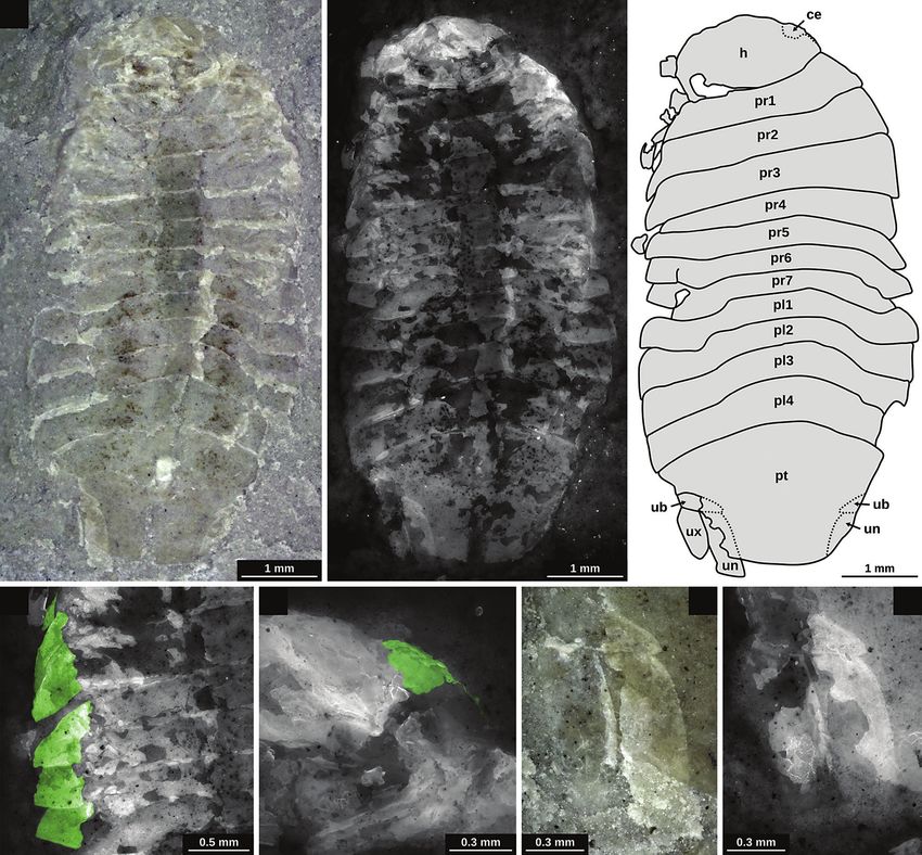
Mario Schädel et al.• Triassic Isopoda – three new species from Central Europe
Figure 2. Temporal occurrences of Triassic fossils of Isopoda based on literature information and the new fossil records within this study. Scale in
million years. The depicted time-spans (horizontal thick grey lines) do not refer to the longevity of the species, but to the temporal uncertainty of each
occurrence. Abbreviations: Ind. – Induan; Jura. – Jurassic; Olen. – Olenekian; Perm. – Permian.
Mader 1984, Gall & Grauvogel-Stamm 2005). Palaega unlikely that it stems from the ‘Keuper’ (Erfurt Formation,
pumila can be easily identified as a representative of Scu Ladinian, Longobardian substage) since ceratids are
tocoxifera. Despite the generic name, no close relation- extremely rare in sediments of this age (Hagdorn 2015).
ship to other species with the same (form-) genus name is Thus, more likely, the holotype of Isopodites triasinus
generally assumed (Feldmann & Rust 2006). is from the underlying, older, limestones of the ‘Upper
Protamphisopus wianamattensis Chilton, 1918 from Muschelkalk’ (Meissner Formation, Ladinian, Fassanian
the Middle Triassic (Anisian) Ashfield Shale (Wiannamatta substage). The Fassanian can be correlated with an abso
Group, Sydney basin, Australia) is a phreatoicidean that lute age of ca. 241.5–239.1 million years (Ogg 2012),
likely lived in a freshwater environment (Chilton 1918, consistent with the International Chronostratigraphic
Wilson & Edgecombe 2003). Protamphisopus baii Fu Chart, v. 2019/5 (Cohen et al. 2013, updated).
et al., 2010 from the Middle Triassic (Anisian) Luoping Isopodites triasinus has originally been interpreted as
fauna (Guanling Formation, Yunnan Province, China) is a representative of the group Sphaeroma (Picard 1858); von
also a representative of Phreatoicidea and likely lived in Ammon (1882) suggested a less specific interpretation and
a marine environment (Fu et al. 2010, Hu et al. 2011). created a new generic name (Isopodites) for the species.
From the Middle Triassic (Meissner Formation, Upper Due to the overall body shape, Isopodites triasinus has been
Anisian) of Southern Germany there is one specimen which interpreted as a representative of Cymothoida (Gerstaecker
has not been formally described as a species (Schöllmann & Ortmann 1901, Van Straelen 1928). Hessler (1969)
et al. 2015). This specimen has been interpreted as suggested a position within Sphaeromatidae due to the
a representative of Cymothoida based on the similarity reduction in the number of pleon segments. Gerstaecker
to some of its representatives (Schöllmann et al. 2015, & Ortmann (1901) had raised questions on the original
but see discussion below). The trace fossil Sinusichnus interpretation by Picard regarding the identity of what
seilacheri (ichnotaxon) occurs in the same stratigraphic Picard has referred to as the head. Gerstaecker & Ortman
range and is interpreted to be caused by representatives of argued that this body part likely is the anteriormost tergite
Isopoda (Knaust et al. 2016). instead of the head.
Isopodites triasinus (Picard, 1858), originally described Ferrensicus magransi Calzada & Urquiola, 1994 from
as Sphaeroma triasina Picard, 1858, has been found near the Middle Triassic (Ladinian, Longobardian substage)
the town Schlotheim (Thuringia, Germany) from the dolomitic sediments of Alcover (Tarragona province,
upper part of the Erfurt Formation (‘Keuperübergänge’; Spain) (Calzada & Urquiola 1994, Calvet & Tucker 1995).
Picard 1858). Since the holotype is located on the shell of Ferrensicus magransi has been interpreted as a repre
a ceratid cephalopod ‘Ammonites nodosus’, it seems very sentative of Archaeoniscidae Haack, 1918 (ingroup of
147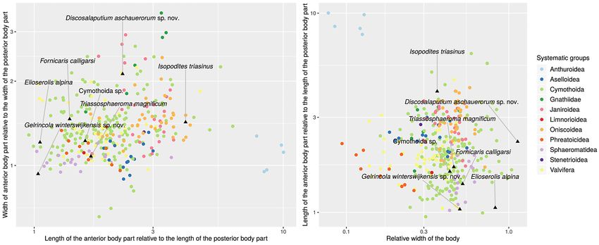
Bulletin of Geosciences • Vol. 95, 2, 2020
Sphaeromatidea) (Calzada & Urquiola 1994, Calzada et ecological specialisations range from wood boring (Daniel
al. 2011). Alongside with Ferrensicus magransi, Calzada et al. 1991), over burrowing (Matsui et al. 2011), herbivory
& Urquiola (1994) mentioned the presence of further, (Salemaa 1987), scavenging (Lowry & Dempsey 2006),
likely not conspecific, isopods from the same locality and and preying (Kaneko & Omori 2003) to parasitism (van
age (not figured). der Wal et al. 2019). Morphological features that indicate
From the middle Late Triassic (Norian) there are three parasitism are also present in the fossil record. Parasitism
species of about the same age (Alaunian to Savatian sub and the development of true larval forms are also key
stage). Two of the species, Elioserolis alpina Basso & Tin- features for the evolution of certain body shapes such as
tori, 1994 and Triassphaeroma magnificum Basso & Tintori, e.g. in Cymothoidae, Epicaridea and Gnathiidae (Smit &
1994 have been interpreted as representatives of Sphaero Davies 2004, Boyko & Wolff 2014, Nagler et al. 2017,
matidea (Basso & Tintori 1994, Brandt et al. 1999). These Schädel et al. 2019). Aside from parasitism, also other
two species come from the same locality and were found ecological factors contributed to the evolution of certain
in the ‘Calcare di Zorzino’ limestone near Zogno (Bergamo, lineages and subsequently also for the evolution of body
Italy). Elioserolis alpina has been interpreted as a repre shapes, such as the colonisation of the deep sea (Lins et al.
sentative of Serolidae due to the rough similarity in the 2012) or interstitial environments (Kim et al. 2017).
overall shape (Basso & Tintori 1994); this interpretation Here, we present three new fossil forms of Isopoda.
has been rejected by Brandt et al. (1999) who stated that All specimens are Triassic in age and come from two
no interpretation further than Sphaeromatidea is possible. field sites in central Europe (Polzberg, Winterswijk).
The other species of similar age, Fornicaris calligarisi We discuss the preserved morphological features from
Wilson & Selden in Selden et al., 2016, has also been a phylogenetic perspective. We also summarize the fossil
found in northern Italy (ca. 240 km distance, Fig. 1) and record of the group up until the end of the Triassic and
is from the ‘Dolomia di Forni’ Formation near Forni di discuss phylogenetic affinities of already described
Sotto (Province of Udine). Fornicaris calligarisi has been Triassic species. Furthermore, we attempt to analyse the
interpreted as a representative of Paramunnidae (ingroup evolution of body shapes based on Triassic and extant
of Janiroidea and Asellota) (Selden et al. 2016). Apart specimens.
from the published specimens there are probably more
Triassic asellotans from Italy pending examination (Paolo
Schirolli, personal communication, mentioned in Selden Material and Methods
et al. 2016).
The fossil record for the Jurassic as well as for the Material
Cretaceous and the Cenozoic is far more extensive. For
the Jurassic there are more than 35 formally described Three specimens form the basis of the study. Two originate
species (Meyer & Münster 1840; Milne Edwards 1843; from the Polzberg locality and one from the Winterswijk
Westwood 1854; Ammon 1882; Carter 1889; Woodward locality.
1890; Stolley 1910; Remeš 1912; Van Straelen 1928; Reiff The two specimens from the Polzberg locality were
1936; Frentzen 1937; Bachmayer 1955; Radawanski 1995; found by Birgitt Aschauer (private collector, Waidhofen
Grant-Mackie et al. 1996; Polz 1998, 2005a ,b; Guinot et an der Ybbs, Austria) and donated to the collection of
al. 2005; Polz et al. 2006; Etter 2014; Jones et al. 2014; the Natural History Museum Vienna (Naturhistorisches
Gašparič et al. 2015; Keupp & Mahlow 2017). Already in Museum Wien), accession numbers NHMW 2020/0003/
the Jurassic more ingroups of Isopoda become apparent 0001 and NHMW 2020/0003/0002. The specimen
such as some ingroups of Cymothoida (Etter 2014, Nagler from the Winterswijk locality was collected by Herman
et al. 2017) or Sphaeromatidae (Bachmayer 1955, Rada Winkelhorst (private collector, Aalten, Netherlands) and
wanski 1995). With the occurrence of fossil-rich amber donated to the Museum Naturalis, Leiden, accession
deposits in the Cretaceous, also terrestrial forms of Iso number RGM.792591.
poda (Oniscoidea) appear in the fossil record (Broly et al.
2015). Geological setting of the Polzberg locality
Modern representatives of Isopoda comprise a large
variety of different body shapes, comprising a wide range The fossil locality Polzberg (after the village near the site
from long and slender forms such as e.g. in Anthuroidea and the Polzberg Graben) also known under the alternative
(Wägele 1981) to disc-shaped forms such as e.g. in some name Schindelberg (after the adjoining mountain) is
lineages of Sphaeromatidea (Wägele 1989, Brandt & located in the municipality of Gaming, Lower Austria
Poore 2003). Distinct disc-shaped forms are also present (state), Austria. The Polzberg Graben is surrounded by the
in the fossil record (Polz 1998). Also, from an ecological mountains Föllbaumberg and Schindelberg. Two scientific
perspective there is much diversity within Isopoda. Their excavations have been carried out in 1886 (Stur 1887)
148Mario Schädel et al.• Triassic Isopoda – three new species from Central Europe
and 1909 (Glaessner 1931). Since then, there have been The fossil remains that have been so far reported
no further excavations at the original site (underground from the Winterswijk site include ray-finned fishes (Acti
quarrying). Recently, a new site yielding the same strata nopterygia; Maxwell et al. 2016), sharks (Euselachii;
has been discovered by amateur palaeontologists Birgitt Oosterink 2001) and representatives of Sauropterygia
and Karl Aschauer. (Klein 2012, Klein et al. 2015), as well as brachiopods
The fossiliferous sediments at the site are dark, organic (Brachiopoda; Oosterink 1986), various representatives of
rich laminated marls of the Reingraben Formation. The Mollusca, such as snails (Gastropoda), clams and relatives
stratum in which the fossils were found is also referred to (Bivalvia), and representatives of Cephalopoda (Oosterink
as “Aon Schichten”, “Aon Schiefer” or “Aonoides Schie 1986) and Scyphozoa (Oosterink & Winkelhorst 2013).
fer” – referring to the Trachyceras aon and/or the Tra Arthropod remains comprise representatives of Xiphosura
chyceras aonoides ammonite stratigraphic zones (Teller (Hauschke et al. 2009), Decapoda (Klompmaker & Fraaije
1891, Glaessner 1931, Hornung & Brandner 2005). The 2011) and Insecta (van Eldijk et al. 2017). Remains of
Reingraben shales can be dated to a Julian (Early Carnian, plants are mostly represented by palynomorphs (pollen
Upper Triassic) age (Hornung & Brandner 2005). The and spores; Herngreen et al. 2005b). In addition, numerous
Julian (substage of the Carnian stage) can be correlated ichnofossils have also been identified, including burrowing
with an absolute age of 235.4 to 237 million years (Ogg traces (Knaust 2013), terrestrial track ways (Diedrich
2012). 2001) and swimming traces (Schulp et al. 2017). An initial
The proximity to the town Lunz am See led to the stratigraphy of the Winterswijk quarry was published
attribution terms like “Lunz Lagerstätte”. This is problem by Oosterink (1986), who identified 39 stratigraphic
atic, as the Lunz Lagerstätte, which is famous for its horizons. Subsequently, additional layers unearthed near
abundant plant remains (e.g. Pott & Krings 2010), is of the top of the profile were identified by the Working Group
a different age and there is also no close geological con Muschelkalk Winterswijk and numbered in accordance
nection between the sites (Forchielli & Pervesler 2013). with the system of Oosterink (1986) (Maxwell et al. 2016).
The fossil remains that have been reported so far from The specimen RGM.792591 described here was found by
the Polzberg site include ray-finned fishes (Actinopterygia; HW in layer 43, this layer is particularly noteworthy since
Abel 1906, Griffith 1977), a lungfish (Dipnoi; Teller it has already yielded several insect remains in addition
1891), sea urchins (Echinoida; Glaessner 1931), as well as to exquisitely preserved fish fossils (Maxwell et al. 2016,
numerous representatives of Mollusca, such as different van Eldijk et al. 2017). There seem to be some unresolved
forms of Cephalopoda (Ammonoidea; Doguzhaeva et al. issues regarding the precise dating of the top section of
2007a, and Colleoidea; Doguzhaeva et al. 2006, 2007b), the quarry to which layer 43 belongs (Maxwell et al.
but also snails (Gastropoda) and clams and relatives 2016, van Eldijk et al. 2017). Hagdorn & Simon (2010)
(Bivalvia) (including abundant planktic representatives considered the top section of the stratigraphy to belong to
of the group Halobia, ‘Halobia shale’; Glaessner 1931). the Illyrian substage of Anisian, based on the occurrence
Eucrustacean remains include thylacocephalans (Glaessner of the bivalve Neoschizodus orbicularis. However, they
1931, Forchielli & Pervesler 2013), paenaeoidean prawns, acknowledged that this dating is not compatible with the
glypheoideans and polychelidan lobsters (Glaessner palynological results obtained by Herngreen et al. (2005a,
1931). Also rare plant remains have been found from the b), which indicate that the top section of the stratigraphy
same sediment (B. Aschauer, personal communication). belongs to the Bithynian substage of the Anisian.
Regardless, specimen RGM.792591 can thus be correlated
Geological setting of the Winterswijk locality with confidence to be Anisian. Although it would seem
most probable that layer 43 is of Bithynian age as the
Early Anisian (Middle Triassic) sediments belonging to palynological date assigned by Herngreen et al. (2005a,
the Lower Muschelkalk Vossenveld Formation are b) is based on two separate stratigraphic marker species,
exposed in the Winterswijk quarry in the easternmost further stratigraphic studies are required. The Anisian can
Netherlands (Hagdorn & Simon 2010). These outcrops of be correlated with an absolute age of 245.9 to 237 million
finely laminated micritic limestone consist of alternating years (Ogg 2012).
marly limestones, dolomites, clayey marls and dolomitic
clay layers (Borkhataria et al. 2006, Klein et al. 2015, Imaging
Maxwell et al. 2016). The sediments were deposited near
the margin of the epicontinental Germanic Basin and The specimens from the Polzberg locality were photo
alternately document shallow marine environments and graphed using a Canon EOS 70D DSLR camera with
intertidal mudflats with algal laminates and polygonal a Canon MP-E 65 mm macro objective and MT24 twin
mudcracks (Klein 2012, Oosterink & Winkelhorst 2013, flashes. In some cases, a grid of multiple images was
Klein et al. 2015). recorded to overcome the limit of field of view at higher
149Bulletin of Geosciences • Vol. 95, 2, 2020
magnifications. Stereoscopic images were recorded by tilt Results
ing the macrophotography setup relative to the specimen.
The specimen from the Winterswijk locality was All new taxonomic names were registered in the Zoo-
photographed using a Keyence BZ9000 digital microscope. Bank database (International Commission on Zoological
Incident white light was used to obtain microscopic Nomenc lature, http://zoobank.org) prior to the publi-
images and incident fluorescent light was used to obtain cation.
epifluorescence microscopic images. For the fluorescence
microscopy the fossils were excited by light of 470 nm
wavelength (GFP-filter) and 545 nm wavelength Systematic part
(TRITC-filter) (e.g. Metz et al. 2015). To overcome the
limitations of depth of field and field of view, for both Euarthropoda sensu Walossek (1999)
kinds of illumination, a grid of image stacks with images Eucrustacea sensu Walossek (1999)
of different levels of focus was recorded (e.g. Haug et al. Peracarida Calman, 1904
2011). Isopoda Latreille, 1817
Scutocoxifera Dreyer & Wägele, 2002
Image processing
Obtusotelson gen. nov.
Images of different levels of focus were combined to overall
in-focus images (‘focal plane merging’) using CombineZP http://zoobank.org/10472CD8-97E6-4B84-9795-374E63
(GPL). Grids of in-focus images were combined to larger AA05C2
images (‘panoramic stitching’) using TrakEM (ImageJ,
GPL). The resulting images were optimised for colour and Type species. – Obtusotelson summesbergeri sp. nov. (type
contrast using GIMP (GPL). Drawings were done using and only species).
Inkscape (GPL). The map was created using QGIS (GPL)
data from naturalearthdata.com (public domain). The map Etymology. – From Latin obtusus (= blunt) and telson,
was exported as a vector file and postprocessed using referring to the shape of the pleotelson in the holotype of
Inkscape (GPL). the type species.
Measurements and analysis Diagnosis. – Not applicable since monotypic.
Measurements were performed using Adobe Acrobat Remarks. – The name Obtusotelson is introduced, exclu-
Reader, ImageJ and Inkscape (using the Bezier tool, the sively, to allow a representation of the species summe-
distance between the cursor and the last click is given in sbergeri sp. nov. under a Linnean framework (by serving
the status bar). Measurements (Suppl. files 1, 2) of extant as the generic name of the type species) (e.g. Béthoux
specimens were gathered from Nicholls (1942, 1943) and 2009). Obtusotelson does not represent a monophyletic
Richardson (1905); measurements of fossil specimens group of organisms, since only a single species is included
were gathered from Basso & Tintori (1994), Picard (1858), (monotypic). Yet, Obtusotelson may serve as a ‘group
Schöllmann et al. (2015) and Selden et al. (2016). Graphs name in advance’ if, in the future, another species is
were created using R including the packages readr, ggplot2, found to be the sister-species to the type species of Obtuso-
ggrepel and gridExtra (Suppl. file 3). telson.
Fossils of Protamphisopus baii and Protamphisopus
wianamattensis, as well as one of the herein presented Obtusotelson summesbergeri sp. nov.
fossils, are laterally compressed and thus could not be Figure 3A–C
measured for dorsal/ventral aspect of the body shape. Fer
reniscus magransi is not included in the analysis, because http://zoobank.org/E449512B-FDF7-45F6-AB88-E41404
there appears to be a mismatch between the reconstructive 666FEA
drawing in the original description (Calzada & Urquiola
1994) and a more recent photograph of the specimen Types. – Holotype and only type, NHMW 2020/0003/0001,
(Institut Cartogràfic i Geològic de Catalunya 2018, Naturhistorisches Museum Wien (Natural History Museum
collection number 52506), that could greatly affect the Vienna), Austria.
outcome of the analysis. Isopodites triasinus is only figured
with an apparently highly stylised, miniature scaled, Type horizon and locality. – Reingraben Formation,
drawing and the measurement must thus be interpreted Julian (Carnian), 235.4 to 237 million years (Ogg 2012);
with caution. Polzberg, Gaming, Lower Austria, Austria.
150Mario Schädel et al.• Triassic Isopoda – three new species from Central Europe
A
B
C
Figure 3. Holotype of Obtusotelson summesbergeri sp. nov. (NHMW 2020/0003/0001); A – macroscopic image, positive side (part); B – macroscopic
image, negative side (counterpart); C – drawing based on part and counterpart of the holotype. Abbreviations: a – antenna; h? – putative remains of the
head; p1–7 – pereopods 1–7; pl1–5 – pleon segments 1–5; pr1–7 – pereon segments 1–7; pt – pleotelson; un – uropod endopod; ux – uropod exopod.
Material. – Holotype only. corners; pleotelson with median ridge, posterior margin
of pleotelson straight in the median portion (‘truncated’
Etymology. – After Dr. Herbert Summesberger, retired appearance); uropod posteriorly extending to the level of
palaeontologist who worked at the Natural History Mu the posterior margin of the pleotelson, endopod with
seum in Vienna. straight distal margin.
Diagnosis. – Body about half as wide as long; coxal plates Description. – Preservation of the holotype: Strongly
triangular; dactyli with truncated distal ends; pleon tergites comp ressed remains of the exoskeleton; specimen
with concave posterior margins and pointed posterolateral accessible in dorsolateral view; remains located on two
151Bulletin of Geosciences • Vol. 95, 2, 2020
corresponding slabs of rock matrix (part and counterpart); Table 2. Measurements of Obtusotelson summesbergeri sp. nov.
counterpart with only few pieces of exoskeleton; head (NHMW 2020/0003/0001).
not preserved or represented by inconspicuous fragments;
Body part Length Width
remains of putative antennal elements visible on the
6.93 mm
counterpart; seven appendages of the pereon (pereopods) complete body 15.29 mm
(preserved part)
preserved on the left side of the body (not all of them might pereon segment 1 1.75 mm (along the midline) not accessible
be legs of the left body side).
pereon segment 2 1.26 mm (along the midline) not accessible
D e s c r i p t i o n o f t h e h o l o t y p e : The body is
composed of a functional head (ocular segment and pereon segment 3 1.24 mm (along the midline) not accessible
postocular segments 1–6, cephalothorax) and a trunk pereon segment 4 1.16 mm (along the midline) not accessible
(postocular segments 7–19). The trunk is divided into two pereon segment 5 1.02 mm (along the midline) not accessible
tagmata: the pereon (postocular segments 7–13) and the pereon segment 6 0.74 mm (along the midline) not accessible
pleon (postocular segments 14–19). The last segment of the
pereon segment 7 0.53 mm (along the midline) not accessible
pleon is conjoined with the telson, forming a pleotelson.
pleon segment 1 0.81 mm (along the midline) not accessible
Body longer than wide, about half as wide as long.
Measurements are listed in Tab. 2. pleon segment 2 0.59 mm (along the midline) not accessible
The trunk bears one pair of appendages per segment on pleon segment 3 0.40 mm (along the midline) not accessible
the ventral side. The appendages of the pereon segments pleon segment 4 0.67 mm (along the midline) not accessible
(pereopods) consist of seven elements (from proximal to pleon segment 5 0.84 mm (along the midline) not accessible
distal: coxa, basipod, ischium, merus, carpus, propodus,
3.75 mm
dactylus). pleotelson 3.88 mm
(preserved part)
Pereon with all seven segments bearing distinct dorsal uropod endopod
1.80 mm 0.63 mm
sclerites (tergites). Pereon segments 2 to 7 with coxa con- length
joined with the tergite of the same segment forming uropod exopod 1.53 mm (preserved part) not accessible
a scale-like lateral sclerite (coxal plate). Tergite of pereon
segment 1 with rounded lateral margin; posterior margin
slightly convex, no coxal plates. Tergite of pereon segment view (posterior side); carpus triangular in the accessible
2 slightly shorter than preceding tergite, about the same view (posterior side), smaller than the merus; propodus
width; posterior margin slightly convex; with coxal plate; elongated, much longer than wide; dactylus shorter and
coxal plate triangular with pointed posterolateral corner, narrower than propodus, with truncated distal end. Third
ridge parallel to the anterolateral margin. Tergite of pereon preserved leg with merus, carpus, propodus and dactylus
segment 3 about as long as preceding tergite, slightly wider preserved; sub-similar to the preceding leg; distal part of
than preceding tergite; posterior margin almost straight; the propodus not preserved; dactylus with truncated distal
with coxal plate; coxal plate triangular with pointed end. Fourth preserved leg with merus, carpus, propodus
posterolateral corner, ridge parallel to the anterolateral and dactylus preserved; overall much slenderer than the
margin. Tergite of pereon segment 4 about as long as preceding leg; preserved part of the merus short; carpus
preceding tergite, slightly wider than preceding tergite; elongated, longer and slenderer than in the preceding
posterior margin almost straight; coxal plate not preserved. legs; propodus elongated, longer than carpus; dactylus
Tergite of pereon segment 5 about as long as preceding much shorter and narrower than propodus and with
tergite, about the same width as the preceding tergite; coxal truncated distal end. Fifth preserved leg with merus and
plate triangular with pointed posterolateral corner, straight carpus preserved; merus short; carpus elongated similar
posterior margin. Tergite of pereon segment 6 slightly to the carpus of the preceding leg. Sixth preserved leg
shorter than the preceding tergite, about the same width with carpus, propodus and dactylus preserved; carpus
as the preceding tergite; posterior margin slightly concave; elongated; propodus elongated with dorsal side slightly
coxal plate not preserved. Tergite of pereon segment 7 convex, distinctly longer than propodus of leg 4; dactylus
shorter than the preceding tergite; coxal plate not preserved. much shorter and narrower than propodus and with
Anteriormost preserved leg with merus, carpus, pro truncated distal end. Seventh preserved leg with only one
podus and dactylus preserved; merus with oblique distal elongated element of uncertain affinity (counterpart).
margin in accessible view (posterior side); carpus triangu- Pleon with all seven segments bearing distinct dorsal
lar and smaller than the merus; propodus long and with sclerites (tergites). Tergite of pleon segment 1 longer than
convex dorsal side; dactylus much shorter and nar the preceding tergite of pereon segment 7. Tergite of pleon
rower than propodus and with truncated distal end. segment 2 longer than the preceding tergite; posterior
Second preserved leg with merus, carpus, propodus and margin convex or straight in the median part and concave
dactylus preserved; merus triangular in the accessible in the lateral parts, posterolateral corner pointed. Tergite of
152Mario Schädel et al.• Triassic Isopoda – three new species from Central Europe
pleon segment 3 shorter than the tergite of the preceding distinct from the tergites. The distinctness and relative size
segment; posterior margin convex or straight in the median of these sclerites (about as long or longer than the lateral
part and concave to the lateral side; lateral margin straight; margin of the corresponding tergite) suggest that these
posterolateral corner distinctly pointed. Tergite of pleon are coxal plates rather than tergopleura or lateral aspects
segment 4 longer than the preceding tergite, posterior of ring-shaped coxae (cf. illustrations in Gruner 1954 and
margin straight to slightly convex in the median part Dreyer & Wägele 2002). Coxal plates are autapomorphic
and concave to the lateral side; lateral margin straight; for Scutocoxifera (Dreyer & Wägele 2002). The truncated
posterolateral corner distinctly pointed. Tergite of pleon distal ends of the dactyli indicate the presence of prominent
segment 5 longer than the preceding tergite; posterior claws. A pair of two claws on the distal end of the dactylus
margin straight to slightly convex in the median part and is plesiomorphic for Scutocoxifera (Wägele 1989).
concave in the lateral part. Pleotelson roughly trapezoid
in dorsal view, with straight lateral margins and straight Discosalaputium gen. nov.
posterior margin (‘truncate’ sensu e.g. Bruce 1986),
anterior side wider than the posterior side, anterior margin http://zoobank.org/C93F3340-35B0-48FF-8B81-93E4DE
with straight median part and concave lateral parts, ridge 43890D
along the midline, without conspicuous ornamentation.
Appendages of the pleotelson (uropods) consisting Type species. – Discosalaputium aschauerorum sp. nov.
of a proximal element (basipod) and two distal elements (type and only species).
(endopod, median and exopod, lateral) that both originate
from the basipod. Endopod broad, wider in the distal part. Etymology. – From Latin discus (= disc) and salaputium
with ‘truncated tip’ (with straight distal margin), median (= manakin), referring to the overall shape and size of the
margin slightly convex. Exopod about as long as the holotype of the type species.
endopod or slightly shorter.
Diagnosis. – Not applicable since monotypic.
Remarks. – The legs that are only present in the anterior
part of the body indicate a tagmatisation of the trunk into Remarks. – The name Discosalaputium is introduced,
two functional units (pereon and pleon; autapomorphy of exclusively, to allow a representation of the species
Malacostraca (Walossek 1999). The posteriormost pair aschauerorum sp. nov. under a Linnean framework (by
of appendages, together with the (pleo-) telson, forms serving as the generic name of the type species) (e.g. Bé
a tail fan (autapomorphy of Eumalacostraca) (Ax 2000). thoux 2009). Discosalaputium does not represent a mono-
Apomorphic features of Isopoda are often not visible phyletic group of organisms, since only a single species
in fossil specimens. Features that are not exclusive for is included (monotypic). Yet, Discosalaputium may serve
Isopoda but part of the ground pattern of Isopoda are: as a ‘group name in advance’ if, in the future, another
posteriormost pleon segment conjoined with the telson species is found to be the sister-species to the type species
(‘pleotelson’) (Wägele 1989, Brusca & Wilson 1991). Head of Discosalaputium.
shield forms a capsule, not constituted by postocular
segment 7 (postocular segment 7 can constitute to the head Discosalaputium aschauerorum sp. nov.
shield in some ingroups of Isopoda) (Ax 2000, Haug 2011). Figure 4A–D
A dorsoventral flattened body is often stated as apomorphic
for Isopoda (e.g. Wägele 1989). However, a dorsoventral http://zoobank.org/CD2A1601-94A6-4271-957F-A2658D
flattened body cannot be reconstructed for the ground 0EEFBA
pattern of Isopoda without assuming convergence in the
earliest lineages within Isopoda (Brusca & Wilson 1991, Types. – Holotype and only type, NHMW 2020/0003/0002,
Wilson 1996). Nevertheless, a dorsoventral flattened body Naturhistorisches Museum Wien (Natural History Museum
can be reconstructed as an apomorphy for an ingroup of Vienna), Austria.
Isopoda (Isopoda nec Phreatoicidea). In Obtusotelson
summesbergeri sp. nov. a flattened body shape is not Type horizon and locality. – Reingraben Formation,
conspicuous, as legs of only one body side are preserved. Julian (Carnian), 235.4 to 237 million years (Ogg 2012);
Yet, in the posterior part of the body, especially in the Polzberg, Gaming, Lower Austria, Austria.
pleotelson, it is apparent that the outline of the right body
side is not constituted by the midline of the body and thus, Material. – Holotype only.
that the body is indeed dorsoventrally flattened.
In the holotype of Obtusotelson summesbergeri sp. Etymology. – In honour of the private collectors and
nov. the pereon segments 2–5 bear lateral sclerites that are amateur palaeontologists Karl and Birgitt Aschauer, who
153Bulletin of Geosciences • Vol. 95, 2, 2020
provided access to two of the herein presented specimens curved ridge on the expansion from centre of the base of
and donated them to the collection of the Geological the expansion to the posterolateral corner, posteromedian
Service of Austria. corner with distinct angle. Pereon segment 3 wider than
preceding segment, shape similar to preceding segment but
Diagnosis. – Body with semicircular outline in dorsal anterior margin overall straight with convex proportion in
view, about as wide as long, dorsoventrally flattened; the centre. Pereon segment 4 about as wide as preceding
coxal plates with rounded anterolateral side and angled segment, shape similar to preceding segment. Pereon
posterolateral corner; pleon without free (distinct, not segment 5 about as wide as preceding segment, shape
conjoined with other sclerites) tergites; pleotelson roughly similar to preceding segment but without concave portion
triangular; uropods large. in the posterior margin. Pereon segment 6 narrower than the
preceding segment, anterior margin evenly convex, pos-
Description. – Preservation of the holotype: Strongly terior margin strongly concave, lateral expansion narrower
compressed remains of the exoskeleton; specimen access than in preceding segments and pointing posterolaterally.
ible in dorsal view; remains located on two corresponding Pereon segment 7 narrower than the preceding segment,
slabs of rock matrix (part and counterpart). Head and shape similar to preceding segment but with strongly
appendages of the trunk (except for the posteriormost convex anterior margin, lateral expansion narrow and
ones) not preserved. pointing more posteriorly than in the preceding segment.
D e s c r i p t i o n o f t h e h o l o t y p e : The body is Pleon segments 1–5 not discernible in the holotype.
composed of a head (ocular segment and postocular Pleon segment 6 conjoined with the telson forming the
segments 1–6, not preserved in the holotype) and a trunk pleotelson. Pleotelson roughly triangular in dorsal view,
(postocular segments 7–19). The trunk is divided into two anterior margin straight in the median part, anterior margin
functional tagmata: the pereon (postocular segments 7–13) oblique (anteromedian to posterlateral) in the lateral part.
and the pleon (postocular segments 14–19). Body with Uropod large, located lateral to the posterolateral
semicircular outline in dorsal view, about as wide as long. margin of the pleotelson, anterolateral corner close to the
Measurements are listed in Tab. 3. lateral expansion of pereon segment 7; elements of the
Pereon segment 1 with concave anterior margin and uropod (basipod, endopod and exopod) not discernible.
convex posterior margin, lateral part not distinct from
median part (no coxal plate). Lateral margin anterolaterally Remarks. – The posteriormost pair of appendages, together
projected and straight to slightly convex. Pereon segment with the (pleo-) telson, forms a tail fan (autapomorphy
2 distinctly wider than preceding segment, with concave of Eumalacostraca; Ax 2000). Although the low relief
anterior margin, posterior margin overall straight with con preservation of the holotype does not allow to estimate
cave part in the median part; lateral expansions distinct from the exact dorsoventral aspect of the living animal, due
the (rest of) the tergite by a notch on the posterior margin to the positioning of the holotype, it can be concluded
(possibly representing coxal plates); lateral expansions with that Discosalaputium aschauerorum sp. nov. was wider
slightly convex lateral margin, anterolateral corner rounded, than high. A dorsoventrally flattened body can be seen
as apomorphic for a monophyletic ingroup of Isopoda
Table 3. Measurements of Discosalaputium aschauerorum sp. nov. (Isopoda nec Phreatoicidea; see discussion above). The
(NHMW 2020/0003/0002). lateral expansions in the pereon are distinct from the
tergite by a notch on the posterior margin. Also, the shape
Body part Length Width of the expansion and the shape and position of the ridge
on the dorsal side of the expansion resembles much that
7.67 mm 8.30 mm (reconstructed
complete body
(preserved part) from one body side) of representatives of Scutocoxifera, indicating that the
pereon segment 1 not accessible 6.13 mm
expansion is likely a coxal plate, which is autapomorphic
for Scutocoxifera (Dreyer & Wägele 2002). However,
pereon segment 2 0.52 mm 7.98 mm
this would imply that the coxal plate is conjoined with
pereon segment 3 0.64 mm 4.19 mm the tergite, as it is e.g. in most adult land-living isopods
8.38 mm (reconstructed (Oniscoidea; Gruner 1954). To test this assumption it
pereon segment 4 0.63 mm
from one body side)
would be necessary to inspect the coxal area on the ventral
8.28 mm (reconstructed
pereon segment 5 0.59 mm
from one body side)
side of the body. All dorsal sclerites in the holotype of
8.18 mm (reconstructed
Discosalaputium aschauerorum sp. nov. can be identified
pereon segment 6 0.53 mm as parts of the pereon, due to the likely presence of coxal
from one body side)
pereon segment 7 0.48 mm 3.95 mm plates. This implies that aside from the pleotelson, all
dorsal sclerites of the pleon are reduced or conjoined
pleotelson 2.40 mm 3.68 mm
with the pleotelson. A reduction in the number of free
154Mario Schädel et al.• Triassic Isopoda – three new species from Central Europe
A B
C D
Figure 4. Holotype of Discosalaputium aschauerorum sp. nov. (NHMW 2020/0003/0002); A – macroscopic image, positive side (part), dotted lines
mark areas where artificial matrix has been added digitally for aesthetic reasons; B – macroscopic image, negative side (counterpart); C – red-cyan
stereo anaglyph based on macroscopic images, positive side (part); D – drawing based on part and counterpart of the holotype. Abbreviations: pr1–7 –
pereon segments 1–7; pt – pleotelson; un – uropod endopod.
pleon segments, in combination with a semicircular body aschauerorum sp. nov., do only occur in Sphaeromatoidea
outline, can be seen in various lineages of Sphaeromatidea Brandt & Poore 2003 (see Fig. 5A–B, D–E). The absence
(e.g. Plakarthriidae, Bathynataliidae and some lineages of of free dorsal pleon sclerites could be interpreted as
Sphaeromatidae) (Wägele 1989, Brusca & Wilson 1991, apomorphic for Sphaeromatoidea. Yet, because no other
Brandt & Poore 2003). Yet, conditions where not even one apomorphic features are visible in the holotype, we suggest
pleon tergite is distinct from the telson are extremely rare to be careful with a possible affinity of Discosalaputium
(e.g. Ancinus belizensis Kensley & Schotte, 1987). aschauerorum sp. nov. with Sphaeromatoidea.
Very wide uropods, in combination with a semicircular The morphology of the lateral aspect of the tergites (or
outline of the body, like in the holotype of Discosalaputium the coxal plates) in Discosalaputium aschauerorum sp.
155Bulletin of Geosciences • Vol. 95, 2, 2020
nov. is very similar to that in Elioserolis alpina. Yet, in the Type species. – Gelrincola winterswijkensis sp. nov. (type
reconstruction of Elioserolis alpina by Basso & Tintori and only species).
(1994) there are two free pleon tergites (the photograph is
of bad quality and the specimen should be restudied as some Etymology. – From Latin Gelria (= Gelderland, province of
parts of the dorsal morphology are unclear). Also there is the Netherlands where the field site is located) and incola
an age gap of 18 million years between the occurrences of (= inhabitant), referring to the field site of the holotype of
both species, making it unlikely that they are conspecific the type species.
in the sense of a biological species concept.
Diagnosis. – Not applicable since monotypic.
Cymothoida Wägele, 1989
Remarks. – The name Gelrincola is introduced, exclusively,
Gelrincola gen. nov. to allow a representation of the species winterswijkensis sp.
nov. under a Linnean framework (by serving as the generic
http://zoobank.org/F7EA70DD-56B0-411C-AE07-0EE68107E515 name of the type species) (e.g. Béthoux 2009). Gelrincola
A B
C
D E
Figure 5. Habitus of different representatives of Sphaeromatidae, pleon and telson region marked with a darker shade of grey. • A – Platysphaera
membranata, redrawn from Bruce 1994. • B – Discidina banawarra, redrawn from Bruce 1994. • C – Discosalaputium aschauerorum sp. nov.,
reconstruction, dotted lines mark body parts which are not preserved in the holotype. • D – Apemosphaera naranagi, redrawn from Bruce 1994. •
E – Cassidinidea clarkae, redrawn from Schotte & Kensley 2005.
156Mario Schädel et al.• Triassic Isopoda – three new species from Central Europe
A B C
D E F G
Figure 6. Holotype of Gelrincola winterswijkensis sp. nov. (RGM.792591); A – microscopic image, white light microscopy; B – microscopic image,
epifluorescence microscopy; C – drawing based on multiple microscopic images; D – detail of the left lateral side of the pereon, (green – coxal plates);
E – detail of the lateral head region, epifluorescence microscopy, (green – compound eye); F – detail of the left uropod, white light microscopy;
G – detail of the left uropod, epifluorescence microscopy. Abbreviations: ce – compound eye; h – head; pl1–4 – pleon segments 1–4; pr1–7 – pereon
segments 1–7; pt – pleotelson; ub – uropod basipod; un – uropod endopod; ux – uropod exopod.
does not represent a monophyletic group of organisms, Types. – Holotype and only type, RGM.792591, Museum
since only a single species is included (monotypic). Yet, Naturalis, Leiden, Netherlands.
Gelrincola may serve as a ‘group name in advance’ if, in
the future, another species is found to be the sister-species Type horizon and locality. – Lower Muschelkalk Vossen
to the type species of Gelrincola. veld Formation, Anisian, 245.9 to 237 million years (Ogg
2012); Winterswijk, Gelderland, Netherlands.
Gelrincola winterswijkensis sp. nov.
Figure 6A–G Material. – Holotype only.
http://zoobank.org/534AEBC1-F99E-43D1-B09D-6030 Etymology. – After the name of the town ‘Winterswijk’,
A3A0185C the field site where the holotype was found.
157Bulletin of Geosciences • Vol. 95, 2, 2020
Diagnosis. – Body obround in dorsal view, about half as posteriorly increasing in width, anterior margin slightly
wide as long; head more than twice as long as the sub concave, posterior margin straight, lateral margin antero
sequent pereon tergite; pleon segment 5 without free tergite; laterally projected and straight to slightly convex. Pereon
pleotelson wider than long; uropod basipod triangular with segment 2 wider than preceding segment, anterior margin
median angle located ventral to the pleotelson; uropod straight, posterior margin straight throughout most of the
endopod posteriorly protruding beyond the posterior width and concave at the lateral-most part. Pereon segment 3
margin of the pleotelson. similar to preceding segment but posterior margin slightly
concave. Pereon segment 4 shorter than the preceding
Description. – Preservation of the holotype: Strongly segment, narrower or about as wide as preceding segment.
compressed remains of the exoskeleton; specimen access- Pereon segment 5 about as long as preceding segment,
ible in dorsal view. wider than preceding segment, anterior margin convex,
D e s c r i p t i o n o f t h e h o l o t y p e : The body is posterior margin concave, lateral margins rounded. Pereon
composed of a head (ocular segment and postocular segment 6 similar to preceding segment. Pereon segment 7
segments 1–6) and a trunk (postocular segments 7–19). The similar to preceding segment. Pleon segment 1 longer and
trunk is divided into two functional tagmata: the pereon wider than the preceding segment, anterior margin convex
(postocular segments 7–13) and the pleon (postocular in the median part and concave in the lateral part, posterior
segments 14–19, dorsal sclerite of one segment missing in margin concave in the median part and convex in the lateral
the holotype). Pleon segment 6 conjoined with the telson part, anterolateral corner rounded, posterolateral corner
forming the pleotelson. Body in dorsal view with straight angled. Pleon segment 2 similar to preceding segment but
lateral sides and rounded anterior and posterior side, about slightly narrower. Pleon segment 3 longer and narrower
half as wide as long. Measurements are listed in Tab. 4. than preceding segment, anterior margin convex in the
Head half circular in dorsal view, posterior margin median part and slightly concave (less than in the preceding
slightly convex; compound eyes at the lateral side of the segments) in the lateral part, anterolateral corner rounded,
head, touching the lateral outline in dorsal projection. posterolateral corner angled. Pleon segment 4 longer and
Pereon segment 1 about half of the length of the head, narrower than preceding segment, anterior margin convex
much wider than long, distinctly broader than the head, in the median part and slightly concave in the lateral part,
posterior margin concave.
Table 4. Measurements of Gelrincola winterswijkensis sp. nov. Pleotelson narrower and about three times longer than
(RGM.792591). preceding segment, about one and a half times wider than
long, anterior margin convex; posterior margin trapezoid
Body part Length Width to semi-circular in dorsal view, posterior side ‘truncated’
3.65 mm
(median portion of the posterior margin almost straight.
complete body 7.30 mm Uropods inserting laterally on the pleotelson at about
(pleon segment 1)
head 0.99 mm 1.92 mm mid-length of the pleotelson, about 60% as long as the
pleotelson; basipod short and wide, triangular in shape,
pereon segment 1 0.42 mm (along the midline) 2.72 mm
proximal margin straight, distal margin slightly convex,
pereon segment 2 0.47 mm (along the midline) 3.29 mm
lateral margin straight, median side angled; endopod
pereon segment 3 0.51 mm (along the midline) 3.40 mm parallelogram-shaped, much longer than wide, distal end
pereon segment 4 0.37 mm (along the midline) 3.12 mm ‘truncated’ (straight distal margin), lateral margins straight,
pereon segment 5 0.37 mm (along the midline) 3.50 mm median margin with weak denticulate pattern in the prox-
pereon segment 6 0.32 mm (along the midline) 3.50 mm
imal part; exopod about as wide as the endopod, preserved
part much shorter than the endopod, median margin
pereon segment 7 0.29 mm (along the midline) 3.24 mm
slightly convex. It should be noted that, to our knowledge,
pleon segment 1 0.34 mm (along the midline) 3.65 mm this specimen constitutes the oldest known isopod fossil
pleon segment 2 0.34 mm (along the midline) 3.61 mm from the Netherlands.
pleon segment 3 0.44 mm (along the midline) 3.48 mm
3.78 mm Remarks. – The posteriormost pair of appendages, together
pleon segment 4 0.61 mm (along the midline) (reconstructed from with the (pleo-) telson, forms a tail fan (autapomorphy
one body side) of Eumalacostraca, Ax 2000). Although the low relief
pleotelson 1.90 mm 2.95 mm preservation of the holotype does not allow to estimate
uropod basipod 0.21 mm 0.63 mm the exact dorsoventral aspect of the living animal, due to
uropod endopod 0.95 mm 0.35 mm the positioning of the holotype, it can be concluded that
uropod exopod 0.68 mm 0.32 mm
Gelrincola winterswijkensis sp. nov. was wider than high.
A dorsoventrally flattened body can be seen as apomorphic
158Mario Schädel et al.• Triassic Isopoda – three new species from Central Europe
Figure 7. Scatter plots, showing ratios of morphometric measurements of extant and Triassic representatives of Isopoda. Triassic fossils marked by
black triangles; extant specimens marked by small circles, both plots correspond to the legend on the right side.
for a monophyletic ingroup of Isopoda (Isopoda nec from the Triassic fossil record. Some phreatoicideans have
Phreatoicidea; see discussion above). The posteriormost equally slender bodies, however their pleon and telson
pleon segment is conjoined with the telson (‘pleotelson’) and region is proportionally longer (Fig. 7, right plot). There
the head shield is not constituted by postocular segment 7 is a reconstruction of the dorsal view of Protamphisopus
(pereon segment 1). Both of those features are present wianamattensis in Nicholls (1942). Yet, due to the incom
in the ground pattern of Isopoda (Wägele 1989, Brusca pleteness of this reconstruction, we decided not to include
& Wilson 1991, Ax 2000). The presence of coxal plates measurements from it. However, it can be assumed that
(Fig. 6D) is autapomorphic for Scutocoxifera (Dreyer & Protamphisopus wianamattensis falls within the same
Wägele 2002). The basipod of the uropod is triangular range as the included extant specimens (we tried to cover
and the median angle is located ventral to the pleotelson as much of the morphological diversity as possible).
(Fig. 6F, G). This condition is apomorphic for Cymothoida All of the Triassic specimens fall within the range of
(Wägele 1989). extant forms of Isopoda and do not plot in areas that are
dominated by a single ingroup of Isopoda. Consequently,
no indications of relationships can be drawn from
Morphological comparison of the fossils this analysis. The posterior body region of Gelrincola
to extant representatives of Isopoda winterswijkensis sp. nov. is wide compared to the majority
of measured specimens. This might be a preservational
For this morphological inspection which is graphically artefact, as the specimen is a compression fossil and the
represented by Fig. 7, the morphology of the isopod body lateral aspect of the anterior body part (coxal plates) may
is drastically simplified. Only the length and width of two have behaved differently from the lateral aspect of the
regions of the body are considered: anterior region (head posterior body region (tergopleura of the pleon). The
capsule and pereon), posterior region (pleon and telson). relative width of the complete body in Discosalaputium
Some ingroups of Isopoda with extant representatives aschauerorum sp. nov. is quite extreme. This is partly also
(based on arbitrary samples of extant representatives) a result of the missing head in the holotype (measurements
have little variance in the observed variables, resulting were performed, assuming a short head that does not
in relatively dense point clouds in the plots (Fig. 7, e.g. extend beyond the anteriormost part of the first pereon
Asseloida, Oniscoidea and Phreatoicidea). Other groups tergite). In Isopodites triasinus the number of free pleon
such as Valvifera and Cymothoida have relatively more tergites is strongly reduced (likely only one free pleon
variance in those variables, resulting in less dense point tergite). This results in a comparably short posterior body
clouds in the plots. Anthuroidea is the only isopod region, though it is by far not as short as in Anthuroidea.
ingroup which is graphically distinctly separated from the The species Obtusotelson summesbergeri sp. nov. was not
remainder groups (Fig. 7). This is a result of a unique body included in this analysis because, due to the preservation of
shape. Anthuroideans have a long and slender body with the holotype, it was not possible to reconstruct the aspects
a very short pleon. There is no such body shape known by which the other species are compared.
159You can also read