Update on Diagnosing and Reporting Malignant Pleural Mesothelioma
←
→
Page content transcription
If your browser does not render page correctly, please read the page content below
Clinical Science
Narrative Review
Acta Medica Academica 2021;50(1):xx-xx
DOI: 10.5644/ama2006-124.XX
Update on Diagnosing and Reporting Malignant Pleural Mesothelioma
Ivana Savic1, Jeffrey Myers2
Institute of Pathology, Faculty of Medicine, University of Belgrade, Serbia, 2Department of Pathology, University of Michigan,
1
Ann Arbor, Michigan, United States
Correspondence: ivanasavic86@yahoo.com; Tel.: + 381 11 364 33 409
Received: 6 March 2021; Accepted: 4 April 2021
Abstract
In this review, we summarize current approaches to diagnosis of malignant pleural mesothelioma, focusing on the distinction
from benign mesothelial proliferations and other malignant tumors. Current recommendations for reporting histological sub-
type and tumor grade are also reviewed. Particular emphasis is placed on immunohistochemical and molecular tools that may
help in establishing the diagnosis of mesothelioma with greater confidence. Immunohistochemical stains for BRCA1-associated
protein (BAP1) and methylthioadenosine phosphorylase (MTAP) and homozygous deletion of p16 using fluorescence in situ
hybridization (FISH) are emphasized as important methods for distinguishing benign from malignant mesothelial prolifera-
tions. Conclusions. Diffuse malignant pleural mesothelioma is a heterogeneous group of aggressive pleural tumors for which
histological classification plays an increasingly important role in patient management. Stage and resectability remain key drivers
of therapeutic strategies and outcomes. There is an increasingly robust suite of diagnostic tools, including immunohistochemical
stains for BAP1 and MTAP and p16 FISH, for differentiating benign from malignant mesothelial proliferations in cytology and
tissue specimens.
Key Words: Malignant Pleural Mesothelioma Diagnostics Immunohistochemistry Fluorescence in Situ Hybridization.
Introduction phoma has been identified as another environ-
mental risk factor for MPM.
Malignant mesothelioma originates from meso- Mesothelioma most commonly affects individ-
thelial cells that line serosal surfaces (i.e., pleura, uals 60 years of age or older, with a male predomi-
pericardium, peritoneum, tunica vaginalis). Pleura nance. MPM is very rare in the pediatric popula-
is the most frequently affected site, accounting for tion, with fewer than 300 cases reported in chil-
70% to 80% of incident cases. Malignant pleural dren. MPM commonly presents as an otherwise
mesothelioma (MPM) is the most frequent prima- unexplained persistent pleural effusion. Features
ry malignant tumor of the pleura, and is character- that tend to favor malignant over benign pleural
ized by aggressive behavior with mean survivals of disease include chest wall pain, hemorrhagic ef-
9 to 12 months. fusion, circumferential pleural thickening that
Incidence and mortality from MPM is highly includes involvement of mediastinal pleura, and
variable from one geographic region to the next, nodular pleural thickening on computed tomog-
and is heavily influenced by the prevalence of raphy (CT) scans of the chest (1).
mining and commercial applications of asbestos Malignant mesothelioma is a locally aggressive
and the long latency periods between exposure tumor that infiltrates the chest wall and lung paren-
and disease onset. In addition to occupational or chyma. Distant metastases are common in late stage
household asbestos exposure, thoracic radiation in disease. Autopsy studies demonstrate extrapleural
patients with breast carcinoma or Hodgkin lym- metastases in almost 90% of individuals (2). Nodal
Copyright © 2021 by the Academy of Sciences and Arts of Bosnia and Herzegovina.
Ivana Savic and Jeffrey Myers: Malignant Pleural Mesothelioma: A Review

metastases are a rare presenting manifestation of         1). Ancillary studies, such as immunohistochem-
MPM and must be distinguished from benign nod-            istry for BRCA1-associated protein (BAP1) and
al inclusions of mesothelial cells in patients with be-   methylthioadenosine phosphorylase (MTAP), and
nign pleural or pericardial effusions (3).                p16 fluorescence in situ hybridization (FISH), are
    The aim of this review article was to summarize       often required to establish a cytological diagnosis
the current approaches to diagnosis of malignant          of MPM with greater confidence, and can substan-
pleural mesothelioma, with a particular emphasis          tially improve diagnostic sensitivity.
on its distinction from benign mesothelial prolif-
erations and other malignant tumors using immu-
nohistochemistry and molecular analyses.
                                                          Histology
                                                          The 2015 WHO classification of pleural tumors
Cytology                                                  divides diffuse malignant mesothelioma into three
                                                          main histological subtypes: Epithelioid (60%-
Cytological examination of pleural fluid is often         80%), sarcomatoid (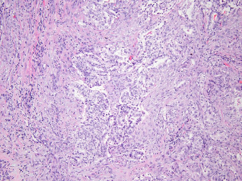
Acta Medica Academica 2021;50(x):xx-xx

                                                                in routine practice and is not included in current
                                                                cancer reporting templates from the College of
                                                                American Pathologists. In their previously refer-
                                                                enced proposal, an international multidisciplinary
                                                                group recommended a two-tier system of grading
                                                                based on nuclear grade and necrosis. In this pro-
                                                                posed system, low-grade MPM comprises nuclear
                                                                grade 1 with or without necrosis and nuclear grade
                                                                2 without necrosis; high-grade is reserved for tu-
                                                                mors with nuclear grade 2 and necrosis or nuclear
                                                                grade 3 with or without necrosis (10). In making a
                                                                recommendation for a practice not yet widely ad-
                                                                opted the authors suggest that tumor grading may
Figure 1. Epithelioid mesothelioma. Low magnification           be of benefit in stratifying patients for clinical tri-
photomicrograph showing tubulopapillary mesothelioma
composed of relatively bland non-mucinous cuboidal cells        als or adding greater precision to the risk stratifica-
forming a combination of tubules (left center), papillae (up-   tion currently provided by histological subtyping.
per left) and small irregularly shaped solid groups and cords   While this may eventually emerge as a standard re-
(hematoxylin and eosin stain; original magnification 40×.       porting element, in our view it should be optional
                                                                for pathology reporting given the absence of com-
                                                                pelling evidence regarding its value outside of a
lopapillary, solid, and trabecular, while micropap-
                                                                research setting. It also should be emphasized that
illary, adenomatoid (microcystic), clear cell, tran-
                                                                this proposed grading system was recommended
sitional, deciduoid, small cell, and lymphohistio-
                                                                only for epithelioid mesothelioma; other types of
cytic variants are relatively rare. Pleomorphic sub-
                                                                MM (sarcomatoid MM, and sarcomatoid parts in
types are defined as epithelioid mesotheliomas in
                                                                biphasic MM) are inherently more aggressive and
which more than 10% of tumor cells show marked
                                                                therefore high grade by definition.
nuclear pleomorphism; this subtype is associ-
ated with the worst overall survival (8.1 months)
among epithelioid MPM (11). The international                   Sarcomatoid Mesothelioma
proposal includes recommendations for reporting
                                                                Sarcomatoid mesothelioma is less frequent but
percentages of the various architectural patterns
                                                                more aggressive than epithelioid mesothelioma,
and cell types for extrapleural pneumonectomy
                                                                with mean survivals of 3.5-8 months (12). Dif-
and extended pleurectomy/decortication surgical
                                                                ferentiating between epithelioid and sarcomatoid
specimens, a reporting practice for which there is
                                                                MM is important because of stage-dependent
no evidence regarding its value and therefore not
                                                                differences in treatment approach (13). Sarcoma-
widely adopted outside of a research setting.
                                                                toid mesothelioma is characterized by neoplastic
                                                                spindle cells exhibiting variable numbers of mito-
Grading of Epithelioid Mesothelioma                             ses and degrees of cytologic atypia (Figure 2). The
                                                                cells are typically arranged in vaguely fascicular
Histological subtype and TNM stage drive thera-
                                                                growth patterns thus resembling soft tissue sar-
peutic strategies in patients with MPM. Several
                                                                comas (“sarcomatoid”). Histological subtypes of
retrospective case series have demonstrated lim-
                                                                sarcomatoid MPM include conventional (44%),
ited utility of histologic grading for epithelioid
                                                                desmoplastic (34%), sarcomatoid with desmoplas-
mesotheliomas, usually based on some combi-
                                                                tic areas (21%), sarcomatoid with heterologous
nation of nuclear grade, mitotic rate, and necro-
                                                                elements (1%), and lymphohistocytoid mesothe-
sis. Grading of MPM has not yet been adopted
                                                                liomas (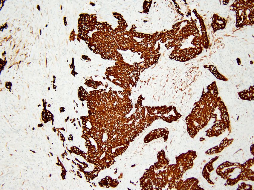
Ivana Savic and Jeffrey Myers: Malignant Pleural Mesothelioma: A Review

Figure 2. Sarcomatoid mesothelioma. Intermediate mag-                                                                   A
nification photomicrograph showing neoplastic spindles
cells arranged in a loosely organized fascicular growth pat-
tern with invasion into chest wall soft tissue at upper left
(hematoxylin and eosin stain; original magnification 132×).

Desmoplastic mesothelioma is a common subtype
and is the most challenging to distinguish from
benign fibrosing pleuritis, sometimes referred to
as fibrous pleurisy. Desmoplastic MPM is paucic-
ellular, with random variation in cellularity across
                                                                                                                        B
a relatively narrow range. The areas showing an
abrupt increase in cellularity comprise mildly                 Figure 3. Desmoplastic mesothelioma. A) Low magnifica-
atypical spindle cells with enlarged hyperchro-                tion photomicrograph showing thickened parietal pleura
                                                               with random variation in cellularity and invasion of chest
matic nuclei arranged in a “patternless pattern of             wall fat (hematoxylin and eosin stain; original magnifica-
Stout” with abundant collagenous stroma (Figure                tion 27×). B) Intermediate magnification photomicrograph
3). Keys to diagnosis are a combination of this dis-           showing more cellular zone in which neoplastic spindle
tinctive storiform histology, invasion of chest wall           cells are arranged in the vaguely storiform “patternless pat-
                                                               tern of Stout” (hematoxylin and eosin stain; original magni-
soft tissue and/or lung parenchyma, bland necro-
                                                               fication 100×).
sis characterized by dropout of basophilic nuclei,
focal areas with frankly malignant sarcomatoid
                                                               Biphasic Mesothelioma
histology, and/or distant metastases (15). Immu-
nohistochemical stains for cytokeratins are of lim-            Biphasic mesothelioma is defined as showing a
ited value since non-neoplastic reactive spindled              combination of epithelioid and sarcomatoid his-
mesothelial cells are also positive, but can be help-          tologies, with each component comprising more
ful in identifying areas of chest wall invasion (16).          than 10% of the tumor (Figure 4). A sarcomatoid
                                                               component of less than 80% in biphasic MM has
Transitional mesothelioma, traditionally consid-               been linked to improved survival. Interobserver
ered a rare architectural and cytological subtype              agreement in diagnosis of biphasic mesothelioma
of epithelioid MPM, comprises cohesive plump                   is moderate (Kappa = 0.45), suggesting that updat-
spindle cells with elongated ambiguous cytomor-                ing the definition of biphasic MPM is needed to
phology. Recent studies indicate that transitional             support more consistent risk stratification (19).
mesothelioma is genetically more closely related               Although fibrous stroma in epithelioid MPM is
to sarcomatoid MPM, and recommend that it be                   typically scant, it is sometimes florid and thus can
considered a subgroup of sarcomatoid mesothe-                  mimic biphasic MPM. Cases in which it is uncer-
lioma (17, 18).                                                tain whether the sarcomatoid component repre-
                                                               sents a benign florid stromal reaction or a prolif-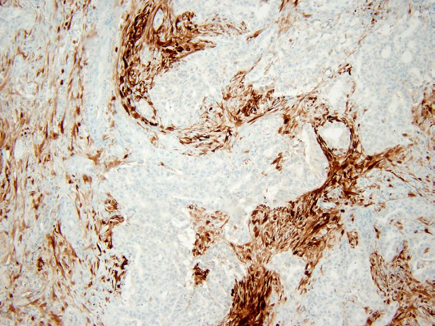
Acta Medica Academica 2021;50(x):xx-xx

                                                                  It is important to first establish a working di-
                                                              agnosis based on routinely stained sections and
                                                              knowledge of the clinical and radiological findings
                                                              before deciding on immunohistochemical stains
                                                              likely to be of value. Choice of immunohistochem-
                                                              ical markers to distinguish MPM from other enti-
                                                              ties with epithelioid phenotypes depends heavily
                                                              on the histologic subtype being considered (epi-
                                                              thelioid or sarcomatoid), location of the neoplasm
                                                              (pleura or peritoneum), and the types of tumors
                                                              included in the differential diagnosis (e.g., squa-
Figure 4. Biphasic mesothelioma. Photomicrograph show-        mous cell carcinoma, adenocarcinoma, epithelioid
ing a glandular epithelial component and a less differenti-   hemangioendothelioma, melanoma). Given that
ated stromal component comprising spindle cells (hema-        none of the markers have 100% specificity, a lim-
toxylin and eosin stain; original magnification 100×).        ited panel that includes antibodies with sensitivity
                                                              or specificity of at least 80% is recommended (22).
eration of neoplastic mesothelial cells may be re-
                                                              An immunohistochemical panel should contain
solved by demonstrating homozygous deletion of
                                                              at least two mesothelial markers and two markers
p16 using a FISH technique (20).
                                                              appropriate to the working diagnoses established
                                                              on the basis of routinely stained sections and any
Immunohistochemical Stains for Diagnosis                      pertinent history including previously diagnosed
                                                              malignancies. For confirmation of mesothelial
Histopathological diagnosis of MPM begins with
                                                              origin in patients suspected of having epitheli-
careful examination of routinely stained sections
                                                              oid or biphasic MPM, calretinin, WT-1 (nuclear
in an appropriate clinical and radiological context.
                                                              staining only), cytokeratin 5/6, and D2-40 (podo-
Immunohistochemical stains can be extremely
                                                              planin) are useful markers (23). Markers useful
helpful in distinguishing MPM from other ma-
                                                              for tumors in which metastatic carcinoma is a di-
lignancies capable of diffuse pleural involvement
                                                              agnostic possibility include MOC31, BG8, CEA,
that may mimic mesothelioma (“pseudomesothe-
                                                              claudin 4, and BerEP4. MOC31 and BerEP4 target
liomatous”), and in separating MPM from benign
                                                              the same transmembrane glycoprotein (EpCAM),
mesothelial proliferations. Pancytokeratin stains
                                                              and therefore the final choice of markers should
may be useful in separating MPM from other
                                                              include one, rather than both of them. In addition
non-epithelial mimics, such as metastatic mela-
                                                              to two general carcinoma markers, immunostains
noma or diffuse high-grade lymphomas confined
                                                              that are specific for certain carcinoma subtypes
to the pleura and pleural space. They should be
                                                              may be helpful. This is dependent not only on the
interpreted with caution, given that reactive spin-
                                                              histologic findings but also on relevant clinical and
dle cells of mesothelial origin are also keratin and
                                                              radiological information (i.e., previous malignan-
calretinin positive (Figure 5). A small minority of
                                                              cies or suspicion of other primary sites at presen-
sarcomatoid MPM may be keratin negative. Epi-
                                                              tation). In patients suspected of having metastatic
thelioid MPM is typically positive for cytokeratin 7
                                                              adenocarcinoma for which no primary is known,
as are many of the entities frequently considered in
                                                              TTF-1 may be helpful since lung is the most fre-
the differential diagnosis, which may limit its utility.
                                                              quent source for metastatic adenocarcinomas with
Epithelioid MPMs are also frequently positive for
                                                              a pseudomesotheliomatous growth pattern (24).
high molecular weight cytokeratins using antibod-
                                                              Major differential diagnoses and immunohisto-
ies directed against cytokeratins 5 and 6; staining
                                                              chemical markers useful for differentiating MPM
for high molecular cytokeratins is less common in
                                                              from other malignant neoplasms are summarized
sarcomatoid types (21).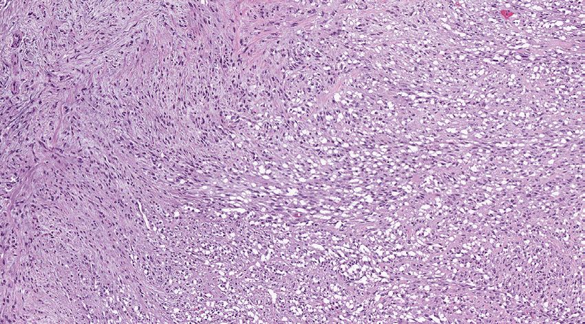
Ivana Savic and Jeffrey Myers: Malignant Pleural Mesothelioma: A Review

A                                                               B

C                                                               D

Figure 5. Metastatic adenocarcinoma of the lung. A) Photomicrograph showing glandular neoplasm infiltrating the pa-
rietal pleura with a variably cellular stromal response resembling biphasic MPM (hematoxylin and eosin stain; original
magnification 100x). B-D) Photomicrographs showing immunostains performed on this pleural biopsy. Both the metastatic
adenocarcinoma and the stromal cells, which include non-neoplastic spindle cells of mesothelial origin, are positive for
cytokeratin 7 (B). Staining for TTF-1 (C) is limited to the adenocarcinoma, and calretinin (D) to non-neoplastic mesothelial
cells (original magnification 100×).

in the Table 1. Immunohistochemistry for BAP1                  pressed cytokeratins; sensitivity increased with use
is used primarily to separate benign from malig-               of an antibody cocktail (e.g., AE1/AE3 ± CAM5.2),
nant mesothelial proliferations (see following sec-            more extensive tumor sampling, and adequate tis-
tion, Separating malignant from benign mesothelial             sue fixation (14). Keratin staining may be negative
proliferations), but can also be helpful in selected           in the osteosarcomatous or chondrosarcomatous
circumstances for distinguishing MPM from car-                 components of sarcomatoid MPM with heterolo-
cinomas in malignant pleural effusion cytology                 gous elements (22). Immunohistochemical mark-
specimens, with high sensitivity (87%) and speci-              ers are less useful for distinguishing sarcomatoid
ficity (98%) (25).                                             MPM from sarcomatoid carcinomas. Calretinin is
    Immunohistochemistry for sarcomatoid MPM                   negative in >50% of sarcomatoid mesotheliomas,
often begins with cytokeratin stains to establish an           as is nuclear staining for WT-1 (21). Strong diffuse
epithelioid phenotype and exclude soft tissue sar-             staining for GATA-3 is nearly universal in sarco-
comas. In a large retrospective case series of over            matoid MPM and, along with radiological distri-
300 cases, 93% of sarcomatoid mesotheliomas ex-                bution of disease (i.e., localized versus diffuse),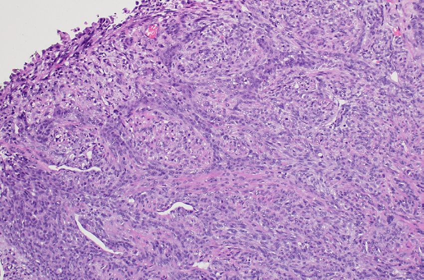
Acta Medica Academica 2021;50(x):xx-xx

Table 1. Immunohistochemical Stains Useful for Separating MPM from Malignant Mimics

                                                            Markers more commonly expressed in
 Histologic MPM type    Mesothelial markers
                                                            non-mesothelial neoplasms
                                                            Lung adenocarcinoma (TTF-1 +, napsin A +)
                                                            Adenocarcinoma, NOS (CEA+, claudin 4+, MOC31/Ber-EP4+, BG8+)

                        CK AE1/3 +, calretinin +, WT-1 +,   Squamous cell lung carcinoma (p40 + MOC-31/Ber-EP4+)
 Epithelioid
                        CK5/6 +, mesothelin +, D2-40 +      Renal cell carcinoma (PAX8 +, CAIX +)
                                                            Breast carcinoma (ER +, PR+, GCDFP-15+, mammaglobin +, GATA3 +)
                                                            Epithelioid hemangioendothelioma (CD31 +, CD34 +, FLI-1 +, ERG +)
                                                            Sarcomatoid carcinoma (CK AE1/3 + CAM 5.2 +, GATA3 -).
                        CK AE1/3 +, CAM5.2 +, D2-40 +,
 Sarcomatoid                                                Angiosarcoma (CD31 +, CD34 +, ERG +)
                        calretinin +, WT-1 +, GATA3 +
                                                            Synovial sarcoma (CD99 +, TLE-1 +)
MPM=Malignant pleural mesothelioma.

can be especially helpful for this frequently chal-                  surface fibrin and granulation tissue in which cap-
lenging differential diagnosis (26).                                 illary sized vascular spaces are arranged in parallel
                                                                     perpendicular to the pleural surface.
                                                                         Loss of BAP1 expression and homozygous de-
Separating Malignant from Benign                                     letion of p16 have become diagnostic methods for
Mesothelial Proliferations                                           separating benign from malignant mesothelial
There are a number of histological features help-                    proliferations with greater frequency and accura-
ful in separating MPM from benign mesothelial                        cy, thus increasing diagnostic sensitivity for MM.
proliferations. Invasion of chest wall soft tissues                  A growing number of studies attest to their practi-
and/or pulmonary parenchyma is the single most                       cal value in the diagnostic process (27).
helpful finding in establishing a diagnosis of MPM                       BAP1 is a cellular enzyme with tumor suppres-
(Figure 6) (1, 16). Cytokeratin stains may be help-                  sor functions. It is involved in cycle-cell progres-
ful in demonstrating invasion not otherwise eas-                     sion, repairing ionizing radiation-induced DNA
ily observed with routinely stained sections alone.                  damage, regulation of gene expression and chroma-
Reactive mesothelial cells do not invade the sur-                    tin remodeling. Early studies showed lack of BAP1
rounding tissues, but “pseudo invasion” may occur                    immunoreactivity due to somatic BAP1 genetic al-
when benign mesothelial cells are entrapped in the                   terations, such as deletions or point mutations, in
fibrosis characteristic of fibrosing pleuritis result-               more than 40% of MPMs (28). More recent studies
ing in a distinctive pattern of layering resembling                  show loss of BAP1 expression in nearly 75% of epi-
the annual growth rings in trees (Figure 7). In-
flammation of the pleura with associated mesothe-
lial hyperplasia tends to have a predictably zonal
distribution of cellularity in which the cellularity is
greatest adjacent to the pleural space and gradually
or abruptly diminishes as it approaches the chest
wall interface. Benign mesothelial proliferations
may include papillary structures but they lack the
complex stratification characteristic of MPM and
instead comprise simple, non-arborizing struc-
tures lined by a single layer of cells. In addition,
reactive proliferations often are accompanied by                     Figure 6. Epithelioid mesothelioma with chest wall invasion
                                                                     (hematoxylin and eosin stain; original magnification 19×).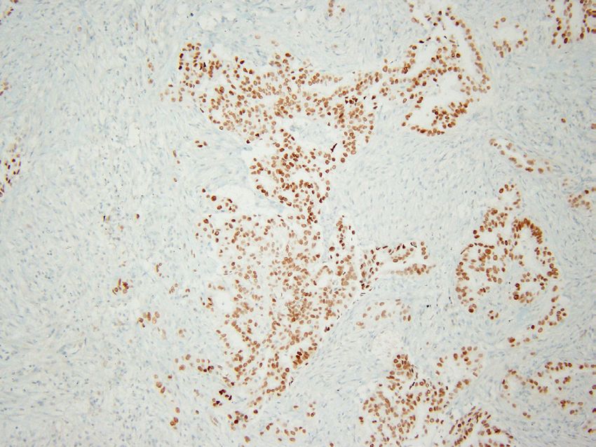
Ivana Savic and Jeffrey Myers: Malignant Pleural Mesothelioma: A Review

                                                                 sociated with younger age at onset and improved
                                                                 median survival in MPM, although BAP1 expres-
                                                                 sion as a prognostic biomarker remains controver-
                                                                 sial (29). Loss of BAP1 expression is manifested as
                                                                 complete, and rarely partial, loss of nuclear stain-
                                                                 ing in tumor cells with positive staining of internal
                                                                 controls (i.e., inflammatory cells and stromal cells)
                                                                 (Figure 8). BAP1 loss occurs in both sporadic and
                                                                 familial MPM, the latter linked to germline BAP1
                                                                 mutations (30, 31). BAP1 loss has consistently
                                                                 shown 100% specificity for distinguishing ma-
                                                                 lignant from benign mesothelial proliferations;
                                                                 benign reactive mesothelial proliferations always
                                                                 retain nuclear BAP1 expression. BAP1 expression
                                                                 is also retained in adenomatoid tumors, a lesion
                                                                 that only rarely occurs in the chest although ad-
                                                                 enomatoid tumor-like histology has been well de-
                                                                 scribed in MPM (32). In a comparison of two ret-
                                                                 rospective cohorts, Erber showed that BAP1 loss
                                                                 occurred only in mesotheliomas and was retained
                                                                 in all 42 genital adenomatoid tumors (33). BAP1
A                                                                immunohistochemistry is a powerful addition to
                                                                 the growing portfolio of diagnostic tools for atypi-
                                                                 cal mesothelial proliferations, but it is important
                                                                 to remember that the variable sensitivity of BAP1
                                                                 loss in MPM limits its negative predictive value:
                                                                 retention of BAP1 expression by itself cannot be
                                                                 used to exclude a diagnosis of mesothelioma.
                                                                     Loss of nuclear expression of 5-hydroxymeth-
                                                                 ylcytosine (5-hmC) has shown promise as an ad-
                                                                 ditional immunohistochemical stain for distin-
B                                                                guishing malignant from benign mesothelial pro-
Figure 7. Fibrosing pleuritis (“fibrous pleurisy”) with layer-   liferations with high (92%) sensitivity and 100%
ing of entrapped mesothelial cells. A) Low magnification         specificity, although this has not yet been widely
photomicrograph showing fibrous thickening of parietal           adopted (34). Several other markers, including
pleura and a sharp interface with chest wall adipose tissue.     desmin, epithelial membrane antigen (EMA), p53,
Entrapped mesothelial cells are arranged in a linear fashion
in the lower third without the random variation in cellular-
                                                                 IMP3, GLUT-1, CD146, and CD147, have shown
ity and invasive growth patterns more characteristic of me-      only limited diagnostic value and are unlikely to
sothelioma (hematoxylin and eosin stain; original magnifi-       be useful in individual cases (1).
cation 20×). B) Photomicrograph of immunohistochemical               Homozygous deletion of 9p21 is an important
stain for cytokeratins (AE1/AE3 and CAM5.2 cocktail) show-
                                                                 method for separating benign from malignant
ing the layering of entrapped mesothelial cells and the ab-
sence of chest wall invasion (original magnification 40×).       mesothelial proliferations. This region comprises
                                                                 genes for two cyclin-dependent inhibitor kinases,
                                                                 CDKNA2A (p16) and CDKN2B, and MTAP. CDK-
thelioid MPM with lower sensitivities on the order
                                                                 N2A is present in normal cells where it is involved
of 50% for biphasic and 10% or less for sarcoma-
                                                                 in cell cycle regulation. Deletion of p16 is present
toid MPM. Loss of BAP1 expression has been as-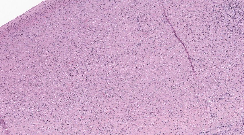
Acta Medica Academica 2021;50(x):xx-xx

                                                                in as many as 90% of sarcomatoid mesotheliomas,
                                                                and about 70% of desmoplastic subtypes, but tends
                                                                to be less common in epithelioid and biphasic me-
                                                                sotheliomas. Practically, p16 deletion is useful for
                                                                distinguishing benign mesothelial proliferations
                                                                from MPM, but cannot reliably distinguish MPM
                                                                from other carcinomas in which p16 may also be
                                                                deleted (35, 36).
                                                                    Homozygous p16 deletion is demonstrated
                                                                with a FISH technique using centromere 9 and
                                                                CDKN2A probes, and can be applied to both cy-
                                                                tology and histology specimens. Overall sensitiv-
A
                                                                ity of p16 FISH in effusion cytology specimens is
                                                                between 56% and 79% with a high (100%) positive
                                                                predictive value given 100% specificity. False nega-
                                                                tive p16 FISH may occur due to admixed reactive
                                                                mesothelial cells that may be morphologically in-
                                                                distinguishable from malignant mesothelial cells
                                                                (22). In histology specimens, the sensitivity of p16
                                                                FISH for epithelioid and biphasic MM ranges be-
                                                                tween 45% and 85%. Homozygous p16 deletion
                                                                has been associated with shorter overall survival.
                                                                    MTAP, a tumor suppressor gene co-located
                                                                with CDKN2A, is often deleted with p16, making
B                                                               immunohistochemical staining for MTAP protein
                                                                a reasonable surrogate for p16 FISH (37). Nega-
                                                                tive cytoplasmic staining for MTAP in tumor cells
                                                                with positive cytoplasmic and nuclear staining in
                                                                positive internal controls, such as inflammatory
                                                                and stromal cells, favors the diagnosis of MM with
                                                                100% specificity and a sensitivity of around 45% in
                                                                tissue sections and cytology cell blocks. Combina-
                                                                tion with BAP1 immunohistochemistry increases
                                                                sensitivity to around 75% to 80% (37).
                                                                    Other emerging methods that are used less fre-
                                                                quently, in part because of technological challeng-
                                                                es that serve as barriers to access, include hemi-
C                                                               zygous deletion of neurofibromatosis type 2 (NF2
Figure 8. Immunohistochemical markers for separating me-        gene) on 22q12 and gene expression arrays (38,
sothelioma from benign mesothelial proliferations. A) High      39). Bruno and colleagues showed that expression
magnification photomicrograph showing two- dimensional
groups of atypical mesothelial cells in a cell block prepared
                                                                analysis of 117 genes using a nanoString System in
from pleural fluid (hematoxylin and eosin; original magnifi-    a relatively small number of cases performed bet-
cation 400×). B-C) Immunohistochemical stains performed         ter than BAP1 and p16 FISH, with overall sensitiv-
on the same cell block showed loss of nuclear staining for      ity of 95.6% and 100% specificity (38). There are
BAP1 (B) and loss of both cytoplasmic and nuclear staining      no currently recommended predictive biomarkers
for MTAP (C) with appropriately staining internal controls
(original magnification 400×).                                  for patients with MPM, although that may change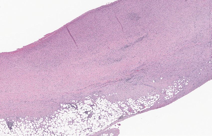
Ivana Savic and Jeffrey Myers: Malignant Pleural Mesothelioma: A Review

as research continues with the hope of identify-       ously diagnosed malignancies. There is an increas-
ing more personalized treatment strategies (10). A     ingly robust suite of diagnostic tools, including
number of clinical trials using immune checkpoint      immunohistochemical stains for BAP1 and MTAP
inhibitors are underway and have shown limited         and p16 FISH, for differentiating benign from ma-
utility for PD-L1 testing in identifying those most    lignant mesothelial proliferations in cytology and
likely to respond.                                     tissue specimens. Mesothelioma in situ has been
                                                       recognized as a distinct clinicopathological entity
                                                       for which more evidence is required to understand
Malignant Mesothelioma in Situ                         its natural history and treatment strategies that are
Malignant mesothelioma in situ is a preinvasive le-    proportional and targeted to the risk.
sion defined as a single layer of atypical mesothe-
                                                       Conflict of Interest: The authors declare that they have no
lial cells lining the pleural surface and character-   conflict of interest.
ized by loss of BAP1 and/or MTAP expression us-
ing immunohistochemistry. Criteria for diagnosis
include recurrent pleural effusions, lack of pleural   References
thickening and nodularity on chest imaging, and        1. Churg A, Galateau-Salle F. The separation of benign and
either no or only incidental findings at video-as-        malignant mesothelial proliferations. Arch Pathol Lab
sisted thoracoscopic surgery (VATS) or thoracoto-         Med. 2012;136(10):1217-26.
my. Given these criteria, diagnosis requires a mul-    2. Finn RS, Brims FJH, Gandhi A, Olsen N, Musk AW,
                                                          Maskell NA, et al. Postmortem findings of malignant
tidisciplinary approach (40). Churg et al. showed         pleural mesothelioma: a two-center study of 318 patients.
that seven of ten patients with well-defined meso-        Chest. 2012;142(5):1267-73.
thelioma in situ developed MPM during a follow-        3. Ogata S, Hiroi S, Tominaga A, Aida S, Kobayashi A,
up period of 12-92 months (40).                           Tamura K, et al. Malignant pleural mesothelioma initially
                                                          diagnosed on cervical lymph node biopsy. Pathol Int.
                                                          2009;59(8):592-4.
Conclusions                                            4. Henderson DW, Reid G, Kao SC, van Zandwijk N, Klebe
                                                          S. Challenges and controversies in the diagnosis of meso-
Diffuse MPM is a heterogeneous group of aggres-           thelioma: Part 1. Cytology-only diagnosis, biopsies, im-
sive pleural tumors for which histological clas-          munohistochemistry, discrimination between mesothe-
                                                          lioma and reactive mesothelial hyperplasia, and biomark-
sification plays an increasingly important role
                                                          ers. J Clin Pathol. 2013;66(10):847-53.
in patient management and survival. Update of          5. Michael CW, Bedrossian CC. The implementation of liq-
epithelioid mesothelioma includes pleomorphic             uid-based cytology for lung and pleural-based diseases.
subtypes, in which more than 10% of tumor cells           Acta Cytol. 2014;58(6):563-73.
show marked nuclear pleomorphism; this subtype         6. Hjerpe A, Ascoli V, Bedrossian CW, Boon ME, Creaney
is associated with the worst overall survival among       J, Davidson B, et al. Guidelines for the cytopathologic
                                                          diagnosis of epithelioid and mixed-type malignant me-
epithelioid MPM. Considering that transitional
                                                          sothelioma. Complementary statement from the Interna-
mesothelioma is genetically closely related to sar-       tional Mesothelioma Interest Group, also endorsed by the
comatoid MPM, recent studies recommend it to              International Academy of Cytology and the Papanicolaou
be within a subgroup of sarcomatoid mesothelio-           Society of Cytopathology. Acta Cytol. 2015;59(1):2-16.
mas. Stage and resectability remain key drivers of     7. Chirieac LR, Hung YP, Foo WC, Hofer MD, VanderLaan
therapeutic strategies and outcomes. Given that           PA, Richards WG, et al. Diagnostic value of biopsy sam-
                                                          pling in predicting histology in patients with diffuse malig-
none of the immunohistochemical markers has               nant pleural mesothelioma. Cancer. 2019;125(23):4164-71.
100% specificity, an immunohistochemical panel         8. Verma V, Ahern CA, Berlind CG, Lindsay WD, Shaba-
should contain at least two mesothelial markers           son J, Sharma S, et al. Survival by Histologic Subtype of
and two markers appropriate to the working diag-          Malignant Pleural Mesothelioma and the Impact of Sur-
noses established based on routinely stained sec-         gical Resection on Overall Survival. Clin Lung Cancer.
                                                          2018;19(6):e901-12.
tions and any pertinent history including previ-
Acta Medica Academica 2021;50(x):xx-xx
9. Marchevsky AM, Khoor A, Walts AE, Nicholson AG, diagnosis of biphasic mesothelioma. Ann Diagn Pathol.
Zhang YZ, Roggli V, et al. Localized malignant mesothe- 2017;26:31-7.
lioma, an unusual and poorly characterized neoplasm of 21. Marchevsky AM, LeStang N, Hiroshima K, Pelosi G, Atta-
serosal origin: best current evidence from the literature noos R, Churg A, et al. The differential diagnosis between
and the International Mesothelioma Panel. Mod Pathol. pleural sarcomatoid mesothelioma and spindle cell/pleo-
2020;33(2):281-96. morphic (sarcomatoid) carcinomas of the lung: evidence-
10. Nicholson AG, Sauter JL, Nowak AK, Kindler HL, Gill based guidelines from the International Mesothelioma
RR, Remy-Jardin M, et al. EURACAN/IASLC Proposals Panel and the MESOPATH National Reference Center.
for Updating the Histologic Classification of Pleural Me- Hum Pathol. 2017;67:160-8.
sothelioma: Towards a More Multidisciplinary Approach. 22. Husain AN, Colby TV, Ordonez NG, Allen TC, Attanoos
J Thorac Oncol. 2020;15(1):29-49. RL, Beasley MB, et al. Guidelines for Pathologic Diagnosis
11. Kadota K, Suzuki K, Sima CS, Rusch VW, Adusumilli PS, of Malignant Mesothelioma 2017 Update of the Consen-
Travis WD. Pleomorphic epithelioid diffuse malignant sus Statement From the International Mesothelioma In-
pleural mesothelioma: a clinicopathological review and terest Group. Arch Pathol Lab Med. 2018;142(1):89-108.
conceptual proposal to reclassify as biphasic or sarcoma- 23. Ordonez NG. Application of immunohistochemistry in
toid mesothelioma. J Thorac Oncol. 2011;6(5):896-904. the diagnosis of epithelioid mesothelioma: a review and
12. Galetta D, Catino A, Misino A, Logroscino A, Fico M. update. Hum Pathol. 2013;44(1):1-19.
Sarcomatoid mesothelioma: future advances in diagnosis, 24. Attanoos RL, Gibbs AR. ‘Pseudomesotheliomatous’ carci-
biomolecular assessment, and therapeutic options in a nomas of the pleura: a 10-year analysis of cases from the
poor-outcome disease. Tumori. 2016;102(2):127-30. Environmental Lung Disease Research Group, Cardiff.
13. Ismail-Khan R, Robinson LA, Williams CC Jr, Garrett CR, Histopathology. 2003;43(5):444-52.
Bepler G, Simon GR. Malignant pleural mesothelioma: a 25. Davidson B, Totsch M, Wohlschlaeger J, Hager T, Pina-
comprehensive review. Cancer Control. 2006;13(4):255-63. monti M. The diagnostic role of BAP1 in serous effusions.
14. Klebe S, Brownlee NA, Mahar A, Burchette JL, Sporn Hum Pathol. 2018;79:122-6.
TA, Vollmer RT, et al. Sarcomatoid mesothelioma: a 26. Berg KB, Churg A. GATA3 Immunohistochemistry for
clinical-pathologic correlation of 326 cases. Mod Pathol. Distinguishing Sarcomatoid and Desmoplastic Mesothe-
2010;23(3):470-9. lioma From Sarcomatoid Carcinoma of the Lung. Am J
15. Mangano WE, Cagle PT, Churg A, Vollmer RT, Roggli VL. Surg Pathol. 2017;41(9):1221-5.
The diagnosis of desmoplastic malignant mesothelioma 27. Churg A, Naso JR. The Separation of Benign and Malig-
and its distinction from fibrous pleurisy: a histologic and nant Mesothelial Proliferations: New Markers and How to
immunohistochemical analysis of 31 cases including p53 Use Them. Am J Surg Pathol. 2020;44(11):e100-12.
immunostaining. Am J Clin Pathol. 1998;110(2):191-9. 28. Bott M, Brevet M, Taylor BS, Shimizu S, Ito T, Wang L, et
16. Galateau-Salle F, Churg A, Roggli V, Travis WD, World al. The nuclear deubiquitinase BAP1 is commonly inacti-
Health Organization Committee for Tumors of the Pleu- vated by somatic mutations and 3p21.1 losses in malignant
ra. The 2015 World Health Organization Classification of pleural mesothelioma. Nat Genet. 2011;43(7):668-72.
Tumors of the Pleura: Advances since the 2004 Classifica- 29. Farzin M, Toon CW, Clarkson A, Sioson L, Watson N,
tion. J Thorac Oncol. 2016;11(2):142-54. Andrici J, et al. Loss of expression of BAP1 predicts longer
17. Schulte JJ, Husain AN. Update on the pathologic diagno- survival in mesothelioma. Pathology. 2015;47(4):302-7.
sis of malignant mesothelioma. Transl Lung Cancer Res. 30. Betti M, Casalone E, Ferrante D, Romanelli A, Grosso F,
2020;9(3):917-23. Guarrera S, et al. Inference on germline BAP1 mutations
18. Galateau Salle F, Le Stang N, Tirode F, Courtiol P, Nich- and asbestos exposure from the analysis of familial and
olson AG, Tsao MS, et al. Comprehensive Molecular and sporadic mesothelioma in a high-risk area. Genes Chro-
Pathologic Evaluation of Transitional Mesothelioma As- mosomes Cancer. 2015;54(1):51-62.
sisted by Deep Learning Approach: A Multi-Institutional 31. Cigognetti M, Lonardi S, Fisogni S, Balzarini P, Pellegrini
Study of the International Mesothelioma Panel from V, Tironi A, et al. BAP1 (BRCA1-associated protein 1) is
the MESOPATH Reference Center. J Thorac Oncol. a highly specific marker for differentiating mesothelioma
2020;15(6):1037-53. from reactive mesothelial proliferations. Mod Pathol.
19. Dacic S, Le Stang N, Husain A, Weynand B, Beasley MB, 2015;28(8):1043-57.
Butnor K, et al. Interobserver variation in the assessment 32. Weissferdt A, Kalhor N, Suster S. Malignant mesothelio-
of the sarcomatoid and transitional components in bipha- ma with prominent adenomatoid features: a clinicopatho-
sic mesotheliomas. Mod Pathol. 2020;33(2):255-62. logic and immunohistochemical study of 10 cases. Ann
20. Wu D, Hiroshima K, Yusa T, Ozaki D, Koh E, Sekine Y, Diagn Pathol. 2011;15(1):25-9.
et al. Usefulness of p16/CDKN2A fluorescence in situ 33. Erber R, Warth A, Muley T, Hartmann A, Herpel E,
hybridization and BAP1 immunohistochemistry for the Agaimy A. BAP1 Loss is a Useful Adjunct to DistinguishIvana Savic and Jeffrey Myers: Malignant Pleural Mesothelioma: A Review
Malignant Mesothelioma Including the Adenomatoid- 37. Hida T, Hamasaki M, Matsumoto S, Sato A, Tsujimura
like Variant From Benign Adenomatoid Tumors. Appl T, Kawahara K, et al. Immunohistochemical detection of
Immunohistochem Mol Morphol. 2020;28(1):67-73. MTAP and BAP1 protein loss for mesothelioma diagno-
34. Chapel DB, Husain AN, Krausz T. Immunohistochemical sis: Comparison with 9p21 FISH and BAP1 immunohis-
evaluation of nuclear 5-hydroxymethylcytosine (5-hmC) tochemistry. Lung Cancer. 2017;104:98-105.
accurately distinguishes malignant pleural mesothelioma 38. Bruno R, Ali G, Giannini R, Proietti A, Lucchi M, Chella
from benign mesothelial proliferations. Mod Pathol. A, et al. Malignant pleural mesothelioma and mesothelial
2019;32(3):376-86. hyperplasia: A new molecular tool for the differential di-
35. Illei PB, Rusch VW, Zakowski MF, Ladanyi M. Homo- agnosis. Oncotarget. 2017;8(2):2758-70.
zygous deletion of CDKN2A and codeletion of the me- 39. Kinoshita Y, Hamasaki M, Yoshimura M, Matsumoto S,
thylthioadenosine phosphorylase gene in the majority of Iwasaki A, Nabeshima K. Hemizygous loss of NF2 de-
pleural mesotheliomas. Clin Cancer Res. 2003;9(6):2108- tected by fluorescence in situ hybridization is useful for
13. the diagnosis of malignant pleural mesothelioma. Mod
36. Hwang HC, Pyott S, Rodriguez S, Cindric A, Carr A, Mi- Pathol. 2020;33(2):235-44.
chelsen C, et al. BAP1 Immunohistochemistry and p16 40. Churg A, Galateau-Salle F, Roden AC, Attanoos R, von
FISH in the Diagnosis of Sarcomatous and Desmoplastic der Thusen JH, Tsao MS, et al. Malignant mesothelioma
Mesotheliomas. Am J Surg Pathol. 2016;40(5):714-8. in situ: morphologic features and clinical outcome. Mod
Pathol. 2020;33(2):297-302.You can also read