7 nicotinic acetylcholine receptor mediates right ventricular fibrosis and diastolic dysfunction in pulmonary hypertension
←
→
Page content transcription
If your browser does not render page correctly, please read the page content below
⍺7 nicotinic acetylcholine receptor mediates right ventricular
fibrosis and diastolic dysfunction in pulmonary hypertension
Alexander Vang, … , Jin O-Uchi, Gaurav Choudhary
JCI Insight. 2021. https://doi.org/10.1172/jci.insight.142945.

 Research In-Press Preview Cardiology Pulmonology

Graphical abstract

Find the latest version:
https://jci.me/142945/pdf
a7 Nicotinic Acetylcholine Receptor Mediates Right Ventricular Fibrosis and Diastolic

 Dysfunction in Pulmonary Hypertension

 Alexander Vang1*, Denielli da Silva Goncalves Bos1,2*, Ana Fernandez-Nicolas1,2*, Peng

 Zhang1,2, Alan R. Morrison1,2, Thomas J. Mancini1, Richard T. Clements1,3, Iuliia Polina4,

Michael W. Cypress4, Bong Sook Jhun4, Edward Hawrot5, Ulrike Mende2,6, Jin O-Uchi4, Gaurav

 Choudhary1,2
1
 Vascular Research Laboratory, Providence VA Medical Center, Providence, RI
2
 Department of Medicine, Alpert Medical School of Brown University, Providence RI
3
 Biomedical & Pharmaceutical Sciences, University of Rhode Island, Kingston, RI
4
 Department of Medicine, University of Minnesota, Minneapolis, MN
5
 Department of Molecular Pharmacology, Physiology, and Biotechnology, Alpert Medical School

of Brown University, Providence RI
6
 Cardiovascular Research Center, Lifespan Cardiovascular Institute, Rhode Island Hospital,

Providence, RI

*Equal Contribution

Corresponding Author

Gaurav Choudhary, MD

Providence VA Medical Center

830 Chalkstone Avenue, Building 35

Providence, RI, 02908

Phone: +1-401-273-7100 x12029

Email: gaurav_choudhary@brown.edu

Conflict of interest statement: The authors have declared that no conflict of interest exists.

 1
Abstract

Right ventricular (RV) fibrosis is a key feature of maladaptive RV hypertrophy and dysfunction

and is associated with poor outcomes in pulmonary hypertension (PH). However, mechanisms and

therapeutic strategies to mitigate RV fibrosis remain unrealized. Previously, we identified that

cardiac fibroblast α7 nicotinic acetylcholine receptor (α7 nAChR) drives smoking induced RV

fibrosis. Here we sought to define the role of α7 nAChR in RV dysfunction and fibrosis in the

settings of RV pressure overload as seen in PH. We show that RV tissue from PH patients has

increased collagen content and ACh expression. Using experimental rat model of PH, we

demonstrate that RV fibrosis and dysfunction are associated with increases in ACh and α7 nAChR

expression in the RV but not in the LV. In vitro studies show that α7 nAChR activation leads to

an increase in adult ventricular fibroblast proliferation and collagen content mediated by a Ca2+/

epidermal growth factor receptor (EGFR) signaling mechanism. Pharmacological antagonism of

nAChR decreases RV collagen content and improves RV function in the PH model. Further, mice

lacking α7 nAChR exhibit improved RV diastolic function and have lower RV collagen content

in response to persistently increased RV afterload, compared to wild-type controls. These finding

indicate that enhanced α7 nAChR signaling is an important mechanism underlying RV fibrosis

and dysfunction, and targeted inhibition of α7 nAChR is a novel therapeutic strategy in the setting

of increased RV afterload.

 2
Introduction

 Myocardial fibrosis is a process of pathological extracellular matrix (ECM) remodeling,

mediated by activation of cardiac fibroblasts (CFs) (1-3). In response to stress and injury, CFs

transform into a more proliferative and hyperactive phenotype with elevated production of

extracellular matrix proteins such as collagen (4) resulting in cardiac fibrosis. Mechanisms of

increased cardiac fibrosis and fibroblast proliferation/transformation have predominantly been

studied in the left ventricle (LV). They include TGFβ signaling, the renin-angiotensin-aldosterone

system signaling, adrenergic and endothelin G-protein coupled receptor signaling, growth factor-

mediated tyrosine kinase signaling, and a number of other inflammatory-based pathways (1, 4).

Far less is known about the mechanisms driving RV fibrosis (5-7). This is notable since we and

others have demonstrated that some established therapies targeting LV fibrosis, such as

angiotensin receptor antagonism, do not attenuate RV fibrosis (8, 9).

 RV fibrosis is a key histological feature observed in maladaptive RV hypertrophy and

dysfunction. RV dysfunction and failure are associated with poor outcomes in a variety of highly

prevalent cardiopulmonary diseases such as heart failure and COPD that are associated with

increased RV afterload due to pulmonary hypertension (PH). The poor outcomes related to RV

function are most pronounced in pulmonary arterial hypertension (PAH), a disease characterized

by progressive pulmonary vascular remodeling resulting in a substantial increase in RV afterload,

eventually leading to maladaptive RV hypertrophy, RV dysfunction and RV failure (10). In

preclinical models of PH, RV fibrosis is associated with reduced RV function and cardiac output

(11), impaired contractility (12) and stiffness (13) of the RV myocardium. Analogous to animal

models, patients with PH and decompensated RV failure have significant RV fibrosis at autopsy

(14). Moreover, the extent of RV fibrosis quantified by MRI in patients with PH correlates with

 3
both RV ejection fraction and RV end diastolic volume (15). Conversely, little fibrosis is observed

in compensated models of RV hypertrophy, despite having similar levels of afterload (11, 14).

While RV fibrosis is an important component of decompensated RV in settings of PH, it remains

to be determined if mitigating RV fibrosis would improve RV structure and function (6, 16).

Moreover, the mechanisms that initiate RV fibrosis and therapeutic targets remain unclear (5, 7).

 We have shown that the mechanism that promotes CF proliferation and RV fibrosis related

to cigarette smoke exposure is mediated through activation of α7 nicotinic acetylcholine receptor

(α7 nAChR) in CF (17). Non-neuronal a7 nAChR has been described in a variety of cell types

and in response to ligand (ACh, Nicotine) binding, these pentameric ligand-gated ion channels

undergo a conformational change that results in opening up of the ion conducting pore (18). The

a7 nAChR has a high Ca2+ permeability compared to other nAChR isoforms and results in

activation of downstream Ca2+ dependent signaling pathways (18). Pro-proliferative effects of a7

nAChR activation have been reported in cancer cells (19-21), vascular smooth muscle cells (22,

23), adventitial fibroblasts (22), and endothelial cells (24, 25), and CF (17) predominantly in

settings of nicotine and/or cigarette smoke exposure. Little is known about the pro-proliferative

and pro-fibrotic roles of a7 nAChR in the absence of nicotine exposure. We hypothesized that a7

nAChR on CF mediates RV fibrosis in settings of increased RV afterload. Therefore, in this study,

we investigate the role of α7 nAChR and its natural ligand, ACh, in the development of RV fibrosis

and dysfunction using human RV tissue and clinically relevant animal models of PH and increased

RV afterload. We report that ACh-a7 nAChR signaling is upregulated in the RV in the setting of

PH and that antagonism of a7 nAChR results in improved RV fibrosis and function. These novel

findings serve as a conceptual basis for a7 nAChR inhibition as potential therapy for maladaptive

RV fibrosis in PH.

 4
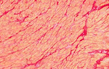
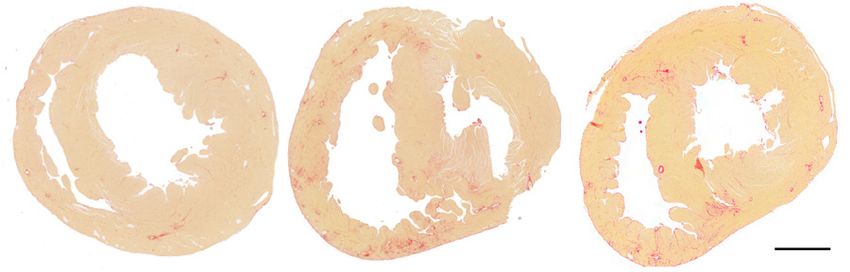
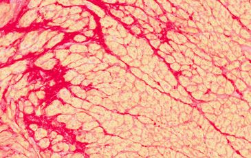
Results

ACh-α7 nAChR signaling is upregulated in the RV in PH.

 In order to assess the role of α7 nAChR signaling in RV fibrosis and dysfunction, we

studied a rat PH model at two time points (3 and 7 weeks, Figure 1A) that develops severe PH

(Figure 1B) with increased RV end diastolic pressure (Figure 1C), RV hypertrophy (Figure 1D),

RV systolic and diastolic dysfunction (Figure 1E and 1F, respectively), and reduced cardiac output

(Figure 1G) in response to SU5416/hypoxia (SuHx) (8, 26). In this PH model, except for an

increase in LV ejection fraction at the 7 weeks timepoint, no significant morphological and

functional changes were observed in the LV, as assessed by echocardiography and hemodynamic

measurements, respectively (TABLE S1 and Supplement Figure S1A-D). Significant fibrotic

remodeling in the RV (but not in the LV) was observed concomitant with RV dysfunction in the

PH rats in comparison to healthy controls (Figure 1H-I, and Supplemental Figure 1E). There was

no significant effect of sex in RV collagen content at either timepoint (two-way ANOVA with sex

as a source of variation, p=0.70 for the 3 wk timepoint, and p=0.67 for the 7 wk timepoint).

 We next assessed the changes in ACh signaling in the RV under PH using ventricular

myocardium from control and PH animals. ACh levels were increased (Figure 2A) and the activity

of ACh degrading enzyme, acetylcholinesterase (AChE), was decreased (Figure 2B) in the RV

(but not in the LV) from PH animals compared to controls. Moreover, ACh levels significantly

correlated with the fibrosis marker, hydroxyproline content, in the RV (Supplementary Figure S2).

We did not find significant changes in expression of ACh synthesis proteins including vesicular

acetylcholine transporter (VAChT), choline acetyltransferase (ChAT), and choline transporter

(ChT) (Supplemental Figure S3 A-C). These results suggest that increased ACh content in the RV

in PH is predominantly via impaired degradation rather than an increase in ACh synthesis. In

 5
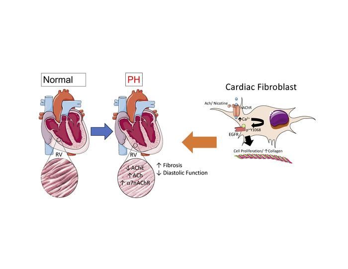
addition to increased ACh, the RV from PH rats exhibited increased protein and mRNA expression

of α7 nAChR (Figure 2C, Supplementary Figure S3D) at the 7-week timepoint. There was no

significant difference in α7 nAChR expression levels in the LV between control and PH at either

time point (Supplementary Figure S3E-F). While α7 nAChR was predominantly expressed in the

CF, overall expression of nAChR remained unaltered in isolated RVCF from PH rats compared to

controls at 7-week time point (Figure 2D). However, a trend for increased expression was noted

in RV cardiomyocytes (Figure 2D). We next assessed the cell membrane expression of α7 nAChR

by staining non-permeabilized RV CF, and found that RV CF from PH animals had higher cell

surface expression of α7 nAChR compared to those isolated from control rats (Figure 2E). In

summary, increased RV ACh-α7 nAChR signaling in PH was associated with RV hypertrophy,

fibrosis, and dysfunction (Figure 2F).

 In RV myocardium from end-stage human PAH patients (Table S2), we found increased

expression collagen and ACh (Figure 3A-B) with no significant differences in expression of α7

nAChR and ACh synthesis proteins (ChT, VAChT and ChAT) (Figure 3C, Supplemental Figure

S4). We did note variability in α7 nAChR protein and mRNA expression in the human RV tissue

likely related to sampling bias and/or underling clinical condition of the patients.

Immunofluorescent staining of human RV tissue demonstrated that α7 nAChR is predominantly

expressed in CF (Figure 3D).

ACh induces cardiac fibroblast activation via α7 nAChR-mediated EGFR transactivation.

 We hypothesized that upregulation of ACh-α7 nAChR signaling contributes to CF

activation in the RV in PH. We stimulated primary RV CF isolated from control and PH rats (7-

week timepoint) with ACh, a natural ligand for α7 nAChR, and found that ACh significantly

 6
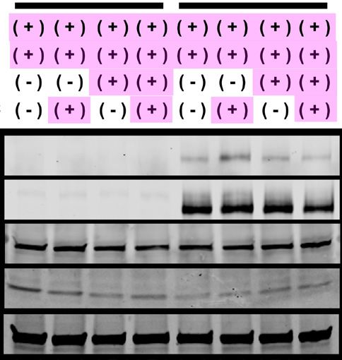
increased CF proliferation and collagen content (Figure 4A-B) compared to vehicle treatment.

Moreover, the relative increases in ACh-mediated CF proliferation and collagen content were

higher in CF from PH compared to control rats (Figure 4A-B). The effect of ACh on CF

proliferation and collagen content were blocked by an α7 nAChR specific antagonist α-

bungarotoxin (α-BTX) (Figure 4C-D), or a non-selective and non-competitive nAChR antagonist

mecamylamine (Mec; Supplement Figure S5A), but not by a muscarinic receptor antagonist

atropine (Figure 4E-F and Supplementary Figure S5B), demonstrating that the ACh effect on CF

proliferation is mediated via α7 nAChR. Similar to ACh, conditioned media from cardiomyocytes

isolated from RVs of PH animals (7 wk timepoint) induced a significant increase in both CF

proliferation and collagen content compared to media from cardiomyocytes isolated from control

animals (Figure 4G-H). These changes induced by conditioned media were attenuated in the

presence of α-BTX suggesting that cardiomyocyte derived ACh results in CF proliferation and

fibrosis in RV in settings of PH.

 We next investigated the mechanism of how α7 nAChR-mediated signaling promotes CF

activation. Some reports have suggested potential interactions between α7 nAChR signaling and

epidermal growth factor receptor (EGFR) signaling (28). EGFR plays an important role in cell

survival and proliferation in a number of cell types including CF (29). Therefore, we investigated

if activation of nAChR would lead to EGFR activation in CF. Using immunocytochemistry, we

found that ACh or nicotine stimulation increased EGFR phosphorylation in adult CF and this

phosphorylation was significantly inhibited in the presence of a nAChR antagonist,

Mecamylamine (Mec, Figure 5A-B). Ach- and nicotine-mediated EGFR phosphorylation was also

confirmed by Western blotting of protein lysates from the CF (Figure 5C). A similar result

regarding EGFR phosphorylation and its inhibition was confirmed by Western blot in HEK-293T

 7
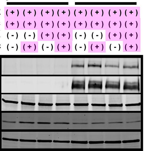
cells overexpressing EGFR, α7 nAChR, and its chaperone protein TMEM35/NACHO

(Supplemental Figure S6) (30) using a-BTX (Figure 5D-E). In contrast, activation of α7 nAChR

was not associated with phosphorylation of another tyrosine kinase growth factor receptor,

fibroblast growth factor receptor (FGFR, Supplementary Figure S7). Moreover, ACh- and

nicotine-induced CF proliferation and collagen synthesis were abolished by knocking down of

EGFR using siRNA (Figure 5F-G and Supplementary Figure S8), or pretreatment with selective

EGFR inhibitor AG1478 (Figure 5H-I and Supplement Figure S9). Together, these results suggest

that α7 nAChR-mediated CF activation is dependent on EGFR transactivation.

 Since homomeric α7 nAChR proteins form ligand-gated cation (e.g., Ca2+) channels at the

plasma membrane (27), we performed live cell imaging with cell-permeable Ca2+-sensitive dyes

to confirm the functional expression of α7 nAChR. We demonstrate a transient increase of

cytosolic Ca2+ concentration ([Ca2+]c) in response to the nAChR specific agonist, nicotine, in adult

rat CFs that was abolished in the presence of the α7 nAChR blocker α-BTX (Figure 6A and

Supplement Figure S10). α7 nAChR-mediated Ca2+ mobilization serves as an important second

messenger in other cell types. Therefore, we tested if cytosolic Ca2+ elevation is mechanistically

involved in α7 nAChR-dependent EGFR transactivation. We found that pretreatment with the cell-

permeable Ca2+ chelator BAPTA-AM significantly inhibited α7 nAChR stimulation-mediated

EGFR phosphorylation (Figure 6B-C).

 In summary, our data demonstrate that ACh stimulation promotes CF proliferation via α7

nAChR and Ca2+-mediated EGFR transactivation in primary adult CFs (Figure 6D), and this is

likely the underlying mechanism by which increased ACh results in cardiac fibrosis in the RV in

the setting of PH in vivo.

Inhibition of nAChR improves RV function in experimental PH.

 8
Since our in-vitro data strongly suggested a significant role of ACh-α7 nAChR signaling

in causing CF proliferation and collagen synthesis, we next investigated the role of ACh-α7

nAChR signaling in PH-induced RV dysfunction in vivo by treating PH rats (SuHx model) with

nAChR antagonist Mec (20 mg/kg/d intraperitoneal for 3 weeks, Figure 7A). Mec was

administered after the establishment of PH which was confirmed by echocardiogram (TABLE S3).

We found that Mec treatment significantly reduced RV hypertrophy (Figure 7B), RV collagen

content (Figure 7C-D) and EGFR phosphorylation (Figure 7E-F) in comparison to the vehicle-

treated group. These RV changes were associated with significantly improved stroke volume and

RV diastolic function (RV e’) as assessed by echocardiography (TABLE S3) and significantly

reduced RV systolic and end diastolic pressures in the Mec-treated group compared to the vehicle

group (Figure 7G-H). In order to assess load-independent parameters of RV function, RV pressure-

volume analysis (Figure 7I-J) was performed. We found that treatment with Mec resulted in a trend

towards RV afterload reduction (Figure 7K) and significantly reduced RV end diastolic stiffness

(Figure 7L), without a significant effect in RV contractility (Figure 7M). There were no changes

in tail blood pressure, LV pressures (Supplementary Figure 11) or LV systolic function (TABLE

S3). Lung histology demonstrated a trend towards decrease in vascular medial thickness in Mec-

treated PH rats compared to vehicle-treated PH rats, while the levels of vessel occlusion were not

altered (Supplemental Figure S12). Taken together, these findings support our hypothesis that

nAChR signaling plays an important role in causing RV fibrosis and dysfunction and that

pharmacologically blocking nAChR can be a potential therapeutic option to mitigate RV fibrosis

and improve RV diastolic function in PH. In order to assess, if Mec inhibition remains effective

in attenuating RV fibrosis and collagen content in more advanced stage of PH, we performed

another treatment experiment starting Mec administration at 5-week timepoint for 3 weeks

 9
(Supplementary Figure S13). We found that later administration of Mec resulted in similar findings

along with reduction in RV collagen and improvement of RV diastolic function (Supplementary

Figure S13, TABLE S4).

RV pressure overload induces RV fibrosis via α7 nAChR

 In order to further delineate that RV fibrosis in PH is mediated via α7 nAChR in the heart,

we utilized α7 nAChR knock out mice (KO: α7nAChR-/-, Supplemental Figure S14) subjected to

fixed RV afterload by pulmonary artery banding (PAB) (Figure 8A). The KO mice grow to normal

size and show no obvious physical or cardiac deficits (31) (TABLE S5). Both wild-type (WT:

α7nAChR+/+) and KO mice subjected to PAB showed a similar pressure gradient across the

banding site and a similar increase in RV systolic pressure compared to sham-operated animals

(TABLE S5, Figure 8B-C). Although both WT and KO mice after PAB developed similar levels

of RV hypertrophy, afterload, and increased ACh content (Figure 8D-E), significant RV diastolic

dysfunction and fibrosis were only observed in WT mice (Figure 8F-H) but not in the KO mice.

There were no significant changes in LV pressures, LV morphology and function among all

groups. (TABLE S5, Supplement Figure S15). This set of experiments clearly demonstrates that

α7 nAChR in the heart mediates RV fibrosis and dysfunction in the setting of increased RV

afterload.

 10
Discussion

 RV fibrosis, dysfunction, and failure due to persistently increased afterload is the

predominant cause of morbidity and mortality in patients with PH, and defining mechanisms

leading to RV dysfunction that may be targeted therapeutically represents a critical unmet need in

the field. Patients with PH demonstrate increased RV collagen deposition and increased RV

expression of ACh. Using experimental models of PH and increased RV afterload, we demonstrate

here that increased RV afterload is not only associated with increased RV ACh /α7 nAChR

signaling but that increased CF proliferation and collagen content depend on the activation of the

α7 nAChR/Ca2+/EGFR signaling axis. Moreover, we demonstrate that both therapeutic inhibition

of α7 nAChR during PH and lack of α7 nAChR gene in the setting of stable increased RV afterload

reduces RV collagen content and consequently improves RV diastolic function. Hence, targeted

inhibition of α7 nAChR as a therapeutic strategy may provide a novel approach to improve the

adverse RV remodeling associated with increased morbidity and mortality in patients with PH.

 LV hypertrophy and failure have been associated with an increase in cholinergic trans-

differentiation (32) and an induction of ACh production by cardiomyocytes in order to preserve

homeostasis of cardiac function through cardiomyocyte muscarinic receptors (33, 34). We

demonstrate here that RV ACh levels are increased in the setting of RV afterload. The ACh in the

RV is partly derived from RV cardiomyocytes in PH as demonstrated by our experiments using

conditioned media from cardiomyocytes. The mRNA expression of key ACh synthesis proteins

did not change in PH, but the AChE activity in the RV was consistently lower in PAH patients in

PH animals compared to respective controls. These observations indicate that the increase in ACh

is likely through decreased degradation via AChE rather than ACh synthesis. The underlying

mechanisms regulating AChE activity in RV in settings of PH remains unclear and needs further

 11
investigation. Nonetheless, our results are consistent with prior data, showing decreases in RV

AChE both in RV tissue from PAH patient and in a preclinical model of PH (35). It was reported

that increasing ACh levels by further inhibition of AChE was associated with restoration of

autonomic function, salutary effects on pulmonary vascular remodeling and resulting

improvement in RV function (35). In contrast, our data demonstrate increased ACh significantly

correlate with RV fibrosis marker and may also adversely contribute to increased RV collagen and

diastolic dysfunction by activation of CF α7 nAChR.

 α7 nAChR is a ligand-gated ion channel expressed in a number of non-neuronal tissues

including CF and upon activation can result in CF proliferation and collagen production (17). The

expression of α7 nAChR is increased in the RV in experimental PH. At a cellular level,

cardiomyocytes have substantially lower expression of α7 nAChR compared to CF both in control

and PH, but show a trend towards increased expression in PH. In contrast, isolated RV CF from

PH did not show an increase in α7 nAChR mRNA expression suggesting that the observed increase

in α7 nAChR in the whole RV lysates was likely as a result of an increase in CF number and an

increase in cardiomyocyte α7 nAChR expression. Despite unchanged α7 nAChR transcript levels,

RV CF from PH animals had higher proliferation and collagen production in response to ACh.

This was likely as a result of increased expression of α7 nAChR at the plasma membrane of CF

and/or increased protein stability of α7 nAChR in the CF. It is known that protein expression and

membrane trafficking of nAChR can be regulated by ligand presence, a number of chaperone

proteins and alteration in proteasomal degradation (36). Further studies are needed to elucidate the

underlying mechanism of increase in α7 nAChR protein despite similar transcript in PH CF.

 As mentioned, α7 nAChR has high Ca2+ permeability relative to other nAChR isoforms.

We demonstrate that α7 nAChR activation in CF results in an increase in intracellular Ca2+. We

 12
further show that α7 nAChR activation by its ligands can transactivate EGFR, and subsequently

increases CF proliferation and collagen production. In vivo, inhibition of nAChR resulted in a

decrease in EGFR phosphorylation in the RV CF in experimental PH. These data support an

important role of EGFR transactivation in mediating the effects of nAChR activation. EGFR can

be activated by binding to its ligand or intracellularly via transactivation by various signaling

proteins, and/or post-translational modifications including phosphorylation. While it is well known

that transactivation of EGFR by G-protein coupled receptors can activate CF and induce cardiac

fibrosis (29, 37, 38), to the best of our knowledge nAChR mediated transactivation of EGFR in

CF has not been previously reported. Since pretreatment of [Ca2+]c chelator BAPTA-AM

eliminated the effects of α7 nAChR activation on EGFR, [Ca2+]c mobilization via ligand-mediated

α7 nAChR opening appears to be required for EGFR transactivation via the α7 nAChR. This

mechanism is consistent with prior data on the involvement of Ca2+-dependent kinase(s) or

calmodulin-dependent kinase(s) in the mechanism of EGFR activation (39-41). However, further

studies are required to delineate the precise signaling mechanism of α7 nAChR-mediated EGFR

transactivation in CF and identify the specific Ca2+-dependent molecules participating in this

pathway.

 The significance of cardiomyocyte α7 nAChR expression and whether it is adaptive or

maladaptive needs to be investigated in future studies. Inhibition or gene-deletion of α7 nAChR

did not affect RV systolic function or RV mass, suggesting that α7 nAChR does not mediate the

potential beneficial effects of ACh on RV systolic function. Interestingly, we observed that in

addition to the effects on RV collagen, nAChR inhibition was associated with decreased RV

systolic pressure and reduced RV hypertrophy. These changes were associated with a trend in

decreased vascular remodeling in the lungs. It has been shown that activation of α7 nAChR results

 13
in proliferation of in vascular smooth muscle cells, vascular adventitial fibroblasts (22) and

endothelial cells (42), and an increase in PA pressure (43). It is likely that these vascular effects

led to reductions in RV systolic pressure upon treatment with nAChR antagonist, potentially

confounding effects on RV remodeling. To overcome this concern, we used the PA banding model

of fixed RV afterload to confirm that lack of α7 nAChR was still associated with both decreased

collagen content and improved diastolic function in the RV despite having similar RV mass.

Collectively, the data support that in setting of increased RV afterload, ACh in the RV plays both

a homeostatic role by improving cardiomyocyte function and reducing inflammation (35), and a

maladaptive role by increasing CF proliferation and collagen production via α7 nAChR stimulation

in CF. Therefore, therapeutic approaches targeting the maladaptive receptor signaling rather than

targeting the ligand may have a more beneficial impact.

 While PAH is rare, PH is a prevalent condition associated with several cardiopulmonary

diseases. Up to half of all echocardiograms and over 80% of patients undergoing right heart

catheterizations reveal elevated pulmonary artery pressures (44-47). Even mildly elevated

pulmonary artery pressure can be associated with RV dysfunction (48). The presence of RV

dysfunction is consistently associated with poor prognosis (49). In addition to RV systolic

function, the presence of RV diastolic stiffness (diastolic dysfunction) is also related to clinical

PAH progression at both baseline and follow-up after treatment (50). Furthermore, RV diastolic

stiffness corrected for RV wall thickness was increased in patients with poor prognosis (50),

suggesting that factors independent of RV hypertrophy play a role in relationship of RV diastolic

function and outcomes. RV fibrosis is associated with maladaptive RV remodeling in patients and

preclinical animal models and is related to impaired RV diastolic stiffness (11, 14).

 14
While RV fibrosis and function improve by reducing in PA pressures, therapeutic strategies

to improve RV diastolic function and RV fibrosis in the context of fixed RV afterload are limited

(5). This is particularly important because despite increasing availability of therapies for PAH, the

average improvement in mean PA pressures with treatment remain modest (51). Preclinical studies

have identified some therapeutic strategies like beta-blockers (52), iloprost (53) and inhibitors of

p38 MAPK signaling (54, 55) that have reduced RV fibrosis and improved RV function despite

persistent increase in afterload. In contrast, treatment with pirfenidone or galectin-3 inhibitor

reduced RV fibrosis without improving RV function (56) suggesting heterogeneity between anti-

fibrotic therapies. It has also been reported that in PAH, both myofibril and fibrosis mediated

stiffness may contribute to increased RV myocardial stiffness (13, 57). While we did not evaluate

myofibril properties in the current study, we have previously shown that nicotine had no effect on

contractile properties of isolated primary cardiomyocytes (17). Therefore, the nAChR mediated

diastolic dysfunction is likely mediated though the receptors on the CF and consequent fibrosis.

 Our study introduces inhibition of α7 nAChR signaling as a novel approach to potentially

mitigate maladaptive RV fibrosis and improve RV diastolic function. However, additional studies

will be required to identify the most effective and safe α7 nAChR blocker and investigate the

potential clinical significance of targeting this pathway in patients to improve symptoms and

outcomes related to RV dysfunction.

 In conclusion, we demonstrate that CF α7 nAChR signaling plays an important role in

mediating RV fibrosis and dysfunction in settings of elevated RV afterload and may be

therapeutically targeted to improve RV fibrosis and function in patients with pulmonary

hypertension.

 15
METHODS

Materials:

 Reagents. All chemicals and reagents used were purchased from Sigma-Aldrich except α-

BTX (Abcam, Cambridge, MA), Lipofectamine3000 and Lipofectamine RNAiMAX (Thermo

Fisher Scientific, Waltham, MA), Collagenase II and Deoxyribonuclease (Worthington

Biochemicals Corporation, Lakewood, NJ), and BAPTA-AM (Invitrogen and Cayman Chemical,

Ann Arbor, Michigan), and heparin (MWI Animal Health, Shakopee, MN). A pool of three 19-25

nt siRNAs against rat EGFR (sc-108050) and the control siRNA (sc-37007) were purchased from

Santa Cruz Biotechnology (Dallas, TX).

 Antibodies. See Supplementary TABLE S6.

 Plasmids. Human α7 nAChR in pcDNA3.1 (a gift from Sherry Leonard & Henry Lester;

Addgene plasmid # 62276; http://n2t.net/addgene:62276; RRID:Addgene_62276) (58), GFP-

tagged EGFR in pEGFP-N1 (a gift from Alexander Sorkin; Addgene plasmid # 32751;

http://n2t.net/addgene: 32751; RRID:Addgene_32751) (59); Flag-tagged FGFR1 in pWZL Neo

Myr (from Drs. William Hahn & Jean Zhao, Addgene plasmid #20486 ;

http://n2t.net/addgene:20486: RRID:Addgene_20486)(60); pWZL-Neo-Myr-Flag-DEST (from

Jean Zhao, Addgene plasmid #15300: http://n2t.net/addgene:15300; RRID:Addgene_15300)(60);

pcDNA3.1(+) (Invitrogen, Carlsbad, CA); and PEGFP-C1 (Clonetech); and Myc-tagged human

transmembrane protein 35 (TMEM35/NACHO) in pLV[Exp]-mCherry-NSE (kindly provided by

Dr. Phu Tran at University of Minnesota).

 Human tissue. Autopsy human RV samples were obtained from Cleveland Clinic and two

control subjects were obtained from BioChain Institute (Newark, CA). See Supplementary Table

S2 for more information.

 16
Animal Models:

 Rat model of pulmonary hypertension. Male and Female adult Sprague-Dawley rats (150-

175g, strain: 001) were purchased from Charles River (Wilmington, MA). PH was induced as

previously described (8) by a single subcutaneous injection of a vascular endothelial growth factor

inhibitor (SU5416, 20 mg/kg body mass; Cayman Chemical, Ann Arbor, MI) that was dissolved

in a diluent containing 0.5% carboxymethylcellulose, 0.9% NaCl, 0.4% Polysorbate 80, and 0.9%

benzyl alcohol, followed by 3 weeks of normobaric hypoxia exposure (10% FiO2; Biospherix Ltd.,

Parish, NY) and subsequently housing them at normoxic conditions for additional 4 weeks. The

control group received a diluent injection and was housed in normoxic condition until the end of

study (see scheme in Figure 1A). At the end of week 3 and 7, the animals underwent a transthoracic

echocardiogram followed by invasive hemodynamics and were then euthanized under isoflurane

by exsanguination for tissue collection.

 α7 nAChR knockout mice model and pulmonary artery banding. Chrna7 null mice (α7

nAChR-/-) bred into the C57BL/6J background and originally derived by the laboratory of Dr.

Arthur Beaudet (31), were obtained from Jackson Laboratories (stock 003232). The mice were

subsequently maintained and bred at the Providence VA Medical Center Veterinarian Medical

Unit. The genetic background of the mice was confirmed by PCR using the following primers

(Integrated DNA Technologies): common primer (5′-TTCCTGGTCCTGCTGTGTTA-3′), wild-

type primer (390 bp) (5′ATCAGATGTTG-CTGGCATGA-3′), and mutant primer (187 bp) (5′-

CCCTTTATAGATTCGCCC TTG-3′) as described by Jackson Laboratories. C57BL/6J mice

were used as experimental controls. Pulmonary artery banding (PAB) was used as a RV pressure

overload model. 6-8 weeks old male and female mice were anesthetized and subsequently

intubated (BioTex Research, Houston, TX), ventilated at a constant ventilator pressure of 13-15

 17
mmHg with 1.5-2% isoflurane with balanced medical oxygen and PEEP at 2 cm of H2O

(MiniVent, Harvard Apparatus, Holliston, MA). A thoracotomy was performed between the

second and third intercostal space to access the pulmonary artery. A titanium clip (Teleflex,

Morrisville, NC) was applied to create an occlusion of 0.6 mm resulting in a 50% reduction in

luminal diameter as previously described (61) The thoracic cavity was sutured and the animal was

allowed to recover from anesthesia before being placed back in its cage. Post-operative analgesia

was achieved with buprenorphine (0.1 mg/kg, sq, b.i.d.) for at least 72 hr. For sham controls,

animals underwent the same procedure without placing the 0.6 mm titanium clip around the

pulmonary artery trunk. After two weeks, animals underwent a transthoracic echocardiogram,

invasive hemodynamics, and then euthanized under isoflurane by exsanguination for tissue

collection. Some animals from week 7 were used for the isolation of primary RV cardiomyocytes

and RV cardiac fibroblasts (RVCF).

 Mecamylamine treatment. 3wk and 5wk PH rats were randomized into vehicle (DMSO) or

mecamylamine (Mec) treatment (20 mg/kg/d, intraperitoneal) groups. After three weeks of drug

administration, animals underwent terminal procedures before being euthanized.

 Tissue collection. Animals were euthanized via exsanguination under anesthesia. Heart and

lungs were removed. The heart was separated into RV, LV, and septum, weighed, and flash frozen

in liquid nitrogen. In some animals, the heart was embedded in OCT for cryosectioning. The

pulmonary vasculature was perfused via the pulmonary artery (PA) with saline to flush out blood

in the lungs. The right side of the lungs was flash frozen in liquid nitrogen and the left side was

fixed in 10% neutral-buffered formalin at 20 cm H2O, followed by submersion overnight. Tissue

were then paraffin-embedded, sectioned (5 μm sections), and stained.

 18
The animals were randomly selected for different assays. The differences in “n” s reflect

the different processing of the tissue samples for the assays as well as availability and integrity of

tissue due to length of storage. There was some missing data in the echocardiograms because not

all parameters could be reliably collected in all animals due to technical reasons in performing

echocardiography. Separate individuals performed the echocardiograms and performed the

measurements and analyses of the echocardiograms. Both were blinded to experimental condition.

In Vitro Models:

 Cardiac Fibroblast and Cardiomyocyte isolation. Cardiomyocytes and CF were isolated

as previously described (62). Briefly, adult male Sprague-Dawley rats (250-300g) were euthanized

with either Ketamine (70 mg/kg) and Dexmedetomidine (0.5 mg/kg) or exsanguination under

isoflurane. Hearts were immediately excised, trimmed of extra-aortic tissue, and retrogradely

perfused for 2 mins in Krebs-Henseleit (KH) Buffer at 37°C and then switched to enzyme buffer

1 (KH buffer containing 0.3 mg/mL collagenase II, 0.3 mg/mL hyaluronidase, and 50 μM CaCl2)

for 23-25 min. After perfusion, the ventricular tissue was excised, minced with scissors, and further

digested in enzyme buffer 2 (enzyme buffer 1 supplemented with trypsin IX 0.6 mg/mL,

deoxyribonuclease 0.6 mg/mL, and increased CaCl2 to 500 μM) at 37°C for 18 min in a shaking

water bath. The digestion was stopped with the addition of 10 mL of DMEM supplemented with

10% FBS, penicillin, and streptomycin (complete media), filtered through a 200 μm nylon mesh,

and centrifuged at 500 rpm for 5 min. The pellet was collected for cardiomyocytes and rod-shaped

cardiomyocytes were purified by going through 0.6% BSA solution. Purified cardiomyocytes were

then washed and plated on laminin-coated dishes for subsequent applications. The supernatant

containing CF was centrifuged at 2,000 rpm for 5 min. The resulting pellet was resuspended in

complete media and plated into four 10 cm dishes. The media was changed after two hours to

 19
remove cellular debris and unbound cells. Cells were cultured to confluency within 2-3 days before

being passaged for in vitro experiments. Only Passage 1 (P1) cells were used in this study.

 HEK293T cell culture and transfection. HEK293T cells (kindly provided by Dr. Keigi

Fujiwara at University Texas MD Anderson, Houston, TX) were maintained in Dulbecco's

modified Eagle's medium (DMEM) (GE Healthcare, Little Chalfont, UK) supplemented with 4.5

g/L glucose, 1 mM sodium pyruvate and 1% L-glutamine, 10% fetal bovine serum (Gibco), 100

U/ml penicillin, 100 µg/mL streptomycin (Mediatech/Corning, Corning, NY, and Genesee

Scientific, El Cajon, CA) at 37ºC with 5% CO2 in a humidified incubator. For transfection,

HEK293T cells were dissociated using Accutase (Innovative Cell Technologies, San Diego, CA)

and plated on 100 mm dishes in 75-80% confluence condition one day before transfection.

 Stable HEK293T cells carrying EGFR-GFP and FGFR-Flag were generated by

transfecting with EGFR-GFP in PEGFP-N1 and FGFR1 in pWZL Neo Myr Flag, respectively,

using Fugene HD transfection reagent (Promega, Madison, WI), and selecting with 1,600 μg/mL

G-418 (Mediatech/Corning, Corning NY) as done previously (63, 64). Cells stably expressing GFP

or Flag were used for control. One month after starting selection under 1600 μg/mL G-418, cell

colonies were isolated under the microscope and were separately maintained. Expression levels of

EGFR-GFP , GFP, FGFR-Flag and Flag were confirmed by Western blotting. For EGFR/FGFR

transactivation assays, stable cell lines were transiently transfected with α7nAChR and Myc-

tagged NACHO using Xfect Transfection Reagent (Takara Bio, Shiga, Japan) per manufacturer’s

protocol. 24 hr after the transfection, cells were detached from the dish using Accutase and replated

into the 150 mm dishes for experiments after additional 24 hrs.

 EGFR Transactivation Assays. 72hr-post transfected HEK293T cells were pretreated with

either α-BTX or BAPTA-AM for 10 mins before activation of nAChR by nicotine. Cells were then

 20
collected for immunoblotting. To stimulate FGFR1 in HEK29T cells, cells were treated with 1

ng/ml recombinant human FGF in the presence of 90 μg/ml heparin for 10 min before cells were

collected for immunoblotting.

 Cardiac Cell Co-Culture Experiments. Cardiomyocytes were isolated and cultured as

described above. After two days, media from RV cardiac myocytes was collected and used to treat

quiesced ARCF (balanced with M199 media) that were pretreated with α-BTX or vehicle. After

24 hr, cells were trypsinized and counted via hemocytometer. In separate experiments, BrdU

incorporation and collagen content were also measured (see “Cell Proliferation Assays” section

below for detail).

 Knockdown of EGFR siRNA Experiments in CFs. ARCFs isolated from rats were seeded

at 50-70% confluency. Cells were transfected with either EGFR siRNA (sc-108050) or Scrambled

siRNA (sc-37007) using Lipofectamine RNAiMAX Transfection Reagent according to

manufacturer’s protocol. After 24 hrs, media was replaced with complete media, followed by

serum-free media for another 24, before cells were treated with vehicle, ACh, or Nic. After 24 hr

treatment, cells were trypsinized and collected for protein assay, collagen content measurement,

and cell counting.

In vivo functional assessments:

 Echocardiographic measurement. Animals were anesthetized with continuous isoflurane

inhalation (1.5-2%) and transthoracic echocardiography was performed as previously described

(65). A 40 MHz linear array transducer was used (Vevo 2100, VisualSonics, Toronto, ON,

Canada). Two-dimensional, Doppler, and M-mode recordings were obtained to measure LV

fractional shortening and LV and RV dimensions. The pulsed-wave Doppler recording at the right

ventricular overflow tract was used to measure pulmonary acceleration time (PAT). Tricuspid

 21
annular plane systolic excursion (TAPSE) was measured by use of M-mode across the tricuspid

valve annulus at the RV free wall. TAPSE was determined by measuring the excursion of the

tricuspid annulus from its highest position to the peak descent during ventricular systole. Tissue

Doppler was used to measure the early diastolic velocity of the septum (at mitral annulus) and RV

lateral wall (at tricuspid annulus).

 Invasive hemodynamic measurements. RV systolic pressure (RVSP), LV systolic pressure

(LVSP), and pulmonary artery pressure (PAP) were measured under isoflurane anesthesia (1.5-

2%) using an open-chest technique. Access to the heart was achieved with a laparotomy followed

by dissection of the diaphragm exposing the thoracic cavity. High-fidelity Millar pressure-volume

1.0F catheter for mice (PVR-1030) or 2.0F catheter for rats (SPR-869) was inserted through a

puncture into the apex of the LV and subsequently into the apex of the RV. Steady state recording

were acquired for at least 30 seconds followed by at least three successful attempts for vena cava

occlusion (2 kHz sampling rate; LabChart 8, ADInstruments, Colorado Springs, CO). The catheter

was then guided into the pulmonary artery just beyond the pulmonic valve to record the PAP in

select animals. Analyses of PV loops were performed off-line. Stoke volume (in RVU) derived

from the conductance catheter was calibrated by the echocardiogram stroke volume (in ml or µl).

RVSP and RVEDP were automatically determined from the steady state measurement, as well as

arterial elastance (Ea). From vena cava occlusion, end-systolic elastance (Ees; RV contractility)

and end-diastolic elastance (Eed; RV stiffness) were determined. The RV arterial coupling was

calculated by the Ees/Ea ratio (66).

Histology and Microscopy:

 Pulmonary vascular remodeling. Pulmonary artery muscularization was determined by the

analysis of H&E-stained sections of rat lung tissue as previously described (67). Slides were

 22
scanned into Aperio ScanScope CS (20X magnification; Leica Biosystems Inc., Buffalo Grove,

IL). The inner and outer lumen diameters of vessels between 30-150 µM were measured in a

blinded fashion and percent vessel thickness (reflective of muscularization) was determined with

the following equation:

 ( − )
 % ℎ = . 9 100

30 vessels were averaged per animal and each animal was considered as n=1. Vessels were also

scored for occlusive lesions as open, partially occluded, and fully occluded by a blinded technician.

Analysis was represented as percentage of each score divided by total amount of vessels counted.

 Picrosirius red staining. Collagen content was visualized by picrosirius red staining of the

RV free wall. Slides were scanned using Aperio ScanScope CS (20X magnification).

 Immunofluorescence staining and quantification. Cryosections of the rodent RV free wall

(5 μm) were fixed with 4% paraformaldehyde, permeabilized with Triton-X (0.1%), blocked with

10% goat serum in phosphate-buffered saline (PBS) and incubated with primary antibodies against

pEGFR (1:50) and vimentin (1:100) overnight, followed by appropriate secondary antibodies for

1 hour (Jackson ImmunoResearch Laboratories, West Grove, PA). Sections were also stained with

wheat germ agglutinin (WGA, 1:50, W32566, Invitrogen) and Hoechst (33342, 1:1000 in PBS;

Thermo Fisher Scientific) counterstaining. Image acquisition was performed on Zeiss LSM780

confocal microscope system (Jena, Germany). Quantification was performed using ImageJ

software. The area positive for pEGFR was divided by total area, measured over three randomly

areas in the RV. FFPE human RV sections were deparaffinized and rehydrated as follow: 100%

xylene for 10 min twice, 100% ethanol for 10 min twice, 95% ethanol for 5 min twice, 70% ethanol

for 5 min once, 50% ethanol for 5 min once, and water for 5 min twice. The slides were then

transferred into 200 ml of pre-warmed (94°C–96°C) 1X target retrieval solution (S1699, Agilent

 23
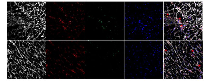
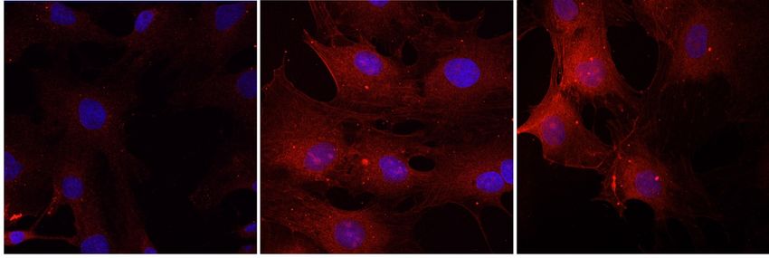
Dako, Santa Clara, CA) and steamed for 30 mins, and then allowed to cool at room temprature.

The sections were then washed in 1X TBS for 10 min, blocked with a mix of 10% goat serum and

5% BSA in 1X TBS for 1 hr, and incubated with primary antibodies against vimentin (1:50) and

α7 nAChR (1:50) overnight. On the next day, the sections were rinsed in 1X TBS and incubated

with fluorescent secondary antibodies (1:2000) for 2 hr, followed by staining of nuclei with

Hoechst (1:2000) and mounted with ProLong Gold Antifade (Thermo Fisher Scientific). Image

acquisition was performed on Zeiss LSM780 confocal microscope.

 Adult rat CF immunofluorescence staining and quantification. Cells were plated at ~70%

confluence on glass coverslips for 24 h. Cells were fixed with 4% paraformaldehyde,

permeabilized with 0.1% Triton-X (for pEGFR staining), and blocked in 5% goat serum for 30

min. After blocking, samples were incubated with anti-pEGFR antibody (1:100) or α7 nAChR

(1:50), washed, and then incubated for 1 h with appropriate fluorescent secondary antibody (1:200-

1:300, Jackson ImmunoResearch Laboratories), nuclear counterstained with Hoechst or DAPI, and

then and mounted with Prolong Gold Antifade. Images were obtained with either a confocal Zeiss

LMS780 laser-scanning microscope (20x or 40x) of Nikon Eclipse E400 IF microscope. Confocal

images were analyzed using Zen Blue (Zeiss) and ImageJ software. For each cell surface marker,

slides were imaged at identical wide-field (magnification, scan speed, laser power, detector gain,

and pinhole diameter) settings. The intensity of pEGFR was measured outlining the cells. Some

images were then analyzed on NS Elements (Nikon, Tokyo Japan) by measuring intensity of FITC

channel divided over the measured area. 3-5 images per animals were used and averaged together

as one.

 In vitro live cell α7 nAChR staining via BTX. Adult rat CF and cardiomyocytes were plated

onto glass chamber slides overnight and then incubated with α-BTX-FITC (1:2000, B13422,

 24
Thermo Fisher Scientific) for two hours in a 37°C humidified chamber. Cells were then fixed with

4% PFA, counterstained with Hoechst, and then mounted and imaged on a confocal Zeiss LMS780

laser-scanning microscope.

 Calcium imaging. Changes in [Ca2+]c in response to nicotine stimulation were observed in

live ARCFs using a Nikon TE2000 live cell epifluorescence microscope (Nikon) equipped with

Retiga EXi camera (QImaging, Surrey, BC, Canada). Cells were plated on glass-bottom 35 mm

dishes (Matek, Ashland, MA and Matsunami Glass USA Inc., Bellingham, WA), loaded with a

Ca2+-sensitive dye Fluo-3 (Biotium, Fremont, CA), and used for observation. Cell culture medium

was replaced by Tyrode solution (mm): NaCl, 136.9; KCl, 5.4; CaCl2, 2; MgCl2, 0.5; NaH2PO4,

0.33; HEPES, 5; glucose, 5; pH 7.40 adjusted with NaOH.

Cell Proliferation Assays:

 Cells were trypsinized and counted as previously described (17). Cells seeded with equal

density were allowed to adhered overnight and then serum-starved with serum-free DMEM

supplemented 10 µg/ml insulin, 5.5 µg/ml transferrin, 5 ng/ml sodium selenite (ITS, Corning,

Corning, NY), and penicillin and streptomycin. After 24 h, cells were pretreated with inhibitors

for 30 min and then treated with agonists for another 24 hr. Cells were trypsinized and counted

with a hemocytometer. For BrdU incorporation experiments, cells were seeded equally in a 96

well plate, serum starved for 24 hrs, and then treated with inhibitors/agonists and with BrdU label,

concomitantly. After 24 hr, cells were fixed and assayed for BrdU content per manufacturer’s

directions (Sigma-Aldrich).

Biochemical and Molecular Biology Assays:

 Collagen content measurement. Collagen content was measured using Sircol collagen dye

binding assay according to manufacturer’s instructions (Accurate, Westbury, NY). Equal amounts

 25
of protein or homogenates were mixed with equal volume of Sircol dye reagent for 30 mins with

agitation. The collagen-dye complex was centrifuged at 12,000 rpm for 10 mins and unbound dye

was aspirated. The remaining pellet was washed with ice cold acid-salt wash reagent (acetic acid,

sodium chloride, and surfactants). Samples were then centrifuged again at 12,000 rpm for 10 min.

The wash reagent was aspirated and the remaining pellet was dissolved by adding the alkali reagent

(0.5 M sodium hydroxide). After 5 mins incubation, collagen content was measured for absorbance

at 555 nm.

 ELISA assays. Levels of ACh (mouse: NBP2-66389, Novus Biologicals; rat: LS-F27870,

LifeSpan BioSciences) and acetylcholinesterase activity (KA4132, Novus Biologicals) were

measured in homogenized tissue per manufacturer’s directions.

 Quantitative RT-PCR. Total RNA was extracted from tissue with RNeasy Mini Kit

(Qiagen). The cDNA was made by High-Capacity cDNA Reverse Transcription Kit (Thermo

Fisher Scientific) and used for quantitative PCR (qPCR) reactions. qPCR was performed on Step

One Plus Real-time PCR System (Applied Biosystems, Foster City, CA) with the SsoAdvanced

Universal SYBR Green Supermix (Bio-Rad). Data for each gene was normalized to GAPDH (rat)

or actin (human and mouse). Real-Time PCR was performed using the primer sets listed in

Supplemental TABLE S7.

 Western blotting. Animal tissue were homogenized at 4°C in homogenization buffer (20

mM HEPES, 250 mM sucrose, 100 mM NaCl, 0.2 mM EDTA, 0.2 mM EGTA and supplemented

with protease and phosphatase inhibitors) as previously described (68, 69) Homogenates were

centrifuged at 5,000 rpm for 10 min at 4°C. The pellet was discarded, and supernatant was then

centrifuged at 15,000 rpm for 10 mins at 4°C. The resulting supernatant was collected for protein

analysis. Total protein concentration was determined with DC Protein Assay (Bio-Rad

 26
Laboratories, Hercules, CA). HEK293T cells were lysed with 1X lysis buffer (Cell Signaling

Technology) containing protease inhibitor cocktail (Sigma-Aldrich) and 1 mM

phenylmethylsulfonyl fluoride (PMSF). Protein concentrations were determined by Pierce BCA

Protein Assay Kit (Thermo Fisher Scientific).

 Equal amounts of total proteins were resolved on separating gels by SDS-PAGE followed

by transfer to polyvinylidene difluoride or nitrocellulose membrane and immunoblots were

visualized with Bio-Rad Imager Chemidoc or with Odyssey infrared imaging system (LI-COR

Biotechnology). Quantitative densitometry was performed using ImageJ (NIH, available online at

http://rsb.info.nih.gov/ij/).

Statistics:

Statistical analyses were performed using GraphPad Prism 9.1.0.221 (La Jolla, CA,

www.graphpad.com). Data are presented as mean±SEM; p-values < 0.05 were considered

statistically significant. Normality of data was checked and either log-transformation or non-

parametric testing was performed if data were not normally distributed. Mann-Whitney test,

Unpaired Student’s t-test or ANOVA with Bonferroni Post Hoc Test were used when appropriate.

Figure legends specify the tests used for each experiment.

Study Approval:

 All animal protocols were approved by the Institutional Animal Care and Use Committees

at the Providence VA Medical Center, Rhode Island Hospital, and University of Minnesota.

Collection and sharing of human tissue were approved by the Cleveland Clinic Institutional

Review Board and adhered to all required ethical standards for human subject research. Use of

human tissue was approved by the Providence VA Medical Center Institutional Review Board.

 27
AUTHORS CONTRIBUTIONS

GC, JO, AV: Designing research studies; AV, DaSGB, AFN, PZ, TJM, IP, MWC, BSJ, JO, GC:

conducting experiments and acquiring data; AV, DaSGB, AFN, PZ, ARM, JO, RTC, UM, GC

analyzing or interpreting data; EH: providing reagents; and AV, DaSGB, AFN, PZ, ARM, RTC,

BSJ, EH, UM, BSJ, JO, GC contributed to writing and editing the manuscript.

The method used in assigning the authorship order among co–first authors was based on relative

contributions in experimental design, conducting experiments, analyses and interpretation of data,

and writing of the manuscript.

ACKNOWLEDGMENTS

The authors would like to thank Dr. Serpil Erzurum, Learner Institute of Cleveland Clinic, for

providing specimens of human hearts, Amanda Costa Sousa for her assistance with

echocardiography data acquisition; Nouaying R. Kue, Alexander Park, and Julia Feord for

technical support, and Dr. Phu Tran at University of Minnesota for TMEM35/NACHO plasmid.

This material was the result of work supported with resources and the use of facilities at the

Providence VA Medical Center and the CVPB (under grant P20GM103652). GC was supported

by NHLBI R01HL128661, VA BLSR&D grant I01CX001892, and NHLBI R01HL148727. ARM

was supported by NHLBI R01HL139795 and NIGMS P20GM103652. JO is supported by NIH

R01HL136757 and P30GM1114750. UM was supported by NHLBI R01HL114784. BSJ was

supported by AHA 18CDA34110091. RTC was supported by NHLBI R01HL135236. The views

expressed in this article are those of the authors and do not reflect the position nor policy of the

Department of Veterans Affairs and the National Institutes of Health.

 28
REFERENCES:

1. Frangogiannis NG. Cardiac fibrosis: Cell biological mechanisms, molecular pathways and

 therapeutic opportunities. Mol Aspects Med. 2019;65:70-99.

2. Frangogiannis NG. Fibroblasts and the extracellular matrix in right ventricular disease.

 Cardiovasc Res. 2017;113(12):1453-64.

3. Tallquist MD, and Molkentin JD. Redefining the identity of cardiac fibroblasts. Nat Rev

 Cardiol. 2017;14(8):484-91.

4. Travers JG, et al. Cardiac Fibrosis: The Fibroblast Awakens. Circ Res. 2016;118(6):1021-

 40.

5. Andersen S, et al. Right Ventricular Fibrosis. Circulation. 2019;139(2):269-85.

6. Bogaard HJ, and Voelkel NF. Is Myocardial Fibrosis Impairing Right Heart Function? Am

 J Respir Crit Care Med. 2019;199(12):1458-9.

7. Egemnazarov B, et al. Right ventricular fibrosis and dysfunction: Actual concepts and

 common misconceptions. Matrix Biol. 2018;68-69:507-21.

8. Clements RT, et al. Treatment of Pulmonary Hypertension With Angiotensin II Receptor

 Blocker and Neprilysin Inhibitor Sacubitril/Valsartan. Circ Heart Fail.

 2019;12(11):e005819.

9. Taraseviciene-Stewart L, et al. Simvastatin causes endothelial cell apoptosis and attenuates

 severe pulmonary hypertension. Am J Physiol Lung Cell Mol Physiol. 2006;291(4):L668-

 76.

10. Simonneau G, et al. Haemodynamic definitions and updated clinical classification of

 pulmonary hypertension. Eur Respir J. 2019;53(1).

 29
11. Bogaard HJ, et al. Chronic pulmonary artery pressure elevation is insufficient to explain

 right heart failure. Circulation. 2009;120(20):1951-60.

12. Kusakari Y, et al. Impairment of Excitation-Contraction Coupling in Right Ventricular

 Hypertrophied Muscle with Fibrosis Induced by Pulmonary Artery Banding. PLoS One.

 2017;12(1):e0169564.

13. Rain S, et al. Right Ventricular Myocardial Stiffness in Experimental Pulmonary Arterial

 Hypertension: Relative Contribution of Fibrosis and Myofibril Stiffness. Circ Heart Fail.

 2016;9(7).

14. Gomez-Arroyo J, et al. Differences in right ventricular remodeling secondary to pressure

 overload in patients with pulmonary hypertension. Am J Respir Crit Care Med.

 2014;189(5):603-6.

15. Mehta BB, et al. Detection of elevated right ventricular extracellular volume in pulmonary

 hypertension using Accelerated and Navigator-Gated Look-Locker Imaging for Cardiac

 T1 Estimation (ANGIE) cardiovascular magnetic resonance. J Cardiovasc Magn Reson.

 2015;17:110.

16. Simpson CE, and Hassoun PM. Myocardial Fibrosis as a Potential Maladaptive Feature of

 Right Ventricle Remodeling in Pulmonary Hypertension. Am J Respir Crit Care Med.

 2019;200(6):662-3.

17. Vang A, et al. Effect of alpha7 nicotinic acetylcholine receptor activation on cardiac

 fibroblasts: A mechanism underlying RV fibrosis associated with cigarette smoke

 exposure. Am J Physiol Lung Cell Mol Physiol. 2017:ajplung 00393 2016.

18. Albuquerque EX, et al. Mammalian nicotinic acetylcholine receptors: from structure to

 function. Physiol Rev. 2009;89(1):73-120.

 30
You can also read