A partially demineralized allogeneic bone graft: in vitro osteogenic potential and preclinical evaluation in two different intramembranous bone ...
←
→
Page content transcription
If your browser does not render page correctly, please read the page content below
www.nature.com/scientificreports

                OPEN             A partially demineralized
                                 allogeneic bone graft: in vitro
                                 osteogenic potential and preclinical
                                 evaluation in two different
                                 intramembranous bone healing
                                 models
                                 Pierre Tournier1,2, Jérôme Guicheux3,4, Arnaud Paré1,5, Aymeric Maltezeanu1,
                                 Thibaut Blondy6, Joëlle Veziers3,4, Caroline Vignes1,4, Manon André1, Julie Lesoeur1,4,
                                 Ana Barbeito2, Raphaël Bardonnet2, Christophe Blanquart6, Pierre Corre3, Valérie Geoffroy1,7,
                                 Pierre Weiss3,7* & Alexis Gaudin3,7

                                 In skeletal surgical procedures, bone regeneration in irregular and hard-to-reach areas may present
                                 clinical challenges. In order to overcome the limitations of traditional autologous bone grafts and bone
                                 substitutes, an extrudable and easy-to-handle innovative partially demineralized allogenic bone graft
                                 in the form of a paste has been developed. In this study, the regenerative potential of this paste was
                                 assessed and compared to its clinically used precursor form allogenic bone particles. Compared to the
                                 particular bone graft, the bone paste allowed better attachment of human mesenchymal stromal cells
                                 and their commitment towards the osteoblastic lineage, and it induced a pro-regenerative phenotype
                                 of human monocytes/macrophages. The bone paste also supported bone healing in vivo in a guide
                                 bone regeneration model and, more interestingly, exhibited a substantial bone-forming ability when
                                 implanted in a critical-size defect model in rat calvaria. Thus, these findings indicate that this novel
                                 partially demineralized allogeneic bone paste that combines substantial bone healing properties and
                                 rapid and ease-of-use may be a promising alternative to allogeneic bone grafts for bone regeneration
                                 in several clinical contexts of oral and maxillofacial bone grafting.

                                 In oral and maxillofacial bone grafting procedures, autologous bone grafting is still considered to be the reference
                                 technique. This technique has a high success rate as a result of the combination of osteoconductivity (the ability
                                 to provide a structural support for bone growth), osteogenicity (the promotion of osteoblastic differentiation of
                                 progenitor cells), and osteoinductivity (the induction of bone growth in large bone defects or heterotopic sites,
                                 mainly due to growth factors in the bone and marrow environment)1–4. Despite these advantages, autologous
                                 bone grafts (ABGs) suffer from numerous serious peri- and post-operative drawbacks. Among these, the potential
                                 donor site morbidities (e.g., pain, hematoma, blood loss, nerve injury, the risk of bone fracture), the additional
                                 operative time, the limited available bone (e.g., iliac crest, calvarial bone), and the occasional need for a second
                                 surgical team are the main limitations of this procedure and consequently hindering the clinical benefits. To

                                 1
                                  INSERM, UMR 1229, RMeS, Regenerative Medicine and Skeleton, Université de Nantes, Oniris, 1 Place Alexis
                                 Ricordeau, 44042 Nantes, France. 2BIOBank SAS, Lieusaint, France. 3INSERM, UMR 1229, RMeS, Regenerative
                                 Medicine and Skeleton, CHU Nantes, Université de Nantes, Oniris, 1 Place Alexis Ricordeau, 44042 Nantes,
                                 France. 4INSERM, UMS 016, CNRS 3556, Structure Fédérative de Recherche François Bonamy, SC3M Facility, CHU
                                 Nantes, Université de Nantes, 44042 Nantes, France. 5Service de Chirurgie Maxillo‑faciale, Plastique et Brulés,
                                 Hôpital Trousseau, CHU de Tours, 37170 Tours, France. 6Université de Nantes, Univ Angers, INSERM, CNRS,
                                 CRCINA, 44000 Nantes, France. 7These authors contributed equally: Valérie Geoffroy, Pierre Weiss and Alexis
                                 Gaudin. *email: pierre.weiss@univ-nantes.fr

Scientific Reports |   (2021) 11:4907                | https://doi.org/10.1038/s41598-021-84039-6                                                  1

                                                                                                                                              Vol.:(0123456789)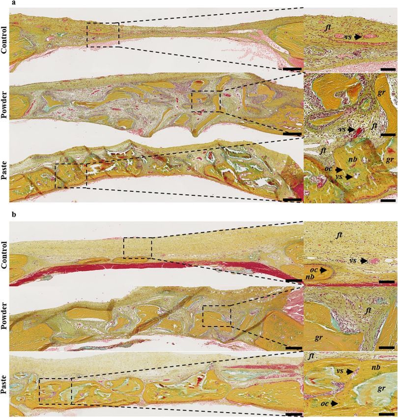
www.nature.com/scientificreports/
overcome these limitations, several alternatives such as alloplastic and xenogeneic bone substitutes or allogeneic
bone grafts are used in oral and maxillofacial surgeries5–7.
Allogeneic bone grafts are reliable and have been used extensively for decades as an alternative to ABGs8,9.
The amount of allogeneic bone is virtually unlimited, and it can be obtained in various sizes and shapes (e.g.,
blocks, blades, paste, putty, powder, chips, injectable forms). The duration of the surgery is shortened compared
to ABG-based reconstructions because there is no need for a bone harvesting procedure. Moreover, allogeneic
bone grafts exhibit the same bone healing capacities as ABGs in multiple indications such as bone augmentation
before dental implant placement (e.g., sinus floor elevation, ridge augmentation)10,11.
Although allogeneic bone grafts are mainly used in powder or block forms, they exhibit a lack of versatility
and/or handling, especially in specific indications related to pre-implant surgery, such as sinus floor elevation,
guided bone regeneration in horizontal/vertical augmentation, and alveolar socket preservation. To address these
unmet clinical needs, a novel ready-to-use, easy-to-handle, extrudable, and moldable human bone graft for bone
regenerative medicine has been developed12. This allogeneic bone paste is made of partially demineralized bone
particles, consisting of a mineralized core (Sup. Fig. 1, upper panel, white arrows) surrounded by a demineralized
bone matrix (Sup. Fig. 1, upper panel, black arrows). This bone paste does not require any mixing, rehydration,
or reconstitution prior to its use, which is a key feature for clinical use. Such a bone paste could replace the bone
substitutes that are in powder form, particularly for the irregular or hard-to-reach areas frequently encountered
in oral and maxillofacial bone surgery. However, aside from its handling and physical characteristics, proof of
concept of its bone regenerative capacity should be established prior to its clinical transfer. In this context, the
present work aimed to provide a thorough assessment of the bone healing capacity of this allogeneic bone paste
compared with an allogeneic particular bone graft before its partial demineralization.
The purpose of this study was hence to assess the effects of the bone paste in vitro (1) on the attachment and
osteoblastic commitment of primary human mesenchymal stromal cells from bone marrow (hBM-MSCs), (2)
on the polarization of primary human monocytes isolated from circulating blood, and (3) to test and validate the
in vivo capacity of this innovative bone paste to support and promote bone healing in two preclinical models: a
guided bone regeneration (GBR) and a critical size defect (CSD) model in rat calvaria to mimic intramembranous
bone healing of oral and maxillofacial defects.
Results
Attachment and osteoblastic commitment of hBM‑MSCs. The attachment of hBM-MSCs was first
investigated after short periods (1, 3, and 6 h) of contact with the particular bone graft or the bone paste. After
1 h of contact, the cells on the surface of the particular bone graft exhibited a round shape, while they had an
elongated and spread-out shape on the surface of bone paste. Interestingly, at 3 h and 6 h, all of the cells exhibited
an elongated and spread-out shape (Fig. 1a) irrespective of the graft. These data strongly suggest that the bone
paste allows faster cell adhesion compared to the particular bone graft. Quantification of the DNA content of the
adherent cells revealed no difference after 1 h of contact. However, after 3 and 6 h of contact, the DNA content
was significantly higher (60% and 70%, respectively) for the cells cultured in contact with the bone paste com-
pared to the cells in contact with the particular bone graft (Fig. 1b).
To determine whether the particular bone graft or the bone paste may affect the osteoblastic differentiation of
hBM-MSCs, the cells were cultured in contact with the bone grafts in culture medium with or without osteogenic
factors for 14 and 21 days. As seen by the SEM analyses, the osteogenic factors did not influence the colonization
of the particular bone graft or the bone paste by the hBM-MSCs. Under all of the culture conditions, the cells had
elongated cellular bodies and cytoplasmic expansions (Fig. 1c, white arrows). Of interest, the cohesion between
the bone paste allowed the cells to create a network from particle to particle, with or without osteogenic factors,
at all of the time points (Fig. 1c). It is also worth noting that this phenomenon was not observed with the cells
cultured in contact with the particular bone graft. Finally, to evaluate the osteogenic commitment of the hBM-
MSCs cultured in contact with the particular bone graft or the bone paste, the ALP activity was measured in cell
lysates, and it was found to be significantly higher in the cells cultured in contact with the bone paste compared
to the cells in contact with the particular bone graft: 3.3 and 2.7 times higher, respectively, in the absence of
osteogenic factors and 7.3 and 4.6 times higher, respectively, in the presence of osteogenic factors after 14 and
21 days of culture (Fig. 1d).
Intramembranous bone healing in a GBR model. To mimic clinical indications of guided bone regen-
eration (e.g., alveolar socket preservation), the ability of the bone paste to support bone healing was first assessed
in a GBR model in rat calvaria. The µ-CT analyses of the control defects at 7 weeks post-surgery revealed a
significant degree of centripetal bone ingrowth from the edges of the defects (Fig. 2a), as expected in this GBR
model. In the grafted defects, homogenous bone ingrowth (Fig. 2a, yellow arrows) was observed, but the particu-
lar bone graft and the bone paste were still visible in the defects (Fig. 2a, white arrows). The three-dimensional
quantification of the MV/TV, increasing over time, indicated that new bone formation occurred in the control
and in the grafted defects (Fig. 2b). However, the increase in the MV/TV between 0 and 7 weeks was greater in
the group grafted with the bone paste than in the group grafted with the particular bone graft (2.2 vs. 1.6-fold,
respectively).
In all of the defects, the histological observations revealed the collagen-rich and layer-organized nature of the
newly formed bone (yellow matrix), with numerous osteocytes, that was identical to the native bone outside the
defects (Fig. 3). These observations confirmed the µ-CT analyses, with significant bone formation after 7 weeks in
the control defects, yet without continuous newly formed bone tissue (i.e., full closure). In the defects implanted
with the particular bone graft and the bone paste, the grafts were perfectly osteointegrated and still visible (Fig. 3,
gr) and recognizable due to their shape and the absence of osteocytes inside the grafts.
Scientific Reports | (2021) 11:4907 | https://doi.org/10.1038/s41598-021-84039-6 2
Vol:.(1234567890)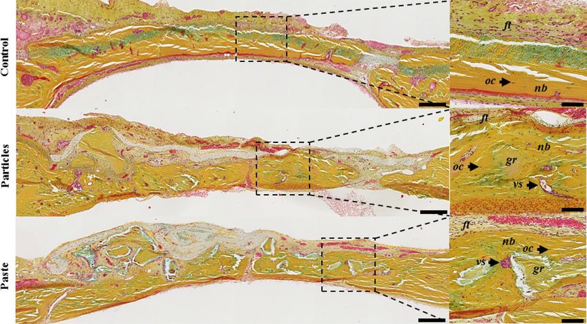
www.nature.com/scientificreports/

                                  Figure 1.  Attachment and osteoblastic commitment of hBM-MSCs cultured in contact with the particular bone
                                  graft or the bone paste. (a) Representative confocal images of stained hBM-MSCs. Red (phalloidin-AF568):
                                  F-actin, blue (Hoechst): nucleus. The particular bone graft and the bone paste were visible as a result of their
                                  blue auto fluorescence. Scale bars: 100 µm. (b) Quantification of the total DNA in hBM-MSCs in contact with
                                  the particular bone graft or the bone paste. (c,d) SEM images and ALP activity in lysates of hBM-MSCs grown
                                  in contact with the particular bone graft or bone paste with or without osteogenic factors for 14 and 21 days.
                                  Scale bars: 20 µm. The results are expressed as means ± the SD (N = 3, n = 3; *p < 0.05, **p < 0.01 ***p < 0.001).

                                  Intramembranous bone healing in a CSD model. The intrinsic bone-forming ability of the bone paste
                                  was then tested using a challenging CSD model of intramembranous bone healing. Based on the reconstructed
                                  µ-CT images, it appeared that no newly formed bone was observable in the control defects or filled with the
                                  particular bone graft at 3 and 7 weeks post-surgery (Fig. 4a,b white arrows). This was confirmed by the MV/TV
                                  quantifications (Fig. 4c). By contrast, in the defects filled with the bone paste, newly formed bone was clearly
                                  visible after 3 and 7 weeks (Fig. 4a,b yellow arrows), thus indicating healing of the defects in the presence of the
                                  bone paste.
                                      The histological observations confirmed the radiological analyses and showed that a barely detectable amount
                                  of new bone had formed at the edges but not at the center of the control defects at 3 weeks (Fig. 5a) and 7 weeks
                                  (Fig. 5b) post-surgery. The same observation was made in the defects filled by the particular bone graft at 3 weeks
                                  (Fig. 5a) and 7 weeks (Fig. 5b) post-surgery. In this group, three weeks after the implantation, the soft tissue
                                  appeared broadly vascularized and highly cellularized in close proximity to the particular bone graft (Fig. 5a).
                                  The surface of these particular bone graft was light blue, probably due to the fact that the surfaces had lost their
                                  collagen content (which appeared yellow by Movat’s pentachrome staining). Seven weeks after the particular

Scientific Reports |   (2021) 11:4907 |                https://doi.org/10.1038/s41598-021-84039-6                                                   3

                                                                                                                                               Vol.:(0123456789)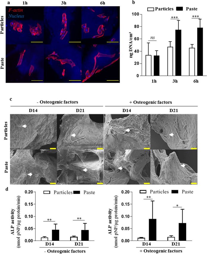
www.nature.com/scientificreports/

                                            Figure 2.  Rat calvaria healing 7 weeks after surgery in a guided bone regeneration model. (a) Representative
                                            images of the reconstructed µ-CT acquisitions (yellow arrows: newly formed bone, white arrows: grafts). The
                                            control represents the defects with the GBR membrane without bone grafts (unfilled defects). Scale bars: 1 mm.
                                            (b) Three-dimensional µ-CT quantitative analysis of the mineral volume (MV) within the tissue volume (TV)
                                            in the defects at the time of surgery (0w, baseline) and 7 weeks (7w) after surgery. The results are expressed as
                                            means ± the SD (n = 6) *p < 0.05, **p < 0.01, ***p < 0.001).

                                            Figure 3.  Rat calvaria healing 7 weeks after surgery in a guided bone regeneration model. Representative
                                            images of the defects with Movat’s pentachrome staining of undecalcified 7-µm thick frontal sections. The
                                            control represents the defects with the GBR membrane without bone grafts (unfilled defects). Scale bars: 250 µm
                                            for the histological full defects and 100 µm for the histological magnification (dashed square). Dark yellow:
                                            collagen in the bone tissue, light red: cytoplasms, dark red: osteoid borders, brown: nuclei, ft: fibrous tissue, oc:
                                            osteocytes, nb: newly formed bone, vs: blood vessels, gr: grafts.

                                            bone graft implantation, the soft tissue appeared to be less vascularized and less cellularized compared to the
                                            defects observed 3 weeks after the implantation. On the other hand, the defects filled with the bone paste exhib-
                                            ited a substantial proportion of newly formed bone, which was already visible 3 weeks post-surgery (Fig. 5a,
                                            nb). Despite the fact that the newly formed bone was distributed heterogeneously in the defects 3 weeks after the
                                            surgery, lamellar bone structures with numerous osteocytes and a number of blood vessels could be observed
                                            (Fig. 5a, nb, vs). Of interest, the bone paste had become fully integrated in the regenerated bone 7 weeks after
                                            the surgery (Fig. 5b, gr), as discernible in the defects with their characteristic acellular mineralized inner core
                                            and demineralized outer part.

          Scientific Reports |   (2021) 11:4907 |                https://doi.org/10.1038/s41598-021-84039-6                                                     4

Vol:.(1234567890)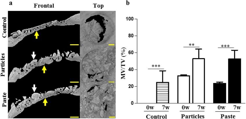
www.nature.com/scientificreports/
Figure 4. Rat calvaria healing at 3 and 7 weeks after surgery in a critical size defect model. (a,b) Representative
images of the reconstructed µ-CT acquisitions (yellow arrows: newly formed bone, white arrows: grafts). The
control represents the unfilled defects. Scale bars: 1 mm. (c) Three-dimensional µ-CT quantitative analysis of the
mineral volume (MV) within the tissue volume (TV) in the defects after 0 weeks (baseline), 3, and 7 weeks of
regeneration. The results are expressed as means ± the SD (n = 6) **p < 0.01, ***p < 0.001).
At this time point, the bone regrowth in the defects filled with the bone paste was much more abundant and
homogeneous compared to at 3 weeks (Fig. 5b), with a limited amount of soft tissue. Moreover, the regenerated
bone (Fig. 5b, nb) exhibited a typical lamellar osseous structure, with numerous osteocytes (Fig. 5b, oc) and
vasculature reminiscent of that observed in native bone tissue outside the grafted area.
Monocyte/macrophages phenotypic changes. Having demonstrated that the bone paste had greater
regenerative properties than the particular bone graft in two complementary models of intramembranous bone
healing, we sought to determine whether monocytes/macrophages may play a pivotal role in the discrepancy
found in the regenerative efficiency of the two types of bone grafts.
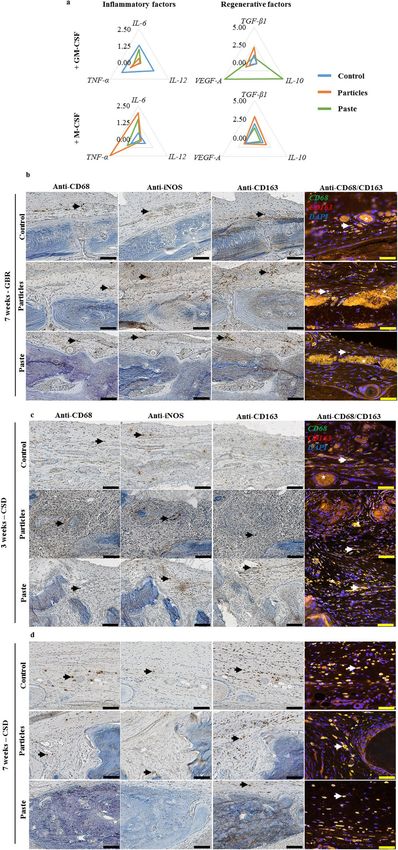
When cultured under pro-inflammatory conditions, the macrophages in contact with the particular bone
graft expressed lower levels of TNF-α, IL-6, and IL-12 mRNA level (classical markers of inflammation) compared
to the control without grafts (Fig. 6a). The macrophages cultured in contact with the bone paste also expressed
lower levels of TNF-α, IL-6, and IL-12 compared to the control. However, they exhibited higher VEGF-A and
IL-10 mRNA expression (classical pro-regenerative markers), thereby suggesting a substantial pro-regenerative
phenotype (Fig. 6a). On the other hand, when cultured under pro-regenerative conditions, the macrophages
exhibited no change in VEGF-A, TGF-β, or IL-10 mRNA expression when in contact with the particular bone
graft or the bone paste compared to the control, thus indicating that the pro-regenerative phenotype of the
macrophages was maintained in these conditions (Fig. 6a). However, the macrophages in contact with the bone
paste also expressed lower IL-6 and TNF-α mRNA levels than those in contact with the particular bone graft,
supporting the role of the bone paste to enhance the regenerative phenotype of the macrophages (Fig. 6a).
To further determine whether this in vitro observation may have a degree of in vivo relevance, the phenotype
of the macrophages was analyzed in the calvarial defects by immunohistochemical labeling [CD68 (total mac-
rophages), iNOS (pro-inflammatory macrophages), and CD163 (pro-regenerative macrophages)].
In the GBR model, CD68+, iNOS+, or CD163+ cells (black arrows), as well as CD68+/CD163+ cells (white
arrows) were found in the control group as well as in the vicinity of the particular bone graft (Fig. 6b). How-
ever, in the group grafted with the bone paste, while C D68+, CD163+, and C D68+/CD163+ cells were found in
Scientific Reports | (2021) 11:4907 | https://doi.org/10.1038/s41598-021-84039-6 5
Vol.:(0123456789)www.nature.com/scientificreports/
Figure 5. Rat calvaria healing at 3 and 7 weeks after surgery in a critical size defect model. Representative
images of the defects with Movat’s pentachrome staining of undecalcified 7-µm thick frontal section, 3 (a) or 7
(b) weeks after the surgery. The control represents the unfilled defects. Scale bars: 250 µm for the full defects,
100 µm for the high-magnification image (dashed square). Dark yellow: collagen in the mature bone, light red:
cytoplasms, dark red: osteoid borders, brown: nuclei, ft: fibrous tissue, oc: osteocytes, nb: newly formed bone, vs:
blood vessels, gr: grafts.
the regenerated tissue, few i NOS+ cells were observed. This strongly suggests that the macrophages had largely
assumed a pro-regenerative phenotype 7 weeks after the implantation.
In the CSD model, after 3 and 7 weeks, C D68+, iNOS+, CD163+, or CD68+/CD163+ cells were present in the
control group (Fig. 6c). In the grafted groups, our data suggest that the macrophages had a more pronounced
pro-regenerative engagement in the defects filled with the bone paste than with the particular bone graft. This
difference was clearly observable after 3 weeks of regeneration. In the defects filled with the particular bone graft,
CD68+ and i NOS+ cells could be seen, while CD163+ and C D68+/CD163+ cells were hard to discern. Conversely,
in the defects filled with the bone paste, CD68+, CD163+, CD68+/CD163+ cells were present, and few iNOS+
cells could be seen. After 7 weeks of regeneration, the unfilled defects and the defects filled with the particular
bone graft contained CD68+, iNOS+, CD163+, and CD68+/CD163+ cells. At this time point, the defects grafted
with bone paste were filled with newly formed bone, and hence few monocytes/macrophages could be seen in
the defects. Indeed, only C D163+ and C D68+/CD163+ cells were present in the soft tissue in the regenerated
defects (Fig. 6d).
Discussion
Allogeneic bone grafts are a suitable and widely used alternative to ABGs in oral and maxillofacial bone graft
procedures. However, allogeneic bone is mainly used in powder or block forms, and its lack of versatility and
adaptability limits its use in irregular or hard-to-reach areas. In this work, the regenerative potential of an easy-
to-handle and ready-to-use moldable allogeneic bone graft in a paste form made of partially demineralized
bone allograft was assessed, with the aim of supplanting the use of allogeneic particular bone grafts in oral and
maxillofacial bone grafting procedures.
Scientific Reports | (2021) 11:4907 | https://doi.org/10.1038/s41598-021-84039-6 6
Vol:.(1234567890)www.nature.com/scientificreports/
Figure 6. Effect of the particular bone graft and the bone paste on monocyte/macrophage polarization. (a)
mRNA expression in human monocytes cultured in the presence of GM-CSF or M-CSF for 3 days in contact
with the particular bone graft or the bone paste. The Y-axis represents the mean relative mRNA expression
level of the specified genes (N = 3). Representative immunohistochemical and immunofluorescent stainings
on successive undecalcified 7-µm thick sections, (b) in the GBR model after 7 weeks, and (c) in the CSD
model after 3 and 7 weeks. Immunohistochemical stainings were performed for CD68, iNOS, CD163, and
immunofluorescence with double labeling of CD68 (green)/CD163 (red) and DAPI (blue) for the nuclei. For
single labeling, positive cells were stained with a brown precipitate (black arrow), for double labeling, double-
positive cells appeared as light yellow (white arrow). Typical auto fluorescent red blood cells were visible (white
stars). Scale bars: 100 µm for the immunohistochemistry, 50 µm for the immunofluorescence images.
Scientific Reports | (2021) 11:4907 | https://doi.org/10.1038/s41598-021-84039-6 7
Vol.:(0123456789)www.nature.com/scientificreports/
Our data first of all demonstrated better attachment of hBM-MSCs to the surface of the bone paste com-
pared to the particular bone graft. The greater attachment could be due to the partial demineralization and
heating of the bone paste, which modifies the surface of the graft and hence also the cell-material interaction.
Although further analyses (e.g., particle topography, kinetics of focal adhesion formation) are required to prop-
erly characterize the nature of the interactional changes, it has been reported that the denaturation of collagen
can expose cryptic adhesion sites and RGD (Arg-Gly-Asp) integrin-binding sites that promote the adhesion
of hBM-MSCs13–18. Additionally, the bone paste was found to remain cohesive for up to 21 days in an aqueous
medium, and it promoted the osteogenic commitment of hBM-MSCs. This latter property is of great relevance
for bone regeneration since the bone paste could act as a cohesive scaffold in the bone defects. This may lead to
better osteointegration and osteoconduction than with independent and non-demineralized bone grafts. The
higher ALP activity observed in hBM-MSCs cultured in contact with the bone paste compared to the particular
bone graft may be due to the different interaction between the cells and the bone grafts, and/or to the presence
of a cellular network at the surface of the bone paste, thereby enhancing the osteogenic commitment of hBM-
MSCs. Moreover, type I collagen is thought to drive osteogenic fate, and the exposure of RGD binding sites is
known to enhance the osteogenic phenotype of mesenchymal stromal c ells18–21.
These positive in vitro effects of the bone paste on the mesenchymal progenitors were also confirmed through
the regenerative potential of the bone paste investigated in a GBR and a CSD model in rat calvaria. In light of the
similar organization and ossification between the cranial and maxillary bones, these calvarial models are widely
used for preclinical studies focused on the development of bone substitutes for oral and maxillofacial s urgeries22.
Additionally, this operating site allows easy and rapid examination of the bone healing properties of the grafted
materials23–25. Moreover, such regenerative properties of bone graft have to be investigated in GBR and CSD
models, as they are representative of bone healing in techniques that are clinically used in several oral and jaw
surgery indications (e.g., sinus lift, socket preservation). However, the atrophic environment of the rat calvaria
(poor vasculature and bone marrow) is a challenge when it comes to achieving bone regeneration.
The covering membrane in the GBR model prevents non-osteoblastic cells from migrating from the surround-
ing soft tissues, thereby indirectly favoring invasion of the bone defects by osteoprogenitors, which improves
the bone regeneration26–28. In the CSD model, as the bone regeneration is slower compared to the GBR model,
an intermediate time point, 3 weeks in addition to 7 weeks of regeneration, was chosen to investigate the early
events that are crucial for bone regeneration in these challenging conditions.
The bone regeneration observed in the defects filled with the bone paste in the GBR model validated the
ability of this graft to support bone healing. Moreover, the more pronounced increase in the MV/TV between
0 and 7 weeks in the group grafted with the bone paste compared to the group grafted with the particular bone
graft can be explained either by the lower MV/TV of the bone paste at the implantation, due to the demineral-
ized part of the bone paste, or by a higher amount of newly formed bone. In the CSD model, the radiological
and histological observations showed that the particular bone graft failed to support bone repair. The fibrous
tissue observed 3 weeks post-implantation suggests that a fibrous encapsulation of the graft occurred, thereby
preventing the bone formation. Longer time points would have been necessary to evaluate the healing properties
of the particular bone graft in this CSD model.
Nonetheless, the absence of calvarial bone healing has been observed for similar time points, 7 weeks post
implantation, with alternative bone filling materials such as synthetic biphasic calcium p hosphate29,30 or xeno-
31
geneic bone which are widely clinically used. Those particular bone grafts substitutes are known to be mainly
osteoconductives, which is not sufficient to allow a bone regrowth in an atrophic site (i.e., low vasculature and the
absence of bone marrow) such as calvaria. The particular bone graft used in this study (i.e., cleaned, grinded and
sterilized bone tissue) displayed insufficient bone healing properties to promote bone regrowth in the CSD model.
Since the particular bone graft had no effect on cell viability (Sup. Fig. 2), the lack of new bone formation in the
particular bone graft-filled defects is likely to be related to the low self-healing capacity of the calvarial bone.
In contrast with the particular bone graft, the bone paste induced bone regeneration. This effect is probably
not due to the release of growth factors32,33 from the demineralized bone matrix, since the heat treatment that the
bone graft was subjected to has been reported to denature growth f actors34. Therefore, we hypothesize that bone
regeneration could be induced by the cell-particle interaction and cellular network that is known to promote
osteoblastic differentiation and to foster bone regeneration35,36. Additionally, the cohesive nature of the bone
paste may prevent it from leaking out of the defect, which is notably advantageous in irregular and hard-to-reach
areas, which are common in oral and maxillofacial surgery.
Lastly, we investigated the phenotype of the macrophages in contact with the particular bone graft and the
bone paste, as these cells are among the first to invade bone defects and interact with grafted materials. Indeed,
pro-inflammatory “M1-like” macrophages in early immune responses37–39 and pro-regenerative “M2-like” mac-
rophages in the later stages of bone h ealing40–42 are both critically required for proper bone regeneration. The
macrophages in contact with the bone paste expressed lower IL-12 and higher IL-10 mRNA levels than those in
contact with the particular bone graft. This result supports the hypothesis that an “M1-like” to “M2-like” phe-
notypic switch known to promote inflammation resolution, wound repair, and bone h ealing43–46 was induced
by the bone paste. In addition, the increase in VEGF-A mRNA expression in the macrophages in contact with
the bone paste may also contribute to acceleration of the bone repair process, since angiogenesis plays a pivotal
role in bone growth and healing47–51.
Our overall immunohistochemical observations also strongly suggest that the bone paste either accelerated
the on-site monocyte/macrophage recruitment and/or their “M1-like” to “M2-like” phenotypic switch compared
to the particular bone graft. Indeed, the small number of i NOS+ cells and the abundance of C D163+ and CD68+/
+
CD163 cells in the defects filled with the bone paste suggest a suitable inflammatory environment for bone
regeneration, with the polarization of the macrophages towards a pro-regenerative phenotype. As the initial
inflammation phase may be shortened in these defects, the mineralization phase of the bone tissue could start
Scientific Reports | (2021) 11:4907 | https://doi.org/10.1038/s41598-021-84039-6 8
Vol:.(1234567890)www.nature.com/scientificreports/
earlier as well and thus explain the higher MV/TV after 7 weeks in the defects filled with the bone paste compared
to the particular bone graft in both the GBR and the CSD model. This is of notably clinical relevance in oral and
jaw surgeries, since shortening of the bone healing time may enhance the speed and stability of osteointegration
of dental implants, thereby leading to a reduction of the associated treatments or temporary prostheses that often
cause significant discomfort for patients.
Conclusions
To the best of our knowledge, this study is the first to demonstrate the bone regenerative capacity of a partially
demineralized bone graft in a paste form in vitro and in vivo. In this study, we successfully demonstrated that a
ready-to-use and easy-to-handle partially demineralized allogeneic bone paste has substantial bone regenerative
properties. The ability of the allogeneic bone paste to induce bone formation by mesenchymal progenitor cells
and macrophages, as well as its radio-opacity, are key features for clinicians. Moreover, as a ready-to-use bone
substitute, this paste does not require any additional compounds prior to its use, which is an advantage when
considering both regulatory approval and potential clinical indications. Our promising preclinical data have
recently led us to initiate an interventional randomized clinical trial for pre-implant guided bone regeneration
in oral surgery (NCT04141215).
Materials and methods
Process to obtain the bone paste. The allogeneic particular bone graft and bone paste were obtained
from human donors who had provided their informed consent, and all of the protocols were approved by the
French ethical committee “Comité de Protection des Personnes” (CPP), in accordance with the 1975 Helsinki
declaration and its subsequent amendments. The bone allografts were harvested during total hip replacement
surgeries and donated to the BIOBank bone bank (Presles-en-Brie, France), under the authorization of the
“Agence Nationale de Sécurité du Médicament et des produits de santé” (ANSM, n°FR07703T-19-01) according
to the relevant French regulations and ethical standards. Cleaning of the femoral heads was ensured by super-
critical CO2 lipid extraction followed by successive immersions in H 2O2, NaOH, and EtOH, leaving the bone
matrix defatted52, virally inactivated53,54, and unaltered55–57. The cleaned femoral heads were crushed to generate
the particular bone graft (0.3 to 1 mm in diameter, Sup. Fig. 1, Sup Vid. 1). The particular bone graft was either
sterilized by 25 kGy gamma irradiation and used as an internal comparator, or it was processed into a paste form.
For this purpose, the particular bone graft was partially demineralized with HCl, then hydrated with water to
make it an extrudable and moldable preparation (Sup. Fig. 1, Sup Vids. 2 and 3), and heated (121 °C) to ensure
partial transformation of the outer demineralized collagen of the particles into gelatin and simultaneous steri-
lization of the entire bone graft18. All of the particular bone graft and the bone paste samples were made from
pooled donor material in order to limit the intra-donor variability.
The bone and paste were imaged with transmitted light (Leica DM12 LED, Wetzlar, Germany) or reflected
light (Leica M125). For all of the X-ray micro-computed tomography (µ-CT) acquisitions, the samples were
scanned under the following conditions: resolution: 8 µm/px, single 360° scan, rotation: 0.710°, averaging frames:
4 (SkyScan-1072, Bruker, Billerica, USA). The three-dimensional datasets were reconstructed with the NRecon
software (Micro Photonics Inc, Allentown, USA). For the scanning electron microscopy (SEM) imaging, the
samples were sputtered with a gold plasma (Sputter Coater Desk Denton Vacuum, Moorestown, USA), and
the images were taken using an electron microscope (Zeiss, Oberkochen, Germany) with a secondary electron
detector set at 10 kV.
Analysis of hBM‑MSCs attachment and osteoblastic commitment. Human BM-MSCs from two
independent donors (one male and one female, PromoCell, Heidelberg, Germany) were amplified in PromoCell
Growth Medium supplemented with 1% penicillin–streptomycin (P/S) (Invitrogen, Paisley, UK) in a humidified
atmosphere at 37 °C, 5% CO2 (Air Liquide, Paris, France). The medium was changed every other day. The cells
were used between passage 2 and 5.
To measure the attachment of hBM-MSCs to the particular bone graft and the bone paste, the grafts (0.125 cc/
well, 66 and 93 mg, respectively) were spread on the bottom of 24-well plates (Ultra-Low Attachment Surface,
Corning). One milliliter of culture medium (DMEM/Ham’s F12 (1:1), high glucose, GlutaMAX (Thermo Fisher
Scientific) with 15% fetal calf serum (FCS, Dominique Dutscher, Brumath, France) and 1% P/S) was added to
each well and the plates were incubated in a humid atmosphere at 37 °C, 5% C O2 for 24 h. The medium was
then removed and 1 × 10⁵ hBM-MSCs were seeded in each well. After 1 h, 3 h, and 6 h of contact between the
hBM-MSCs and the particular bone graft or the bone paste, the cells were extensively rinsed with 1X phosphate
buffered saline (PBS) at 37 °C to remove the non-adherent cells. The DNA content of remaining adherent cells
in contact with the particular bone graft or the bone paste was measured with Quant-iT™ PicoGreen dsDNA
Reagent (Thermo Fisher Scientific) according to the manufacturer’s instructions and then normalized to the
surface of the bone grafts measured by µ-CT as mentioned in subsection 2.1.
For the confocal imaging, the cells were fixed in 4% paraformaldehyde (PFA) for 20 min and then permeabi-
lized with Triton X-100 (1% in 1X PBS) for 10 min at room temperature. The cells were then labeled with 1:200
Alexa Fluor 568 phalloidin (Thermo Fisher Scientific) in 1X PBS at room temperature for 1 h, and the nuclei were
stained with 1:50,000 Hoechst 33258 (Thermo Fisher Scientific) in 1X PBS for 15 min at room temperature. The
cell samples were imaged using a confocal microscope (A1RS, Nikon, Champigny sur Marne, France).
To assess the ability of the particular bone graft and the bone paste to support osteogenic commitment, the
level of alkaline phosphatase (ALP) activity was measured in cell lysates. The particular bone graft and the bone
paste were placed in low-binding 24-well plates as described above, and 1 × 10⁵ hBM-MSCs were seeded in each
well. After 48 h, the medium was replaced with culture medium with or without osteogenic factors (50 µM
Scientific Reports | (2021) 11:4907 | https://doi.org/10.1038/s41598-021-84039-6 9
Vol.:(0123456789)www.nature.com/scientificreports/
Gene Common name Primer names/sequence Supplier
IL-12 Interleukin-12 QT00000364 Qiagen
IL-6 Interleukin-6 QT00083720 Qiagen
TNF-α Tumor necrosis factor-alpha QT01079561 Qiagen
TGF-β Transforming growth factor-beta QT00000728 Qiagen
IL-10 Interleukin-10 QT00041685 Qiagen
F: 5′ GCTGTCTTGGGTGCATTGGA
VEGF-A Vascular endothelial growth factor-A Eurofins
R: 5′ ATGATTCTGCCCTCCTCCTTC
F: 5′ CCTGGAGGCTATCCAGCGTA
B2M Beta2-microglobulin Eurofins
R: 5′ GGATGACGTGAGTAAACCTGAATCT
F: 5′ GTCAACCCCACCGTGTTCTT
PPIA Peptidyl prolyl isomerase A Eurofins
R: 5′ CTGCTGTCTTTGGGACCTTGT
Table 1. Targets, names, and sequences of the primers used for the real-time PCR analysis of mRNA
expression in human monocytes/macrophages. F: forward, R: reverse.
ascorbic acid, 10 mM β-glycerophosphate, and 100 nM dexamethasone (Thermo Fisher Scientific)). The medium
was changed every other day. The ALP activity was measured after 14 or 21 days with an Alkaline Phosphatase
Substrate kit (Bio-Rad, Hercules, USA) and normalized to the total protein content, measured using a BCA
protein assay kit (Thermo Fisher Scientific) according to the manufacturer’s instructions.
Monocyte/macrophage polarization. Monocytes and human serum (HS) were obtained from the clini-
cal transfer platform of Nantes Hospital (PF DTC, CIC 0503). Monocytes were seeded in 12-well plates at a
density of 1.25 × 10⁶ cells in 2.5 mL of culture medium (RPMI 1640, Thermo Fisher Scientific) with 8% HS,
2 mM l-glutamine, 100 IU/mL penicillin, and 0.1 mg/mL streptomycin. To induce pro-inflammatory “M1-like”
macrophages, the medium was supplemented with GM-CSF (granulocyte–macrophage colony-stimulating fac-
tor) at 20 ng/mL (CellGenix GmbH, Freiburg im Breisgau, Germany) and to induce pro-regenerative “M2-like”
macrophages, the medium was supplemented with M-CSF (macrophage colony-stimulating factor) at 50 ng/mL
(Isokine, Kopavogur, Iceland), for 3 days, as previously described58,59. The cells were cultured in contact with the
particular bone graft, the bone paste (0.125 cc/well, 66 and 93 mg, respectively), or in the absence of the graft
as a control. After 3 days, lipopolysaccharide (LPS) (Sigma-Aldrich, St. Louis, USA) at 200 ng/mL was added to
each well and the total RNA was extracted after 6 h.
To investigate the monocyte/macrophage polarization in contact with the particular bone graft and the
bone paste, real-time PCR was used to measure the expression levels of genes encoding specific markers of the
monocyte/macrophage phenotype. Total RNA was prepared with TRIzol Reagent (Thermo Fisher Scientific),
followed by Nucleospin XS RNA columns (Macherey–Nagel, Hoerdt, France) according to the manufacturer’s
instructions. A 0.5 μg quantity of the total RNA was reverse transcribed and analyzed using a Bio-Rad CFX96
detection system using SYBR Select Master Mix (Thermo Fischer Scientific). The relative mRNA expression was
normalized to the expression of the B2M and PPIA housekeeping genes and calculated using the 2 −∆Ct method
with Bio-Rad CFX Manager software. The primer sequences and the names of the target and the housekeeping
genes are indicated in Table 1.
In vivo analysis of the bone regeneration in a guided bone regeneration (GBR) and a critical
size defect model (CSD). Animals and ethical aspects. The study was carried out in compliance with
the ARRIVE guidelines. All methods were carried out in accordance with relevant guidelines and regulations,
the institutional guidelines of the European and French Ethics Committee and approved by the local ethics
committee (Comité d’Ethique en Experimentation Animale, Pays de la Loire (CEEA.PdL) n°06, agreements n°
2016111513349765/8560 and n°201802021521/15410). Seven-week-old syngeneic male Lewis rats were pur-
chased from an approved breeder (Charles River, Écully, France). A concerted effort was made to minimize
physical and psychological suffering and to reduce the number of animals used.
Surgical procedures for the GBR and CSD models. All of the veterinary medicines were purchased from Centra-
vet (Dinan, France). The animals were allowed to acclimate for a week at the animal facility before the surgery.
The acclimated animals were anesthetized by inhalation of an air/isoflurane mixture (4% isoflurane) in a closed
induction chamber. Throughout the surgical procedures, the animals were placed on a heating pad, and the anes-
thesia was maintained with an air/isoflurane mixture (2% isoflurane) through an inhalation mask. The operating
site (the top of the skull) was shaved and the skin was cleaned with sterile water and iodized polyvidone (Beta-
dine). The preoperative analgesia was performed by subcutaneous injection of lidocaine (Xylocaine) at 5 mg/
kg on the operative area, and buprenorphine (Buprecare) at 0.02 mg/kg and meloxicam (Metacam) at 1 mg/
kg were injected subcutaneously in the back of the rats. The skin and the periosteum were incised and pushed
back on the sides, after which full-thickness bilateral paramedian parietal craniotomies were performed using a
5-mm outer diameter trephine (two defects per rat). During the surgery, the defects were abundantly irrigated
with saline solution to avoid cauterization of the defect edges by the trephine. The defects were then either left
Scientific Reports | (2021) 11:4907 | https://doi.org/10.1038/s41598-021-84039-6 10
Vol:.(1234567890)www.nature.com/scientificreports/
unfilled as a negative control or filled with the particular bone graft (rehydrated with 0.9% NaCl, 0.6 mL/cc for
10 min) or the bone paste.
Two experimental procedures were independently carried out: a GBR and a CSD model. For the GBR model,
the defects were covered with a polytetrafluoroethylene membrane. For the CSD model, the defects were left
uncovered. For both models, the skin was sutured (5/0, non-absorbable suture (Ethicon, Bridgewater, USA))
and the animals were returned to their cages with water and food ad libitum. The postoperative analgesia was
provided by subcutaneous injection of buprenorphine at 0.02 mg/kg twice daily for 3 days and meloxicam at
1 mg/kg mixed in their water supply for 5 days. After 7 weeks for the GBR model, and after 3 or 7 weeks for the
CSD model, the animals were euthanized in a CO2 chamber. The calvaria were collected using sharp scissors
inserted through the foramen magnum, and the skulls were cut following the temporal crest to the frontal bone,
above the coronal suture. The calvaria were fixed in 4% PFA for 72 h and then stored in 70% ethanol.
In vivo micro‑computed tomography analyses. To quantify the mineral volume (MV) within the
tissue volume (TV) (i.e., the bone in the defects), the calvaria were scanned using µ-CT, as mentioned in subsec-
tion 2.1. To calculate the MV/TV among the different groups, a hollow cylindrical volume of interest (VOI) of
4.5 mm in diameter and 1 mm in height were selected in the defects. The three-dimensional quantitative results
were expressed as MV/TV (%) after 3 or 7 weeks (CTAn software, Bruker, Madison, WI).
Histological and immunological analyses. Fixed samples were dehydrated by means of a graded series
of ethanol baths. Non-decalcified bone specimens were infiltrated and embedded in Technovit 9100 New (Her-
aeus Kulzer, Les Ulis, France). For each sample, a frontal section was performed through the defects of each
explant using a circular diamond saw (Leica, Wetzlar, Germany) and serial 7-μm thick sections were cut using
a hard tissue microtome (Polycut, Leica). The sections were stained with Movat’s pentachrome (Thermo Fisher
Scientific).
Single immunostainings were performed with three different antibodies (anti-CD68: 1:500 ab125212, anti-
CD163: 1:2,000 ab182422, or anti-iNOS (inducible nitric oxide synthase): 1:250 ab15323 (Abcam, Cambridge,
UK)). The labeling was visualized with a horse anti-rabbit peroxidase-conjugated antibody (Vector, Burlingame,
USA) and 3,3-iaminobenzidine (DAB, Vector). The slides were counterstained with Mayer’s hematoxylin
(Microm Microtech, Brignais, France).
For double CD68/CD163 fluorescent labeling, the samples were processed as described above. The incubation
with CD163 was followed by incubation with a secondary antibody (Alexa Fluor 568-conjugated goat anti-rabbit
1:200, ab17471). The CD68 was then added, followed by Alexa Fluor 488-conjugated goat anti-rabbit (1:200,
ab150081). All of the slides were scanned with a slide reader (NanoZoomer, Hamamatsu).
Statistical analyses. The statistical analyses were performed using GraphPad Prism 5.0 software (Graph-
Pad, San Diego, USA). Statistical significance was determined using two-way ANOVA followed by Bonferroni’s
post-test for multiple group comparisons with different variables, or a t-test for two-group comparisons. Statisti-
cal significance was set at p < 0.05. Unless stated otherwise, the experiments were repeated at least three times.
The results are presented as means ± the SD.
Received: 22 October 2020; Accepted: 9 February 2021
References
1. Ozgul, O. et al. Allogenic versus autogenous bone rings in dental implant surgery: Guidance of stress analysis-Part II. J. Biomater.
Tissue Eng. 8, 448–453. https://doi.org/10.1166/jbt.2018.1763 (2018).
2. Sakkas, A., Wilde, F., Heufelder, M., Winter, K. & Schramm, A. Autogenous bone grafts in oral implantology—is it still a “gold
standard”? A consecutive review of 279 patients with 456 clinical procedures. Int. J. Implant Dent. 3, 23. https://doi.org/10.1186/
s40729-017-0084-4 (2017).
3. Tuchman, A. et al. Autograft versus allograft for cervical spinal fusion: A systematic review. Glob. Spine J. 7, 59–70. https://doi.
org/10.1055/s-0036-1580610 (2017).
4. Urrutia, J. et al. Autograft versus allograft with or without demineralized bone matrix in posterolateral lumbar fusion in rabbits.
Laboratory investigation. J. Neurosurg. Spine 9, 84–89. https://doi.org/10.3171/SPI/2008/9/7/084 (2008).
5. Aludden, H. C., Mordenfeld, A., Hallman, M., Dahlin, C. & Jensen, T. Lateral ridge augmentation with Bio-Oss alone or Bio-
Oss mixed with particulate autogenous bone graft: A systematic review. Int. J. Oral Maxillofac. Surg. 46, 1030–1038. https://doi.
org/10.1016/j.ijom.2017.03.008 (2017).
6. Chavda, S. & Levin, L. Human studies of vertical and horizontal alveolar ridge augmentation comparing different types of bone
graft materials: A systematic review. J. Oral Implantol. 44, 74–84. https://doi.org/10.1563/aaid-joi-D-17-00053 (2018).
7. Tolstunov, L., Hamrick, J. F. E., Broumand, V., Shilo, D. & Rachmiel, A. Bone augmentation techniques for horizontal and verti-
cal alveolar ridge deficiency in oral implantology. Oral Maxillofac. Surg. Clin. N. Am. 31, 163–191. https://doi.org/10.1016/j.
coms.2019.01.005 (2019).
8. Temple, H. T. M. T. Bone allografts in dentistry: A review. Dentistry https://doi.org/10.4172/2161-1122.1000199 (2014).
9. Titsinides, S., Agrogiannis, G. & Karatzas, T. Bone grafting materials in dentoalveolar reconstruction: A comprehensive review.
Jpn. Dent. Sci. Rev. 55, 26–32. https://doi.org/10.1016/j.jdsr.2018.09.003 (2019).
10. Nevins, M. et al. The efficacy of mineralized allograft cortical and cancellous chips in maxillary sinus augmentations. Int. J. Peri-
odontics Restorative Dent. 34, 789–793. https://doi.org/10.11607/prd.1720 (2014).
11. Lima, J. L. D. O. et al. Growth dynamic of allogeneic and autogenous bone grafts in a vertical model. Braz. Dent. J. 29, 325–334.
https://doi.org/10.1590/0103-6440201801994 (2018).
12. Raphaël, B. & Ana, B. Method for Producing a Bone Paste. WO2015/162372 A1 (2018).
Scientific Reports | (2021) 11:4907 | https://doi.org/10.1038/s41598-021-84039-6 11
Vol.:(0123456789)www.nature.com/scientificreports/
13. Pfaff, M. et al. Integrin and Arg-Gly-Asp dependence of cell adhesion to the native and unfolded triple helix of collagen type VI.
Exp. Cell Res. 206, 167–176. https://doi.org/10.1006/excr.1993.1134 (1993).
14. Sawyer, A. A., Weeks, D. M., Kelpke, S. S., McCracken, M. S. & Bellis, S. L. The effect of the addition of a polyglutamate motif to
RGD on peptide tethering to hydroxyapatite and the promotion of mesenchymal stem cell adhesion. Biomaterials 26, 7046–7056.
https://doi.org/10.1016/j.biomaterials.2005.05.006 (2005).
15. Suwa, Y. et al. Thermal denaturation behavior of collagen fibrils in wet and dry environment: Thermal denaturation behavior
of collagen fibrils in wet and dry environment. J. Biomed. Mater. Res. B Appl. Biomater. 104, 538–545. https://doi.org/10.1002/
jbm.b.33418(2016).
16. Porté-Durrieu, M. C. et al. Cyclo-(DfKRG) peptide grafting onto Ti–6Al–4V: Physical characterization and interest towards human
osteoprogenitor cells adhesion. Biomaterials 25, 4837–4846. https://doi.org/10.1016/j.biomaterials.2003.11.037 (2004).
17. Hersel, U., Dahmen, C. & Kessler, H. RGD modified polymers: Biomaterials for stimulated cell adhesion and beyond. Biomaterials
24, 4385–4415. https://doi.org/10.1016/S0142-9612(03)00343-0 (2003).
18. Taubenberger, A. V., Woodruff, M. A., Bai, H., Muller, D. J. & Hutmacher, D. W. The effect of unlocking RGD-motifs in collagen
I on pre-osteoblast adhesion and differentiation. Biomaterials 31, 2827–2835. https://doi.org/10.1016/j.biomaterials.2009.12.051
(2010).
19. Mauney, J. & Volloch, V. Collagen I matrix contributes to determination of adult human stem cell lineage via differential, structural
conformation-specific elicitation of cellular stress response. Matrix Biol. 28, 251–262. https: //doi.org/10.1016/j.matbio .2009.04.002
(2009).
20. Detsch, R. et al. Biofunctionalization of dispense-plotted hydroxyapatite scaffolds with peptides: Quantification and cellular
response. J. Biomed. Mater. Res. A 92A, 493–503. https://doi.org/10.1002/jbm.a.32386 (2010).
21. Chua, P.-H., Neoh, K.-G., Kang, E.-T. & Wang, W. Surface functionalization of titanium with hyaluronic acid/chitosan polyelec-
trolyte multilayers and RGD for promoting osteoblast functions and inhibiting bacterial adhesion. Biomaterials 29, 1412–1421.
https://doi.org/10.1016/j.biomaterials.2007.12.019 (2008).
22. Shirasu, N. et al. Bone formation in a rat calvarial defect model after transplanting autogenous bone marrow with beta-tricalcium
phosphate. Acta Histochem. 112, 270–277. https://doi.org/10.1016/j.acthis.2009.01.003 (2010).
23. Gomes, P. S. & Fernandes, M. H. Rodent models in bone-related research: The relevance of calvarial defects in the assessment of
bone regeneration strategies. Lab. Anim. 45, 14–24. https://doi.org/10.1258/la.2010.010085 (2011).
24. Spicer, P. P. et al. Evaluation of bone regeneration using the rat critical size calvarial defect. Nat. Protoc. 7, 1918–1929. https://doi.
org/10.1038/nprot.2012.113 (2012).
25. McGovern, J. A., Griffin, M. & Hutmacher, D. W. Animal models for bone tissue engineering and modelling disease. Dis. Model.
Mech. 11, 14 (2018).
26. Donos, N., Dereka, X. & Mardas, N. Experimental models for guided bone regeneration in healthy and medically compromised
conditions. Periodontology 2015(68), 99–121. https://doi.org/10.1111/prd.12077 (2000).
27. Retzepi, M. & Donos, N. Guided bone regeneration: Biological principle and therapeutic applications. Clin. Oral Implants Res. 21,
567–576. https://doi.org/10.1111/j.1600-0501.2010.01922.x (2010).
28. Verna, C., Carles, B., Dalstra, M., Wikesjo, U. M. E. & Trombelli, L. Healing patterns in calvarial bone defects following guided bone
regeneration in rats. A micro-CT scan analysis. J. Clin. Periodontol. 29, 865–870. https://doi.org/10.1034/j.1600-051X.2002.29091
2.x (2002).
29. Paré, A. et al. Tailored three-dimensionally printed triply periodic calcium phosphate implants: A preclinical study for craniofacial
bone repair. ACS Biomater. Sci. Eng. 6, 553–563. https://doi.org/10.1021/acsbiomaterials.9b01241 (2020).
30. Corre, P. et al. Direct comparison of current cell-based and cell-free approaches towards the repair of craniofacial bone defects—A
preclinical study. Acta Biomater. 26, 306–317. https://doi.org/10.1016/j.actbio.2015.08.013 (2015).
31. do Lago, E. S., Ferreira, S., Garcia, I. R., Okamoto, R. & Mariano, R. C. Improvement of bone repair with l-PRF and bovine bone
in calvaria of rats. Histometric and immunohistochemical study. Clin. Oral Investig. 24, 1637–1650. https://doi.org/10.1007/s0078
4-019-03018-4 (2020).
32. Urist, M. R. Bone: Formation by autoinduction. Science 150, 893–899. https://doi.org/10.1126/science.150.3698.893 (1965).
33. Urist, M., DeLange, R. & Finerman, G. Bone cell differentiation and growth factors. Science 220, 680–686. https: //doi.org/10.1126/
science.6403986 (1983).
34. Ohta, H. et al. The effects of heat on the biological activity of recombinant human bone morphogenetic protein-2. J. Bone Miner.
Metab. 23, 420–425. https://doi.org/10.1007/s00774-005-0623-6 (2005).
35. Honda, M., Hariya, R., Matsumoto, M. & Aizawa, M. Acceleration of osteogenesis via stimulation of angiogenesis by combination
with scaffold and connective tissue growth factor. Materials 12, 2068. https://doi.org/10.3390/ma12132068 (2019).
36. Stains, J. P. & Civitelli, R. Cell-cell interactions in regulating osteogenesis and osteoblast function. Birth Defects Res. Part C Embryo
Today Rev. 75, 72–80. https://doi.org/10.1002/bdrc.20034 (2005).
37. Huang, R., Wang, X., Zhou, Y. & Xiao, Y. RANKL-induced M1 macrophages are involved in bone formation. Bone Res. 5, 17019.
https://doi.org/10.1038/boneres.2017.19 (2017).
38. Martinez, F. O. & Gordon, S. The M1 and M2 paradigm of macrophage activation: Time for reassessment. F1000Prime Rep. 6,
12703. https://doi.org/10.12703/P6-13 (2014).
39. Italiani, P. & Boraschi, D. From monocytes to M1/M2 macrophages: Phenotypical vs. functional differentiation. Front. Immunol.
5, 22. https://doi.org/10.3389/fimmu.2014.00514 (2014).
40. Gordon, S. Alternative activation of macrophages. Nat. Rev. Immunol. 3, 23–35. https://doi.org/10.1038/nri978 (2003).
41. Zhang, R., Liang, Y. & Wei, S. M2 macrophages are closely associated with accelerated clavicle fracture healing in patients with
traumatic brain injury: A retrospective cohort study. J. Orthop. Surg. 13, 1–9. https://doi.org/10.1186/s13018-018-0926-7 (2018).
42. Okubo, M. et al. M2-polarized macrophages contribute to neovasculogenesis, leading to relapse of oral cancer following radiation.
Sci. Rep. 6, 12. https://doi.org/10.1038/srep27548 (2016).
43. Haselow, K. et al. Bile acids PKA-dependently induce a switch of the IL-10/IL-12 ratio and reduce proinflammatory capability of
human macrophages. J. Leukoc. Biol. 94, 1253–1264. https://doi.org/10.1189/jlb.0812396 (2013).
44. Ma, X. et al. Regulation of IL-10 and IL-12 production and function in macrophages and dendritic cells. F1000Research 4, 1465.
https://doi.org/10.12688/f1000research.7010.1 (2015).
45. Wynn, T. A. & Vannella, K. M. Macrophages in tissue repair, regeneration, and fibrosis. Immunity 44, 450–462. https://doi.
org/10.1016/j.immuni.2016.02.015 (2016).
46. Yu, X.-L. et al. Overexpression of IL-12 reverses the phenotype and function of M2 macrophages to M1 macrophages. Int. J. Clin.
Exp. Pathol. 9, 8963–8972 (2016).
47. Al Subaie, A. E. et al. Anti-VEGFs hinder bone healing and implant osseointegration in rat tibiae. J. Clin. Periodontol. 42, 688–696.
https://doi.org/10.1111/jcpe.12424 (2015).
48. Amirian, J., Linh, N. T. B., Min, Y. K. & Lee, B.-T. Bone formation of a porous Gelatin-Pectin-biphasic calcium phosphate composite
in presence of BMP-2 and VEGF. Int. J. Biol. Macromol. 76, 10–24. https://doi.org/10.1016/j.ijbiomac.2015.02.021 (2015).
49. Strachna, O. et al. Molecular imaging of expression of vascular endothelial growth factor A (VEGF A) in femoral bone grafts
transplanted into living mice. Cell Transplant 23, 901–912. https://doi.org/10.3727/096368912X667015 (2014).
50. Clarkin, C. E. & Gerstenfeld, L. C. VEGF and bone cell signalling: An essential vessel for communication?. Cell Biochem. Funct.
31, 1–11. https://doi.org/10.1002/cbf.2911 (2013).
Scientific Reports | (2021) 11:4907 | https://doi.org/10.1038/s41598-021-84039-6 12
Vol:.(1234567890)You can also read