Androgen deprivation induced OPHN1 amplification promotes castration resistant prostate cancer
←
→
Page content transcription
If your browser does not render page correctly, please read the page content below
ONCOLOGY REPORTS 47: 3, 2022

                Androgen deprivation‑induced OPHN1 amplification
                   promotes castration‑resistant prostate cancer
                          JUNJIANG LIU1*, YUNXIA ZHANG2*, SHOUBIN LI1, FUZHEN SUN1,
                             GANG WANG1, DONG WEI1, TAO YANG1 and SHOUYI GU1

                                 Departments of 1Urology and 2Obstetrics and Gynecology,
                               Hebei General Hospital, Shijiazhuang, Hebei 050051, P.R. China

                                    Received June 8, 2021; Accepted September 27, 2021

                                                  DOI: 10.3892/or.2021.8214

Abstract. Androgen deprivation therapy (ADT) is used to          mouse model, the PCa xenograft with OPHN1 overexpression
treat prostate cancer (PCa). However, ADT may increase the       had a higher growth rate and was more resistant to the ADT
expression of androgen receptor (AR) through the amplifica‑      condition (P
2 LIU et al: OPHN1 PROMOTES ANDROGEN RESISTANCE IN PROSTATE CANCER
has been identified in PCa by Goto et al, and the overexpression were visualized using Kodak XAR film. The probe of OPHN1
of OPHN1 has been revealed to be related to the high Gleason was 5'‑TCTTAGG CGGATG CAGTCA A‑3', the probe of AR
score and poor prognosis of PCa (12). Du et al reported that was 5'‑TTGGAGCATCTGAGTCCAGG‑3', and the probe of
OPHN1 is related to the prognosis of CRPC (13). Therefore, it 18S was 5'‑TCGGAACTGAGGCCATGAT T‑3', to generate
was hypothesized that OPHN1 is amplified in PCa and that the DNA against the target gene by PCR.
overexpression of OPHN1 could contribute to PCa progres‑
sion. In the present study, the potential effects of OPHN1 on Reverse transcription‑quantitative PCR (RT‑qPCR). Total
the pathology of CRPC development were investigated. The RNA was extracted from PCa cells using an RNeasy kit
results revealed that the expression of OPHN1 could enhance (Qiagen, Inc.), following the manufacturer's protocol, and was
PCa cell resistance to ADT. Additionally, OPHN1 could then reverse‑transcribed into cDNA using SuperScript™ III
promote PCa cell proliferation, migration, and resistance to Reverse Transcriptase (Invitrogen; Thermo Fisher Scientific,
apoptosis. Inc.). qPCR (SYBR™ Green; cat. no. 4309155; Thermo Fisher
Scientific, Inc.) was subsequently performed using an ABI
Materials and methods PRISM 7500 Real‑Time PCR System (Applied Biosystems;
Thermo Fisher Scientific, Inc.), according to the manufac‑
Genomic data and analysis. TCGA is a landmark cancer turer's protocol. The cycling conditions for the reaction were
genomics program that provides genomic, epigenomic, tran‑ as follows: An initial hold for 10 min at 95˚C; then 40 cycles of
scriptomic, and proteomic data spanning 33 types of cancer, 15 sec at 95˚C denaturation, 30 sec at an annealing temperature
including PCa (14,15). TCGA database was employed to of 60˚C and a 30‑sec extension at 72˚C. The following primer
evaluate the expression of both AR and OPHN1 in PCa, and 4 pairs were used for the qPCR: GAPDH forward, 5'‑AATGGA
datasets of Pca were recruited: A dataset of metastatic prostate CAAC TGGTCGTGGAC‑3' and reverse, 5'‑CCCTCCAGG
adenocarcinoma (MCTP; 61 samples) (16), another dataset of GGAT CTGTT T G‑3'; AR forward, 5'‑CCAG GGACCATG
metastatic prostate adenocarcinoma (SU2C/PCF Dream Team; TTTTGCC‑3' and reverse, 5'‑CGAAGACGACAAGATGGA
444 samples) (17), a dataset of metastatic castration‑sensitive CAA‑3'; and OPHN1 forward, 5'‑TGGAGACACTCTGAC
PCa (MSK; 424 samples, CRPC) (18), and a dataset of the TGATGAT‑3' and reverse, 5'‑TACCTCGTTGAG CAAT TC
metastatic PCa project (provisional; 75 samples). However, AGC‑3'. Gene expression levels were quantified using the
in these four datasets, there were 965 cases that provided the 2‑ΔΔCq method and normalized to the internal reference gene
genomic data of PCa tumors, which were involved for analysis GAPDH (21). Each experiment was performed in triplicate.
in the present study. Clinical information (data including age,
stage, therapy, and clinical variables) was downloaded from Mouse xenograft models and treatments. All animal care
TCGA database. All copy number variation (CNV) data of procedures and experiments were conducted in accordance
genes (AR and OPHN1) of PCa were obtained and analyzed with the Animal Research: Reporting of In Vivo Experiments
from TCGA Data Portal: cBioPortal (https://www.cbioportal. (ARRIVE) guidelines (22) and approved by Ethics Committee
org/). Normally, a copy number of more than 1 of each gene of Animal Experiments of the Hebei General Hospital (approval
was considered amplification. no. 20200147), Shijiazhuang, China). The experiments were
conducted by the Department of Urology of Hebei General
Cell cultures. The LNCaP (CRL‑1740), 22RV1 (CRL‑2505), Hospital. BALB/c NU/NU nude mice (male; 8 weeks old; body
PC3 (CRL‑1435) and 293T (CRL‑3216) cell lines were weight 20‑30 g; 40 mice in total) were purchased from Hebei
purchased from the American Type Culture Collection Medical University and housed in the Experimental Animal
(ATCC), and maintained in RPMI‑1640 medium (ATCC), Facility (five mice per cage) of the hospital under standard labo‑
or Dulbecco's modified Eagle's medium (DMEM; HyClone; ratory conditions (18‑23˚C; 40‑60% humidity; 12‑h light/dark
Cytiva). This was supplemented with 10% fetal bovine serum cycle) with unlimited access to food and water. Subcutaneous
(FBS; Gibco; Thermo Fisher Scientific, Inc.), penicillin injections of 1x10⁶ cells (regular LNCaP or LNCaP‑transfected
(100 U/ml) and streptomycin (10 mg/ml; both from Invitrogen; with OPHN1 recombinant lentiviral vectors) suspended in
Thermo Fisher Scientific Inc.) and maintained at 37˚C and 100 µl Matrigel (BD Biosciences) were administered in the
5% CO 2. FBS was treated with dextran‑coated charcoal flank of each mouse (at 8 weeks old). Every two days, each
(final concentration 0.25%; Sigma‑Aldrich; Merck KGaA) to tumor volume was measured with a dial caliper, while the body‑
remove any hormonal effects. Bicalutamide (AR antagonist) weights of the mice were measured. The tumor volumes were
was purchased from Sigma‑Aldrich; Merck KGaA (product determined by the following formula: length x width2 x0.5.
no. B9061). Cells were treated with 1 µM bicalutamide in The mice (20/group) were randomly assigned into the
DMSO (19,20). following treatment groups: i) Mice injected with regular
LNCaP cells transfected with empty vectors; and ii) mice
Northern blot analysis. Following treatment of PCa cells with injected with LNCaP cells transfected with OPHN1 recom‑
bicalutamide, total RNA was extracted using an RNeasy kit binant lentiviral vectors. Subsequently, the tumor volumes of
(Qiagen, Inc.) for northern blot analysis. Equal amounts of the both groups were monitored until they exceeded 500 mm 3.
sample (10 µg) were loaded and separated on 1% agarose‑form‑ Thereafter, the mice were castrated by surgery (anesthetized
aldehyde gels by electrophoresis and then transferred to the using 87.5 mg/kg ketamine and 12.5 mg/kg xylazine by intra‑
membranes. The probes were labeled with P32 ‑OPHN1 by peritoneal injection), and the tumor volumes continued to be
random labeling and hybridized overnight at 42˚C for 16 h. monitored. At the end of the experiment, the mice were eutha‑
The internal control used was an 18S rRNA probe. The blots nized via CO2 inhalation (50% of the chamber volume/min),
ONCOLOGY REPORTS 47: 3, 2022                                                    3

and the tumor tissues were collected and weighed. In addition,      filter was collected, fixed (2.5% glutaraldehyde, 10 min, room
the maximum tumor diameters observed were 12.43x9.68 mm             temperature), and stained with a 0.1% crystal violet for 20 min
during the experiments.                                             at room temperature. The filter was observed under a light
                                                                    microscope at a magnification of x100, and the number of cells
Cell viability assay. The PCa cells (LNCaP, 22RV1, and PC3)         that passed through the filter in five random fields was counted
were seeded in 96‑well plates (5,000 cells/well). The LNCaP         as the invasion ability.
and 22RV1 cells were maintained with/without bicalutamide
at a concentration of 1 µM for 72 h at 37˚C, while the PC3 cells    Western blotting. The cell pellets were collected, and a radio‑
were maintained under regular conditions for 72 h at 37˚C.          immunoprecipitation assay (RIPA) buffer (Sigma‑Aldrich;
Subsequently, cell viability was determined via an MTT assay        Merck KGaA) containing protease inhibitors was used to
(Beyotime Institute of Biotechnology), following the manufac‑       extract proteins from the cell pellets. The protein levels were
turer's instructions. The formazan crystals were dissolved in       determined by bicinchoninic acid (BCA) method. Equal
DMSO and the absorbance was measured at 440 nm with a               amounts of protein were loaded on 8‑10% sodium dodecyl
multimode plate reader.                                             sulfate (SDS)‑polyacrylamide gels at 20 µg/lane and were
                                                                    then separated for electrophoresis. Thereafter, the protein on
Cell apoptosis assay and caspase‑3/8 activity assay. The            the gels was transferred to nitrocellulose membranes. Next,
LNCaP and 22RV1 cells received different pre‑treatments             following washing of the membranes using Tris‑Cl‑buffered
with: i) ADT by bicalutamide (1 µM); ii) transfection of            saline (TBST, 0.1% Tween‑20) and blocking with 5% BSA
OPHN1 recombinant lentiviral vectors; iii) ADT + transfec‑          for 1 h at room temperature, the membranes were incubated
tion of OPHN1 recombinant lentiviral vectors; or iv) ADT            overnight with primary antibodies at 4˚C. Subsequently, the
+ transfection of OPHN1 siRNA for 72 h, while the PC3               membranes were washed and incubated with secondary anti‑
cells received pre‑treatments with transfection of OPHN1            bodies: Horseradish peroxidase (HRP)‑conjugated anti‑mouse
siRNA for 72 h. Subsequently, the cells were collected              (1:5,000; cat. no. sc‑2357; Santa Cruz Biotechnology, Inc.)
and resuspended in 500 µl binding buffer. A total of 5 µl           or anti‑rabbit (1:5,000; product no. 7074s, Cell Signaling
Annexin V‑FITC (Beyotime Institute of Biotechnology)                Technology, Inc.) at room temperature for 1 h. The membranes
was added into the cell suspension and incubated for 10 min         were then stained using an enhanced chemiluminescence
at room temperature in the dark. Next, 10 µl of propidium           (ECL) detection kit (Pierce; Thermo Fisher Scientific, Inc.).
iodide (PI; Beyotime Institute of Biotechnology) was added          The signals were detected via a chemiluminescence detection
to the cell suspension, which was then incubated for 15 min         system (Bio‑Rad Laboratories, Inc.). The following primary
on ice or at room temperature in the dark. Following incuba‑        antibodies were used: rabbit polyclonal OPHN1 (1:3,000;
tion, the cell suspensions were loaded onto a FACScan flow          product code ab229655; Abcam) and mouse monoclonal
cytometer (BD Biosciences) to evaluate cell apoptosis. The          GAPDH (1:3,000; cat. no. sc‑32233; Santa Cruz Biotechnology,
number of early apoptotic (Annexin V‑positive) and late             Inc.). GAPDH was used as the loading control. The protein
apoptotic (Annexin V‑ and PI‑positive) cells indicates the total    expression levels were quantified with ImageJ software
percentage of gated cells. The results were analyzed using          (version 1.8.0; National Institutes of Health).
FlowJo software (version 10.6; BD Biosciences) to determine
the apoptotic rate. The Caspase‑3/8 activity was assayed            Plasmid construction and transfection. Human OPHN1 cDNA
using the Caspase‑3 Assay kit and the Caspase‑8 Assay kit           (Origene Technologies, Inc.) was sequenced and subcloned
(cat. no. C1168S and cat. no. C1152, respectively; Beyotime         into the PGL3‑basic vector (OriGene Technologies, Inc.)
Institute of Biotechnology) according to the manufacturer's         with GFP in accordance with the manufacturer's protocol.
instructions. The total protein lysates were collected from cells   The 293T cells were then transfected with a 4 µg lentiviral
using a cell lysis buffer (cat. no. P0013; Beyotime Institute of    vector (2nd generation, lentiviral plasmid: packaging vector:
Biotechnology). Then, a reaction buffer (85 µl), combined           envelope at 4:3:1) and pCMV‑OPHN1‑plasmid constructs
with Leu‑Glu‑His‑Asp‑p‑nitroanilide (5 µl; cat. no. P9728;          using a Lipofectamine 3000 (Thermo Fisher Scientific, Inc.)
Beyotime Institute of Biotechnology), was added to each             transfection reagent for 48 h in accordance with the manu‑
sample and incubated at 37˚C for 2 h. The absorbance was            facturer's protocol. Subsequently, 12‑h post‑transfection, the
measured using a multiplate reader at 450 nm.                       medium was replaced with DMEM supplemented with 5%
                                                                    FCS and incubated at 5% CO2 for 48 h prior to the collection
Cancer invasion assay. The LNCaP, 22RV1 and PC3 cells               of viral supernatant. The conditioned medium was centrifuged
received different pre‑treatments (transfection of OPHN1            at 1,500 rpm for 5 min at 4˚C and then passed through a
recombinant lentiviral vectors or transfection of OPHN1             filter pore (pore size, 0.45 µm; EMD Millipore). The OPHN1
siRNA). A 24‑well Transwell chamber plate (pore size 5.0 µm;        recombinant lentiviral vectors were harvested from the cell
Invitrogen; Thermo Fisher Scientific, Inc.) was used to eval‑       supernatant for further experiments. Subsequently, lentiviral
uate the invasion ability of cancer cells. The upper chamber        vectors were then added with a multiplicity of infection (MOI)
was precoated with 100 µg Matrigel for 30 min at 37˚C, and          of 30. The target cells were incubated at 37˚C with the vectors,
then the cells (200 µl) were added to the upper chamber at          and were used for further experiments after 72 h. Western
a concentration of 1x10⁵ cells/ml, while a 500‑µl RPMI‑1640         blotting was performed to verify the interference efficiency.
culture medium containing 10% FBS was added into the lower
chamber. The cells (LNCaP, 22RV1 and PC3) were maintained           siRNA transfection. Cells (LNCaP, 22RV1 and PC3) were
in the chamber system for 24 h at 37˚C. Subsequently, the           seeded (1x10⁵ cells/well) into 12‑well culture plates and
4 LIU et al: OPHN1 PROMOTES ANDROGEN RESISTANCE IN PROSTATE CANCER Figure 1. Expression levels of AR and OPHN1 in bicalutamide‑treated PCa cells. Both LNCaP and 22RV1 cells were treated with 1 µM bicalutamide for one week, while the same passages of cells treated with DMSO were considered as controls. Subsequently, the cells were collected and total RNA was extracted for analysis. (A) Northern blot analysis of the expression of AR and OPHN1 in LNCaP and 22RV1 cells, as well as the data, revealed that treat‑ ment with bicalutamide clearly increased both AR and OPHN1 level compared with that in parental cells. (B) Relative expression levels of AR and OPHN1 mRNA as determined by reverse transcription‑quantitative PCR. The OPHN1 mRNA level was ~10 times higher in LNCaP cells and ~14 times higher in 22RV1 cells than in controls (parental LNCaP and 22RV1). Data are presented as the mean ± SEM of three replicates (*P
ONCOLOGY REPORTS 47: 3, 2022                                                                 5

Figure 2. Expression of OPHN1 promotes PCa cell resistance to ADT. (A) The transfection of OPHN1 recombinant lentiviral vectors increased OPHN1
expression (OPHN1 over expression, OPHN1‑OE) in the LNCaP, 22RV1, and PC3 cells, while the knockdown of OPHN1 by siRNA (si‑OPHN1) inhibited
OPHN1 expression in the LNCaP, 22RV1, and PC3 cells (representative images are shown), as determined by western blotting. (B) Effects of OPHN1 on the
cell viability of PCa cells. The expression of OPHN1 was overexpressed or knocked down in LNCaP, 22RV1, and PC3 cells, which were then cultured in vitro
under various conditions for 72 h. Thereafter, cell viability rates (%) were determined using an MTT assay. The treatment groups were compared with the
controls. The data revealed that the enhanced expression of OPHN1 promoted the viability of LNCaP, 22RV1 and PC3 cells, while blocking the expression
of OPHN1 by siRNA decreased the viability of LNCaP, 22RV1 and PC3 cells. In addition, under ADT (bicalutamide 1 µM) conditions, the overexpression
of OPHN1 contributed to the survival of cells of LNCaP and 22RV1. Data are presented as the mean ± SEM of three replicates. *P
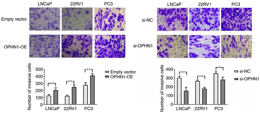
6                                 LIU et al: OPHN1 PROMOTES ANDROGEN RESISTANCE IN PROSTATE CANCER

Figure 3. Expression of OPHN1 promotes the invasion capacity of PCa. The expression of OPHN1 was enhanced by the transfection of recombinant lentiviral
vectors (OPHN1‑OE) or blocked by the transfection of siRNA (si‑OPHN1). Subsequently, the cells (LNCaP, 22RV1 and PC3) were loaded into the Transwell
chambers to evaluate the invasion ability of cells. The number of cells that passed through the filter in five random fields was counted as the invasion ability.
The data revealed that the overexpression of OPHN1 significantly promoted the invasion capacity of the LNCaP, 22RV1 and PC3 cells, with more cells passing
through the filters, whereas knockdown OPHN1 inhibited the invasion capacity of LNCaP, 22RV1 and PC3 cells by decreasing the cell numbers that passed
through the filters. Scale bar, 50 µm. The results are expressed as the mean ± SEM for three replicate determinations. *P
ONCOLOGY REPORTS 47: 3, 2022                                                                     7

Figure 4. Expression of OPHN1 prevents cell apoptosis in vitro. The expression of OPHN1 in PCa cells (LNCaP, 22RV1 and PC3) was overexpressed by the
transfection of recombinant lentiviral vectors (OPHN1‑OE) or blocked by the transfection of siRNA (si‑OPHN1). Then the cells were cultured in vitro under
various conditions for 72 h. (A) Different types of PCa cells (LNCaP and 22RV1) with OPHN1 overexpression or knockdown were treated with ADT for 72 h,
and the PC3 cells with the OPHN1 knockdown by siRNA continued to be cultured for 72 h. Following treatment, all the PCa cells were collected, stained
with Annexin V and PI, and analyzed by FACS. The number of early apoptotic (Annexin V‑positive) and late apoptotic (Annexin V‑ and PI‑positive) cells
indicates the total percentage of gated cells. Representative images and relative quantifications are shown. Compared with the cells without ADT, both the
LNCaP and 22RV1 cells under ADT conditions had a higher percentage of apoptosis. However, overexpression of OPHN1 alleviated the pro‑apoptotic effect
of ADT in both LNCaP and 22RV1 cells. Under ADT conditions, both LNCaP and 22RV1 cells with OPHN1 overexpression displayed a lower percentage of
apoptosis than the LNCaP and 22RV1 cells transfected with empty vectors. In addition, under ADT conditions, the knockdown of the expression of OPHN1
in LNCaP and 22RV1 cells increased the apoptotic rates compared with the cells that were transfected with negative control RNA. In addition, in PC3 cells,
the knockdown of the expression of OPHN1 promoted apoptosis and displayed a higher percentage of apoptosis than cells transfected with negative control
RNA. (B) The data on the activities of caspase‑3 and caspase‑8 were consistent with the apoptotic assay of flow cytometry. ADT promoted the activities of both
caspase‑3 and caspase‑8 in both LNCaP and 22RV1 cells compared with the cells without ADT, which could be alleviated by the overexpression of OPHN1.
Furthermore, under ADT conditions, blocking the expression of OPHN1 increased caspase‑3 and caspase‑8 activities in LNCaP and 22RV1 cells. Blocking the
expression of OPHN1 in PC3 cells promoted the activities of both caspase‑3 and caspase‑8. All the experiments were performed in triplicate, and the data are
presented as the mean ± SEM. *P
8                               LIU et al: OPHN1 PROMOTES ANDROGEN RESISTANCE IN PROSTATE CANCER

Figure 5. Effects of OPHN1 on tumor xenograft growth in mice. LNCaP (1x10⁶ cells) transfected with recombinant lentiviral vectors (OPHN1 overexpression,
OPHN1‑OE) were injected into mice subcutaneously (20 mice/group), and then the tumor growth was monitored, while the LNCaP cells transfected with
empty vectors were injected as controls. (A) The tumor volumes of both groups were monitored until they exceeded 500 mm 3, as revealed with growth curves
for xenografts in each group. The data revealed that the tumors of OPHN1‑OE LNCaP cells had a higher rate of tumor growth (F=14.81; P
ONCOLOGY REPORTS 47: 3, 2022                                                              9

Additionally, further research is required to comprehend the               4. Visakorpi T, Hyytinen E, Koivisto P, Tanner M, Keinänen R,
                                                                              Palmberg C, Palotie A, Tammela T, Isola J and Kallioniemi OP:
potential pathway regulated by OPHN1 in PCa.                                  In vivo amplification of the androgen receptor gene and progres‑
    In summary, in prostate cancer, ADT induced the ampli‑                    sion of human prostate cancer. Nat Genet 9: 401‑406, 1995.
fication of OPHN1, which contributed to the development of                 5. LaTulippe E, Satagopan J, Smith A, Scher H, Scardino P, Reuter V
                                                                              and Gerald WL: Comprehensive gene expression analysis of
CRPC. The overexpression of OPHN1 facilitated PCa survival                    prostate cancer reveals distinct transcriptional programs associ‑
under ADT by contributing to PCa viability, invasion, and                     ated with metastatic disease. Cancer Res 62: 4499‑4506, 2002.
progression. Therefore, targeting OPHN1 could be used to                   6. Jernberg E, Bergh A and Wikstrom P: Clinical relevance
                                                                              of androgen receptor alterations in prostate cancer. Endocr
reverse endocrine therapy resistance in CRPC.                                 Connect 6: R146‑R161, 2017.
                                                                           7. Merson S, Yang ZH, Brewer D, Olmos D, Eichholz A, McCarthy F,
Acknowledgements                                                              Fisher G, Kovacs G, Berney DM, Foster CS, et al: Focal ampli‑
                                                                              fication of the androgen receptor gene in hormone‑naive human
                                                                              prostate cancer. Br J Cancer 110: 1655‑1662, 2014.
Not applicable.                                                            8. Muller FL, Colla S, Aquilanti E, Manzo VE, Genovese G, Lee J,
                                                                              Eisenson D, Narurkar R, Deng P, Nezi L, et al: Passenger dele‑
                                                                              tions generate therapeutic vulnerabilities in cancer. Nature 488:
Funding                                                                       337‑342, 2012.
                                                                           9. Bergmann C, Zerres K, Senderek J, Rudnik‑Schoneborn S,
No funding was received.                                                      Eggermann T, Häusler M, Mull M and Ramaekers VT:
                                                                              Oligophrenin 1 (OPHN1) gene mutation causes syndromic
                                                                              X‑linked mental retardation with epilepsy, rostral ventricular
Availability of data and materials                                            enlargement and cerebellar hypoplasia. Brain 126: 1537‑1544,
                                                                              2003.
                                                                          10. Al‑Owain M, Kaya N, Al‑Zaidan H, Al‑Hashmi N, Al‑Bakheet A,
The datasets used and/or analyzed during the current study                    Al‑Muhaizea M, Chedrawi A, Basran RK and Milunsky A:
are available from the corresponding author on reasonable                     Novel intragenic deletion in OPHN1 in a family causing XLMR
request.                                                                      with cerebellar hypoplasia and distinctive facial appearance.
                                                                              Clin Genet 79: 363‑370, 2011.
                                                                          11. Busa T, Caietta E, Chabrol B, Girard N, Philip N and Missirian C:
Authors' contributions                                                        Large in‑frame intragenic deletion of OPHN1 in a male patient
                                                                              with a normal intelligence quotient score. Clin Dysmorphol 26:
                                                                              47‑49, 2017.
JL and YZ designed and performed experiments, analyzed the                12. Goto K, Oue N, Hayashi T, Shinmei S, Sakamoto N, Sentani K,
data and wrote the manuscript. SL and FS analyzed the data                    Teishima J, Matsubara A and Yasui W: Oligophrenin‑1 is
and performed the experiments. GW and DW performed the                        associated with cell adhesion and migration in prostate cancer.
                                                                              Pathobiology 81: 190‑198, 2014.
experiments. TY and SG contributed to study design. JL and                13. Du M, Tian Y, Tan W, Wang L, Wang L, Kilari D, Huang CC,
YZ confirm the authenticity of all the raw data. All authors                  Wang L and Kohli M: Plasma cell‑free DNA‑based predictors
read and approved the final manuscript.                                       of response to abiraterone acetate/prednisone and prognostic
                                                                              factors in metastatic castration‑resistant prostate cancer. Prostate
                                                                              Cancer Prostatic Dis 23: 705‑713, 2020.
Ethics approval and consent to participate                                14. Cerami E, Gao J, Dogrusoz U, Gross BE, Sumer SO, Aksoy BA,
                                                                              Jacobsen A, Byrne CJ, Heuer ML, Larsson E, et al: The cBio
                                                                              cancer genomics portal: An open platform for exploring multi‑
All animal care procedures and experiments were conducted                     dimensional cancer genomics data. Cancer Discov 2: 401‑404,
in accordance with the Animal Research: Reporting of In Vivo                  2012.
Experiments (ARRIVE) guidelines and approved by Ethics                    15. Gao J, Aksoy BA, Dogrusoz U, Dresdner G, Gross B, Sumer SO,
                                                                              Sun Y, Jacobsen A, Sinha R, Larsson E, et al: Integrative analysis
Committee of Animal Experiments of the Hebei General                          of complex cancer genomics and clinical profiles using the cBio‑
Hospital (Shijiazhuang, China).                                               Portal. Sci Signal 6: pl1, 2013.
                                                                          16. Grasso CS, Wu YM, Robinson DR, Cao X, Dhanasekaran SM,
                                                                              Khan AP, Quist MJ, Jing X, Lonigro RJ, Brenner JC, et al:
Patient consent for publication                                               The mutational landscape of lethal castration‑resistant prostate
                                                                              cancer. Nature 487: 239‑243, 2012.
Not applicable.                                                           17. Abida W, Cyrta J, Heller G, Prandi D, Armenia J, Coleman I,
                                                                              Cieslik M, Benelli M, Robinson D, Van Allen EM, et al: Genomic
                                                                              correlates of clinical outcome in advanced prostate cancer. Proc
Competing interests                                                           Natl Acad Sci USA 116: 11428‑11436, 2019.
                                                                          18. Stopsack KH, Nandakumar S, Wibmer AG, Haywood S,
                                                                              Weg ES, Barnett ES, Kim CJ, Carbone EA, Vasselman SE,
The authors declare that they have no competing interests.                    Nguyen B, et al: Oncogenic genomic alterations, clinical pheno‑
                                                                              types, and outcomes in metastatic castration‑sensitive prostate
References                                                                    cancer. Clin Cancer Res 26: 3230‑3238, 2020.
                                                                          19. Hodgson MC, Astapova I, Hollenberg AN and Balk SP: Activity
                                                                              of androgen receptor antagonist bicalutamide in prostate cancer
 1. Global Burden of Disease Cancer Collaboration, Fitzmaurice C,             cells is independent of NCoR and SMRT corepressors. Cancer
    Allen C, Barber RM, Barregard L, Bhutta ZA, Brenner H,                    Res 67: 8388‑8395, 2007.
    Dicker DJ, Chimed‑Orchir O, Dandona R, et al: Global, regional,       20. Hara T, Miyazaki J, Araki H, Yamaoka M, Kanzaki N, Kusaka M
    and national cancer incidence, mortality, years of life lost, years       and Miyamoto M: Novel mutations of androgen receptor: A
    lived with disability, and disability‑adjusted life‑years for 32          possible mechanism of bicalutamide withdrawal syndrome.
    cancer groups, 1990 to 2015: A systematic analysis for the global         Cancer Res 63: 149‑153, 2003.
    burden of disease study. JAMA Oncol 3: 524‑548, 2017.                 21. Livak KJ and Schmittgen TD: Analysis of relative gene expres‑
 2. Roehl KA, Han M, Ramos CG, Antenor JA and Catalona WJ:                    sion data using real‑time quantitative PCR and the 2(‑Delta Delta
    Cancer progression and survival rates following anatomical                C(T)) method. Methods 25: 402‑408, 2001.
    radical retropubic prostatectomy in 3,478 consecutive patients:       22. Kilkenny C, Browne W, Cuthill IC, Emerson M and Altman DG;
    Long‑term results. J Urol 172: 910‑914, 2004.                             NC3Rs Reporting Guidelines Working Group: Animal research:
 3. Chang SS: Treatment options for hormone‑refractory prostate               Reporting in vivo experiments: The ARRIVE guidelines. Br J
    cancer. Rev Urol 9 (Suppl 2): S13‑S18, 2007.                              Pharmacol 160: 1577‑1579, 2010.
10 LIU et al: OPHN1 PROMOTES ANDROGEN RESISTANCE IN PROSTATE CANCER
23. Kawata H, Ishikura N, Watanabe M, Nishimoto A, Tsunenari T 39. Buttigliero C, Tucci M, Bertaglia V, Vignani F, Bironzo P,
and Aoki Y: Prolonged treatment with bicalutamide induces Di Maio M and Scagliotti GV: Understanding and overcoming the
androgen receptor overexpression and androgen hypersensitivity. mechanisms of primary and acquired resistance to abiraterone
Prostate 70: 745‑754, 2010. and enzalutamide in castration resistant prostate cancer. Cancer
24. Albertson DG: Gene amplification in cancer. Trends Genet 22: Treat Rev 41: 884‑892, 2015.
447‑455, 2006. 40. Watson PA, Chen YF, Balbas MD, Wongvipat J, Socci ND,
25. Monni O, Barlund M, Mousses S, Kononen J, Sauter G, Viale A, Kim K and Sawyers CL: Constitutively active androgen
Heiskanen M, Paavola P, Avela K, Chen Y, Bittner ML and receptor splice variants expressed in castration‑resistant prostate
Kallioniemi A: Comprehensive copy number and gene expres‑ cancer require full‑length androgen receptor. Proc Natl Acad Sci
sion profiling of the 17q23 amplicon in human breast cancer. USA 107: 16759‑16765, 2010.
Proc Natl Acad Sci USA 98: 5711‑5716, 2001. 41. Edwards J, Krishna NS, Grigor KM and Bartlett JM: Androgen
26. Grasso C, Butler T, Rhodes K, Quist M, Neff TL, Moore S, receptor gene amplification and protein expression in hormone
Tomlins SA, Reinig E, Beadling C, Andersen M and Corless CL: refractory prostate cancer. Br J Cancer 89: 552‑556, 2003.
Assessing copy number alterations in targeted, amplicon‑based 42. Zhang X, Hong SZ, Lin EJ, Wang DY, Li ZJ and Chen LI:
next‑generation sequencing data. J Mol Diagn 17: 53‑63, 2015. Amplification and protein expression of androgen receptor
27. Boeva V, Popova T, Lienard M, Toffoli S, Kamal M, gene in prostate cancer cells: Fluorescence in situ hybridization
Le Tourneau C, Gentien D, Servant N, Gestraud P, Rio analysis. Oncol Lett 9: 2617‑2622, 2015.
Frio T, et al: Multi‑factor data normalization enables the detec‑ 43. Du M, Huang CC, Tan W, Kohli M and Wang L: Multiplex digital
tion of copy number aberrations in amplicon sequencing data. PCR to detect amplifications of specific androgen receptor loci
Bioinformatics 30: 3443‑3450, 2014. in cell‑free DNA for prognosis of metastatic castration‑resistant
28. Press MF, Ellis CE, Gagnon RC, Grob TJ, Buyse M, Villalobos I, prostate cancer. Cancers (Basel) 12: 2139, 2020.
Liang Z, Wu S, Bang YJ, Qin SK, et al: HER2 Status in advanced 44. Dicken BJ, Graham K, Hamilton SM, Andrews S, Lai R,
or metastatic gastric, esophageal, or gastroesophageal adenocar‑ Listgarten J, Jhangri GS, Saunders LD, Damaraju S and Cass C:
cinoma for entry to the TRIO‑013/LOGiC trial of lapatinib. Mol Lymphovascular invasion is associated with poor survival in
Cancer Ther 16: 228‑238, 2017. gastric cancer: An application of gene‑expression and tissue
29. Bivin WW, Yergiyev O, Bunker ML, Silverman JF and array techniques. Ann Surg 243: 64‑73, 2006.
Krishnamurti U: GRB7 expression and correlation with HER2 45. Chen X, Cheng H, Pan T, Liu Y, Su Y, Ren C, Huang D, Zha X
amplification in invasive breast carcinoma. Appl Immunohistochem and Liang C: mTOR regulate EMT through RhoA and Rac1
Mol Morphol 25: 553‑558, 2017. pathway in prostate cancer. Mol Carcinog 54: 1086‑1095, 2015.
30. Doi A, Ishikawa K, Shibata N, Ito E, Fujimoto J, Yamamoto M, 46. Chen W, Delongchamps NB, Mao K, Beuvon F, Peyromaure M,
Shiga H, Mochizuki H, Kawamura Y, Goshima N, et al: Liu Z and Dinh‑Xuan AT: High RhoA expression at the tumor
Enhanced expression of retinoic acid receptor alpha (RARA) front in clinically localized prostate cancer and association with
induces epithelial‑to‑mesenchymal transition and disruption of poor tumor differentiation. Oncol Lett 11: 1375‑1381, 2016.
mammary acinar structures. Mol Oncol 9: 355‑364, 2015. 47. Liu J, Wang L, Zhang Y, Li S, Sun F, Wang G, Yang T, Wei D,
31. Katoh M and Katoh M: MGC9753 gene, located within Guo L and Xiao H: Induction of entosis in prostate cancer cells
PPP1R1B‑STARD3‑ERBB2‑GRB7 amplicon on human chro‑ by nintedanib and its therapeutic implications. Oncol Lett 17:
mosome 17q12, encodes the seven‑transmembrane receptor with 3151‑3162, 2019.
extracellular six‑cystein domain. Int J Oncol 22: 1369‑1374, 48. Ahmat Amin MKB, Shimizu A, Zankov DP, Sato A, Kurita S,
2003. Ito M, Maeda T, Yoshida T, Sakaue T, Higashiyama S, et al:
32. Glynn RW, Miller N and Kerin MJ: 17q12‑21‑the pursuit of Epithelial membrane protein 1 promotes tumor metastasis by
targeted therapy in breast cancer. Cancer Treat Rev 36: 224‑229, enhancing cell migration via copine‑III and Rac1. Oncogene 37:
2010. 5416‑5434, 2018.
33. Matsui A, Ihara T, Suda H, Mikami H and Semba K: Gene 49. Zins K, Lucas T, Reichl P, Abraham D and Aharinejad S:
amplification: Mechanisms and involvement in cancer. Biomol A Rac1/Cdc42 GTPase‑specific small molecule inhibitor
Concepts 4: 567‑582, 2013. suppresses growth of primary human prostate cancer xenografts
34. Waltering KK, Urbanucci A and Visakorpi T: Androgen receptor and prolongs survival in mice. PLoS One 8: e74924, 2013.
(AR) aberrations in castration‑resistant prostate cancer. Mol Cell 50. Kobayashi T, Inoue T, Shimizu Y, Terada N, Maeno A, Kajita Y,
Endocrinol 360: 38‑43, 2012. Yamasaki T, Kamba T, Toda Y, Mikami Y, et al: Activation
35. Karantanos T, Corn PG and Thompson TC: Prostate cancer of Rac1 is closely related to androgen‑independent cell prolif‑
progression after androgen deprivation therapy: Mechanisms eration of prostate cancer cells both in vitro and in vivo. Mol
of castrate resistance and novel therapeutic approaches. Endocrinol 24: 722‑734, 2010.
Oncogene 32: 5501‑5511, 2013. 51. Lyons LS, Rao S, Balkan W, Faysal J, Maiorino CA and
36. Feng Q and He B: Androgen receptor signaling in the develop‑ Burnstein KL: Ligand‑independent activation of androgen
ment of castration‑resistant prostate cancer. Front Oncol 9: 858, receptors by Rho GTPase signaling in prostate cancer. Mol
2019. Endocrinol 22: 597‑608, 2008.
37. Chandrasekar T, Yang JC, Gao AC and Evans CP: Mechanisms 52. Chen X, Yin L, Qiao G, Li Y, Li B, Bai Y and Feng F: Inhibition
of resistance in castration‑resistant prostate cancer (CRPC). of Rac1 reverses enzalutamide resistance in castration‑resistant
Transl Androl Urol 4: 365‑380, 2015. prostate cancer. Oncol Lett 20: 2997‑3005, 2020.
38. Zhao XY, Malloy PJ, Krishnan AV, Swami S, Navone NM,
Peehl DM and Feldman D: Glucocorticoids can promote
androgen‑independent growth of prostate cancer cells through a This work is licensed under a Creative Commons
mutated androgen receptor. Nat Med 6: 703‑706, 2000. Attribution-NonCommercial-NoDerivatives 4.0
International (CC BY-NC-ND 4.0) License.You can also read