ANTIMICROBIAL PEPTIDES: A NEW HOPE IN BIOMEDICAL AND PHARMACEUTICAL FIELDS - MPG.PURE
←
→
Page content transcription
If your browser does not render page correctly, please read the page content below
REVIEW
                                                                                                                                                        published: 14 June 2021
                                                                                                                                                doi: 10.3389/fcimb.2021.668632

                                             Antimicrobial Peptides:
                                             A New Hope in Biomedical
                                             and Pharmaceutical Fields
                                             Antonio Moretta 1†, Carmen Scieuzo 1,2†, Anna Maria Petrone 1†, Rosanna Salvia 1,2†,
                                             Michele Dario Manniello 1, Antonio Franco 1,2, Donatella Lucchetti 3, Antonio Vassallo 1,
                                             Heiko Vogel 4, Alessandro Sgambato 3,5* and Patrizia Falabella 1,2*
                                             1 Department of Sciences, University of Basilicata, Potenza, Italy, 2 Spinoff XFlies s.r.l, University of Basilicata, Potenza, Italy,
                                             3 Department of Translational Medicine and Surgery, Università Cattolica del Sacro Cuore, Rome, Italy, 4 Department of
                            Edited by:       Entomology, Max Planck Institute for Chemical Ecology, Jena, Germany, 5 Centro di Riferimento Oncologico della Basilicata
                     Francesc Rabanal,       (IRCCS-CROB), Rionero in Vulture, Italy
         University of Barcelona, Spain

                        Reviewed by:
                 Surajit Bhattacharjya,      Antibiotics are essential drugs used to treat pathogenic bacteria, but their prolonged use
    Nanyang Technological University,        contributes to the development and spread of drug-resistant microorganisms. Antibiotic
                             Singapore
                         Clara Balleste,
                                             resistance is a serious challenge and has led to the need for new alternative molecules less
     Instituto Salud Global Barcelona        prone to bacterial resistance. Antimicrobial peptides (AMPs) have aroused great interest
                     (ISGlobal), Spain       as potential next-generation antibiotics, since they are bioactive small proteins, naturally
                    *Correspondence:         produced by all living organisms, and representing the first line of defense against fungi,
                        Patrizia Falabella
             patrizia.falabella@unibas.it    viruses and bacteria. AMPs are commonly classified according to their sources, which are
                Alessandro Sgambato          represented by microorganisms, plants and animals, as well as to their secondary
         alessandro.sgambato@crob.it
                                             structure, their biosynthesis and their mechanism of action. They find application in
     †
      These authors have contributed
       equally to this work and share
                                             different fields such as agriculture, food industry and medicine, on which we focused our
                       first authorship       attention in this review. Particularly, we examined AMP potential applicability in wound
                                             healing, skin infections and metabolic syndrome, considering their ability to act as
                    Specialty section:
                                             potential Angiotensin-Converting Enzyme I and pancreatic lipase inhibitory peptides as
          This article was submitted to
                   Clinical Microbiology,    well as antioxidant peptides. Moreover, we argued about the pharmacokinetic and
                a section of the journal     pharmacodynamic approaches to develop new antibiotics, the drug development
               Frontiers in Cellular and
                 Infection Microbiology
                                             strategies and the formulation approaches which need to be taken into account in
         Received: 16 February 2021
                                             developing clinically suitable AMP applications.
            Accepted: 10 May 2021
                                             Keywords: drug-resistant microorganisms, antimicrobial peptides, biomedical and pharmacological applications,
           Published: 14 June 2021
                                             pharmacokinetics and pharmacodynamics, drug delivery
                                 Citation:
   Moretta A, Scieuzo C, Petrone AM,
                                             Abbreviations: ACE, Angiotensin-Converting Enzyme I; AMP, Antimicrobial Peptide; APD, Antimicrobial Peptide Database;
    Salvia R, Manniello MD, Franco A,        API, Active pharmaceutical ingredient; DDS, Drug Delivery System; Di-Phe, di-phenylalanine; EGFR, Epidermal Growth
     Lucchetti D, Vassallo A, Vogel H,       Factor Receptor; EPL, ϵ-poly-L-lysine; GMO, Glyceryl Monooleate; HBBD, HG-Based Burn Dressings; hBD, Human b
  Sgambato A and Falabella P (2021)          defensin; HG, Hydrogel; IPTG, Isopropyl b- D-1-Thiogalactopyranoside; LPS, Lipopolysaccharides; MRSA, Methicillin-
Antimicrobial Peptides: A New Hope in        resistant Staphylococcus aureus; PD, Pharmacodynamics; PK, Pharmacokinetics; SARS-CoV-2, Severe Acute Respiratory
Biomedical and Pharmaceutical Fields.        Syndrome Coronavirus 2; SCID, Severe Combined Immunodeficiency; TLR, Toll-like receptor; WHO, World Health
Front. Cell. Infect. Microbiol. 11:668632.   Organization; BPS, block polymeric structure; CB, cubosome; CMC, Critical Micelle Concentration; PA, peptide
       doi: 10.3389/fcimb.2021.668632        amphiphiles; SPPS, Solid Phase Peptide Synthesis.

Frontiers in Cellular and Infection Microbiology | www.frontiersin.org                  1                                              June 2021 | Volume 11 | Article 668632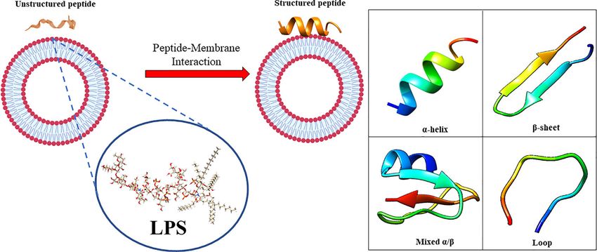
Moretta et al.                                                                                       AMPs: Biomedical and Pharmacological Applications

INTRODUCTION                                                                 AMP Properties and Biosynthesis
                                                                             Natural AMPs are evolutionary conserved gene-encoded
A wide variety of antimicrobial agents are available today and               molecules with structural and functional diversity, which is
they are broadly applied to treat different types of human                   responsible for their wide range of activities against different
infections. Specifically, antibiotics are powerful drugs used for             pathogens in various organisms (Zhang and Gallo, 2016).
treatments of pathogenic bacteria (Lei et al., 2019). However,               However, although displaying considerable diversity in their
their indiscriminate and prolonged use, especially in developing             physio-chemical and structural properties, origins and
countries, in both human and veterinary medicine, as well as in              mechanisms of action, AMPs share some common features
agriculture have contributed to the development and spread of                (Moravej et al., 2018). Indeed, they are mostly short molecules
drug-resistant microorganisms (Huan et al., 2020). As the World              (
Moretta et al.                                                                                                   AMPs: Biomedical and Pharmacological Applications

passage into the cytosol. Indeed, this interaction could change                      structure and do not undergo radical conformational changes as
AMP tertiary structure, and AMP molecules could assume                               helical peptides upon membrane interaction (Mahlapuu et al.,
different conformations, such as monomeric helical or helix-                         2016). It is not easy to clarify the structural conformations of b-
loop-helix structures (Figure 1) (Bhunia et al., 2011).                              sheet AMPs in membranes, because of the potential micelle
    For example, the contact with LPS induces oligomerization of                     aggregations; indeed, a recent report on thanatin peptide,
specific AMPs, such as temporines, through the interaction among                      isolated from insect Podisus maculiventris, showed dimerization
hydrophobic N and C terminal residues, preventing the correct                        of b-sheet structures (Sinha et al., 2017). These dimeric structures
movement throughout the membrane and the correct antimicrobial                       could facilitate the bond with LPS molecules, also at the distal ends,
action (Bhunia et al., 2011). A particular amino acids composition                   fostering bacterial cell associations and agglutination (Sinha et al.,
could prevent this oligomerization, enhancing temporin activity.                     2017). Defensins, a large group of AMPs, which are produced in
This is the case of temporin-1Tl, which is rich in aromatic residues                 macrophages, neutrophils and epithelial cells belong to this class
with two positively charged amino acids (Bhunia et al., 2011). The                   (Mahlapuu et al., 2016). It was observed that the right combination
synergy of temporin-1Tl with other temporins (Temporin A and                         of hydrophobicity, charge density and peptide length influence the
Temporin B), prevent their oligomerization and facilitate the                        antimicrobial activity of AMPs. Changing the amino acids position
correct crossing of the bacterial membrane (Bhunia et al., 2011).                    in the peptide chain or increasing the number of positively charged
Exceptions are related to some AMPs with particular structural                       residues affect the secondary structure of AMPs, and consequently
characteristics, including the peptide MSI-594 (an analogue of                       their biological activity against pathogens (Wu Q. et al., 2018).
magainin), that is unstructured in free solution, but have a folded                  Besides the principle that the amino acid sequence determines the
helical hairpin structure when interact with LPS (Bhattacharjya,                     function of a peptide, it was found that the amino acid composition
2016). The interactions between two helical segments, facilitated by                 (in terms of abundance of residues with specific phyco-chemical
the fifth phenylalanine residue, allows the acquisition of the hairpin                properties) also affects AMP activity as clearly documented for a
structure, implicating its very high activity against bacteria, fungi,               novel class of cationic AMPs known as “cationic intrinsically
and viruses (Domadia et al., 2010; Bhattacharjya, 2016). Another                     disordered antimicrobial peptides’’ or “CIDAMPs” since they are
example of change in conformation after the interaction with LPS, is                 characterized by an intrinsically disordered structure. CIDAMPs
the b-hairpin structures of Tachyplesin I, that becomes more                         have been detected in human skin and other barrier organs (Gerstel
ordered and compact when interacting with LPS (Saravanan et                          et al., 2018; Latendorf et al., 2019) and, carrying a positive net
al., 2012; Kushibiki et al., 2014). Another interesting example is                   charge, have a low percentage of order-promoting amino acids
linked to the human LL-37 AMP, one of the best studied peptides of                   (mostly hydrophobic residues commonly located within the
this group, present in neutrophils and epithelial cells (Mahlapuu et                 hydrophobic core of foldable proteins) and a high percentage of
al., 2016). It has been demonstrated that aromatic-aromatic                          disorder-promoting amino acids (mostly charged and polar
interactions stabilize protein structure in correlation with lipids                  residues, typically found at the surface of foldable proteins). They
(Li et al., 2006) and that LL-37 could undergo a re-orientation                      show microbicidal activity against several microbes, including
depending on the concentration, suggesting also in this case an                      Candida albicans, Staphylococcus aureus and Pseudomonas
oligomerization process (Ding et al., 2013). On the contrary, b-sheet                aeruginosa (Gerstel et al., 2018). The protein hornerin, expressed
peptides are more ordered in aqueous solution because of their rigid                 in the cornified epithelium, seems to be the main source of

                       A                                                                        B

 FIGURE 1 | (A) in aqueous solution, the AMPs are unstructured while after the interaction with biological membrane, particularly with the LPS component, they
 assume the right conformation, which can be (B) a-helical, b-sheet, mixed a-helical/b-sheet, and loop. Figure created with Biorender.com and UCSF CHIMERA
 software (Pettersen et al., 2004).

Frontiers in Cellular and Infection Microbiology | www.frontiersin.org           3                                          June 2021 | Volume 11 | Article 668632
Moretta et al. AMPs: Biomedical and Pharmacological Applications
CIDAMPs, which act as disinfectants, helping to keep the surface of Insights Into the Mechanisms of Action
healthy skin free of infections (Gerstel et al., 2018). of AMPs
AMP biosynthesis can occur in three different ways: classical The prerequisite to develop efficient AMPs as novel candidate
ribosomal synthesis, non-ribosomal synthesis and proteolytic drugs is the understanding of their mode of action. AMPs exert
digestion of proteins (Buda De Cesare et al., 2020). their activity by interaction with microbial cell membranes and
Ribosomally synthesized AMPs, such as histatins and human this interaction is strongly affected by the lipid composition of
b-defensins, are produced by ribosomal translation of specific biological membranes (Wu Q. et al., 2018). Since microbial
mRNAs into the biologically active amino acid sequences in membranes are the primary targets of AMPs, it is difficult for
vertebrates, insects, plants, and bacteria. Non-ribosomally bacteria to develop resistance to AMPs as easily as to
synthesized peptides are produced by large enzymes referred to conventional antibiotics (Boparai and Sharma, 2020).
as non-ribosomal peptide synthases, which incorporate non- Membrane interactions are mediated by electrostatic forces
proteinogenic amino acids into the sequence, and are found in between positively charged AMPs and negatively charged
filamentous fungi and bacteria (Actinomycetes and Bacilli). microbial surfaces. The teichoic acids in the cell wall of Gram-
Finally, some AMPs, called cryptic peptides, are generated by positive bacteria and the LPS in the outer membrane of Gram-
proteolytic cleavage of bigger proteins with other functions. For negative bacteria supply electronegative charge to the microbial
example, the histone H2A of the Asian toad (Duttaphrynus surfaces, strengthening the interaction with AMPs (Boparai and
melanostictus) is processed by the enzymatic activity of pepsin Sharma, 2020). On the contrary, the outer layer of eukaryotic
C producing buforin I, which in turn is processed by an membranes is composed by zwitterionic phosphatidylcholine
endopeptidase to generate buforin II (Buda De Cesare et al., and sphingomyelin, which do not favor AMP interaction
2020). Interestingly, many AMPs are produced as inactive because of their neutral charge at physiological pH. Based on
precursors and are active after proteolytic cleavage. Therefore, their mode of action, AMPs are divided into “membrane acting
their activity is not only dependent on their own expression but peptides”, which destabilize bacterial membranes causing their
also on the presence of appropriate proteases (Mahlapuu et al., disruption, and “non-membrane acting peptides”, which are able
2016). The expression of AMPs can be constitutive or inducible to translocate across the membranes without damaging them but
by specific external factors (Mahlapuu et al., 2016; Lei et al., destabilizing normal cell functions (Boparai and Sharma, 2020)
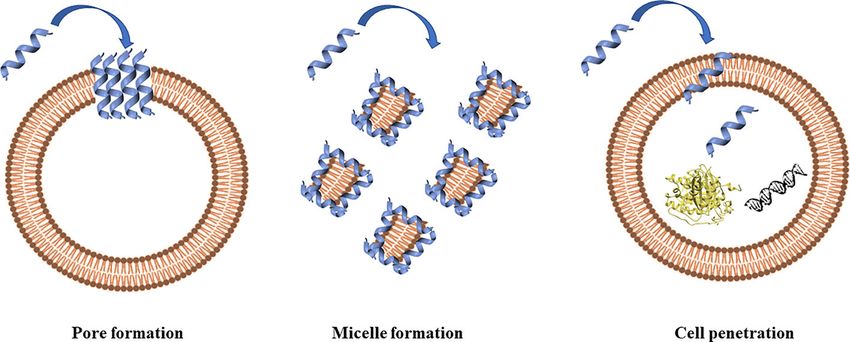
2019). Some AMPs are expressed during the whole cellular (Figure 2).
lifetime but are stored at high concentration as precursors in Three models have been proposed to explain the
granules and are released upon infection in the site of infection or permeabilization of bacterial membranes by AMPs: barrel-
inflammation (Mahlapuu et al., 2016). P9A and P9B are stave model, toroidal-pore model and carpet model (Raheem
examples of inducible peptides, whose expression can be and Straus, 2019). Thanks to their positive net charge, AMPs are
induced in silkmoth (Bombyx mori) hemolymph by able to interact with components of bacterial membranes,
vaccination with Enterobacter cloacae, as demonstrated by resulting in the disruption of the lipidic bilayer with cell death.
Hultmark and colleagues (Hultmark et al., 1980). In addition, AMP insertion can be perpendicular, as in the barrel-stave
Bals et al. (1999) reported that defensin production from model, or perpendicular with the interaction with the head
epithelial cells of multiple mouse organs increases upon groups of lipids that provokes a deflection in the membrane
infection with P. aeruginosa PAO1. (toroidal model) (Brogden, 2005). AMPs can also dispose
A B C
FIGURE 2 | Antimicrobial peptides can act through a membranolytic and non-membranolytic mechanism. In the membranolytic mechanism AMPs can lead to
(A) pore formation on the cell membrane or (B) micelle formation on the cell membrane. In the non-membranolytic mechanism, (C) AMPs can penetrate cell
membranes and interact with intracellular targets, such as DNA and proteins. Figure created with Biorender.com and UCSF CHIMERA software (Pettersen et al., 2004).
Frontiers in Cellular and Infection Microbiology | www.frontiersin.org 4 June 2021 | Volume 11 | Article 668632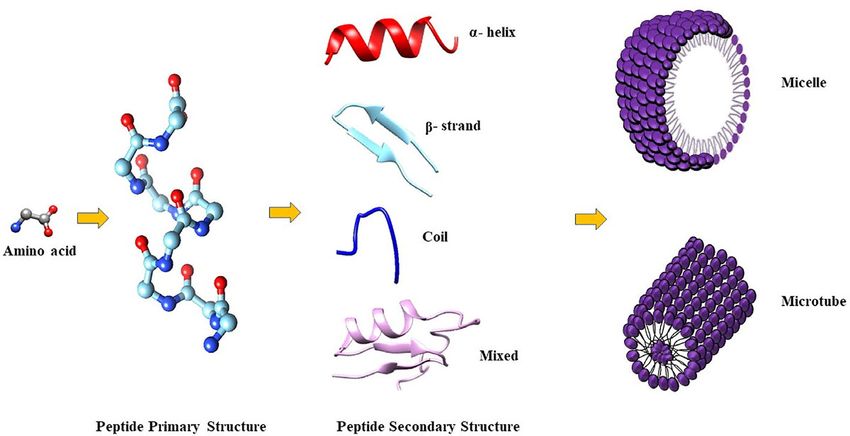
Moretta et al.                                                                                        AMPs: Biomedical and Pharmacological Applications

parallel to the membrane, covering it completely, and forming, at             essential components of the defense systems of all life forms, from
the same time, micelles with the starting broken membranes                    bacteria to plants and invertebrate and vertebrate species,
(carpet model), as proposed by Gazit and colleagues in 1996                   including mammals (Jenssen et al., 2006; Borah et al., 2020).
(Gazit et al., 1996). Moreover, defensins interact with LPS in                   They are naturally produced in the body of both lower and
Gram-negative bacteria and peptidoglycan in Gram-positive                     higher organisms and their production is cell specific and may be
bacteria (Pachó n-Ibá ñez et al., 2017). Defensins have LPS-                constitutive or inducible in response to pathogenic challenges
neutralizing activity in different bacteria (Lee et al., 2010)                (Borah et al., 2020). In multicellular organisms, AMPs are mostly
despite the chemical structure of LPS varies among them. LPS                  localized to specific sites that are normally more exposed to
can self-aggregate forming oligomers above a Critical Micelle                 microbes, such as the skin and mucosa epithelia (Jenssen et al.,
Concentration (CMC) because of its amphiphilic nature, a                      2006). The primary role of these defense peptides is the killing of
concentration of LPS, or any surfactant, above which it                       invading pathogens; however, in higher organisms they act also
aggregates in micelles. It has been demonstrated that the                     as modulators of the innate immune response (Jenssen et al.,
association of defensin analogues and other peptides, such as                 2006). AMPs are commonly classified according to their sources,
gramicidin A, melittin, LL-37 and polymyxin B, with LPS leads                 which are represented by microorganisms, plants, and animals.
to the disintegration of LPS aggregates. Moreover, it was                        Below, we give an overview of various naturally occurring
observed that defensins amino acids (such as Arg, Trp, and                    AMPs and the potential clinical application of some of them.
Tyr) are involved in the stabilization of the peptide-pathogen
surface complexes (Zhang et al., 2016).                                       Microorganisms as Source of AMPs
    The interaction with LPS has been demonstrated to be                      Bacteria and fungi are reservoirs of AMPs (Huan et al., 2020).
essential for AMPs like gramicidin S and polymyxin B to exert                 Among the numerous AMPs, the first isolated and characterized
their mechanism of action for bacterial killing (Zhang et al.,                were those produced by bacteria (Jenssen et al., 2006). AMPs
2000). Bhunia and colleagues studied the structure of MSI-594                 from bacteria are not produced for the purpose to protect against
peptide in LPS micelles. They observed that the peptide is                    infections, but rather as a competition strategy (Jenssen et al.,
unstructured in solution, while it adopts a helix-loop-helix                  2006). With their activity they kill other microbes competing for
structure in complex with LPS, suggesting how AMPs could                      nutrients in the same niches, ensuring the survival of individual
overcome the LPS barrier (Bhunia et al., 2009). A mutant form of              bacterial cells (Jenssen et al., 2006). Bacterial AMPs, also called
MSI-594 peptide, substituting Phe5 with Ala amino acid,                       bacteriocins, are represented by a heterogeneous family of small
displays a limited permeabilization through the LPS layer                     ribosomally synthesized molecules with strong antimicrobial
suggesting that peptide conformation is essential to disrupt                  activity at specific concentrations (Soltani et al., 2021). These
LPS (Domadia et al., 2010).                                                   molecules, produced by Gram-positive and Gram-negative
    Other examples of AMPs acting by perturbation of microbial                bacteria, are effective against many pathogenic bacteria and are
membrane structure are the fungal peptide alamethicin, the                    extraordinarily active compared to their eukaryotic counterparts
amphibian AMP aurein 1.2, and several defensins (Machado                      (Jenssen et al., 2006; Soltani et al., 2021). For example, AMPs
and Ottolini, 2015; Shahmiri et al., 2017; Su et al., 2018) AMPs              isolated from Pseudomonas spp display activity against several
acting through a non-membranolytic mechanism, thus displaying                 bacterial species, such as S. aureus, E. coli, Salmonella, Shigella,
intracellular activities (such as inhibition of nucleic acids, proteins       showing both general antibacterial and specific antibiofilm
or cell wall synthesis), include buforin II and indolicidin that bind         activity (Fontoura et al., 2008; Mohammadi-Barzelighi et al.,
to DNA (Scocchi et al., 2016), teixobactin that binds to                      2019). Mersacidin, isolated by Bacillus spp, shows in vivo
peptidoglycan precursor lipid II (Chiorean et al., 2020), Bac5                bactericidal activity against Methicillin-resistant S. aureus
that interacts with ribosomes (Mardirossian et al., 2018) and                 (MRSA) equivalent to that of vancomycin (Jenssen et al., 2006).
Temporin-L, which binds FtsZ protein inhibiting Escherichia coli                  AMPs are also produced by human microbiota. Host-
cell division (Di Somma et al., 2020). A recent study performed by            microbiota crosstalk is based on AMPs secretion by phagocytic
Moura et al. demonstrated that the AMP thanatin interacts with                and epithelial cells and microbiota of the human gut, skin, and
LptC-LptA proteins, which belong to the Lpt complex, involved                 oral cavity; these peptides contribute to microbial and ecological
in the LPS transport, exploiting an inhibitory activity (Moura et             balance (Magana et al., 2020). An example of these human
al., 2020). Thanatin interaction with Lpt complex prevents LPS                microbiota AMPs is the thiopeptide lactocillin produced by the
translocation to the outer membrane, modifying its stability and              vaginal commensal Lactobacillus gasseri and acting against
permeability and favoring the cell agglutination process (Dash                Gram-positive bacteria, including S. aureus and Gardnerella
and Bhattacharjya, 2021).                                                     vaginalis (He et al., 2020).
                                                                                  Several filamentous fungi produce AMPs which are similar to
Sources of AMPs and Their Potential                                           plant and animal defensins. Examples of cysteine-rich defensin-
Applications in Clinical Practice                                             like AMPs in ascomycetes are AFP from Aspergillus giganteus,
The survival of organisms in an environment where pathogens are               PAF from Penicillium chrysogenum, ANAFP from Aspergillus
widely distributed, solely depends on their defense mechanisms.               niger, AcAFP and AcAMP from Aspergillus clavatus
The inborn immunity of organisms involves endogenic peptides                  (Montesinos, 2007; Hegedüs and Marx, 2013). All these fungal
which supply a quick and viable method for safeguard against                  peptides have antifungal activity against filamentous
microbial attacks (Borah et al., 2020) AMPs are universal and                 ascomycetes, including animal and plant opportunistic and

Frontiers in Cellular and Infection Microbiology | www.frontiersin.org    5                                     June 2021 | Volume 11 | Article 668632
Moretta et al. AMPs: Biomedical and Pharmacological Applications
pathogens, such as Aspergillus fumigatus, Fusarium sp., and Plants as Source of AMPs
Botrytis sp. (Hegedüs and Marx, 2013). Bioactive peptides are essential components of plants defense
On the basis of their antimicrobial properties and their safety mechanisms, with extraordinary physiological importance,
and tolerability, some of these natural AMPs have potential providing fast protection against bacterial and fungal infections
therapeutic applications. The bacteriocin nisin, produced by (Jenssen et al., 2006; Ló pez-Meza et al., 2011; Salas et al., 2015).
Lactococcus lactis, has been extensively studied being used as Plant AMPs not only display microbicide activities but are also
food preservative (Soltani et al., 2021). Nisin is the only involved in cellular signaling (Salas et al., 2015). Several active
bacteriocin legally approved as biopreservative and is used in peptides have been extracted and isolated from roots, flowers,
the dairy industry to control contamination from Listeria strains seeds, stems and leaves and are classified based on their amino
(Soltani et al., 2021). Because of its broad-spectrum activity acids sequence, position and number of cysteine residues
against both Gram-positive and Gram-negative pathogens, involved in the disulfide bridge formation (Ló pez-Meza et al.,
nisin is approved for clinical use as an alternative to antibiotics 2011). Ten families of plant AMPs have been described (Ló pez-
(Dijksteel et al., 2021). Several studies have reported the Meza et al., 2011) and the best-studied groups are defensins,
suitability of nisin in the treatment of several infection thionins and snakins (Jenssen et al., 2006; Ló pez-Meza et al.,
diseases, such as mastitis (Cao et al., 2007; Ferná ndez et al., 2011; Huan et al., 2020). The first plant-derived AMP is
2008), oral (Shin et al., 2015; Mitra et al., 2019), respiratory (De purothionin, which displays activity against Corynebacterium
Kwaadsteniet et al., 2009) and skin (Heunis et al., 2013) fascians, Pseudomonas solanacearum, Corynebacterium
infections. Johnson et al. (1978) have been the first to poinsettia (de Caleya et al., 1972). Plant defensins are cysteine-
demonstrate that there were fewer numbers of streptococci in rich AMPs, with four disulphide bridges and a globular structure
the dental plaque of monkeys that received nisin in their foods. (Salas et al., 2015); they are basic peptides, composed by 45 to 54
Moreover, more recent studies support the antimicrobial abilities amino acid residues, ubiquitous in the plant kingdom, displaying
of nisin against oral pathogenic bacteria relevant to periodontal activities against bacteria and fungi. The PvD1 peptide is a
diseases and caries. Indeed, Tong et al. (2010) showed that nisin defensin from Phaseolus vulgaris, which inhibits growth of
A is able to inhibit the growth of cariogenic bacteria. Cao et al. yeasts, such as Candida albicans, Candida tropicalis and
(2007) demonstrated that a nisin‐based formulation was effective Saccharomyces cerevisiae (Mello et al., 2011). Thionins,
in the treatment of clinical mastitis in lactating dairy cows caused composed by 45 to 47 amino acids, are basic peptides found in
by different mastitis pathogens. Mastitis is a common several plant tissues, which are toxic to bacteria and
inflammatory disease in lactating women, which causes phytopathogenic fungi (Ló pez-Meza et al., 2011). Snakins are
breastfeeding cessation (Foxman et al., 2002). S. aureus and small peptides with 12 cysteine residues forming six disulphide
Staphylococcus epidermidis are two common agents that cause bridges, essential for their biological activity (Meneguetti et al.,
mastitis‐associated infections (Foxman et al., 2002). Nisin 2017). Snakin-Z from Ziziphus jujuba, composed by 31 amino
peptide causes bacterial growth inhibition by membrane pores acids, is more toxic for fungi than bacteria (Meneguetti et al.,
formation and by interrupting the cell wall biosynthesis through 2017). Finally, different AMPs have been identified in avocado
specific lipid II interaction (Prince et al., 2016). fruit and in fruits of Capsicum, which for their antimicrobial
Another example of bacterially derived AMPs used in clinics properties could be used in the treatment of infections caused by
as alternative to antibiotics is gramicidin, which is a mix of S. aureus and E. coli strains (Liu et al., 2006; Guzmá n-Rodrı́guez
gramicidin A, B and C. They are AMPs naturally produced by et al., 2013; Taveira et al., 2014).
Bacillus brevis, with activity against several Gram-positive Considering their efficiency and broad-spectrum activity,
bacteria, inducing membrane depolarization and consequently plant AMPs may represent a promising alternative to
cell lysis (David and Rajasekaran, 2015; Yang and Yourself, conventional antibiotics for counteracting infections (da Silva
2018). Gramicidin is a constituent of Neosporin®, a triple and Machado, 2012).
antibiotic used in ophthalmic and topical preparations (Hallett
et al., 1956). Gramicidin S is used in the treatment of wound Animals as Source of AMPs
infection and of the root canal of teeth due to the tetracycline Animal AMPs are produced at the sites that are constantly
resistant Enterococcus faecalis biofilms formation (Berditsch et exposed to microbes, such as skin and mucosal barriers
al., 2016). The bacterium Streptomyces roseosporus is a rich (Ló pez-Meza et al., 2011). Various AMPs have been isolated
source of the anionic AMP daptomycin, which shows from invertebrates and many vertebrate species (including fish,
bactericidal activity against Gram-positive pathogens (Ball et amphibians, and mammals).
al., 2004). Daptomycin exerts its bactericidal action by formation In invertebrates the innate immune system is extremely
of membrane pores, membrane depolarization and inhibition of efficient since they lack an adaptive immune system, and in
cell wall synthesis (Taylor and Palmer, 2016). This peptide has this regard, AMPs play a key role in protection against foreign
been approved and marketed as anionic AMP for the treatment microbial attacks (Jenssen et al., 2006). Invertebrates can
of skin infections caused by Gram-positive bacteria (Wang et produce a wide range of proteins and peptides which are
al., 2014). found in phagocytes, in epithelial cells and in hemolymph
Considering the great variety of AMPs existing in nature, it has to (plasma and hemocytes) (Jenssen et al., 2006). The b-hairpin-
be expected that other novel nature-inspired peptides, like peptides tachyplesin (Nakamura et al., 1988) and
pharmacological active, might find clinical applications in the future. polyphemusin (Miyata et al., 1989) (from horseshoe crab), and
Frontiers in Cellular and Infection Microbiology | www.frontiersin.org 6 June 2021 | Volume 11 | Article 668632Moretta et al. AMPs: Biomedical and Pharmacological Applications
melittin (from bee venom) (Raghuraman and Chattopadhyay, Amphibians, especially frogs, are a rich source of AMPs. Most
2007) are examples of invertebrate AMPs. of the amphibian AMPs have been isolated from the frog skin.
A recent study has demonstrated that a pretreatment with These biologically active molecules are released from cutaneous
Tachyplesin III on mice protects them against P. aeruginosa and glands and excreted towards the skin surface following pathogen
Acinetobacter baumannii infection, reduces the production of stimulations (Patockaa et al., 2018). The prototypic and the most
pro-inflammatory cytokines (IL-1b, IL-6, and TNF-a) and famous AMP from frogs is the a-helical magainin (Zasloff,
induces the macrophage phagocytosis, fundamental to exert 1987), which is active against yeasts, fungi, bacteria, and
bacterial clearance, in a dose-dependent manner (Qi et al., viruses (Borah et al., 2020). Esculentins, nigrocins, brevinins,
2019). All these findings must be confirmed in human temporins are some of the best characterized peptides produced
clinical trials. by frogs of the genus Rana (Patockaa et al., 2018). The basic
More than 200 AMPs have been isolated in insects (Li et al., esculentin-1 peptide, composed by 46 amino acid residues and a
2012). The number of these bioactive molecules varies between disulphide bridge, exhibits strong activity against several human
species. Hermetia illucens and Harmonia axyridis produce up to pathogens, such as C. albicans, P. aeruginosa, E. coli and S.
50 AMPs, while they are not found in other species, such as aureus (Patockaa et al., 2018).
Acyrthosiphon pisum (Huan et al., 2020; Moretta et al., 2020). Esculentin was in vitro tested on human lung epithelium to
AMPs are produced mainly in the fat body and blood cells determine the toxicity, finding a good tolerability in terms of
(hemocytes) of insects and then are secreted into the hemolymph inflammatory effects. Then, it was studied in a mouse model, in
(Jenssen et al., 2006; Huan et al., 2020). Based on their amino which a lung-infection was induced with P. aeruginosa:
acid sequences and antimicrobial activities, insect AMPs are promising results showed a strong reduction in bacterial load
divided into several groups: cecropins, defensins, proline-rich not only in lungs but also in spleen, indicating a decrease in
and glycine-rich peptides (Manniello et al., 2021). Cecropin was systemic spread of bacteria (Chen C. et al., 2017).
the first insect AMP discovered in the hemolymph of the pupae Brevinin-2Ta was tested on mice infected with Klebsiella
of Hyalophora cecropia (Steiner et al., 1981). Cecropins, which pneumoniae. In this study, it was demonstrated that the peptide
are described only in the order Diptera and Lepidoptera, are decreases the bacterial load, altering the microorganism structures
linear peptides with a-helix and without cysteines, composed by in infection sites and it also showed the ability to faster angiogenesis
around 35 amino acid residues and displaying activity against and granulation tissue maturing process, obtaining comparable
Gram-positive and Gram-negative bacteria (Wu Q. et al., 2018). results to classical antibiotics. For this reason, this peptide is a good
Insect defensins are inducible peptides which display strong candidate for pre-clinical studies, even if some modifications are
activity against Gram-positive bacteria and less against Gram- needed in order to decrease its hemolytic power (Liu et al., 2017).
negative bacteria. They are composed by 29-34 amino acid Liu et al. (2017), hypothesized that amino acid substitutions in the
residues and have been isolated from several insect orders, such primary structure could be the right strategy to reduce the hemolytic
as Coleoptera, Hemiptera Diptera, Trichoptera, Hymenoptera activity, improving, at the same time, the antimicrobial one.
and Odonata (Bulet et al., 1999). Attacins are an example of Regarding anionic AMPs, the temporin-1Ja, carrying a net
glycine-rich AMPs, which show activity against Gram-negative charge of -1, has been isolated from the skin secretions of the
bacteria, including E. coli (Carlsson et al., 1991). This group of Japanese frog Rana japonica (Isaacson et al., 2002). This anionic
peptides is heterologous in size, but their common feature is the peptide revealed moderate activity against E. coli and S. aureus
high content of glycine-residues (10-22%) (Wu Q. et al., 2018), strains. However, it was found that this peptide synergizes with
which affect the tertiary structure and consequently their mode of other temporins, contributing to endotoxin neutralization
action (Li et al., 2012). Diptericin, Coleoptericin, Sarcotoxin IIA (Rosenfeld et al., 2006). AMPs can also protect amphibians
are other glycine-rich AMPs isolated from insects (Ando and from ingested pathogens since they are produced in the
Natori, 1988; Dimarcq et al., 1988; Sagisaka et al., 2001). Although mucosa of the stomach. The Asian toad peptide buforin and
insect AMPs could be a good alternative to conventional buforin II are the best characterized examples in this regard
antibiotics, their clinical use is still limited and most of them (Jenssen et al., 2006). Some of these natural AMPs have been
are just in vitro tested (Manniello et al., 2021). used for the production of synthetic peptides, such as the
Among them, the melittin peptide is, currently, in clinical use Pexiganan, also known as MSI-78. It is a synthetic 22-amino-
for its antimicrobial potency. Composed by 26 amino acids, acid analogue of magainin–2, which has been tested as a topical
melittin is the principal component of venom from the cream for treatment of bacterial infections related to diabetic foot
honeybee Apis mellifera. Melittin has broad spectrum activity, ulcers. It showed promising in vitro broad-spectrum activity (Ge
and its ability to protect in vivo against MRSA infections has been et al., 1999), but it was rejected by FDA because there was no
demonstrated (Choi et al., 2015). It acts by induction of pore advantage compared to conventional antibiotics (Koo and
formation following interaction with membrane surfaces (van Seo, 2019).
den Bogaart et al., 2008). Since it also shows anti-inflammatory Mammalian AMPs have been identified in humans, cattle,
properties (Lee and Bae, 2016), the Food and Drug sheep and other vertebrates (Huan et al., 2020). Some AMPs
Administration (FDA) approved its usage in clinical practice from mammalians have a second major function inducing
(Dijksteel et al., 2021), for relieving pain associated to chemoattraction and activation of host cells to engage in
tendinitis, arthritis, sclerosis multiple (Park et al., 2004; Son et innate host defense (Yang et al., 2001). AMPs can be stored in
al., 2007; Yang et al., 2011). phagocytes and epithelial cells and can be released extracellularly
Frontiers in Cellular and Infection Microbiology | www.frontiersin.org 7 June 2021 | Volume 11 | Article 668632Moretta et al. AMPs: Biomedical and Pharmacological Applications
by degranulation in response to different stimuli, becoming antibiotics against clinical ocular isolates of P. aeruginosa and S.
available at the site of infection (Yang et al., 2001). For aureus (Oo et al., 2010). Moreover, it improves diabetic wound
example, cathelicidins are stored within granules of circulating healing (Mouritzen et al., 2021) and finds applications in the
immune cells as inactive propeptides (Jenssen et al., 2006). treatment of osteo-articular diseases (Yan et al., 2013). The saliva
Cathelicidins and defensins are the main AMPs found in of humans and other primates contains various forms of AMPs,
mammalians, such as humans, horses, rabbits, sheep and mice. among them the histatins, which are small histidine-rich cationic
Cathelicidin family comprises heterogeneous peptides which peptides with antifungal properties. Histatin 5, that is the product
share the N-terminal pro-region but show a variable of histatin 3 proteolytic cleavage, is the most active histatin against
antibacterial peptide in the C-terminal region, displaying several yeasts, such as Cryptococcus neoformans, Candida
different structures, including b-hairpin, a-helical, and arginine dubliniensis and Candida albicans (da Costa et al., 2015).
and proline-rich peptides (Koś ciuczuk et al., 2012). This Histatins exert their activity by targeting the mitochondria,
structural diversity reflets cathelicidin different functions and affecting cell respiration (Kavanagh and Dowd, 2004) and,
their diverse spectrum of antimicrobial and immunomodulatory because of their safety and tolerance, have been successfully
activities (Jenssen et al., 2006). The a-helical BMAP-28 is a tested in topical gels to treat oral fungal infections (Paquette et
bovine AMP of the cathelicidin family which is able to al., 2002). Several efforts have been made to identify fragments of
permeabilize the membranes of several bacteria and fungi at a histatin 5 with pharmaceutical application and have yielded
moderate concentration in vitro (Risso et al., 2002; Benincasa et promising results. An example in this regard is the 12-amino
al., 2006). Only one cathelicidin, the hCAP18 (better known as acid peptide P113, which was evaluated in phase I and phase II
LL-37), is produced in humans and has been isolated from clinical studies as pharmaceutical agent to fight oral candidiasis
specific granules of neutrophil granulocytes. A second group of (Woong et al., 2008; Cheng et al., 2018; Browne et al., 2020).
mammalian AMPs are the defensins, which require proteolytic Tables 1 and 2 summarize, respectively, naturally occurring
processing to acquire their active form (Selsted and Ouellette, AMPs from different sources and those used in clinical practice.
2005). More than 50 defensins have been identified in
mammalian species; some of them are stored in granules of
macrophages, neutrophils and Paneth cells, while others are
produced by mucosal epithelial cells and keratinocytes (Yang AMPs: INNATE WEAPONS AGAINST
et al., 2001). Defensins production can be constitutive, such as DISEASES
for human b-defensin-1 (hBD1), or inducible, such as for hBD2,
whose expression is induced by exposure to bacteria or microbial Given the broad spectrum of action of the AMPs, their diversity in
components, as LPS (Jenssen et al., 2006). Maiti et al. (2014) sequences and considering the physico-chemical characteristics
studied mice mortality after the infection with Salmonella related to their several sources, they can find application in
typhimurium, demonstrating that the administration of hBD1, different fields. Specifically, below we addressed the suitability of
hBD2, or a combination of both, lead to an increased mice AMPs in the biomedical and pharmacological fields, also taking
mortality and a decreased S. typhimurium load in peritoneal into account the pharmacokinetic and pharmacodynamic
fluid, liver and spleen. approaches to develop new molecules with antimicrobial activity.
The anionic peptide Dermcidin, discovered in epithelial and The excessive use of antibiotics in clinical treatment has
neutrophil granules of humans, is one of the most studied human increased pathogens resistance to these compounds (Aminov,
anionic AMPs. This peptide is proteolytically processed in sweat 2010). The pharmaceutical industry is trying to solve this
producing several truncated peptides which display a good problem by looking for new molecules with antibiotic activity
spectrum of antimicrobial activity (Schittek et al., 2001). or by modifying/improving the existing ones. Nevertheless,
There are several examples of mammalian AMPs proposed for pathogens can develop resistance mechanisms that
clinical applications. The acid-pepsin digestion of bovine compromise this strategy. Thus, the need to find new active
lactoferrin results in the release of the peptide lactoferricin, molecules with different mechanisms of action represents one of
which shows the strongest antimicrobial activity among the most urgent challenges in medicine (Parisien et al., 2008).
mammalian lactoferricins (Vorland et al., 1998) and has potent AMPs are among the most promising alternatives to modern
immunological and antitumor properties (Gifford et al., 2005; Yin antibiotics and they have already found clinical applications in
et al., 2013; Arias et al., 2017). It exerts its bactericidal activity on this field, as previously mentioned, alone or in synergy with
Gram-positive and Gram-negative bacteria inducing existing antibiotics. AMPs are susceptible to proteolysis due to
depolarization of the cell membrane, with fusion of negatively their chemical characteristics and their activity is affected by salts
charged liposomes and formation of blebs on the cell surface concentration and pH. For this reason, the most promising
(Ulvatne et al., 2001; Bruni et al., 2016). The bovine lactoferricin applications for AMPs in clinical evaluations are those
displays useful properties for potential applications in human involving topical applications (Hancock and Sahl, 2006). The
medicine. It has been successfully utilized for treatment of endogenous production of AMPs is also relevant and worth
enterohemorrhagic E. coli infections (Kühnle et al., 2019). further studies. For example, sodium butyrate administration has
Because of its antimicrobial and anti-inflammatory properties, been shown to induce the production of intestinal AMPs,
the bovine lactoferricin can be used for treatment of ocular beneficial for the treatment of infectious or inflammatory
infections, since it potentiates the effect of conventional diseases (Guanı́-Guerra et al., 2010).
Frontiers in Cellular and Infection Microbiology | www.frontiersin.org 8 June 2021 | Volume 11 | Article 668632Moretta et al. AMPs: Biomedical and Pharmacological Applications
TABLE 1 | Overview of AMPs from different sources in nature and the current status of research.
AMPs from Microorganism
Class Source Peptide Name Biological activity Studies Reference
Bacteriocin Bacteria Mersacidin Antibacterial In vivo Jenssen et al., 2006
Bacillus spp. Kruszewska et al., 2004
Bacteriocin Bacteria Lactocillin Antibacterial In vitro Magana et al., 2020
Lactobacillus gasseri Donia et al., 2014
Bacteriocin Bacteria Nisin Antibacterial Clinical practice Dijksteel et al., 2021
Lactococcus lactis
Bacteriocin Bacteria Ericin Antibacterial In vitro Sharma et al., 2018
Bacillus subtilis
Defensin Fungi PAF Antifungal In vivo Kaiserer et al., 2003
Penicillium chrysogenum Barna et al., 2008
Marx, 2004
Palicz et al., 2016
Defensin Fungi AFP Antifungal In vitro Hegedüs and Marx, 2013
Aspergillus giganteus Krishnamurthy et al., 2020
AMPs from Plants
Defensin Phaseolus vulgaris PvD1 Antifungal In vitro Mello et al., 2011
do Nascimento et al., 2015
Defensin Persea americana PaDef Antibacterial In vitro Guzmá n-Rodrıǵ uez et al., 2013
Thionin Triticum aestivum a1-purothionin Antibacterial In vitro de Caleya et al., 1972
Oard et al., 2012
Snakin Ziziphus jujuba Snakin-Z Antifungal In vitro Daneshmand et al., 2013
Meneguetti et al., 2017
AMPs from Insects
Cecropin Hyalophora cecropia CecA Antibacterial In vitro Wu Q. et al., 2018
Wang et al., 2017
Cecropin Spodoptera litura Spodopsin Ia Antibacterial Discovery Choi et al., 1997
Defensin Drosophila melanogaster Drosomycin Antifungal In vitro Landon et al., 1997
Fehlbaum et al., 1994
Proline-rich AMPs Apis mellifera Abaecin Antibacterial In vitro Casteels et al., 1990
Luiz et al., 2017
Attacin Hyphantria cunea Attacin-B Antibacterial In vitro Kwon et al., 2008
Glycine-rich AMPs Drosophila melanogaster Diptericin Antibacterial In vitro Verma and Tapadia, 2012
Wicker et al., 1990
AMPs from Animals
Cathelicidin Bovine BMAP-28 Antibacterial In vivo Risso et al., 2002
Benincasa et al., 2003
Brevinin Rana boylii Brevinin-1BYa Antifungal In vivo Conlon et al., 2003
Liu et al., 2017
Cathelicidin Pig Protegrin-1 Antibacterial In vitro Soundrarajan et al., 2019
Huynh et al., 2018
AMPs from Humans
Cathelicidin Human granulocytes hCAP18/LL-37 Antibacterial Clinical trial Leszczynska et al., 2013
Defensin Human monocytes hBD1 Antibacterial In vivo Levó n et al., 2015
hBD2 Maiti et al., 2014
hBD3
Histatin Human saliva Histatin-1 Antibacterial Clinical practice Khurshid et al., 2017
Antifungal
TABLE 2 | List of natural AMPs in clinical practice.
Peptide Name Origin Mechanism of action Indication Reference
Nisin Bacteria Membrane depolarization Bacterial infections Cao et al., 2007
(Lactococcus lactis) Mitra et al., 2019
Gramicidin Bacteria Membrane depolarization/Lysis Bacterial conjunctivitis David and Rajasekaran, 2015
(Brevibacillus brevis)
Melittin Insect Membrane disruption Anti-inflammatory applications Lee and Bae, 2016
(Apis mellifera)
Daptomycin Bacteria Membrane depolarization/Lysis Skin infections Taylor and Palmer, 2016
(Streptomyces roseosporus)
Lactoferricin Mammalians Membrane depolarization Anti-inflammatory applications Oo et al., 2010
Yan et al., 2013
Histatin Humans Inhibition of respiration Fungal infections Paquette et al., 2002
Frontiers in Cellular and Infection Microbiology | www.frontiersin.org 9 June 2021 | Volume 11 | Article 668632Moretta et al. AMPs: Biomedical and Pharmacological Applications
However, AMPs broad spectrum of biological activities peptide highly expressed by keratinocytes at wound sites is
suggests other potential clinical benefits such as for the represented by hBD3 defensin. It promotes cytokine secretion,
treatment of cancer and viral infections as well as in the cell migration and proliferation by phosphorylating EGFR and
immune system modulation (Schweizer, 2009). STAT proteins (Sørensen et al., 2005). It also speeds up the wound
closure when topically applied in a porcine model of infected skin
Involvement of AMPs in Respiratory wounds (Hirsch et al., 2009). Moreover, it has been demonstrated
Diseases that hBD3 exhibits anti-inflammatory activity through the
Infections in the lower respiratory tract are involved in chronic inhibition of TLR (Toll-like receptor) signaling pathways in
inflammatory lung disorders such as cystic fibrosis and chronic immune cells leading to a transcriptional repression of the pro-
obstructive pulmonary disease. In cystic fibrosis patients with a inflammatory genes (Semple et al., 2011).
P. aeruginosa infection, this organism produces AMPs, such as The expression of skin LL-37 peptide is also increased after
pyocins, which inhibit the growth of its closest competitors. wounding (Heilborn et al., 2003), and it seems to be involved in
Thus, the same AMPs could be used as a therapeutic agent to the modulation of angiogenesis. Indeed, LL-37 peptide stimulates
minimize the effects of the infection, besides rooting out other endothelial cells proliferation and neovascularization by
susceptible pathogens. Pyocins derived from P. aeruginosa activating the formyl peptide receptor-like 1 (FPR2/ALX)
strains also have toxic effects on Haemophilus, Neisseria and (Koczulla et al., 2003).
Campylobacter strains and have been successfully used for the Psoriasis vulgaris is an inflammatory skin disease
treatment of peritonitis in mice (Scholl and Martin, 2008; Waite characterized by abnormal epidermal proliferation and a
and Curtis, 2009). cellular infiltrate including neutrophils and T cells (Davidovici
It is of interest that neutrophils and airway epithelial cells et al., 2010). Due to the enhanced proliferation rate of psoriatic
produce AMPs to prevent infection of the respiratory system by keratinocytes associated with a reduction of the cell cycle
pathogens. In cystic fibrosis patients, P. aeruginosa induces the duration, psoriasis has been thought to be an epidermal
secretion of sPLA2-IIA by airways epithelial cells via a Krüppel- disease. However, experiments performed with severe
like transcription factor (KLF)-2-dependent pathway, that lead combined immunodeficiency (SCID) mice indicated that
to the selective death of S. aureus (Rahnamaeian, 2011). psoriatic eruptions are induced by CD4+ cells and T cells are
Moreover, the serum level of the human LL-37 peptide is believed to play a key role in the pathogenesis of psoriasis (Ellis et
higher in patients with lower respiratory tract infections than in al., 1986; Wrone-Smith and Nickoloff, 1996).
healthy people (Majewski et al., 2018). Recently, it has been The keratinocytes within the epidermis of psoriatic plaques
reported that the Esculentin peptide (1−21), active on both P. are abnormal and among the abnormalities there is the excessive
aeruginosa planktonic and biofilm forms, has the ability to production of AMPs which, in vertebrates, are believed to modify
prolong the survival of mouse models with pulmonary host inflammatory responses through different mechanisms
infection. The main AMPs detected in lung tissues and including regulation of cell proliferation, chemotactic and
secretions of cystic fibrosis patients are sPLA2-IIA, neutrophil angiogenic activities (Lai and Gallo, 2009).
a-defensins/HNPs, hBDs and LL-37 (Hiemstra et al., 2016). HNP1, HNP2, HNP3, hBD2 and hBD3 are defensins
Similar phenomena have been described in periodontal identified from lesional psoriatic scale extracts and their
diseases caused by Porphyromonas gingivalis in which the presence could help to explain why a hyperproliferative and
sPLA2-IIA peptide is produced by oral epithelial cells via noninfectious skin disease, such as psoriasis, undergoes less
activation of the Notch-1 receptor and kills oral bacteria cutaneous infections than it would be expected (Harder et al.,
(Balestrieri et al., 2009). 2001; Harder and Schröder, 2005). Studies performed on LL-37
peptide demonstrated that it has both pro-inflammatory and
AMPs in Wound Healing and Skin anti-inflammatory activity, can promote chemotaxis,
Infections angiogenesis and enhance wound repair (Yang et al., 2000;
Skin and soft tissue infections are one the most common Koczulla et al., 2003; Braff et al., 2005; Tokumaru et al., 2005;
microbial infections in humans and AMPs can be a new Mookherjee et al., 2006). Frohm et al. were the first to report that
therapeutic option thanks to their broad-spectrum of biological cathelicidin/LL-37 expression is upregulated in psoriatic
activities, since skin pathogens include bacteria but also epidermis and suggested that this induction increases the
protozoa, fungi and viruses (Sunderkötter and Becker, 2015). antimicrobial defense ability of the disrupted barrier in the
Moreover, AMP preparations have the advantage of high lesions (Frohm et al., 1997). Later, it has been hypothesized
concentration at the target site for topical administration that LL-37 could drive inflammation in psoriasis by allowing
because of their low ability to penetrate into the bloodstream. plasmacytoid dendritic cells (pDCs) to recognize self-DNA
Moreover, AMPs can promote wound healing by modulating cell through TLR9 (Lande et al., 2007).
migration, angiogenesis, chemotaxis, and cytokine release
(Ramos et al., 2011). Angiotensin-Converting Enzyme I (ACE)
For example, the hBD2 is induced by the Epidermal Growth Inhibitory Peptides
Factor Receptor (EGFR) activation and it can increase keratinocyte The angiotensin-converting enzyme I (ACE) is produced by lung
migration and cytokines production (Sørensen, 2016). Another or kidney tissue and the luminal membrane of vascular endothelial
Frontiers in Cellular and Infection Microbiology | www.frontiersin.org 10 June 2021 | Volume 11 | Article 668632Moretta et al. AMPs: Biomedical and Pharmacological Applications
cells. ACE converts inactive decapeptide angiotensin I (ANG I) can promote diseases like obesity, diabetes, and heart disease
into vasoconstrictor octapeptide angiotensin II (ANG II). ANG II (Pizzino et al., 2017). Environmental stressors like pollutants,
is involved in several physiological and pathophysiological heavy metals, xenobiotics, high-fat diet and the progression of
cardiovascular conditions such as atherosclerosis and aging can contribute to an increase in ROS production. Oxidative
hypertension (Wu C. H. et al., 2018). ACE inhibitors are used in stress is also involved in several neurological disorders such as
hypertension treatment, but they may cause serious side effects, Alzheimer’s and Parkinson’s diseases (Singh et al., 2019).
such as cough, rush and edema (Wu C. H. et al., 2018). Hence, it A growing number of antioxidant AMPs have been identified
derives the need to identify new and nontoxic ACE inhibitors, from different sources, including animals, plants and insects
whose activity depends on the amount and type of amino (Balti et al., 2010; Villadó niga and Cantera, 2019; Liang et al.,
acid composition. 2020). Peptide antioxidant activity is related to their sequence
It has been observed that the binding to ACE is influenced by and amino acid composition. Indeed, it has been suggested that
hydrophobic amino acids at the peptide C-terminus (Salampessy isoleucine, leucine and histidine residues could contribute to the
et al., 2017). Moreover, amino acids like alanine, valine, antioxidant activity of fermented anchovy fish extracts (Najafian
isoleucine, isoleucine and glycine – which are hydrophobic and Babji, 2019). A study carried out by Wu et al. on the
residues with aliphatic side chains – at the C-terminus have QMDDQ peptide, from a shrimp protein hydrolysate, showed
been associated with an increase in the ACE inhibitory activity that the antioxidant potency could be related to the high number
(Toopcham et al., 2017). SAGGYIW and APATPSFW are two of active hydrogen sites (Wu et al., 2019). Peptide antioxidant
AMPs able to act as ACE inhibitors potentially suitable as properties are usually expressed as free radical scavenging, metal
antihypertensive peptides. They are produced in wheat gluten ion chelation activity and inhibition of lipid peroxidation (Jiang
hydrolysate by the P. aeruginosa protease and contain et al., 2020). For example, Zhang et al. showed that the VYLPR
tryptophan at the C-terminus (Zhang et al., 2020). This peptide has a protective effect on H2O2-induced cell damage
observation led to the idea that the presence of a tryptophan at (HEK-293 cells) (Zhang et al., 2019). Moreover, Liang et al.
the C-terminus of a peptide could influence the ACE inhibitory investigated antioxidant peptides deriving from a protein
activity by blocking the enzyme active site via weak interactions, hydrolysate of Moringa oleifera seeds and demonstrated their
such as electrostatic, hydrophobic and Van Der Waals protective effects on Chang liver cells exposed to H2O2 oxidative
interactions and hydrogen bonds. damage (Liang et al., 2020). Jiang et al. identified four peptides
Another example is the VEGY peptide, which was isolated AYI(L) and DREI(L) from Jiuzao protein hydrolysates able to
from the marine Chlorella ellipsoidea and has been demonstrated decrease ROS production in HepG2 cells (Jiang et al., 2020).
to exhibit ACE inhibitory activity and to be stable against
gastrointestinal enzymes (Ko et al., 2012). This potential use of
AMPs certainly represents a fruitful avenue of pursuit and will AMPs in Intestine Infection
likely find clinical applications in the future. and Inflammation
The bacterial microflora is essential for human health and the
Pancreatic Lipase Inhibitory Peptides development of the mucosal immune system. In the small
Obesity and fatty acid metabolism disorders are widespread intestine, Paneth cells secrete a-defensins in response to
epidemic. One of the pharmacological strategies to counteract bacterial antigens including LPS and muramyl dipeptide
these issues is the dietary lipid inhibition. The pancreatic lipase (Ayabe et al., 2000). Petnicki-Ocweija et al. showed that the
enzyme hydrolyzes 50–70% of food-derived fat in the human bactericidal activity of crypt secretions of the terminal ileum was
organism and its inhibition is exploited by the Orlistat drug used compromised by NOD2 gene deletion (Petnicki-Ocwieja et al.,
in obesity treatment. However, in long-term treatment, this 2009). The human NOD2 protein is a cytoplasmic receptor for
strategy can cause side effects, such as pancreatic damage and bacterial molecules principally expressed in Paneth cells (Lala
gastrointestinal toxicity (Cheung et al., 2013). For this reason, the et al., 2003) and it was identified as a susceptibility gene for
search of new compounds able to inhibit pancreatic lipase, Crohn’s disease (Hugot et al., 2001). Deficient expression of
without exerting side effects, represents a still alive need to Paneth cell a-defensins (HD5 and HD6) may contribute to the
fight these disorders. Several AMPs have been identified so far pathophysiology of Crohn’s disease (Bevins, 2006). It has been
that are able to show this activity, which depends on the structure demonstrated that mice lacking NOD2, fail to express cryptidins,
and amino acid composition of the peptide (Hüttl et al., 2013). equivalents of human a-defensins (Kobayashi et al., 2005).
CQPHPGQTC, EITPEKNPQLR and RKQEEDEDEEQQRE are Moreover, human a-defensin expression is reduced in Crohn’s
three peptides from purified soybean b‐conglycinin that have disease patients, particularly in those with NOD2 mutations
been demonstrated to inhibit the pancreatic lipase (Lunder et al., (Wehkamp et al., 2005).
2005; Martinez-Villaluenga et al., 2010), and are under hBD1 was the first defensin identified in the human large
investigation for potential clinical applications (Złotek et intestine and in the not-inflamed colon. It was observed a
al., 2020). reduction of hBD1 expression in inflamed mucosa in patients
with inflammatory bowel diseases (Wehkamp et al., 2003).
Peptides With Antioxidant Activity hBD1, hBD2, hBD3 and hBD4 expression has been
Oxidative stress, caused by an imbalance between production demonstrated to be upregulated in colonic enterocytes in
and removal of reactive oxygen species (ROS) in cells and tissues, patients with ulcerative colitis (Fahlgren et al., 2004).
Frontiers in Cellular and Infection Microbiology | www.frontiersin.org 11 June 2021 | Volume 11 | Article 668632You can also read


