Cannabidiol (CBD) Is a Novel Inhibitor for Exosome and Microvesicle (EMV) Release in Cancer
←
→
Page content transcription
If your browser does not render page correctly, please read the page content below
ORIGINAL RESEARCH
                                                                                                                                        published: 13 August 2018
                                                                                                                                   doi: 10.3389/fphar.2018.00889

                                           Cannabidiol (CBD) Is a Novel
                                           Inhibitor for Exosome and
                                           Microvesicle (EMV) Release in
                                           Cancer
                                           Uchini S. Kosgodage 1 , Rhys Mould 2 , Aine B. Henley 2 , Alistair V. Nunn 2 ,
                                           Geoffrey W. Guy 3 , E. L. Thomas 2 , Jameel M. Inal 4 , Jimmy D. Bell 2 and Sigrun Lange 5,6*
                                           1
                                            Cellular and Molecular Immunology Research Centre, School of Human Sciences, London Metropolitan University, London,
                                           United Kingdom, 2 Research Centre for Optimal Health, Department of Life Sciences, University of Westminster, London,
                                           United Kingdom, 3 GW Research, Sovereign House Vision Park, Cambridge, United Kingdom, 4 School of Life and Medical
                                           Sciences, University of Hertfordshire, Hatfield, United Kingdom, 5 Tissue Architecture and Regeneration Research Group,
                                           Department of Biomedical Sciences, University of Westminster, London, United Kingdom, 6 Department of Pharmacology,
                                           University College London School of Pharmacy, London, United Kingdom

                                           Exosomes and microvesicles (EMV) are lipid bilayer-enclosed structures, released by
                                           cells and involved in intercellular communication through transfer of proteins and genetic
                                           material. EMV release is also associated with various pathologies, including cancer,
                                           where increased EMV release is amongst other associated with chemo-resistance
                                           and active transfer of pro-oncogenic factors. Recent studies show that EMV-inhibiting
                          Edited by:
                          Mark Ware,       agents can sensitize cancer cells to chemotherapeutic agents and reduce cancer
            McGill University, Canada      growth in vivo. Cannabidiol (CBD), a phytocannabinoid derived from Cannabis sativa,
                      Reviewed by:         has anti-inflammatory and anti-oxidant properties, and displays anti-proliferative activity.
                       Bernd Groner,
      Georg Speyer Haus, Germany           Here we report a novel role for CBD as a potent inhibitor of EMV release from three
                       Bassam Janji,       cancer cell lines: prostate cancer (PC3), hepatocellular carcinoma (HEPG2) and breast
 Luxembourg Institute of Health (LIH),
                                           adenocarcinoma (MDA-MB-231). CBD significantly reduced exosome release in all
                         Luxembourg
                                           three cancer cell lines, and also significantly, albeit more variably, inhibited microvesicle
                *Correspondence:
                      Sigrun Lange         release. The EMV modulating effects of CBD were found to be dose dependent (1
         S.Lange@westminster.ac.uk         and 5 µM) and cancer cell type specific. Moreover, we provide evidence that this may
                 Specialty section:
                                           be associated with changes in mitochondrial function, including modulation of STAT3
       This article was submitted to       and prohibitin expression, and that CBD can be used to sensitize cancer cells to
 Pharmacology of Anti-Cancer Drugs,        chemotherapy. We suggest that the known anti-cancer effects of CBD may partly be
             a section of the journal
          Frontiers in Pharmacology        due to the regulatory effects on EMV biogenesis, and thus CBD poses as a novel and
         Received: 12 October 2017         safe modulator of EMV-mediated pathological events.
            Accepted: 23 July 2018
                                           Keywords: exosomes, microvesicles (MVs), cannabidiol (CBD), peptidylarginine deiminase (PAD), cancer,
         Published: 13 August 2018
                                           inflammation, mitochondria, combinatory treatment
                              Citation:
Kosgodage US, Mould R, Henley AB,
       Nunn AV, Guy GW, Thomas EL,
  Inal JM, Bell JD and Lange S (2018)
                                           INTRODUCTION
Cannabidiol (CBD) Is a Novel Inhibitor
 for Exosome and Microvesicle (EMV)
                                           Extracellular vesicles released from cells are classified into exosomes, microvesicles and apoptotic
                     Release in Cancer.    bodies (György et al., 2011). Exosomes and microvesicles (EMVs) are lipid-bilayer structures
              Front. Pharmacol. 9:889.     that carry molecules characteristic of their parental cells to recipient cells, mediating intercellular
       doi: 10.3389/fphar.2018.00889       communication and affecting various physiological and pathological processes including cell

Frontiers in Pharmacology | www.frontiersin.org                                 1                                             August 2018 | Volume 9 | Article 889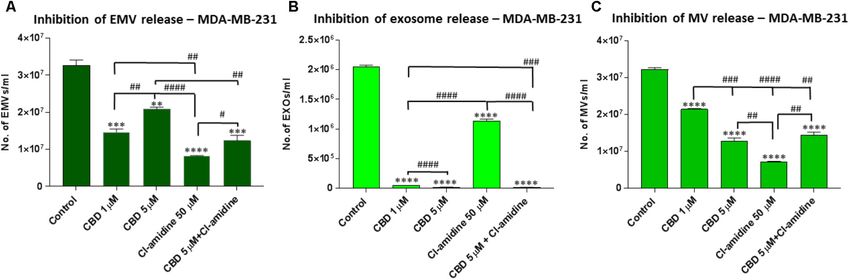
Kosgodage et al.                                                                                                  Cannabidiol Inhibits Exosomes in Cancer

migration, differentiation and angiogenesis (Ansa-Addo et al.,                contributing to disease pathology, EMVs are being considered as
2010; Muralidharan-Chari et al., 2010; Turola et al., 2012;                   therapeutic vehicles themselves (György et al., 2015; Moore et al.,
Colombo et al., 2014; Batrakova and Kim, 2015; Kholia et al.,                 2017).
2016).                                                                            It has been shown that EMV shedding from cancer cells
    Microvesicles (MVs) are phospholipid-rich cell-membrane                   aids increased active drug efflux and thus contributes to their
derived vesicles (100–1000 nm) released as part of normal                     resistance to chemotherapeutic agents (Bebawy et al., 2009; Tang
physiology as well as during apoptosis or upon stimulation                    et al., 2012; Jorfi and Inal, 2013; Pascucci et al., 2014; Jorfi et al.,
(Piccin et al., 2007; Inal et al., 2013). The release of MVs can              2015; Saari et al., 2015; Soekmadji and Nelson, 2015; Aubertin
be mediated via calcium ion influx through stimulation of cation              et al., 2016; Koch et al., 2016; Muralidharan-Chari et al., 2016).
channels such as the ATP-gated P2X7, through pores created by                 Recent studies on pharmacological inhibition of EMV release
sublytic complement, or via calcium released by the endoplasmic               have shown that such interventions could be a new strategy to
reticulum (Turola et al., 2012; Raposo and Stoorvogel, 2013;                  render cancer cells more susceptible to anticancer drug treatment
Stratton et al., 2015a,b). This increase in cytosolic calcium                 (Tang et al., 2012; Federici et al., 2014; Jorfi et al., 2015; Koch
results in cytoskeletal reorganization and membrane asymmetry,                et al., 2016; Muralidharan-Chari et al., 2016; Kosgodage et al.,
followed by subsequent MV blebbing (Inal et al., 2012, 2013;                  2017). Such approaches have recently been shown to be effective
Kholia et al., 2015; Kosgodage et al., 2017; Tricarico et al.,                in vivo, demonstrating that the application of EMV inhibitors can
2017). MV formation can also be caused by mitochondrial stress,               effectively sensitize tumors to chemotherapy (Jorfi et al., 2015;
which leads to increased membrane permeability and leakage                    Koch et al., 2016; Muralidharan-Chari et al., 2016), reduce drug
of reactive oxygen species (ROS), cytochrome C and apoptosis                  efflux (Federici et al., 2014; Koch et al., 2016; Muralidharan-Chari
inducing factor into the cytoplasm. This results in the formation             et al., 2016) and reduce the dose of anti-cancer drug required to
of the apoptosome – which, during pseudoapoptosis, can be                     limit tumor growth in vivo (Jorfi et al., 2015). Pharmacological
formed into MVs for the export of hazardous agents (Inal et al.,              non-toxic agents that can selectively manipulate extracellular
2013).                                                                        vesicle release may thus be relevant not only to cancer but also
    Exosomes (30–100 nm) are generated intracellularly as they                to other pathologies involving EMV release (Lange et al., 2017).
are formed after the invagination of the endosome membrane,                       Cannabidiol (CBD) (Mechoulam et al., 2002), a
resulting in intraluminal vesicle formation and the appearance                phytocannabinoid derived from Cannabis sativa, is anxiolytic
of multivesicular endosomes, which then release exosomes from                 (Blessing et al., 2015) and has analgesic, anti-inflammatory,
the plasma membrane via exocytosis (Kowal et al., 2014; van                   antineoplastic and chemo-preventive activities (Martin-Moreno
Niel et al., 2018). Crucial cellular components for exosomal                  et al., 2011; Pisanti et al., 2017). CBD has been shown to
biogenesis are ESCRT (endosomal sorting complexes required                    have a plethora of molecular targets, including the classical
for transport), sphingolipid ceramide, syntetin and syndecan, and             endocannabinoid system, while effects that do not involve the
tetraspanins (Théry et al., 2002; Baietti et al., 2012; Colombo et al.,       classical cannabinoid system are also gaining increased attention
2013; Costa, 2017; Hessvik and Llorente, 2018). The secretion of              (Ibeas Bih et al., 2015; Pisanti et al., 2017). CBD is generally safe at
exosomes is also affected via purinergic receptors such as P2X7               therapeutic doses, shows biphasic effects on the immune system,
(Qu et al., 2007), by microenvironmental pH (Federici et al.,                 and has demonstrated anti-cancer activity in vivo (Bergamaschi
2014) and calcium (Savina et al., 2003; Kramer-Albers et al.,                 et al., 2011; Massi et al., 2013; Haustein et al., 2014; Velasco
2007).                                                                        et al., 2016). Critically, CBD has been shown to be effective
    Exosome and microvesicles are emerging as novel therapeutic               in various EMV-linked pathologies (Velasco et al., 2016), and
targets in treatment of disease as they have been shown to                    seems to modulate mitochondrial function, including ATP, ROS
contribute to inflammatory processes (Foers et al., 2017) and                 and proton leak, as well as uptake and release of calcium (Ryan
the progression of numerous pathologies including autoimmune                  et al., 2009; Mato et al., 2010; Rimmerman et al., 2013; Cui et al.,
diseases (Antwi-Baffour et al., 2010; Withrow et al., 2016; Perez-            2017). These observations may be relevant as mitochondria are
Hernandez et al., 2017), cancers (Luga et al., 2012; Jorfi et al.,            key in modulating calcium signaling (Szabadkai and Duchen,
2015; Kholia et al., 2015; Stratton et al., 2015a; Sung et al.,               2008; Rizzuto et al., 2012) and importantly, altered calcium
2015; Tkach and Théry, 2016; Moore et al., 2017; Sung and                     signaling and mitochondrial function are hallmarks of many
Weaver, 2017) and neurodegenerative diseases (Colombo et al.,                 cancers (Boland et al., 2013; Stefano and Kream, 2015; Monteith
2012; Gupta and Pulliam, 2014; Porro et al., 2015; Basso and                  et al., 2017). This study therefore aimed to investigate putative
Bonetto, 2016). In cancer patients, elevated EMV levels have                  modulatory effects of CBD on EMV release and to further
for example been demonstrated in the blood (Ginestra et al.,                  establish whether CBD had combinatory effects with the recently
1998; Kim et al., 2003; Zwicker et al., 2009) and EMVs can also               described EMV-inhibitor Cl-amidine (Luo et al., 2006; Kholia
aid tumor spread and survival as they transport various micro                 et al., 2015; Kosgodage et al., 2017). For proof of principle we
RNAs, pathological growth factor receptors and soluble proteins               used three cancer cell lines, prostate cancer (PC3), hepatocellular
(Muralidharan-Chari et al., 2010; Inal et al., 2012; Hoshino                  carcinoma (HEPG2) and breast adenocarcinoma (MDA-
et al., 2015). Circulating EMVs in various body fluids such                   MB-231). Here we show effects of CBD on EMV release, on
as cerebrospinal fluid, urine and blood, may in addition serve                mitochondrial function, as well as on STAT3 expression, which
as reliable biomarkers of pathophysiological processes (Piccin                amongst other is associated with mitochondrial respiration and
et al., 2007; Inal et al., 2012, 2013; Porro et al., 2015). Besides           Ca2+ regulation in the mitochondrion (Wegrzyn et al., 2009;

Frontiers in Pharmacology | www.frontiersin.org                           2                                           August 2018 | Volume 9 | Article 889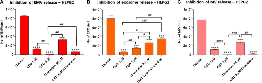
Kosgodage et al.                                                                                            Cannabidiol Inhibits Exosomes in Cancer

Yang et al., 2015; Yang and Rincon, 2016), alongside modulatory            for CBD (Bergamaschi et al., 2011); while Cl-amidine (a kind
effects on prohibitin, a pleiotropic protein involved in cellular          gift from Prof P.R. Thompson, UMASS) was used at 50 µM
proliferation and mitochondrial housekeeping (Peng et al., 2015;           concentration (in PBS) as previously determined as an optimal
Ande et al., 2017). Our findings suggest a new link between the            dose for maximum EMV inhibition in several cancer cell lines
emerging understanding of anti-cancer effects of CBD and its               (Kholia et al., 2015; Kosgodage et al., 2017). For testing of a
modulatory effects on EMV biogenesis in cancer cells, described            combinatory effect on EMV release, CBD was applied at 5 µM
here for the first time.                                                   together with Cl-amidine at 50 µM concentrations. After the 1 h
                                                                           incubation period, the supernatants from each well were collected
                                                                           from the cell preparations, transferred to sterile 1.5 ml Eppendorf
MATERIALS AND METHODS                                                      tubes (kept on ice) and centrifuged at 200 g for 5 min at 4◦ C to
                                                                           remove the cell debris. The resulting supernatants were kept on
Cell Cultures                                                              ice and subsequently treated for isolation of EMVs, as described
Human prostate adenocarcinoma (PC3 and ECACC), human                       below, to include both exosomes and MVs based on previously
hepatocellular carcinoma (HEPG2 and ECACC) and human                       established protocols (Lötvall et al., 2014; Kholia et al., 2015;
breast adenocarcinoma (MDA-MB-231; a kind gift from Dr T.                  Kosgodage et al., 2017; Witwer et al., 2017).
Kalber, UCL) cell lines were maintained at 37◦ C/5% CO2 , in
growth medium containing 10% EMV-free Foetal Bovine Serum                  Isolation of EMVs
(FBS) and RPMI (Sigma, United Kingdom). The cells were                     Exosome and microvesicles were isolated from the CBD,
split every 3–5 days, depending on confluence, washed twice                Cl-amidine, and CBD plus Cl-amidine treated cell culture
with EMV-free Dulbecco’s Phosphate Buffered Saline (DPBS),                 supernatants, as well as from the control treated cells (DMSO
prepared as described before (Kosgodage et al., 2017) and                  or PBS), by differential centrifugation as follows: First, whole
detached by incubation for 10–15 min at 37o C with 0.25%                   cells were removed by spinning at 200 g/5 min at 4◦ C. The
(v/v) trypsin/EDTA, followed by two washes by centrifugation               supernatants were then collected and further centrifuged at
using EMV-free DPBS at 200 g/5 min. Before the start of every              4,000 g for 60 min at 4◦ C, to remove cell debris. The resulting
experiment, cell numbers and viability were determined by Guava            supernatants were thereafter collected and centrifuged again at
ViaCount assay (Guava Millipore) and exponentially growing                 25,000 g for 1 h at 4◦ C. The resulting EMV pellets were collected
cells with viabilities of ≥95% were used.                                  and the supernatants were discarded. Next, the isolated EMV
                                                                           pellets were resuspended in sterile-filtered (0.22 µm) EMV-
Cell Viability Assays                                                      free Dulbecco’s PBS (DPBS) and thereafter centrifuged again at
The Guava EasyCyte 8HT flow cytometer (Millipore) and                      25,000 g for 1 h at 4◦ C to remove proteins that may have bound to
ViaCount assay (Guava Millipore) were used to count and                    the EMV surface. The DPBS supernatant was thereafter discarded
determine viability of cells before the start of every experiment          and the resulting isolated EMV pellets were resuspended in
and to assess cell viability after treatment with EMV inhibitors, as       200 µl of sterile EMV-free DPBS for further nanoparticle
previously described (Jorfi et al., 2015; Kosgodage et al., 2017).         tracking analysis (NTA), using the Nanosight (LM10; Nanosight,
Cell viability after cisplatin treatment (see 2.9) was assessed            Amesbury, United Kingdom). Each experiment was repeated
by MTT [3-(4,5-dimethylthiazol-2-yl)-2,5-diphenyltetrazolium               three times and performed in triplicate.
bromide] assay, performed according to the manufacturer’s
instructions (Sigma, United Kingdom).                                      Nanoparticle Tracking Analysis (NTA,
                                                                           NanoSight LM10)
Effects on EMV Biogenesis Using CBD                                        To determine size distribution of isolated EMVs, nanoparticle
and Cl-Amidine                                                             tracking analysis (NTA), based on the Brownian motion of
For assessment of effects of CBD and Cl-amidine on EMV                     vesicles in suspension (Soo et al., 2012), was used. A Nanosight
generation, PC3, HEPG2 and MDA-MB-231 cells were seeded                    LM10, equipped with a sCMOS camera and a 405 nm diode
at a density of 3.8 × 10⁵ cells/well, in triplicate, in 12-well            laser, was used to enumerate the EMVs. The NTA software 3.0
microtiter plates, using pre-warmed serum- and EMV-free RPMI               was used for data acquisition and processing according to the
1640 (Sigma-Aldrich, United Kingdom). To ensure that the                   manufacturer’s instructions (Malvern). The ambient temperature
medium was EMV free, it was centrifuged at 70,000 g/24 h and               was set at 23◦ C, while background extraction and automatic
filtered through a 0.22 µm pore size membrane before use. For              settings were applied for the minimum expected particle size,
testing of putative inhibitory or modulatory effects on EMV                minimum track length and blur. For calibration, silica beads
release, the cells were then incubated with CBD (1 or 5 µM),               (100 nm diameter; Microspheres-Nanospheres, Cold Spring, NY)
Cl-amidine (50 µM) or with a combination of CBD (5 µM)                     were used. The samples were diluted 1:50 in sterile-filtered, EMV-
and Cl-amidine (50 µM), for 60 min at 37◦ C/5% CO2 , while                 free DPBS. To maintain the number of particles in the field
control cells were treated with either DMSO (0.001%) or PBS for            of view approximately in-between 20 and 40, the minimum
CBD and Cl-amidine, respectively. The following concentrations             concentration of samples was set at 5 × 10⁷ particles/ml. For
of CBD (GW Pharmaceuticals, United Kingdom) were used: 1                   capturing, the screen and camera gain were set at 8 and 13,
or 5 µM in 0.001% DMSO, based on clinically relevant doses                 respectively; while for processing, the settings were at nine

Frontiers in Pharmacology | www.frontiersin.org                        3                                        August 2018 | Volume 9 | Article 889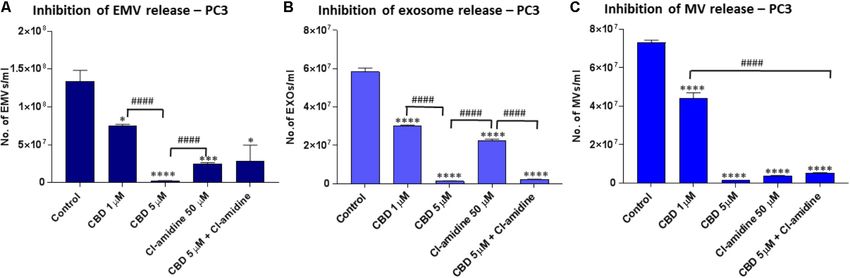
Kosgodage et al.                                                                                             Cannabidiol Inhibits Exosomes in Cancer

and three for screen gain and detection threshold, respectively,            while for each cell lysate preparation, 10 µl were loaded per
as according to the manufacturer’s instructions (Malvern).                  lane. For immunoblotting, proteins were transferred to 0.45 µm
Five × 30 s videos were recorded for each sample, measurements              nitrocellulose membranes (BioRad) using semi-dry Western
with at least 1,000 completed tracks were used for analysis and the         blotting at 15 V constant for 1 h, even transfer was assessed
resulting replicate histograms were averaged. Each experiment               using Ponceau S staining (Sigma) and the membranes were
was repeated three times and performed in triplicate.                       blocked in 5% bovine serum albumin (Sigma) in tris-buffered-
    For verification of the presence of exosomes within the 30–             saline (TBS) containing 0.01% Tween-20 (Sigma) for 1 h at
100 nm sized vesicle peak, according to NTA analysis, and                   room temperature. Incubation with anti-human CD63 (ab68418,
MVs within the 101–900 nm sized vesicle peak, according to                  Abcam, United Kingdom, 1/1000 in TBS-T) was carried out
NTA analysis (Supplementary Figures 1A,B), MVs were pelleted                overnight at 4◦ C, thereafter the blots were washed three times for
first, from the EMV supernatants, by centrifugation at 11,000 g             10 min in TBS-T and incubated thereafter in secondary antibody
for 30 min at 4◦ C, and thereafter the presence of MVs was                  (HRP-conjugated anti rabbit IgG1, 1/4000, BioRad) for 1 h at
assessed by flow cytometry for Annexin V-FITC binding as                    room temperature. The blots were washed five times for 10 min
a measure of phosphatidylserine exposition characteristic for               in TBS-T, followed by one wash in TBS before visualization
MVs (Supplementary Figure 1C). The remaining supernatant                    with ECL (Amersham, United Kingdom). Membranes were
was further centrifuged for the isolation of the smaller sized              imaged using the UVP transilluminator (UVP BioDoc-ITTM
exosomes (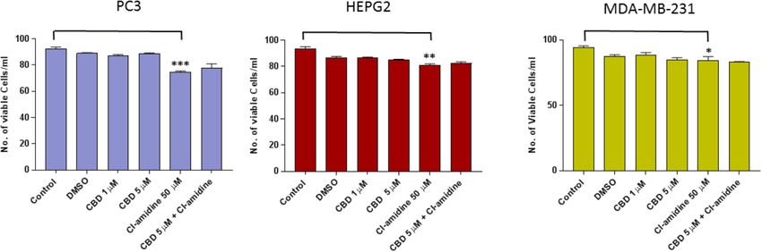
Kosgodage et al.                                                                                             Cannabidiol Inhibits Exosomes in Cancer

Biosciences) supplemented with glucose and 1% sodium                        In addition, longer-term (24 h) treatment effects of CBD on
pyruvate, pH 7.4 at 37◦ C. Thereafter, oligomycin, carbonyl                 cancer cell viability was further assessed for HEPG-2 and MDA-
cyanide-4-(trifluoromethoxy) phenylhydrazone (FCCP, 0.2 µM)                 MB-231 cells, showing dose-depended reduction in cell viability
and antimycin/rotenone (0.25 µM) were added to the sensor                   compared to control DMSO treated cells as follows: In HEPG2
plate prior to the commencement of calibration and the assay.               cells, 1 µM CBD resulted in a 38.8% decrease in cell viability
Calculations were normalized to protein level, as calculated by             (p < 0.001), and 5 µM CBD in a 47.2% decrease in cell viability
Bradford assay directly after the experimental procedure. Each              (p < 0.001) compared to DMSO treated control cells. In MDA-
experiment was repeated 3–5 times, with technical replicates of             MB-231 cells, 1 µM CBD resulted in a 12.9% decrease in cell
four per plate.                                                             viability (p < 0.05), and 5 µM CBD in a 35.8% decrease in cell
                                                                            viability (p < 0.01) compared to DMSO treated control cells
Effect of CBD on Cisplatin-Mediated                                         (Supplementary Figure 2).
Apoptosis of HEPG2 and MDA-MB231
Cancer Cells                                                                EMV Release Profiles Vary Between PC3,
HEPG2 and MDA-MB231 cells were grown as a monolayer in                      HEPG2, and MDA-MB-231 Untreated
T75 flasks (Nunc, United States) until 80% confluent. The media
                                                                            Cancer Cells
was removed, the cells washed in DPBS and fresh medium added,
                                                                            A range in the total amount of EMVs (
Kosgodage et al. Cannabidiol Inhibits Exosomes in Cancer FIGURE 1 | CBD does not affect cell viability of PC3, HEPG2, and MDA-MB-231 cells after 1 h treatment. The Guava EasyCyte 8HT flow cytometer (Millipore) and ViaCount assay were used to count and determine viability of CBD treated cells compared to EMV inhibitor Cl-amidine and DMSO treated control cells after 1 h incubation (∗ p ≤ 0.05;∗∗ p ≤ 0.01;∗∗∗ p ≤ 0.001). FIGURE 2 | CBD significantly inhibits total EMV, exosome and MV release from HEPG2 cells. Inhibitory effects of CBD alone and in combination with Cl-amidine on extracellular vesicle release from HEPG2 cancer cells are presented as histograms which are based on size exclusion analysis by Nanosight Tracking Analysis (NTA). EMVs represent all vesicles 0–900 nm (A); exosomes are vesicles
Kosgodage et al. Cannabidiol Inhibits Exosomes in Cancer
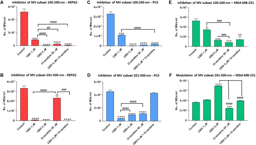
subset, 5 µM CBD showed a stronger inhibitory effect (99.4%; compared to DMSO treated controls (98.8, 95.9, and 94.4%,
p = 0.0003) than 1 µM CBD (82.0%; p = 0.0006) compared respectively; p = 0.0001 for all groups compared to control). CBD
to control. Cl-amidine reduced this smaller subset by 98.1% at 1 µM also showed significant inhibition compared to control
(p = 0.0004) compared to control, and combinatory treatment (71.8%; p = 0.0007), but significantly less inhibition on this MV
of 5 µM CBD and Cl-amidine had a 98.2% inhibitory effect subset than 5 µM CBD alone, Cl-amidine alone or CBD (5 µM)
(p = 0.0003) compared to control treated cells (Figure 5A). and Cl-amidine in combination (p = 0.0001, p = 0.0001 and
For the shedding of the larger 201–500 nm sized vesicles, CBD p = 0.0002, respectively; Figure 5C).
was a more effective inhibitor in HEPG2 cells than Cl-amidine, For the shedding of the larger MV subset of 201–500 nm sized
with 1 µM CBD showing 97.1% (p = 0.0001) and 5 µM CBD vesicles, CBD was more effective at the lower dose of 1 µM than
100% (p = 0.0002) inhibitory effect, respectively, compared to at 5 µM (p = 0.0001). Cellular release of this MV subset was
controls. In this larger MV subset, Cl-amidine showed only 30% reduced by 92% in 1 µM CBD treated cells (p = 0.0001), and
inhibition (p = 0.0356) compared to control. The combinatory by 81.2% in 5 µM CBD treated cells (p = 0.0001) compared to
application of 5 µM CBD and Cl-amidine had a similar inhibitory controls, while Cl-amidine alone reduced this subset of MVs by
effect (99.7%; p = 0.0001) as 5 µM CBD alone (Figure 5B). 64.0% (p = 0.0002). When used in combination, 5 µM CBD with
Cl-amidine did not show significant inhibition of this MV subset
compared to control (4% inhibition; p = 0.4250) (Figure 5D).
CBD Effectively Inhibits Exosome and
Microvesicle Release From PC3 Cells
Pre-treatment of PC3 with both 1 and 5 µM CBD, for 60 min
CBD Effectively Inhibits Exosome and
before EMV isolation, resulted in a significant reduction of total Microvesicle Release From MDA-MB-231
EMV release compared to the DMSO treated control cells (44.5 Cells
and 98.1% reduction of EMV release for 1 and 5 µM CBD, Pre-treatment of MDA-MB-231 with both 1 and 5 µM CBD for
respectively; p = 0.0149; p = 0.0008, respectively) (Figure 3A). 60 min before EMV isolation resulted in a significant reduction
The inhibitory effect by 5 µM CBD on total EMV release of total EMV release compared to the control treated cells
was greater than observed with our previously most efficient (53.4%; p = 0.0001 and 42.9%; p = 0.0001, respectively) but
EMV inhibitor Cl-amidine, which was used for comparison was a less potent total EMV inhibitor than Cl-amidine (75.9%;
(p = 0.0001), while Cl-amidine had a significantly stronger EMV p = 0.0001 compared to control). When using CBD (5 µM) in
inhibitory effect than 1 µM CBD (p = 0.0001). When using CBD combination with Cl-amidine, a significantly (p = 0.0052) higher
(5 µM) in combination with Cl-amidine no additive change in inhibition was observed compared to 5 µM CBD alone, while
total EMV inhibition was found compared to single inhibitors there was no significant difference compared to 1 µM CBD
(Figure 3A). treatment (p = 0.2474). Compared to control treated cells the
Analysis of inhibitory effects on exosome sized vesicles combinatory treatment resulted in a 55.1% reduction of total
(Kosgodage et al. Cannabidiol Inhibits Exosomes in Cancer FIGURE 3 | CBD significantly inhibits total EMV, exosome and MV release from PC3 cells. Inhibitory effects of CBD alone and in combination with Cl-amidine on extracellular vesicle release from PC3 cancer cells are presented as histograms which are based on size exclusion analysis by Nanosight Tracking Analysis (NTA). EMVs represent all vesicles 0–900 nm (A); exosomes are vesicles
Kosgodage et al.                                                                                                           Cannabidiol Inhibits Exosomes in Cancer

  FIGURE 5 | CBD modulates different MV subpopulations released from PC3, HEPG2 and MDA-MB-231 cells. Inhibitory effects of CBD alone, Cl-amidine or CBD in
  combination with Cl-amidine on 100–200 nm and 201–500 nm sized microvesicles, based on size exclusion analysis by Nanosight Tracking Analysis (NTA). Inhibition
  of 100–200 nm MV release in (A) PC3; (C) HEPG2; and (E) MDA-MB-231 cancer cells. Inhibition of 201–500 nm MV release in (B) PC3; (D) HEPG2; and
  (F) MDA-MB-231 cancer cells. The experiments were repeated three times and the data presented are mean ± SEM of the results (∗ p ≤ 0.05; ∗∗ p ≤ 0.01;
  ∗∗∗ p ≤ 0.001; ∗∗∗∗ p ≤ 0.0001 versus Control; Differences between CBD and Cl-amidine treatment group is further indicated as #p ≤ 0.05; ##p ≤ 0.01;

  ###p ≤ 0.001; ####p ≤ 0.0001).

was also confirmed to exclude contamination by cellular debris in                   CBD treated MDA-MB-231 cells ATP production levels were
the exosome isolates (not shown).                                                   60.5 ± 12.8 pMoles/min (p ≤ 0.05) and in 5 µM CBD treated cells
                                                                                    ATP production levels were 69.2 ± 16.8 pMoles/min (p ≤ 0.05)
Mitochondrial Function Alteration                                                   (Figure 7B). PC3 cells showed a decreasing trend in ATP
Analysis in MDA-MB-231 and PC3 Cells                                                with increased dose of CBD, albeit not statistically significant
                                                                                    compared to control treated PC3 cells (106.0 ± 9.4 pMoles/min).
Following CBD Treatment                                                             In 1 µM CBD treated PC3 cells ATP production levels were
Mitochondrial analysis, using the Seahorse Bionalayser,                             87.8 ± 9.1 pMoles/min and in 5 µM CBD treated cells ATP
measured mitochondrial respiration along with several key                           production levels were 81.2 ± 11.9 pMoles/min (Figure 7E).
mitochondrial factors associated with mitochondrial function                           A significant dose dependent increase in proton leak was
through oxygen consumption (Figure 7). In MDA-MB-231                                observed for both concentrations of CBD in MDA-MB-231 cells
cells, basal mitochondrial OCR (oxygen consumption rate)                            as follows: 1 µM CBD: 21.6 ± 3.2 pMoles/min (p ≤ 0.01), and
was significantly increased, compared to non-treated controls                       5 µM CBD: 32.2 ± 9.5 pMoles/min (p ≤ 0.01), compared to
(50.4 ± 13.5 pMoles/min), following 1 h CBD treatment at                            untreated cells (5.8 ± 3.3 pMoles/min) (Figure 7C). In PC3 cells
1 µM (104.1 ± 23.7 pMoles/min; p ≤ 0.05) and 5 µM CBD                               proton leak was somewhat, but not significantly, reduced in the
(129.6 ± 36.4 pMoles/min; p ≤ 0.05) (Figure 7A). In PC3                             presence of 1 µM (36.4 ± 3.6 pMoles/min) and 5 µM CBD
cells, basal mitochondrial OCR showed a decreasing trend                            (35.1 ± 2.8 pMoles/min) compared to untreated cells (40.9 ± 3.7
with increased dose of CBD, following 1 h CBD treatment at                          pMoles/min) (Figure 7F).
1 µM (124.2 ± 9.6 pMoles/min; p ≤ 0.05) and 5 µM CBD
(116.3 ± 13.1 pMoles/min), while not statistically significant
compared to non-treated control (146.8 ± 12.9 pMoles/min)
                                                                                    CBD Modulates Expression of
(Figure 7D).                                                                        Mitochondrial Associated Proteins
   Dose dependent changes in relative ATP production levels                         Prohibitin and STAT3
were observed in both cancer cell lines in response to CBD                          Protein isolates from HEPG2, PC3 and MDA-MB231 cells were
treatment compared to untreated cells. Compared to untreated                        further assessed for changes in two mitochondrial associated
MDA-MB-231 cells (37.5 ± 8.9 pMoles/min), in 1 µM                                   proteins; prohibitin and STAT3, following CBD (5 µM)

Frontiers in Pharmacology | www.frontiersin.org                                 9                                              August 2018 | Volume 9 | Article 889
Kosgodage et al. Cannabidiol Inhibits Exosomes in Cancer
FIGURE 6 | CD63 exosomal marker is reduced following 1 h CBD treatment in HEPG2, PC3, and MDA-MB-231 cancer cells. The results from the NTA analysis
were confirmed by Western blotting for the exosomal CD63 marker, which was reduced in all three cell lines following 1 h treatment with 5 µM CBD: (A) CD63
expression is reduced in CBD treated versus DMSO control treated HEPG2 cancer cells; (B) CD63 expression is reduced in CBD treated versus DMSO control
treated PC3 cells; (C) CD63 expression is reduced in CBD treated versus DMSO control treated MDA-MB-231 cells. All EMV preparations were performed in equal
buffer volume (50 µl) and all cell lysates were prepared in equal buffer volume (50 µl) between all samples, for accurate presentation of amounts of vesicles isolated
and amounts of cells grown and collected per flask. For EMV isolates, 20 µl of sample was loaded per lane, while for β-actin detection in the corresponding cell
lysates, 10 µl of sample was loaded per lane. The relative detection of CD63 in EMVs released from the corresponding cell preparation is indicated by “R,” in relation
to β-actin detection in the corresponding cell isolate, for comparison between CBD treatments versus DMSO control.
treatment and compared to DMSO treated controls. In all three exosome release significantly and also had significant, albeit more
cancer cell lines, levels of prohibitin were reduced, although variable, modulating effects on MV release. This novel function of
more marked changes were noted in the PC3 (Figure 8A) and CBD on EMV release, revealed here for the first time, may be of
HEPG2 (Figure 8B) cells compared to the MDA-MB-231 cells high relevance for optimized therapeutic application in various
(Figure 8C). In all three cancer cell lines, STAT3 (phospho Y705) EMV-mediated pathologies.
was also reduced after 1 h CBD (5 µM) treatment; again this There is a considerable interest in using EMV inhibitors to
reduction was higher in PC3 and HEPG2 cells (Figures 8D,E), sensitize cancers to chemotherapy. Previous work, using the
compared to the MDA-MB-231 cells (Figure 8F). calpain inhibitor calpeptin for MV inhibition, in combination
with chemotherapy drugs fluorouracil and docetaxel, reduced
CBD Sensitizes HEPG2 and the effective chemotherapeutic dose needed by 100-fold to
MDA-MB-231 Cancer Cells to on produce comparable reduction in tumor volume in vivo. The
Cisplatin-Mediated Apoptosis same study also showed that methotrexate is released from
cancer cells in MVs (Jorfi et al., 2015). Similar findings of
In both HEPG2 and MDA-MB-231 cancer cells, CBD increased
drug efflux and sensitisation to gemcitabine in response to
cisplatin-mediated apoptosis (Figure 9). In HEPG2 cells,
MV inhibition were established in pancreatic cancer in vitro
compared to untreated control cells, cisplatin treatment alone
and in vivo upon MV inhibition via ERK-mediated pathways
resulted in 57.3% cell viability (p < 0.01). However, this effect was
(Muralidharan-Chari et al., 2016); and to doxorubicin and
significantly enhanced (p < 0.001) if cells were first treated with 1
pixantrone treatment upon exosome inhibition via inhibition
and 5 µM CBD (54.5 and 39.1%, respectively), prior to cisplatin
of ATP-transporter A3 in B-cell lymphoma models (Koch
(Figure 9A). In MDA-MB-231 cells, compared to untreated
et al., 2016). Also, chemotaxis of cancer cells has been shown
control cells, cisplatin treatment alone resulted in 47.3% cell
to be promoted by exosome secretion, but to be diminished
viability (p < 0.001), while 21.3 and 8.3% cell viability was
by knockdown of the exosome regulator Rab27a (Sung and
observed for cells treated with 1 or 5 µM CBD prior to cisplatin
Weaver, 2017). Inhibition of exosome secretion has been shown
treatment (p < 0.01). CBD treatment alone led to significant
to cause defective tumor cell migration (Sung et al., 2015),
changes in cell viability, but to a much lesser extent than those
while exosomes isolated from gastric tumor cells were shown
observed when cells were first treated with CBD followed by
to induce tumor cell migration and promotion in receiving
cisplatin (Figures 9A,B).
cells (Wu et al., 2016). Previously, our work identified a novel
pathway of MV release involving peptidylarginine deiminases
DISCUSSION (PADs) and the effective inhibition of PAD-mediated EMV
release using Cl-amidine (Kholia et al., 2015; Kosgodage et al.,
This study reveals a novel finding for CBD; it can selectively 2017), suggesting implications in a number of pathologies
inhibit the release of subsets of EMVs, from cancer cell lines. (Lange et al., 2017). In addition, we have also recently
The different cancer cell lines tested here (prostate cancer PC3, shown that several new candidate EMV inhibitors, including
hepatocellular carcinoma HEPG2 and breast adenocarcinoma bisindolylmaleimide-I, imipramine and Cl-amidine, are more
MDA-MB-231) varied in the proportional amounts of total potent EMV inhibitors than calpeptin (Kosgodage et al., 2017)
EMVs, MVs and exosomes released under standard conditions and those they sensitize cancer cells to chemotherapeutic agents.
(Supplementary Figure 3). Nonetheless, across this range of This further call for the identification of novel EMV inhibitors,
EMV release profiles, we found that CBD consistently inhibited which are safe for application in vivo, such as CBD now
Frontiers in Pharmacology | www.frontiersin.org 10 August 2018 | Volume 9 | Article 889Kosgodage et al. Cannabidiol Inhibits Exosomes in Cancer
FIGURE 7 | Mitochondrial function alteration following 1 h CBD treatment in MDA-MB-231 and PC3 cancer cells. MDA-MB-231 and PC3 cells were treated with 1
or 5 µM CBD for 1 h prior to mitochondrial functional analysis using the Seahorse Bioanalyser for the following parameters: (A,D) Basal mitochondrial respiration;
(B,E) Quantification of ATP production; (C,F) Proton leak. Data shown is repeated three times (for MDA-MB-231 cells) or five times (for PC3 cells) with four technical
replicates per plate. Data is represented as mean ± SEM. ∗ p < 0.05, ∗∗ p < 0.01, ∗∗∗ p < 0.001 versus untreated control cells.
identified here. Indeed, as we have shown here, by significantly to CBD and may also be a sign of pseudoapoptotic responses,
increasing cisplatin mediated apoptosis, CBD showed a similar where increased membrane permeability and leakage of reactive
ability to other EMV inhibitors of sensitizing cancer cells to oxygen species (ROS) and other apoptotic factors is still low
chemotherapy. enough for the cell to turn the apoptosome into MVs for export
CBD-mediated inhibition of EMV release, observed in the of hazardous agents (Mackenzie et al., 2005; Inal et al., 2013).
present study, was more effective for some EMV subsets and Indeed, a dose-dependent increase in ROS levels in response to
cancer cells than Cl-amidine, our most potent EMV inhibitor to 1 h CBD treatment (Supplementary Figure 5) alongside a dose-
date (Kosgodage et al., 2017). One intriguing finding in our study dependent increase in proton leak, mitochondrial respiration
is the selectivity of CBD on different EMV subsets in the three and ATP levels (Figures 7A–C) were observed in this cancer
different cancer cell lines, which also varied with concentration cell line in particular. In the PC3 cells on the other hand, the
(1 and 5 µM). In PC3 cells, 5 µM of CBD was the most reduced EMV release observed in all EMV subsets, tallied in
effective inhibitor of total EMVs, exosomes, total MVs and the with a trend of reduced ATP production and reduced proton
smaller MV subset (100–200 nm), while 1 µM CBD was most leak as well as lowered mitochondrial respiration, indicating an
effective at inhibiting the larger MV subset (201–500 nm). In absence of pseudoapoptotic responses, as clearly reflected also in
the HEPG2 hepatocellular carcinoma cells 5 µM CBD had the the significant reduction of the 201–500 nm MV subset in the PC
main impact on total EMV and MV release, while 1 µM CBD cancer cells.
most significantly affected exosome release. Overall, the potency In the current study we have found that while reducing EMVs,
of CBD to inhibit all subsets of EMVs tested here was most CBD also modulates mitochondrial function and the expression
marked in the HEPG2 cells. In MDA-MB-231 cells the inhibitory of mitochondrial associated proteins prohibitin and STAT3.
effect of CBD was particularly marked for exosome release, while Although studies on direct links between EMV release and
total MV release was less inhibited by CBD compared to Cl- mitochondrial changes are relatively limited, requiring further
amidine. Recent studies in this invasive breast cancer cell line investigation, EMV generation has previously been linked to
have suggested an active role for exosomes in increased cell this organelle (Mackenzie et al., 2005; Qu et al., 2007; Lopez
movement and metastasis (Harris et al., 2015). The increase in et al., 2008; Morel et al., 2010; Dubyak, 2012; Soto-Heredero
MVs released in the size range of 201–500 nm in response to et al., 2017). Both changes in mitochondrial calcium buffering
CBD treatment was specific for the MDA-MB-231 cells. This and dynamics, including ROS, ATP and proton leak, have
may indicate a higher sensitivity of this particular cancer cell line previously been shown to be linked to MV formation (Mackenzie
Frontiers in Pharmacology | www.frontiersin.org 11 August 2018 | Volume 9 | Article 889Kosgodage et al. Cannabidiol Inhibits Exosomes in Cancer FIGURE 8 | CBD modulates expression of prohibitin and STAT3 in cancer cells. PC3, HEPG2, and MDA-MB-231 cells were tested for changes in mitochondrial associated prohibitin and STAT3 expression following 1 h treatment with CBD (5 µM). Levels of prohibitin were reduced in all three cancer cell lines while this reduction was more marked in PC3 (A) and HEPG2 (B) cells compared to MDA-MB-231 (C). STAT3 (phospho Y705) was also reduced after 1 h CBD (5 µM) in both PC3 (D) and HEPG2 cells (E), while MDA-MB-231 cells showed a similar, albeit less marked trend (F). Beta-actin is shown as an internal loading control and “R” indicates the change of prohibitin and pSTAT3 expression relative to β-actin levels, respectively, for comparison between CBD treatment and DMSO control. FIGURE 9 | CBD sensitizes HEPG2 and MD-MB-231 cancer cells to cisplatin-mediated apoptosis. HEPG2 (A) and MDA-MB-231 (B) cells were treated with 1 or 5 µM CBD for 24 h prior to further 24 h incubation with cisplatin (CSP, 100 µM). Cell viability was assessed by MTT assay. Data shown is repeated three times with three technical replicates per plate. Data is represented as mean ± SEM. ∗ p < 0.05, ∗∗ p < 0.01 versus untreated control cells. et al., 2005) and to affect ATP-mediated release of MVs and mediating Cyt C release; this causes cytoskeletal degradation exosomes (Qu et al., 2007; Dubyak, 2012). Mitochondrial stress and formation of the apoptosome (Dale and Friese, 2006; Lopez can also lead to MV formation via pro-apoptotic Bax and Bak, et al., 2008; Inal et al., 2012). In scenarios of minimal damage which insert into the mitochondrial outer membrane resulting the cell can use the apoptosome to form a MV and export in its depolarisation and increased membrane permeability. This the hazardous agents via pseudoapoptosis (Inal et al., 2012). leads to ROS, cytochrome C (Cyt C) and apoptosis inducing Furthermore, pseudoapoptosis has been shown to involve rapid factor (AIF) leakage into the cytoplasm and eventual apoptosis. reversible mitochondrial depolarization, mitochondrial swelling Where apoptosis is triggered by the extrinsic pathway, such as and changes in mitochondrial and cytosolic calcium (Mackenzie stimulation of FasL, activation of caspase 8 leads to cleavage et al., 2005). In cancer cells, a previous study has for example of Bid, tBid then translocating to the mitochondrial membrane, shown a ten-fold increase in the release of a 333–385 nm MV Frontiers in Pharmacology | www.frontiersin.org 12 August 2018 | Volume 9 | Article 889
Kosgodage et al.                                                                                               Cannabidiol Inhibits Exosomes in Cancer

subset in pseudoapoptotic response to sublytic C5b-9 stimulation              been shown to cause chemoresistance, while knock-down of
(Stratton et al., 2015a). Mitochondrial permeability has been                 prohibitin sensitized cancer cells to chemotherapeutic treatment
shown to be important also for MV shedding from platelets,                    (Tortelli et al., 2017). Inhibition of prohibitin has also been
where the natural phenol and Bax activator gossypol promoted                  shown to repress cancer cell malignancy progression in hypoxia
mitochondrial depolarization, PS exposure and MV release (Dale                (Cheng et al., 2014). The observed reduction in prohibitin
and Friese, 2006). As it is now thought that many oncogenes and               observed here, following CBD treatment, may thus be an
tumor suppressors control calcium flow into the mitochondrion,                important factor in contributing to the sensitisation of cancer
one key emerging target in cancer treatment is mitochondrial                  cells to chemotherapeutic agents, as previously shown for CBD
control of calcium signaling (Danese et al., 2017). Previously,               in glioblastoma (Torres et al., 2011), in addition to affecting EMV
effects of CBD on modulating mitochondrial calcium buffering                  release due to changes in mitochondrial function caused partly by
and mitochondrial function have been described (Ryan et al.,                  prohibitin and STAT3 downregulation following CBD exposure.
2009; Mato et al., 2010; De Petrocellis et al., 2011; Shrivastava                 Using a combined application of CBD (5 µM) with Cl-amidine
et al., 2011; Rimmerman et al., 2013; Fisar et al., 2014; Cui et al.,         resulted in different effects on the various EMV subsets and
2017), including on mitochondrial swelling, ROS production and                varied between the three cancer cell lines. In general, Cl-amidine
mitochondrial potential (Mato et al., 2010). STAT3 is indeed                  did not have additive effects on the inhibitory effect of EMV
implicated in mitochondrial calcium control (Yang et al., 2015;               release compared to CBD alone, while the combinatory treatment
Garama et al., 2016; Yang and Rincon, 2016) and the reduction                 was more effective on some subsets than Cl-amidine alone, as was
in STAT3 in cancer cells observed here following CBD treatment                observed on exosome release in PC3 cells and on MV release
may thus have modulatory effects on EMV release. A reduction                  in HEPG2 cells. However, the difference between cancer cell
of STAT3 by CBD has previously been shown in glioblastoma                     types to combinatory treatment did not significantly affect the
cells where it was for example related to the inhibition of                   larger MV subset in PC3 cells, while both CBD and Cl-amidine
self-renewal (Singer et al., 2015). Prohibitin is ubiquitously                alone did. Similarly, combinatory treatment did not show more
expressed in many cell types and involved amongst other in                    effect than CBD or Cl-amidine alone on exosome release from
energy metabolism, proliferation and apoptosis (Peng et al.,                  HEPG2 cells. Interestingly, in MDA-MB-231 cells, Cl-amidine
2015; Ande et al., 2017). It acts as a scaffold protein in the                counteracted the increased CBD-mediated release observed for
inner mitochondrial membrane and is thus important for the                    the larger MV subset (201–500 nm), when used in combination,
regulation of mitochondrial architecture (Merkwirth et al., 2012).            bringing the amount of vesicles release down to similar levels
Prohibitin is critical for mitochondrial house-keeping including              as for the control treated cells. Overall our results suggest that
mitochondrial dynamics, morphology and biogenesis as well as                  the two EMV inhibitors act on different pathways involved in
stabilizing the mitochondrial genome (Peng et al., 2015). Here                MV and exosome release. While previously, Cl-amidine has
we show, for the first time, that prohibitin is reduced in cancer             been shown to act on MV biogenesis via increased cytoskeletal
cells following CBD treatment. The slight variability in reduction            actin deimination and nuclear PAD translocation, indicative for
of prohibitin in response to CBD between the cancer cell lines                changes in histone deimination (Kholia et al., 2015), CBD may
tested here tallies in with the observed differences in effectivity of        act in part through modulation of mitochondrial metabolism
CBD to inhibit EMVs from these different cancer cells. A similar              as described here. Accordingly, and depending on which EMV
correlation was found between expression changes in STAT3                     subset is being targeted, our results indicate that tailored
and inhibition of EMV release following CBD treatment, as                     approaches for selective EMV inhibition could be developed for
both PC3 and HEPG2 cells showed more reduction in STAT3                       various EMV mediated pathologies. The expanded repertoire of
levels following CBD treatment, alongside a more pronounced                   EMV inhibiting agents, including CBD now revealed here, along
inhibitory effect on total EMV release, compared to MDA-MB-                   with its sensitizing effects on cancer cells to cisplatin-mediated
231 cells; which, while showing overall significant reduction                 apoptosis, indicates a therapeutic potential for sensitisation of
in EMVs and reduced levels of STAT3 and prohibitin, these                     cancer cells to chemotherapy, as has been demonstrated for other
effects were somewhat less marked than in the other two cancer                promising EMV inhibitors (Tang et al., 2012; Federici et al.,
types. The EMV modulatory effects of CBD could thus be                        2014; Jorfi et al., 2015; Koch et al., 2016; Muralidharan-Chari
partly mediated by the above observed mitochondrial changes.                  et al., 2016; Kosgodage et al., 2017). Importantly, such EMV-
In addition, CBD has also been shown for example to stimulate                 modulating agents could be used to allow for lower dose of
mitochondrial uptake of calcium, followed by a decrease and a                 chemotherapeutic drug for effective inhibition of tumor growth
matching sudden increase in intracellular calcium (Ryan et al.,               in vivo (Jorfi et al., 2015; Muralidharan-Chari et al., 2016). The
2009), indicating thus also putative dynamic effects on EMV                   ability of CBD to inhibit EMV release may indeed be a hitherto
release. Notably, in PC3 cells, a CBD-dose-dependent trend was                overlooked contributing factor in the beneficial effects of CBD
observed for reduced ATP production, which correlated with                    observed in cancer therapy, where the exact mechanisms still
the overall reduction observed in total EMVs, exosomes and                    remain to be unraveled (Torres et al., 2011; Ramer et al., 2012,
MVs in response to CBD treatment, compared to DMSO treated                    2014; Massi et al., 2013; Vara et al., 2013; Haustein et al., 2014;
control cells. Furthermore, prohibitin has previously been shown              Velasco et al., 2016; Pisanti et al., 2017), as for example in
to protect cancer cells from ER stress and chemotherapy-induced               glioma models, where CBD has been shown to enhance effects
cell death (Cheng et al., 2014; Tortelli et al., 2017). Prohibitin            of temozolomide (Torres et al., 2011). Modulating EMV release
accumulation in mitochondria and de novo accumulation has                     may thus be an important therapeutic approach, also to prevent

Frontiers in Pharmacology | www.frontiersin.org                          13                                        August 2018 | Volume 9 | Article 889
Kosgodage et al.                                                                                                                           Cannabidiol Inhibits Exosomes in Cancer

metastasis, where tumor derived exosomes have been shown to                                     SL and AN wrote the manuscript. All authors contributed equally
be involved in preparation of the pre-metastatic niche (Hoshino                                 to the critical reviewing of the manuscript.
et al., 2015).

CONCLUSION                                                                                      FUNDING
                                                                                                This work was supported in parts by the IAPP project
A new mode of action for CBD in cancer, via modulation of EMV
                                                                                                612224 (EVEStemInjury), from the REA FP7, Project No.
release, is revealed here for the first time. The findings presented
                                                                                                LSC09R R3474 to JI, a University of Westminster Start-up
in this study serve as a first proof of principle for CBD-mediated
                                                                                                Grant CB513130 to SL and an unrestricted grant from GW
inhibition and modulation of EMV biogenesis, and shows
                                                                                                Pharmaceuticals.
cancer-type and dose specific effects. As CBD modulation of
mitochondrial functions is well established, the effects observed
here on changes in EMV release, mitochondrial function and
mitochondrial associated proteins, alongside sensitisation of                                   ACKNOWLEDGMENTS
cancer cells to cisplatin mediated apoptosis, provide a platform
                                                                                                The authors would like to thank Prof. P.R. Thompson, UMASS,
for further research on detailed mechanistic pathways of CBD’s
                                                                                                for providing Cl-amidine and Dr. T. Kalber, UCL, for providing
mode of action on EMV biogenesis and cellular communication.
                                                                                                the MDA-MB-231 cell line.
Furthermore, this work opens up wide ranging research into
novel therapeutic avenues in EMV-mediated pathologies.
                                                                                                SUPPLEMENTARY MATERIAL
AUTHOR CONTRIBUTIONS
                                                                                                The Supplementary Material for this article can be found
UK, RM, AH, and SL carried out the experiments. AN, GG, ET, JI,                                 online at: https://www.frontiersin.org/articles/10.3389/fphar.
JB, and SL contributed to experimental design and data analysis.                                2018.00889/full#supplementary-material

REFERENCES                                                                                      Boland, M. L., Chourasia, A. H., and Macleod, K. F. (2013). Mitochondrial
                                                                                                   dysfunction in cancer. Front. Oncol. 3:292. doi: 10.3389/fonc.2013.00292
Ande, S. R., Xu, Y. X. Z., and Mishra, S. (2017). Prohibitin: a potential therapeutic           Cheng, J., Gao, F., Chen, X., Wu, J., Xing, C., Lv, Z., et al. (2014). Prohibitin-2
   target in tyrosine kinase signaling. Signal. Trans. Target Ther. 2:17059.                       promotes hepatocellular carcinoma malignancy progression in hypoxia based
   doi: 10.1038/sigtrans.2017.59                                                                   on a label-free quantitative proteomics strategy. Mol. Carcinog. 53, 820–832.
Ansa-Addo, E. A., Lange, S., Stratton, D., Antwi-Baffour, S., Cestari, I.,                         doi: 10.1002/mc.22040
   Ramirez, M. I., et al. (2010). Human plasma membrane-derived vesicles halt                   Colombo, E., Borgiani, B., Verderio, C., and Furlan, R. (2012). Microvesicles: novel
   proliferation and induce differentiation of THP-1 acute monocytic leukemia                      biomarkers for neurological disorders. Front. Physiol. 29:63. doi: 10.3389/fphys.
   cells. J. Immunol. 185, 5236–5246. doi: 10.4049/jimmunol.1001656                                2012.00063
Antwi-Baffour, S., Kholia, S., Aryee, Y. K., Ansa-Addo, E. A., Stratton, D.,                    Colombo, M., Moita, C., van Niel, G., Kowal, J., Vigneron, J., Benaroch, P., et al.
   Lange, S., et al. (2010). Human plasma membrane-derived vesicles inhibit the                    (2013). Analysis of ESCRT functions in exosome biogenesis, composition and
   phagocytosis of apoptotic cells – possible role in SLE. Biochem. Biophys. Res.                  secretion highlights the heterogeneity of extracellular vesicles. J. Cell Sci. 126,
   Commun. 398, 278–283. doi: 10.1016/j.bbrc.2010.06.079                                           5553–5565. doi: 10.1242/jcs.128868
Aubertin, K., Silva, A. K., Luciani, N., Espinosa, A., Djemat, A., Charue, D.,                  Colombo, M., Raposo, G., and Théry, C. (2014). Biogenesis, secretion, and
   et al. (2016). Massive release of extracellular vesicles from cancer cells after                intercellular interactions of exosomes and other extracellular vesicles. Annu.
   photodynamic treatment or chemotherapy. Sci. Rep. 6:35376. doi: 10.1038/                        Rev. Cell Dev. Biol. 30, 255–289. doi: 10.1146/annurev-cellbio-101512-
   srep35376                                                                                       122326
Baietti, M. F., Zhang, Z., Mortier, E., Melchior, A., Degeest, G., Geeraerts, A., et al.        Costa, J. (2017). Glycoconjugates from extracellular vesicles: structures, functions
   (2012). Syndecan-syntenin-ALIX regulates the biogenesis of exosomes. Nat.                       and emerging potential as cancer biomarkers. Biochim. Biophys. Acta 1868,
   Cell Biol. 14, 677–685. doi: 10.1038/ncb2502                                                    157–166. doi: 10.1016/j.bbcan.2017.03.007
Basso, M., and Bonetto, V. (2016). Extracellular vesicles and a novel form of                   Cui, C., Merritt, R., Fu, L., and Pan, Z. (2017). Targeting calcium signalling
   communication in the brain. Front. Neurosci. 10:127. doi: 10.3389/fnins.2016.                   in cancer therapy. Acta Pharm. Sin. B 7, 3–17. doi: 10.1016/j.apsb.2016.
   00127                                                                                           11.001
Batrakova, E. V., and Kim, M. S. (2015). Using exosomes, naturally-equipped                     Dale, G. L., and Friese, P. (2006). Bax activators potentiate coated-platelet
   nanocarriers, for drug delivery. J. Control. Release 219, 396–405. doi: 10.1016/j.              formation. J. Thromb. Haemost. 4, 2664–2669. doi: 10.1111/j.1538-7836.2006.
   jconrel.2015.07.030                                                                             02211.x
Bebawy, M., Combes, V., Lee, E., Jaiswal, R., Gong, J., Bonhoure, A., et al. (2009).            Danese, A., Patergnani, S., Bonora, M., Wieckowski, M. R., Previati, M., Giorgi, C.,
   Membrane microparticles mediate transfer of P-glycoprotein to drug sensitive                    et al. (2017). Calcium regulates cell death in cancer: roles of the mitochondria
   cancer cells. Leukemia 23, 1643–1649. doi: 10.1038/leu.2009.76                                  and mitochondria-associated membranes (MAMs). Biochim. Biophys. Acta
Bergamaschi, M. M., Queiroz, R. H., Zuardi, A. W., and Crippa, J. A. (2011). Safety                1858, 615–627. doi: 10.1016/j.bbabio.2017.01.003
   and side effects of cannabidiol, a Cannabis sativa constituent. Curr. Drug Saf. 6,           De Petrocellis, L., Ligresti, A., Moriello, A. S., Allarà, M, Bisogno, T.,
   237–249. doi: 10.2174/157488611798280924                                                        Petrosino, S., et al. (2011). Effects of cannabinoids and cannabinoid-
Blessing, E. M., Steenkamp, M. M., Manzanares, J., and Marmar, C. R. (2015).                       enriched Cannabis extracts on TRP channels and endocannabinoid metabolic
   Cannabidiol as a potential treatment for anxiety disorders. Neurotherapeutics                   enzymes. Br. J. Pharmacol. 163, 1479–1494. doi: 10.1111/j.1476-5381.2010.
   12, 825–836. doi: 10.1007/s13311-015-0387-1                                                     01166.x

Frontiers in Pharmacology | www.frontiersin.org                                            14                                                  August 2018 | Volume 9 | Article 889
Kosgodage et al. Cannabidiol Inhibits Exosomes in Cancer Dubyak, G. R. (2012). P2X7 receptor regulation of non-classical secretion from in patients with gastric cancer: possible role of a metastasis predictor. Eur. J. immune effector cells. Cell Microbiol. 14, 1697–1706. doi: 10.1111/cmi.12001 Cancer 39, 184–191. doi: 10.1016/S0959-8049(02)00596-8 Federici, C., Petrucci, F., Caimi, S., Cesolini, A., Logozzi, M., Borghi, M., et al. Koch, R., Aung, T., Vogel, D., Chapuy, B., Wenzel, D., Becker, S., et al. (2016). (2014). Exosome release and low pH belong to a framework of resistance of Nuclear trapping through inhibition of exosomal export by indomethacin human melanoma cells to cisplatin. PLoS ONE 9:e88193. doi: 10.1371/journal. increases cytostatic efficacy of doxorubicin and pixantrone. Clin. Cancer Res. pone.0088193 22, 395–404. doi: 10.1158/1078-0432.CCR-15-0577 Fisar, Z., Singh, N., and Hroudova, J. (2014). Cannabinoid-induced changes in Kosgodage, U. S., Trindade, R. P., Thompson, P. R., Inal, J. I., and Lange, S. respiration of brain mitochondria. Toxicol. Lett. 231, 62–71. doi: 10.1016/j. (2017). Chloramidine/bisindolylmaleimide-I-mediated inhibition of exosome toxlet.2014.09.002 and microvesicle release and enhanced efficacy of cancer chemotherapy. Int. Foers, A. D., Cheng, L., Hill, A. F., Wicks, I. P., and Pang, K. C. (2017). Extracellular J. Mol. Sci. 18:E1007. doi: 10.3390/ijms18051007 vesicles in joint inflammation. Arthritis Rheumatol. 69, 1350–1362. Kowal, J., Tkach, T., and Thery, C. (2014). Biogenesis and secretion of exosomes. doi: 10.1002/art.40076 Curr. Opin. Cell Biol. 29, 116–125. doi: 10.1016/j.ceb.2014.05.004 Garama, D. J., White, C. L., Balic, J. J., and Gough, D. J. (2016). Mitochondrial Kramer-Albers, E. M., Bretz, N., Tenzer, S., Winterstein, C., Mobius, W., Berger, H., STAT3: powering up a potent factor. Cytokine 87, 20–25. doi: 10.1016/j.cyto. et al. (2007). Oligodendrocytes secrete exosomes containing major myelin and 2016.05.019 stress-protective proteins: trophic support for axons? Proteom Clin. Appl. 1, Ginestra, A., La, P., Saladino, F., Cassarà, D., Nagase, H., and Vittorelli, M. L. 1446–1461. doi: 10.1002/prca.200700522 (1998). The amount and proteolytic content of vesicles shed by human cancer Lange, S., Gallagher, M., Kholia, S., Kosgodage, U., Hristova, M., Hardy, J., et al. cell lines correlates with their in vitro invasiveness. Anticancer. Res. 18, (2017). Peptidylarginine deiminases - roles in cancer and neurodegeneration 3433–3437. and possible avenues for therapeutic intervention via modulation of exosome Gupta, A., and Pulliam, L. (2014). Exosomes as mediators of neuroinflammation. and microvesicle (EMV) Release? Int. J. Mol. Sci. 18:1196. doi: 10.3390/ J. Neuroinflamm. 11:68. doi: 10.1186/1742-2094-11-68 ijms18061196 György, B., Hung, M. E., Breakefield, X. O., and Leonard, J. N. (2015). Therapeutic Lopez, J. J., Salido, G. M., Pariente, J. A., Rosado, J. A. (2008). Thrombin induces applications of extracellular vesicles: clinical promise and open questions. activation and translocation of Bid, Bax and Bak to the mitochondria in human Annu. Rev. Pharmacol. Toxicol. 55, 439–464. doi: 10.1146/annurev-pharmtox- platelets. J. Thromb. Haemost. 6, 1780–1788. doi: 10.1111/j.1538-7836.2008. 010814-124630 03111.x György, B., Szabó, T. G., Pásztói, M., Pál, Z., Misják, P., Aradi, B., et al. (2011). Lötvall, J., Hill, A. F., Hochberg, F., Buzás EI, Di Vizio, D., Gardiner, C., et al. Membrane vesicles, current state-of-the-art: emerging role of extracellular (2014). Minimal experimental requirements for definition of extracellular vesicles. Cell Mol. Life. Sci. 68, 2667–2688. doi: 10.1007/s00018-011-0689-3 vesicles and their functions: a position statement from the international society Harris, D. A., Patel, S. H., Gucek, M., Hendrix, A., Westbroek, W., and for extracellular vesicles. J. Extracell. Vesicles 3:26913. doi: 10.3402/jev.v3. Taraska, J. W. (2015). Exosomes released from breast cancer carcinomas 26913 stimulate cell movement. PLoS One 10:e0117495. doi: 10.1371/journal.pone. Luga, V., Zhang, L., Viloria-Petit, A. M., Ogunjimi, A. A., Inanlou, M. R., Chiu, E., 0117495 et al. (2012). Exosomes mediate stromal mobilization of autocrine Wnt-PCP Haustein, M., Ramer, R., Linnebacher, M., Manda, K., and Hinz, B. (2014). signaling in breast cancer cell migration. Cell 151, 1542–1556. doi: 10.1016/j. Cannabinoids increase lung cancer cell lysis by lymphokine-activated killer cells cell.2012.11.024 via upregulation of ICAM-1. Biochem. Pharmacol. 92, 312–325. doi: 10.1016/j. Luo, Y., Arita, K., Bhatia, M., Knuckley, B., Lee, Y. H., Stallcup, M. R., et al. bcp.2014.07.014 (2006). Inhibitors and inactivators of protein arginine deiminase 4: functional Hessvik, N. P., and Llorente, A. (2018). Current knowledge on exosome and structural characterization. Biochemistry 45, 11727–11736. doi: 10.1021/ biogenesis and release. Cell Mol. Life. Sci. 75, 193–208. doi: 10.1007/s00018-017- bi061180d 2595-9 Mackenzie, A. B., Young, M. T., Adinolfi, E., and Surprenant, A. (2005). Hoshino, A., Costa-Silva, B., Shen, T. L., Rodrigues, G., Hashimoto, A., Tesic Pseudoapoptosis induced by brief activation of ATP-gated P2X7 receptors. Mark, M., et al. (2015). Tumour exosome integrins determine organotropic J. Biol. Chem. 280, 33968–33976. doi: 10.1074/jbc.M502705200 metastasis. Nature 527, 329–335. doi: 10.1038/nature15756 Martin-Moreno, A. M., Reigada, D., Ramírez, B. G., Mechoulam, R., Ibeas Bih, C., Chen, T., Nunn, A. V., Bazelot, M., Dallas, M., and Whalley, Innamorato, N., Cuadrado, A., et al. (2011). Cannabidiol and other B. J. (2015). Molecular targets of cannabidiol in neurological disorders. cannabinoids reduce microglial activation in vitro and in vivo: relevance Neurotherapeutics 12, 699–730. doi: 10.1007/s13311-015-0377-3 to Alzheimer’s disease. Mol. Pharmacol. 79, 964–973. doi: 10.1124/mol.111. Inal, J. M., Ansa-Addo, E. A., Stratton, D., Kholia, S., Antwi-Baffour, S. S., Jorfi, S., 071290 et al. (2012). Microvesicles in health and disease. Arch. Immunol. Ther. Exp. Massi, P., Solinas, M., Cinquina, V., and Parolaro, D. (2013). Cannabidiol as (Warsz) 60, 107–121. doi: 10.1007/s00005-012-0165-2 potential anticancer drug. Br. J. Clin. Pharmacol. 75, 303–312. doi: 10.1111/j. Inal, J. M., Kosgodage, U., Azam, S., Stratton, D., Antwi-Baffour, S., and Lange, S. 1365-2125.2012.04298.x (2013). Blood/plasma secretome and microvesicles. Biochim. Biophys. Acta Mato, S., Victoria Sanchez-Gomez, M., and Matute, C. (2010). Cannabidiol induces 1834, 2317–2325. doi: 10.1016/j.bbapap.2013.04.005 intracellular calcium elevation and cytotoxicity in oligodendrocytes. Glia 58, Jorfi, S., Ansa-Addo, E. A., Kholia, S., Stratton, D., Valley, S., Lange, S., et al. (2015). 1739–1747. doi: 10.1002/glia.21044 Inhibition of microvesiculation sensitizes prostate cancer cells to chemotherapy Mechoulam, R., Parker, L. A., and Gallily, R. (2002). Cannabidiol: an overview of and reduces docetaxel dose required to limit tumor growth in vivo. Sci. Rep. some pharmacological aspects. J. Clin. Pharmacol. 42, 11S–19S. doi: 10.1002/j. 5:13006. doi: 10.1038/srep13006 1552-4604.2002.tb05998.x Jorfi, S., and Inal, J. M. (2013). The role of microvesicles in cancer progression Merkwirth, C., Martinelli, P., Korwitz, A., Morbin, M., Brönneke, H. S., Jordan, and drug resistance. Biochem. Soc. Trans. 41, 293–298. doi: 10.1042/BST20 S. D., et al. (2012). Loss of prohibitin membrane scaffolds impairs mitochondrial 120273 architecture and leads to tau hyperphosphorylation and neurodegeneration. Kholia, S., Jorfi, S., Thompson, P. R., Causey, C. P., Nicholas, A. P., Inal, J. M., PLoS Genet. 8:e1003021. doi: 10.1371/journal.pgen.1003021 e al. (2015). A novel role for peptidylarginine deiminases in microvesicle Monteith, G. R., Prevarskaya, N., and Roberts-Thomson, S. J. (2017). The calcium- release reveals therapeutic potential of PAD inhibition in sensitizing prostate cancer signalling nexus. Nat. Rev. Cancer 17, 367–380. doi: 10.1038/nrc. cancer cells to chemotherapy. J. Extracell. Vesicles 4:26192. doi: 10.3402/jev.v4. 2017.18 26192 Moore, C., Kosgodage, U., Lange, S., and Inal, J. M. (2017). The emerging Kholia, S., Ranghino, A., Garnieri, P., Lopatina, T., Deregibus, M. C., Rispoli, P., role of exosome and microvesicle- (EMV-) based cancer therapeutics and et al. (2016). Extracellular vesicles as new players in angiogenesis. Vascul. immunotherapy. Int. J. Cancer 141, 428–436. doi: 10.1002/ijc.30672 Pharmacol. 86, 64–70. doi: 10.1016/j.vph.2016.03.005 Morel, O., Toti, F., Jesel, L., and Freyssinet, J. M. (2010). Mechanisms of Kim, H. K., Song, K. S., Park, Y. S., Kang, Y. H., Lee, Y. J., Lee, K. R., et al. (2003). microparticle generation: on the trail of the mitochondrion! Semin. Thromb. Elevated levels of circulating platelet microparticles, VEGF, IL-6 and RANTES Hemost. 36, 833–844. doi: 10.1055/s-0030-1267037 Frontiers in Pharmacology | www.frontiersin.org 15 August 2018 | Volume 9 | Article 889
You can also read