DDR1-induced neutrophil extracellular traps drive pancreatic cancer metastasis
←
→
Page content transcription
If your browser does not render page correctly, please read the page content below
RESEARCH ARTICLE

                                         DDR1-induced neutrophil extracellular
                                         traps drive pancreatic cancer metastasis
                                         Jenying Deng,1 Yaan Kang,2 Chien-Chia Cheng,3,4 Xinqun Li,1 Bingbing Dai,1 Matthew H. Katz,1
                                         Taoyan Men,1 Michael P. Kim,1,5 Eugene A. Koay,6 Huocong Huang,7 Rolf A. Brekken,7,8
                                         and Jason B. Fleming9
                                         Department of Surgical Oncology, 2Department of Experimental Therapeutics, 3Department of Molecular and Cellular
                                         1

                                         Oncology, 4Functional Genomics Core, 5Genetics, Division of Basic Science Research, 6Department of Radiation Oncology,
                                         7
                                             Department of Surgery and Hamon Center for Therapeutic Oncology Research, Division of Surgical Oncology, and
                                         8
                                             Department of Pharmacology, The University of Texas Southwestern Medical Center, Dallas, Texas, USA. 9Department of
                                         Gastrointestinal Oncology, H. Lee Moffitt Cancer Center, Tampa, Florida, USA.

                                                    Pancreatic ductal adenocarcinoma (PDAC) tumors are characterized by a desmoplastic reaction
                                                    resulting in dense deposition of collagen that is known to promote cancer progression. A
                                                    central mediator of protumorigenic collagen signaling is the receptor tyrosine kinase discoid
                                                    domain receptor 1 (DDR1). DDR1 is a critical driver of a mesenchymal and invasive cancer
                                                    cell PDAC phenotype. Previous studies have demonstrated that genetic or pharmacologic
                                                    inhibition of DDR1 reduces PDAC tumorigenesis and metastasis. Here, we investigated whether
                                                    DDR1 signaling has cancer cell nonautonomous effects that promote PDAC progression and
                                                    metastasis. We demonstrate that collagen-induced DDR1 activation in cancer cells is a major
                                                    stimulus for CXCL5 production, resulting in the recruitment of tumor-associated neutrophils
                                                    (TANs), the formation of neutrophil extracellular traps (NETs), and subsequent cancer cell
                                                    invasion and metastasis. Moreover, we have identified that collagen-induced CXCL5 production
                                                    was mediated by a DDR1/PKCθ/SYK/NF-κB signaling cascade. Together, these results highlight
                                                    the critical contribution of the collagen I–DDR1 interaction in the formation of an immune
                                                    microenvironment that promotes PDAC metastasis.

                                         Introduction
                                         Pancreatic ductal adenocarcinoma (PDAC) is now the third leading cause of cancer death in the United
                                         States. The majority of patients with PDAC are found to have metastatic disease at diagnosis, and only
                                         approximately 10% of patients survive over 5 years (1, 2). A major contributor to the dismal prognosis
                                         of PDAC is its unique stroma. PDAC is characterized by a desmoplastic reaction accompanying the pro-
                                         gression of the disease, resulting in the deposition of a dense extracellular matrix (ECM) (3). One of the
                                         major ECM components is collagen, which can promote cancer cell survival and facilitate invasion (4,
                                         5). However, the mechanisms through which collagen functions to promote PDAC progression are not
Conflict of interest: The authors have   clear. The discoid domain receptor (DDR) family, which includes DDR1 and DDR2, is the only recep-
declared that no conflict of interest    tor tyrosine kinase family that is specifically activated by fibrillar collagens (5, 6). Upon ligation with
exists.                                  collagen, DDRs undergo autophosphorylation and propagate downstream signaling. DDRs have been
Copyright: © 2021, Deng et al. This is   shown to regulate cancer cell survival, adhesion, proliferation, motility, and invasion in different settings
an open access article published under   (7). In PDAC, elevated expression of DDR1 is negatively associated with clinical outcomes (8). In addi-
the terms of the Creative Commons        tion, collagen-DDR1 signaling can induce an invasive phenotype in pancreatic cancer cells through an
Attribution 4.0 International License.   epithelial-mesenchymal transition (EMT) (9, 10). Therefore, DDR1 might be a critical mediator of col-
Submitted: November 16, 2020             lagen-driven tumorigenesis in PDAC. This is supported by a recent study involving genetically ablated
Accepted: July 7, 2021                   DDR1 in a genetically engineered mouse model (GEMM) of PDAC (11). As a result, the progression of
Published: August 23, 2021               tumors was significantly delayed, and the tumors failed to progress into an undifferentiated phenotype.
                                         Moreover, the metastases of the DDR1-deficient tumors were also significantly reduced. We reported
Reference information: JCI Insight.
2021;6(16):e146133.                      in an earlier study that the pharmacological inhibition of DDR1 activation by a novel small-molecule
https://doi.org/10.1172/jci.             inhibitor, 7rh benzamide, inhibited tumorigenesis and enhanced chemosensitivity in orthotopic xenografts
insight.146133.                          and autochthonous pancreatic tumors (12).

                                                                                                                                                                   1
RESEARCH ARTICLE

                                       Although DDR1 induces an invasive cancer cell phenotype that contributes to invasion, metastasis,
                                  and therapy resistance, whether DDR1 can mediate a communication between cancer cells and stromal
                                  cells and alter the tumor microenvironment (TME) is poorly understood. This is particularly important
                                  because metastasis is a complex, multistep process requiring cancer cell migration and survival in a dis-
                                  tant organ (13). Therefore, a TME that facilitates the distant travel and seeding of cancer cells is essential
                                  for tumor progression. The recruitment of immunosuppressive stromal cells, including tumor-associated
                                  macrophages (TAMs) and tumor-associated neutrophils (TANs), by cancer cells is a major contributor
                                  to a metastasis-permissive TME (14). As a critical component of the innate immune system, neutrophils
                                  are first responders to infection and injury (15). Through the generation of reactive oxidants and the acti-
                                  vation of granular constituents and neutrophil extracellular traps (NETs), neutrophils target microbes
                                  and prevent their dissemination (16). In cancer, there is growing evidence that TANs can enhance tumor
                                  progression through NETs (17). NETs are long, thin-stranded, web-like extracellular fibers formed by
                                  neutrophils, consisting of chromatin DNA filaments and specific proteins, such as lactoferrin, myelop-
                                  eroxidase (MPO), histones, and neutrophil elastase. Recent studies have shown that NETs are highly
                                  associated with metastasis in different cancer types (18–23). NETs can remodel the stroma to help tumor
                                  cells invade and induce thrombosis formation, which in turn help tumor cell clusters progress and metas-
                                  tasize (21, 24, 25). NETs also capture and induce apoptosis of cytotoxic T cells (26). Moreover, a recent
                                  study has shown that NETs can activate CCDC25 on cancer cells and enhance cell motility (22). Despite
                                  the important functions of TANs and NETs during cancer metastasis, the pathways that cancer cells use to
                                  induce NET formation remain unclear.
                                       In this study, we investigated whether cancer cells exploit collagen-DDR1 signaling to communicate with
                                  other stromal cells and modulate the TME to promote PDAC progression and metastasis. The results demon-
                                  strate that the activation of cancer cell DDR1 by collagen is an essential step for TAN infiltration and NET for-
                                  mation. Specifically, we identified CXCL5 as a key chemokine in collagen I-induced NET formation and show
                                  that the pharmacologic blockade of DDR1 effectively prevents collagen induction of CXCL5, subsequent NET
                                  formation, and cancer cell invasion. Moreover, we also identified that collagen-induced CXCL5 production is
                                  mediated by a PKCθ/SYK/NF-κB signaling cascade downstream of activated DDR1. Taken together, we con-
                                  clude that collagen stimulates pancreatic cancer cells to produce CXCL5 through a DDR1/PKCθ/SYK/NF-κB
                                  pathway, and as a result, CXCL5 induces TANs to form NETs and promote cancer cell invasion and metastasis.

                                  Results
                                  DDR1 drives metastasis in PDAC. Recent evidence demonstrates that the genetic ablation of DDR1 in a GEMM
                                  of PDAC resulted in a significant reduction of metastasis (11). To further validate this result, we exploited wide-
                                  ly used human PDAC cell lines, primary human PDAC cell lines (MDA-PATC) from patient-derived xenograft
                                  (PDX) tumors generated in our laboratory (27). We first examined the expression of DDRs in PDAC cell lines
                                  and confirmed that 14 of the cell lines expressed high levels of DDR1, while only several expressed DDR2 (Fig-
                                  ure 1A). We orthotopically implanted 2 cell lines with robust DDR1 expression, MDA-PATC 148 and MDA-
                                  PATC 153, into nude mice and harvested liver metastases, which were used to generate matched metastatic
                                  cell lines (PATC 148LM and 153LM). We compared MDA-PATC 148LM and 153LM cells with the matched
                                  parental lines and found that the metastatic clones expressed higher levels of DDR1 (Figure 1A). Comparison
                                  of PDX tumors derived from metastatic or primary human PDAC tumors also revealed that PDX derived from
                                  metastatic tumors expressed higher levels of DDR1 than PDX derived from primary tumors (Figure 1B).
                                       To confirm DDR1-induced cancer cell invasion and metastasis, we generated stable DDR1-de-
                                  ficient clones using different shRNAs against DDR1 in MDA-PATC 148 (MDA-PATC 148KD#32 and
                                  MDA-PATC 148KD#33) and BxPC-3 (BxPC-3KD#32 and BxPC-3KD#33) cells (Supplemental Figure 1A; sup-
                                  plemental material available online with this article; https://doi.org/10.1172/jci.insight.146133DS1).
                                  In addition, we rescued DDR1 expression by introducing an shRNA-resistant DDR1 construct in
                                  MDA-PATC 148KD#32 (MDA-PATC 148KD#32-exDDR1) and BxPC-3KD#32 (BxPC-3#32-exDDR1) cells (Sup-
                                  plemental Figure 1A). The loss of DDR1 resulted in a reduction of invading cells in each cell line
                                  and an effect that was rescued by DDR1 reexpression (Figure 1C). Upon orthotopic implantation of
                                  MDA-PATC 148CTL, MDA-PATC 148KD#32, and MDA-PATC 148KD#32-exDDR1 cells, we found that DDR1
                                  knockdown in the cancer cells had no effect on primary tumor growth but resulted in a significant
                                  reduction in the incidence of liver metastasis (WT, 58.33%; KD#32, 12.5%; P value = 0.0272 in Fisher’s
                                  exact test) (Figure 1, D–F).

JCI Insight 2021;6(16):e146133 https://doi.org/10.1172/jci.insight.146133                                                                          2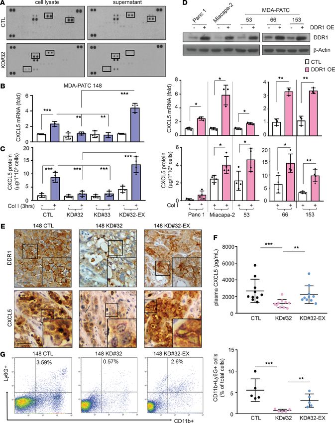
RESEARCH ARTICLE

Figure 1. DDR1 induces liver metastasis in pancreatic cancer. (A) DDR1 and DDR2 expression were analyzed by western blotting in 2 fibroblasts, 14 primary
PDAC cell lines, and 2 metastatic PDAC cell lines, in 3 independent experiments. (B) DDR1 was observed at PDX tumors derived from metastatic or primary
human PDAC tumors by IHC staining using anti–human DDR1 antibody and identified using PE Vectra3. Scale bar: 50 μm. The H-score of DDR1 quantification
was displayed as DBA signals by inForm software. n = 10, unpaired 2-tailed Student’s t test. **P < 0.01. (C) Cell invasion assay in MDA-PATC 148 cells with
knockdown or reexpression DDR1 were used by Matrigel transwell chamber. The invading cells in each chamber were counted under a fluorescence microscope
after cultured 18 hours, and the average number of cells was calculated based on the number of cells found in 6 fields per chamber. Data are mean ± SD. n = 5,
3 independent experiments; 1-way ANOVA with Sidak post hoc testing. *P < 0.05; ***P < 0.001. (D–F) Mice were orthotopically injected with MDA-PATC 148
(control, DDR1–deficient or DDR1-reexpression clones) cells for 9 weeks. (D) H&E staining of pancreas and liver section. Arrow: region of tumor. n = 12. Scale
bar: 50 μm. (E) Tumor size measurement in pancreas. Unpaired 2-tailed Student’s t test. (F) The numbers of liver-met. n = 12; Fisher’s exact test. *P < 0.05.

                                         DDR1 induces CXCL5 production and Ly6G+ neutrophil infiltration. To investigate the influence of cancer
                                    cell DDR1 signaling on the TME, we screened for cancer cell DDR1-induced cytokine production using
                                    a human chemokine antibody array. We found that the activation of DDR1 by collagen induced the pro-
                                    duction of 4 candidate factors, CD130, CXCL8, CXCL5, and MCP-1, which were reduced by knocking
                                    down DDR1 (Figure 2A). We then validated that CXCL5 was a DDR1-induced factor by quantitative PCR
                                    (qPCR) assays (Supplemental Figure 1B). The exposure of parental cancer cells to collagen I for 3 hours
                                    increased mRNA and protein levels of CXCL5 in MDA-PATC 148 and BxPC-3 cells, which was dimin-
                                    ished in knockdown DDR1 cells (Figure 2, B and C, and Supplemental Figure 1C). In addition, the mRNA
                                    and protein expression of CXCL5 was rescued after reexpression of DDR1 in MDA-PATC 148KD#32 and

JCI Insight 2021;6(16):e146133 https://doi.org/10.1172/jci.insight.146133                                                                                    3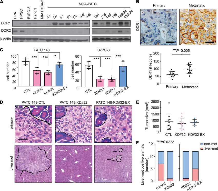
RESEARCH ARTICLE
BxPC-3KD#32 cells (Figure 2B and Supplemental Figure 1C). To confirm this was a DDR1-mediated effect,
we overexpressed DDR1 in an additional 5 human pancreatic cancer cell lines, which resulted in a signifi-
cant increase of CXCL5 expression upon collagen activation (Figure 2D).
CXCL5, also known as epithelial-derived neutrophil-activating peptide 78 (ENA-78), has been previ-
ously shown to induce TAN infiltration and increase metastatic risk in hepatocellular carcinoma (28). We
sought to investigate whether DDR1-derived CXCL5 is associated with TAN infiltration in PDAC. We
first measured the level of CXCL5 and Ly6G+ neutrophil infiltration in tumors derived from MDA-PATC
148 variants (MDA-PATC 148CTL, MDA-PATC 148KD#32, and MDA-PATC 148KD#32-exDDR1). Knocking down
DDR1 reduced the level of CXCL5 in the primary tumor and plasma (Figure 2, E and F). Importantly, we
also observed a reduction of CD11b+Ly6G+ neutrophil infiltration in DDR1-knockdown tumors (Figure
2G). Reexpression of DDR1 (MDA-PATC 148KD#32-ex-DDR1) rescued CXCL5 levels in the tumor and plas-
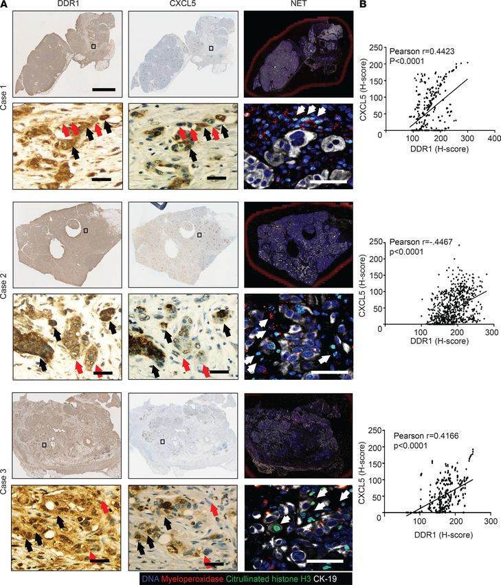
ma as well as CD11b+Ly6G+ TAN infiltration (Figure 2, E–G). To verify this association in a clinically rel-
evant setting, we performed IHC on a tissue microarray of pancreatic cancer PDXs. Scoring from 82 tumor
samples identified a positive correlation between DDR1 expression and CXCL5 production (Pearson’s r =
0.4460; 95% CI, 0.2535–0.6045) and Ly6G+ TAN infiltration (r = 0.2840; 95% CI, 0.07144–0.4720) (Figure
3, A–C). As expected, a positive correlation was also observed between CXCL5 and the infiltration of Ly6G+
TANs (r = 0.6403; 95% CI, 0.4916–0.7527) (Figure 3, A and D). To confirm the results were not an artifact
of immunodeficient mice necessary for PDX generation, we repeated the experiments in a syngeneic model.
KPwmC cells (congenic mouse PDAC cells derived from KrasLSL.G12D/+; p53R172H/+; and Pdx1CreTg/+ [KPwmC] mice)
with stable DDR1 knockdown were generated (KPwmCKD#588 and KPwmC KD#809) (Supplemental Figure 2A)
and orthotopically implanted into immunocompetent C57BL/6J mice. Similar to the xenograft models,
DDR1-deficient KPwmC orthotopic tumors consistently demonstrated decreased CD45+CD11b+Ly6G+ neu-
trophil infiltration and fewer liver metastases (WT, 80%; KD#588, 0%; and KD#809, 20%) as well as the level of
CXCL5 in plasma (Supplemental Figure 2, B–D). Together, these results suggest that DDR1 on cancer cells
drives CXCL5 production, CD45+CD11b+Ly6G+ TAN infiltration, and liver metastasis.
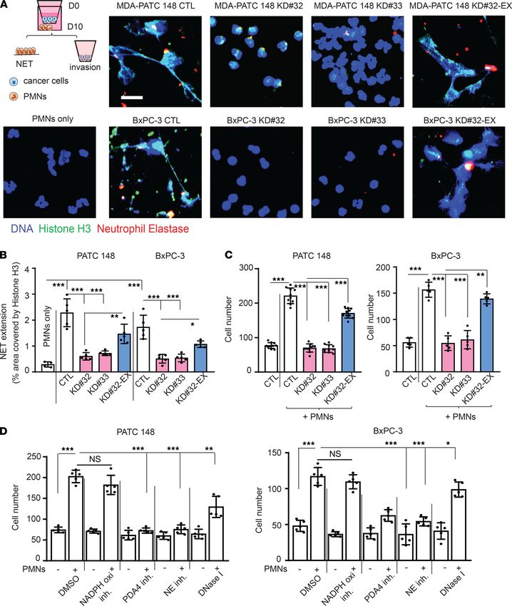
DDR1-induced NET formation enhances pancreatic cancer cell invasion. We then asked if DDR1-induced
TAN infiltration directly enhanced the metastatic capability of cancer cells. We first observed a higher
level Ly6G+ TANs within the TME of tumors derived from cell lines selected for liver metastasis, PATC
148LM and 153LM, compared with tumors derived from the parental lines (Figure 4A). In addition,
PDXs derived from metastatic PDAC tumors were found to have higher numbers of Ly6G+ TANs than
PDXs derived from primary tumors (Figure 4B).
Recent studies have demonstrated NET formation is a major driver of metastasis in breast cancer (21).
In PDAC, NETs have also been frequently observed in tumor tissues (18). Thus, we investigated wheth-
er NETs contributed to DDR1-induced cancer cell invasion. NET formation requires the generation of
reactive oxygen species and nicotinamide adenine dinucleotide phosphate (NADPH) oxidase activity as
well as the activation of peptidylarginine 4 (PAD4) that promotes the decondensation of nuclear DNA by
histone citrullination and the release of MPO/neutrophil elastase (NE) from azurphilic granules (29, 30).
These events can be triggered in vitro by exposing neutrophils to phorbol myristate acetate (PMA) (31). We
cocultured neutrophils harvested from the blood of patients with untreated PDAC, with MDA-PATC 148
or BxPC-3 cells for 18 hours using a Matrigel-coated transwell chamber and found increased NET forma-
tion and citrullinated histone H3 expression (Figure 5, A and B, and Supplemental Figure 3A). Histone
H3 citrullination and NET formation were significantly reduced when the experiments were performed
using PDAC cells lacking DDR1 (Figure 5, A and B, and Supplemental Figure 3A) and were rescued when
DDR1 expression was restored (Figure 5, A and B). When compared with cancer cells alone, the addition
of neutrophils resulted in an almost 3-fold increase in invasion of MDA-PATC 148 and BxPC-3 cells (Fig-
ure 5C). Knocking down DDR1 in the cancer cells significantly reduced neutrophil-mediated cancer cell
invasion (Figure 5C). In contrast, the reexpression of DDR1 recovered the effect of neutrophil-mediated
cancer cell invasion in MDA-PATC 148KD#32-exDDR1 and BxPC-3KD#32-exDDR1 cells (Figure 5C). Interestingly,
the inhibition of PDA4 and NE inhibited NET formation and cancer cell invasion in cocultured neutro-
phils with MDA-PATC 148 and BxPC-3 for 18 hours (Supplemental Figure 3, B and C, and Figure 5D).
However, NADPH oxidase inhibition had no effect on NETs or cancer cell invasion, and DNase I treat-
ment showed only a partial effect compared with the control (Supplemental Figure 3, B and C, and Figure
5D). Taken together, these data suggest that DDR1 on pancreatic cancer cells induces the formation of
NETs, which promote cancer cell invasion through an NAPDH oxidase-independent pathway.
JCI Insight 2021;6(16):e146133 https://doi.org/10.1172/jci.insight.146133 4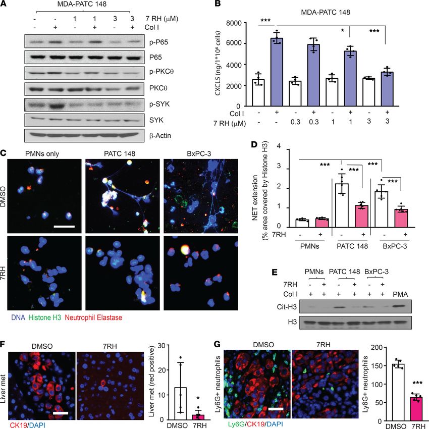
RESEARCH ARTICLE

JCI Insight 2021;6(16):e146133 https://doi.org/10.1172/jci.insight.146133                  5
RESEARCH ARTICLE
Figure 2. DDR1 induces CXCL5 production in pancreatic cancer cells. (A) Chemokine array analysis in cell lysate and supernatant of MDA-PATC 148 cells with
knockdown DDR1. (B and C) MDA-PATC 148 cells with knockdown or reexpressed DDR1 were treated with collagen I for 3 hours. (B) CXCL5 mRNA level by using real-
time PCR. (C) CXCL5 protein level by using ELISA. (D) CXCL5 expression in overexpressed DDR1 in 5 pancreatic cancer cell lines. Upper: DDR1 levels were checked by
western; middle: CXCL5 mRNA level were detected by real-time PCR; lower: CXCL5 protein levels were analyzed by ELISA. (E–G) Mice were orthotopically injected
with MDA-PATC 148 (control, DDR1-deficient or DDR1-reexpression clones) cells for 9 weeks. (E) IHC staining with anti-DDR1 (upper panel) and anti-CXCL5 (bottom
panel) antibodies in pancreas. (F) ELISA showed CXCL5 level in plasma harvest from mice. (G) FACS by using anti-CD11b and anti-Ly6G antibodies to determine the
presence of CD11b+Ly6G+ neutrophils infiltration in pancreas. (B–D) Data are mean ± SD. n = 3–4, 3 independent experiments; (B and C) 1-way ANOVA with Sidak
post hoc testing; (D) Unpaired 2-tailed Student’s t test. *P < 0.05; **P < 0.01. (F and G) n = 5–10 mice, data performed in triplicate; 1-way ANOVA with Sidak post
hoc testing. **P < 0.01; ***P < 0.001. Data show signal after membrane exposed to x ray film for 2 minutes.
Cancer cell DDR1-induced CXCL5 mediates NET formation and NET-induced cancer cell invasion. To
examine the function of CXCL5 in DDR1-mediated NET formation and cancer cell invasion, the inva-
sion assay was repeated in the presence of a CXCL5-neutralizing monoclonal antibody or recombinant
CXCL5. The CXCL5-neutralizing antibody significantly reduced MDA-PATC 148 and BxPC-3 cell–
induced histone H3 citrullination, NET formation, and cancer cell invasion (Figure 6, A, B, D, and
E). Conversely, recombinant human CXCL5 induced histone H3 citrullination, NET formation, and
cancer cell invasion in MDA-PATC 148KD #32 cells (Figure 6, A, C, D, and F). The inhibition of PDA4
and NE blocked recombinant CXCL5-mediated, NET-induced MDA-PATC 148KD#32 cell invasion, but
NADPH inhibition had no effect (Supplemental Figure 4). To evaluate whether DDR1-induced NET
formation and NET-induced cell invasion were mediated by the secretion of soluble factors, we col-
lected conditioned media from 3 in vitro cell culture conditions: cancer cells alone (CCM), neutrophils
alone (NCM), or neutrophils exposed to cancer cell-conditioned media (NCCM). NET formation and
citrullinated histone H3 was induced by CCM from DDR1-expressing cancer cells exposed to collagen
I but not from CCM harvested from DDR1-knockdown cancer cells after collagen I stimulation (Figure
7, A–C). We then repeated the cancer cell invasion assay experiment using NCCM in the presence or
absence of collagen I. NCCM from MDA-PATC 148CTL/collagen I/neutrophil cultures induced cancer
cell invasion in a DDR1-independent manner (Figure 7D). However, NCCM harvested from MDA-
PATC 148KD#32/collagen I/neutrophil cultures failed to increase cancer cell invasion again, regardless
of the DDR1 status (Figure 7E). These carefully designed studies demonstrate that CXCL5 is a solu-
ble factor released into the media from DDR1-positive cancer cells in the presence of collagen; when
exposed to CXCL5, neutrophils generate NETs that promote cancer cell invasion. The dependence
upon DDR1 is demonstrated by the fact that CCM from DDR1-expressing cancer cell/collagen I cul-
tures stimulates NE activity in neutrophils, but NE activity is significantly reduced after exposure to
CCM from DDR1-knockdown clones (Figure 7F). In addition, heat treatment of NCCM harvested
from DDR1 intact cancer cell/collagen I/neutrophils results in a reduction of cancer cell invasion (Fig-
ure 7G), further suggesting that a secreted protein is responsible for the observed effect.
To examine the DDR1/CXCL5 axis and NET formation in human PDAC, we performed IHC and
immunofluorescence staining on primary human PDAC samples. We found that the areas of high DDR1
expression were associated with high intratumoral levels of CXCL5 (black arrows) (Figure 8). Conversely,
within the same tumor we observed that regions of low DDR1 expression were associated with decreased
CXCL5 expression (red arrow). In addition, we found that the NET-like structures were located around
tumor cells (green and red color). Statistically, Pearson’s r correlation analysis indicated a positive associa-
tion between DDR1 and CXCL5 level in 3 cases of pancreatic patient tumor specimens (case 1: r = 0.4423;
case 2: r = 0.4467; and case 3: r = 0.4166).
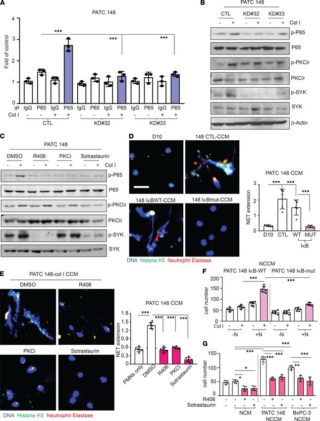
Cancer cells induce CXCL5 and cause NET formation through the DDR1/PKCθ/NF-κB signaling pathway. It has
been reported that CXCL5 is a downstream product of the NF-κB pathway (32) and that the NF-κB pathway
can be activated by DDR1 (33). Therefore, we sought to determine whether the NF-κB pathway is involved in
collagen-induced CXCL5 production. ChIP-qPCR assay demonstrated that the exposure of MDA-PATC 148
cells to collagen I induced binding of the NF-κB P65 subunit to the CXCL5 promoter, and this binding was
reduced significantly after knockdown of DDR1 (Figure 9A). In addition, the exposure of MDA-PATC 148
and BxPC-3 cells to collagen I for 3 hours induced NF-κB P65 translocation into the nucleus (Supplemental
Figure 5A). Consistent with this observation, the activation of NF-κB P65 was prevented by genetic DDR1
knockdown in MDA-PATC 148 and BxPC-3 cells (Supplemental Figure 5A). Furthermore, a phospho-NF-
κB pathway array performed after collagen I stimulation of MDA-PATC 148KD#32 cells demonstrated that the
phosphorylation levels of the top 5 proteins had the greatest decrease (20%–25%) in phosphorylation when
JCI Insight 2021;6(16):e146133 https://doi.org/10.1172/jci.insight.146133 6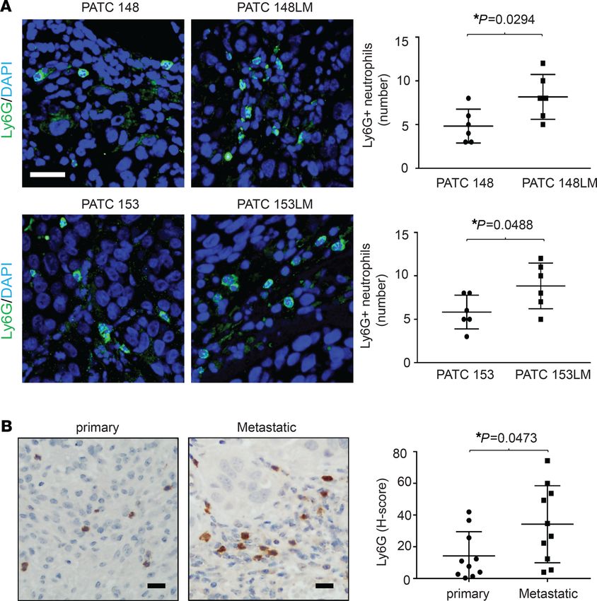
RESEARCH ARTICLE

                                  Figure 3. The correlation of DDR1, CXCL5, and neutrophils infiltration at tissue microarray (TMA) in PDX tumors. (A)
                                  IHC staining showed DDR1, CXCL5, and Ly6G+ neutrophils infiltration at PDX tumors and identified using PE Vectra3.
                                  Scale bar: 50 μm. (B–D) Pearson’s correlation showed relationship of DDR1, CXCL5, and Ly6G by using H-score, which
                                  quantified the DBA signals by inForm software. n = 82.

                                  compared with parental cells: NF-κB P100/52, SYK, NF-κB P65, ZAP-70, and PKCθ (Supplemental Figure
                                  5B). Recent studies have shown that SYK is highly expressed in multiple cancer cell types and that SYK kinase
                                  activity induces cancer cell migration and metastasis (34, 35). Thus, we tested whether DDR1 upregulated
                                  CXCL5 through NF-κB, SYK, and PKCθ and found that knockdown of DDR1 decreased collagen I-stimu-
                                  lated NF-κB, SYK, and PKCθ phosphorylation (Figure 9B). Next, we generated an IκB super-repressor mutant
                                  clone to block the activation of NF-κB in MDA-PATC 148 (MDA-PATC 148IκB-MUT) (Supplemental Figure 5C)
                                  and found that this significantly inhibited collagen I-induced CXCL5 production (Supplemental Figure 5D).
                                  Additionally, pretreatment with SYK and PKC inhibitors prevented collagen I-induced CXCL5 production
                                  and inhibited NF-κB P65 activation in MDA-PATC 148 and BxPC-3 cells (Supplemental Figure 5E and Figure
                                  9C). In addition, PKC inhibitors blocked the effect of collagen I-induced SYK phosphorylation; in contrast,
                                  the SYK inhibitor had no effect on collagen I-induced PKCθ activation (Figure 9C). These results suggest that
                                  collagen I-induced CXCL5 production is mediated by a DDR1/PKCθ/SYK/NF-κB signaling cascade.

JCI Insight 2021;6(16):e146133 https://doi.org/10.1172/jci.insight.146133                                                                                7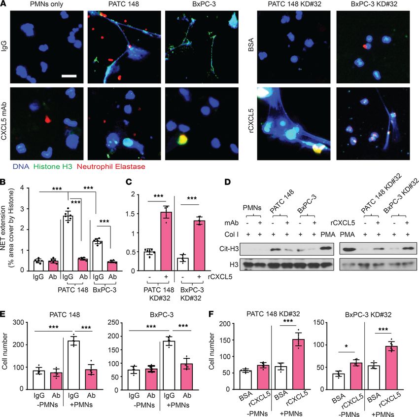
RESEARCH ARTICLE

                                  Figure 4. Metastatic tumors recruit more Ly6G+ neutrophils infiltration than primary tumors. (A) Ly6G+ neutrophils were
                                  observed at tumors derived from primary and match liver-met cell lines by immunofluorescence staining using anti-Ly6G
                                  (green) and DAPI (blue) with a fluorescence microscope. Scale bar: 50 μm. The number of neutrophils were counted in x20
                                  field, 6 fields per slice. Data are mean ± SD. n = 5 mice, unpaired 2-tailed Student’s t test. *P < 0.05. (B) Ly6G+ neutrophils
                                  were observed at PDX tumors derived from metastatic or primary human PDAC tumors by IHC staining using anti-Ly6G
                                  antibody and identified using PE Vectra3. Scale bar: 50 μm. The H-score of Ly6G quantification was displayed as DBA signals
                                  in x20 field, 6 fields per slice by inForm software. Data are mean ± SD. n = 10, unpaired 2-tailed Student’s t test. *P < 0.05.

                                       To confirm the contribution of the PKCθ/SYK/NF-κB axis to DDR1-mediated NET formation, we har-
                                  vested CCM from MDA-PATC 148IκB-WT, MDA-PATC 148IκB-MUT cells, and MDA-PATC 148 cells pretreated
                                  with SYK or PKC inhibitors after exposure to collagen I for 3 hours and incubated neutrophils with the CCM
                                  for 18 hours. CCM from cancer cells incapable of NF-κB activation was associated with decreased NET forma-
                                  tion after collagen I stimulation (Figure 9D). In addition, pretreatment with SYK and PKC inhibitors protected
                                  against MDA-PATC 148 CCM–induced NET formation (Figure 9E). Finally, we tested whether DDR1-me-
                                  diated NET-induced cancer cell invasion was through the PKCθ/SYK/NF-κB axis. NCCM harvested from
                                  MDA-PATC 148IκB-WT/collagen I/neutrophil cultures significantly induced cancer cell invasion while NCCM
                                  from MDA-PATC 148IκB-MUT/collagen I/neutrophil cultures was associated with diminished cancer cell invasion
                                  (Figure 9F). NCCM from MDA-PATC 148 and BxPC-3 cells pretreated with SYK or PKC inhibitors/collagen
                                  I/neutrophil cultures also reduced cancer cell invasion (Figure 9G). Together, these results provide evidence that
                                  DDR1 stimulates CXCL5 production through a PKCθ/SYK/NF-κB signaling cascade and that CXCL5 from
                                  cancer cells induces neutrophils to form NETs and thereby enhances metastasis.
                                       Targeting DDR1 reduces neutrophil-mediated cancer cell invasion. Our previous study demonstrated that
                                  the specific DDR1 inhibitor 7rh benzamide can improve the efficacy of standard-of-care chemotherapy
                                  in PDAC (12). In the current study, we wanted to determine whether inhibition of DDR1 signaling by

JCI Insight 2021;6(16):e146133 https://doi.org/10.1172/jci.insight.146133                                                                                       8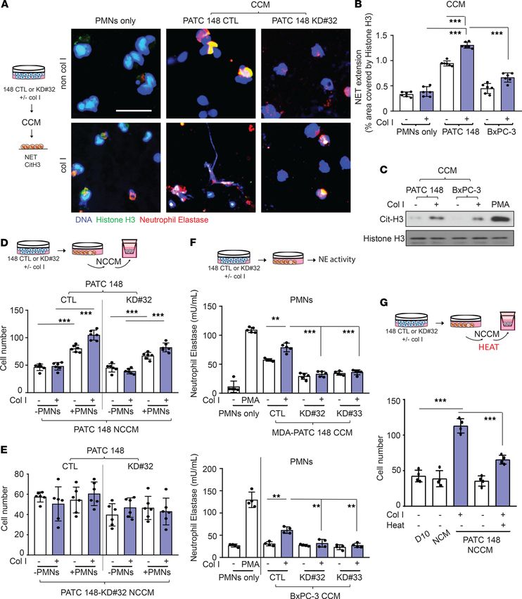
RESEARCH ARTICLE

Figure 5. DDR1-positive pancreatic cancer cells mediated NET formation from neutrophils and enhanced cancer cell invasion. (A–D) Human neu-
trophils were cocultured with DDR1 knockdown or reexpression of MDA-PATC 148 or BxPC-3 by Matrigel transwell chamber, with or without NADPH
oxidase inhibitor, PDA4 inhibitor, NE inhibitor, and Dase I treatment for 18 hours. (A) NET structures were analyzed by immunofluorescence staining
using DAPI (blue), anti-NE (red), and anti-histone H3 (green) mAbs. Scale bar: 50 μm. (B) The NET quantification is displayed as NET histone area
(μm2) per x40 field, 6 fields per group. (C and D) The number of invaded cells analyzed by immunofluorescence staining using DAPI and calculated
based on the number of cells found in 6 fields per chamber. All the data are mean ± SD. n = 5, 3 independent experiments; 1-way ANOVA with Sidak
post hoc testing. *P < 0.05; **P < 0.01; ***P < 0.001.

JCI Insight 2021;6(16):e146133 https://doi.org/10.1172/jci.insight.146133                                                                        9
RESEARCH ARTICLE

Figure 6. CXCL5 involved in DDR1-mediated NET formation and cancer cell invasion. (A–F) Human neutrophils were cocultured with DDR1 knockdown
or reexpression of MDA-PATC 148 or BxPC-3 by Matrigel transwell chamber, with or without anti-CXCL5 neutralized antibody or recombinant CXCL5 treat-
ment, for 18 hours. (A) NET structures were analyzed by immunofluorescence staining using DAPI (blue), anti-NE (red), and anti–histone H3 (green) mAbs.
Scale bar: 50 μm. (B and C) The NET quantification is displayed as NET histone area (μm2) per field, 6 fields per group. (D) Cit-histone H3 expression were
analyzed by western blotting. (E and F) The number of invaded cells were analyzed by immunofluorescence staining using DAPI and calculated based on
the number of cells found in 6 fields per chamber. All the data are mean ± SD. n = 5, 3 independent experiments. (B and E) P values were analyzed by 1-way
ANOVA with Sidak post hoc testing. ***P < 0.001. (C and F) P values were analyzed by unpaired 2-tailed Student’s t test. *P < 0.05; ***P < 0.001.

                                   7rh benzamide would result in a reduction of CXLC5 and subsequent NET formation. We found that
                                   pretreatment with 3 μM of 7rh benzamide in MDA-PATC 148 and BxPC-3 cells significantly reduced
                                   NF-κB, PKCθ, and SYK phosphorylation as well as the production of CXCL5 (Figure 10, A and B, and
                                   Supplemental Figure 6A). As predicted, NET formation and cell invasion were significantly decreased in
                                   7rh benzamide–treated cells (Figure 10, C–E, and Supplemental Figure 6B).
                                       To confirm the effect of 7rh benzamide in vivo, we orthotopically implanted MDA-PATC 148 cells
                                   into nude mice followed by i.p. treatment with 3 mg/kg/day 7rh benzamide for 9 weeks. 7rh benzamide

JCI Insight 2021;6(16):e146133 https://doi.org/10.1172/jci.insight.146133                                                                               10
RESEARCH ARTICLE Figure 7. DDR1-positive pancreatic cancer cells mediated NET formation from neutrophils through a soluble factor secretion and enhanced cancer cell invasion. (A–G) Human neutrophils were cultured with CCM from cancer cells for 18 hours. (A) NET structures were analyzed by immunofluorescence staining using DAPI (blue), anti-NE (red), and anti–histone H3 (green) mAbs. Scale bar: 50 μm. (B) The NET quantification is displayed as NET histone area (μm2) per field, 6 fields per group. (C) Cit-histone H3 expression were analyzed by western blotting. (D and E) The number of invaded cells analyzed by immunofluorescence staining using DAPI and calculated based on the number of cells found in 6 fields per chamber. (F) Neutrophils Elastase activity were showed in human neutrophils with CCM treatment for 18 hours. (G) The number of invaded cells analyzed by immunofluorescence staining using DAPI and calculated based on the number of cells found in 6 fields per chamber. All the data are mean ± SD. n = 4–5, 3 independent experiments; 1-way ANOVA with Sidak post hoc testing. **P < 0.01; ***P < 0.001. JCI Insight 2021;6(16):e146133 https://doi.org/10.1172/jci.insight.146133 11
RESEARCH ARTICLE

Figure 8. The correlation of DDR1, CXCL5, and NET-like structure in samples of patient with PADC. (A) Upper and middle panel: IHC staining showed
DDR1, CXCL5 expression in PDAC patient samples, and identified using PE Vectra3. Scale bar: 3 mm and 50 μm. Bottom panel: NET-like structures were
analyzed by immunofluorescence staining using DAPI (blue), anti-CK19 (white), anti-MPO (green), and anti–citrullinated histone H3 (red) mAbs in samples
of patient with PDAC. Scale bar: 20 μm. (B) Pearson’s correlation showed relationship of DDR1 and CXCL5 by using H-score, which quantified the DBA
signals by inForm software, P < 0.0001.

JCI Insight 2021;6(16):e146133 https://doi.org/10.1172/jci.insight.146133                                                                           12
RESEARCH ARTICLE

JCI Insight 2021;6(16):e146133 https://doi.org/10.1172/jci.insight.146133                 13
RESEARCH ARTICLE

Figure 9. PKCθ/SYK/NF-κB pathway involved in DDR1-induced CXCL5 production, NET formation from neutrophils, and enhanced cancer cell invasion.
(A) qPCR results were used to quantify enrichment of NF-κB P65 at the CXCL5 promoter using ChIP assay in MDA-PATC 148 cells with DDR1 knockdown.
Data are mean ± SD. n = 3, 3 independent experiments; 1-way ANOVA with Sidak post hoc testing. ***P < 0.001. (B) Phospho-NF-κB P65, phospho-PKCθ,
and phospho-SYK were analyzed by western blotting in MDA-PATC 148 cells with DDR1 knockdown. (C) Phospho-NF-κB P65, phospho-PKCθ, and phos-
pho-SYK were analyzed by western blotting in MDA-PATC 148 cells with or without SYK inhibitor and PKC inhibitor pretreatment. (D and E) NET structures
were analyzed by immunofluorescence staining using DAPI (blue), anti-NE (red), and anti–histone H3 (green) mAbs. (D) In MDA-PATC 148 cells with CCM
from MDA-PATC 148 with IκB super-repressor mutation/collagen I, treatment for 18 hours. (E) In MDA-PATC 148 cells with MDA-PATC 148, with or without
SYK inhibitor and PKC inhibitor pretreatment/collagen I, treatment for 18 hours. Scale bar: 50 μm. The NET quantification is displayed as NET histone area
(μm2) per field, 6 fields per group. (F and G) The number of invaded cells were analyzed by immunofluorescence staining using DAPI and calculated based
on the number of cells found in 6 fields per chamber. (F) In MDA-PATC 148 cells with NCCM from MDA-PATC 148 cells with IκB super-repressor mutation/
neutrophils/collagen I, treatment for 18 hours. (G) In MDA-PATC 148 cells with NCCM from MDA-PATC 148/collagen I/SYK or PKC inhibitor, treatment for 18
hours. (D–G) Data are mean ± SD. n = 5–6, 3 independent experiments; 1-way ANOVA with Sidak post hoc testing. *P < 0.05;**P < 0.01; ***P < 0.001.

                                   significantly reduced liver metastasis events (Figure 10F) as well as Ly6G+ neutrophil infiltration within the
                                   primary tumor (Figure 10G).

                                   Discussion
                                   Fibrillar collagen is abundant within the microenvironment of primary tumors and has been identified as phys-
                                   ically adjacent to cancer cells expressing high levels of DDR1 (36). Here we report a potentially novel mech-
                                   anism by which a collagen receptor, DDR1, on PDAC cells interacts with type I collagen to attract tumor-as-
                                   sociated neutrophils, induce NET formation, and facilitate cancer cell invasion and metastasis. In this study,
                                   we mechanistically link DDR1 expression in human PDAC with CXCL5 levels, Ly6G+ neutrophil infiltration,
                                   NET-like structures, and metastatic events. Importantly, in animal models of PDAC, tumors derived from
                                   cancer cells lacking DDR1 had fewer NET-like structures and the animals experienced fewer liver metastases,
                                   suggesting that cancer cell-derived DDR1 contributes significantly to NET-induced tumor metastasis.
                                        NETs have been reported to link neutrophils and metastasis (21). Not only neutrophils but also other
                                   types of leukocytes, including macrophages, mast cells, and eosinophils, form extracellular traps (a process
                                   called ETosis) (37), but NET formation is the most prominent ETosis in cancer. Cancer cell-mediated cyto-
                                   kines and chemokines can prime neutrophils for NET formation by inducing NADPH oxidase activation to
                                   support tumor metastasis. NADPH oxidase and PDA4 inhibitors have been reported to significantly decrease
                                   tumor cell invasion, suggesting that NET-mediated tumor cell invasion requires neutrophil NADPH oxidase
                                   and PDA4 activity (21). Similar to Park and colleagues, we also found that PDA4 and NE activity are import-
                                   ant for pancreatic cancer cell–induced NET formation and NET-mediated cancer cell invasion; however, we
                                   found that NADPH inhibition had no effect on NET-mediated tumor cell invasion, suggesting that PDAC
                                   cells induce NET formation and NET-mediated PATC cell invasion through an NADPH oxidase–indepen-
                                   dent pathway (Supplemental Figure 3, B and C, and Figure 5D). It has been shown that CXCR2-related
                                   chemokines induce NET formation through the NADPH oxidase–independent pathway (38). This is con-
                                   sistent with our findings that CXCL5, a CXCR2 ligand, is expressed by tumor cells in a DDR1-dependent
                                   manner and drives neutrophil activation and NET formation (Figure 2; Figure 6, A–D; and Supplemental
                                   Figure 1). Recent studies have suggested that NET-associated DNA meshes can catch circulating tumor cells
                                   and enhance cell metastasis (19, 39). In addition, NET-associated proteases, NE and matrix metalloprotease
                                   9 (MMP9), awaken dormant cancer cells and facilitate cancer cell metastasis (20, 21). In our studies, we
                                   observed a decrease in NE activity through DDR1 knockdown, NE inhibitors, and heat treatment; all of these
                                   conditions prevented NET-mediated cancer cell invasion, highlighting the contribution of NE during PDAC
                                   cell invasion (Figure 5D; Figure 7, F and G; and Supplemental Figure 4). Taken together, these data support
                                   that collagen I–DDR1 interaction induces CXCL5 production by cancer cells, which promotes neutrophil
                                   infiltration and NET formation to drive NET-associated cancer cell migration.
                                        In patients, elevated serum CXCL5 levels have been statistically associated with liver metastasis and poor
                                   survival (40, 41). CXCL5 recruits neutrophils into the TME (28), and the CXCL5/CXCR2 axis contributes to
                                   tumor growth and metastasis through the activation of PI3K/AKT/GSK-3β/Snail signaling to promote EMT
                                   (42). However, little is known about the mechanism of CXCL5 production. Our studies demonstrate that the
                                   activation of DDR1 signaling by collagen I-induced CXCL5 mRNA and protein expression in PDAC cells in
                                   vitro and in vivo. The STAT3 and NF-B pathways are involved in DDR1 downstream signaling and have been
                                   reported to induce CXCL5 production (32, 43, 44). However, in our system, the inhibition of STAT3 by cucur-
                                   bitacin I had no effect on collagen I-induced CXCL5 production (Supplemental Figure 7). For this reason, we

JCI Insight 2021;6(16):e146133 https://doi.org/10.1172/jci.insight.146133                                                                              14
RESEARCH ARTICLE Figure 10. 7rh treatment reduced NET formation through inhibition of the DDR1/PKCθ/SYK/CXCL5 axis and reduced cancer metastasis. (A and B) MDA-PATC 148 cells were pretreated with 7rh for 30 minutes and then with collagen I for 3 hours. (A) Phospho-NF-κB P65, phospho-PKCθ, and phospho-SYK were analyzed by western blotting. (B) CXCL5 levels were analyzed by ELISA. Data are mean ± SD. n = 4, 3 independent experiments; 1-way ANOVA with Sidak post hoc testing. *P < 0.05; ***P < 0.001. (C–E) Human neutrophils were cocultured with MDA-PATC 148 and BxPC-3 cells by Matrigel transwell chamber for 18 hours. (C) NET structures were analyzed by immunofluorescence staining using DAPI (blue), anti-NE (red), and anti–histone H3 (green) mAbs. Scale bar: 50 μm. (D) The NET quantification is displayed as NET histone area (μm2) per field, 6 fields per group. Data are mean ± SD. n = 6, 3 independent experiments; 1-way ANOVA with Sidak post hoc testing. ***P < 0.001. (E) Cit-histone H3 expression were analyzed by western blotting. (F and G) Mice were orthotopically injected with MDA-PATC 148 cells, with or without 3 mg/kg 7rh treatment for 9 weeks. (F) Liver metastasis was detected by immunofluorescence staining using DAPI (blue) and anti-CK19 (red) mAbs in liver section. Scale bar: 50 μm. The metastasis quantification is displayed as CK-19 positive signals/per x20 field, 6 fields per group. (G) Neutrophils infiltration was detected by immunofluorescence staining using DAPI (blue), anti-CK19 (red), and anti-Ly6G (green) mAbs in pancreas section. Scale bar: 50 μm. The Neutro- phils infiltration quantification is displayed as Ly6G positive signals per x20 field, 6 fields per group. Data are mean ± SD. n = 5, unpaired 2-tailed Student’s t test. ***P < 0.001. JCI Insight 2021;6(16):e146133 https://doi.org/10.1172/jci.insight.146133 15
RESEARCH ARTICLE

                                  focused on the NF-κB pathway and identified a putative signaling cascade from DDR1 to CXCL5 expression
                                  via the PKCθ/SYK/NF-κB axis (Figure 9 and Supplemental Figure 6). This is consistent with descriptions of
                                  how NF-κB is associated with the hallmarks of cancer, including cancer cell proliferation, protection against
                                  apoptosis, and metastasis (45, 46). In addition to NF-κB, PKCθ also promotes cancer metastasis by upregulating
                                  EMT and MMP-1 (47, 48), characteristics that are also consistent with our observations.
                                       Constitutive KRAS and NF-κB activation are signature alterations in PDAC (49). KrasG12D-induced
                                  cytokines and CXCR2-related chemokines have been reported to facilitate myeloid cell infiltration and
                                  tumor progression (50–52). In addition, we previously showed that KrasG12D-activated IL-1α/NF-κB /IL-1α
                                  and p62 feedforward loops are necessary for the induction and maintenance of NF-κB activity (53). Here
                                  we found that BxPC-3 cells, which are Kras WT, could also produce CXCL5 to drive NET formation.
                                  BxPC-3 cells have been reported to bind with high affinity to ECM proteins, including collagen I, and pos-
                                  sess invasive capabilities consistent with our observations (54). Nevertheless, it is possible that Kras-medi-
                                  ated NF-κB could drive CXCL5 production, neutrophil infiltration, and NET formation. MDA-PATC 148
                                  cells, which are Kras-mutant, generated higher levels of CXCL5 with associated NET formation and inva-
                                  sion when compared with BxPC-3 cells (Figure 2C; Supplemental Figure 1C; and Supplemental Figure 5,
                                  A–C). Although KrasG12D may intrinsically drive NF-κB–CXCL5–mediated NET, our work has identified a
                                  potentially novel mechanism by which an abundant stromal molecule, collagen I, can interact with cancer
                                  cells through a targetable receptor (DDR1) to induce CXCL5 production, NET formation, and NET-medi-
                                  ated cancer cell invasion, even in the absence of a Kras mutation.
                                       Therapeutic strategies must also account for stromal cells in the TME, which are important in the pro-
                                  motion of metastasis (55). In the current study, we focus on DDR1 and 2 stromal components: collagen I
                                  and tumor-associated neutrophils. DDR1 amplification is commonly observed in various cancers (56) and is
                                  significantly deregulated in aggressive cancers (57). We observed higher levels of DDR1 in metastatic cell lines
                                  compared with matched primary cell lines (Figure 1, A and B). Emerging evidence suggests that DDR1 is cru-
                                  cial for the progression and metastasis of various solid tumors; as such, targeting DDR1 represents a promising
                                  therapeutic approach (58–60). This is consistent with our repeated observations of a reduction of cell invasion
                                  and metastasis after pharmacologic or genetic inhibition of DDR1 signaling (Figure 1, C–F; Figure 5C; Figure
                                  10, F and G; Supplemental Figure 2, C and D; and Supplemental Figure 6B). The role of neutrophils in solid
                                  tumors remains, however, poorly defined. Although TANs support cancer cell progression and invasion, they
                                  can also kill cancer cells and bacteria within the TME and are necessary to protect the host from infection (61).
                                  For example, the American Society of Clinical Oncology recommends that patients who are undergoing cer-
                                  tain chemotherapy regimens receive prophylactic treatment with granulocyte colony-stimulating factor, which
                                  stimulates neutrophil production to counteract neutropenia (62). It is possible, however, that inducing neutro-
                                  phil production may increase the risk of metastatic spread. Recent work suggests that NET production might
                                  awaken clinically dormant cancer cells after chemotherapy and drive them to become metastatic (20). Our
                                  previous study suggested that a small-molecule inhibitor of DDR1 signaling, 7rh benzamide, would improve
                                  the efficacy of standard-of-care chemotherapy in patients with PDAC (12). In this study we found that 7rh sig-
                                  nificantly reduced cancer cell–induced NET formation in support of our prior observations. Taken together, we
                                  suggest that 7rh, when given in therapeutic doses, could increase the sensitivity of cancer cells to conventional
                                  chemotherapy and inhibit liver metastasis by blocking NET formation. DDR1 is critical in this process and
                                  represents a potential valuable pharmacological target in the treatment of patients with PDAC.

                                  Methods
                                  Cell lines. Pancreatic cancer cell lines — MDA-PATC 43, 50, 53, 66, 69, 102, 108, 124, 148, 153, and 216
                                  — were generated from PDX tumors in our laboratory at various times in the period 2010–2016 (27).
                                  The MDA-PATC 148LM and MDA-PATC 153LM cell lines were generated from liver metastases of
                                  MDA-PATC 148 and 153 cells orthotopically implanted into nude mice, respectively. PANC-1 and BxPC-3
                                  cell lines were obtained from the American Type Culture Collection. WM8865 mouse cells were generat-
                                  ed from KrasLSL.G12D/+; p53R172H/+; and Pdx1CreTg/+ (KPwmC) mice in our laboratory. All cells were cultured in
                                  DMEM that contained 10% FBS, penicillin (100 units/mL), and streptomycin (100 μg/mL; D10 medi-
                                  um) and had been tested monthly for mycoplasma and found negative.
                                       Animal studies. All animal experiments were conducted according to the National Institutes of Health’s
                                  Guide for the Care and Use of Laboratory Animals. The study protocol was approved by the University of
                                  Texas MD Anderson Cancer Center’s institutional animal care and use committee.

JCI Insight 2021;6(16):e146133 https://doi.org/10.1172/jci.insight.146133                                                                       16
RESEARCH ARTICLE
We randomLy assigned 8-week-old nude mice and C57BL/6J mice from the Jackson Laboratory to
different groups: nude mice with MDA-PATC 148CTL and MDA-PATC 148KD#32; and C57BL/6J mice with
KPwmCCTL, KPwmCKD#588, and KPwmCKD#809. We resuspended 1 × 10⁵ cells in 25 μL 1X PBS and added 1
volume of Matrigel (Corning, CB356253). The suspension was then directly injected into the pancreas of
mice. Nine weeks after cancer cell injection, all mice were euthanized, and we collected their plasma and
pancreatic, liver, and spleen tissues.
PDX tumors and tissue microarrays. We followed our previously published protocol for the heterotopic
engraftment of pancreatic patient tumors into immunodeficient mice (63). After harvesting the tissues from
the euthanized mice, we embedded them in paraffin and used core samples to construct a tissue microarray.
Isolation of human neutrophils. To isolate human neutrophils, we obtained peripheral venous blood samples
(10 mL) that were collected from untreated patients with pancreatic cancer and stored at MD Anderson Cancer
Center (IRB: PA11-0670). Isolation of the neutrophils was modified as described previously (64). After the plas-
ma and platelets were removed from the blood, the granulocytes were isolated using a Ficoll-Paque PLUS (GE
Healthcare, 17-1440-03) with density-gradient centrifugation, and the erythrocytes were removed by 2 rounds of
hypotonic lysis with a red blood cell lysis buffer. The viability and purity of the neutrophils were determined by
anti–human CD66b PE (clone G10F5; BD Biosciences, 561650) and anti–human CD16 FITC (clone 3G8; BD
Biosciences, 560996) double staining using FACS (Supplemental Figure 8).
In vitro cell invasion assay. Our cell invasion assay was modified as described previously (21). We suspend-
ed 1 × 10⁵ neutrophils in 100 μL of D10 medium and seeded the cells on poly-L-lysine–coated coverslips
in a 24-well culture plate for 30 minutes. We then removed the medium and nonadhered neutrophils and
added 700 μL of D10 medium with or without IgG, anti-CXCL5 antibody (Abcam, ab9802), BSA, human
recombinant CXCL5 (Abcam, ab9803), DNase I (1.5U), apocynin (10 μM; Abcam, ab120615), the PAD4
inhibitor, Cl-amidine (200 μM; Cayman Chemical, 1043444-18-3), and sivelestat (10 μM; ApexBio, B6189).
We suspended 2 × 10⁵ cancer cells in 500 μL of serum-free DMEM medium (D0 medium) with or without
7rh, IgG, anti-CXCL5 antibody, BSA, human recombinant CXCL5, DNase I, NADPH oxidase inhibitor,
apocynin, PAD4 inhibitor, Cl-amidine, and the neutrophil elastase inhibitor sivelestat and then added the cells
to a rehydrated Matrigel chamber (Corning, 354480). After the cells had been cultured for 18 hours at 37°C,
the invading cancer cells were fixed with 100% cold methanol and stained with 0.05 mg/mL DAPI (BD Bio-
sciences, 564907). The invading cells in each chamber were counted under a fluorescence microscope, and the
average number of cells was calculated based on the number of cells found in 6 fields per chamber.
To generate CCM, we suspended 2 × 10⁶ cancer cells cultured in 2 mL of D0 medium with or without the
SYK inhibitor R406 (10 μM; BioVision, 9682); the PKC inhibitor (30 μM; Santa Cruz Biotechnology, sc-3007);
another PKC inhibitor, sotrastaurin (2 nM; Selleck Chemicals, S2791); and different dosages of the DDR1
kinase inhibitor, 7rh, for 30 minutes at 37°C. We then seeded the cells on a collagen I-coated 6-well culture plate
for 3 hours. Next, we removed and stored the medium at –80°C. NCM was prepared from 1 × 10⁶ neutrophils
cultured in 1 mL of D10 medium with or without phorbol 12-myristate 13-acetate (PMA; Sigma-Aldrich, now
MilliporeSigma, P8139) for 4 hours; human recombinant CXCL5 for 18 hours; then we collected and stored
the medium at –80°C. Neutrophils exposed to NCCM were prepared from 1 × 10⁶ neutrophils cultured in
CCM for 18 hours; then we collected the medium with or without heat denaturing (95°C) for 5 minutes, and
stored it at –80°C. The NCM or NCCM was added to a 24-well culture plate, and the cancer cells were seeded
in D0 medium in a rehydrated Matrigel chamber. After the cells had been cultured for 18 hours at 37°C, the
invading cancer cells were fixed with 100% cold methanol and stained with 0.05 mg/mL DAPI. The invading
cells in each chamber were counted under a fluorescence microscope, and the average number of cells was
calculated based on the number of cells found in 6 fields per chamber.
Detection of NET structure by immunofluorescence staining. Neutrophils grown on coverslips were fixed,
permeabilized with 4% paraformaldehyde and 0.5% Triton X-100 in PBS, and blocked in PBS containing
2.5% FBS and 2.5% BSA for 1 hour at room temperature. The neutrophils were then incubated overnight at
4°C with primary antibodies: anti–histone H3 (1:50 dilution; Cell Signaling Technology, 3680) and either
anti-NE (1:100 dilution; Abcam, ab68672) or anti-myeloperoxidase (1:150 dilution; R&D Systems). After
being washed with PBS, secondary antibodies conjugated with Alexa Fluor 488 (Thermo Fisher Scientific,
A-11094) or TRITC (Thermo Fisher Scientific) were applied to detect primary Ab (1:250 dilution), and
DAPI was used as a counterstain. NET images, defined as areas of colocalized DNA, myeloperoxidase,
and citrullinated histone H3, were observed in five fields of each coverslip using a fluorescence microscope,
and histone area was used to quantify NET extension using ImageJ software.
JCI Insight 2021;6(16):e146133 https://doi.org/10.1172/jci.insight.146133 17RESEARCH ARTICLE
For tissue histology, paraffin-embedded sections of tumors were deparaffinized and rehydrated; antigen
retrieval was in citrate buffer (10 mM sodium citrate, 0.05% Tween 20, pH 6.0; MilliporeSigma), and the
sections were blocked in PBS containing 3% BSA for 1 hour. The tissue sections were then incubated over-
night at 4°C with antibodies against myeloperoxidase (1:150 dilution; R&D Systems, AF3667), citrullinated
histone H3 antibody (1:100 dilution; Abcam, ab5103), and CK-19 (1:500; Abcam). The tissue sections were
then stained with secondary antibodies conjugated with Alex488, TRITC, or Cy5 to detect primary Ab (1:250
dilution), and DAPI was used as a counterstain. NET structure was identified using an automated multispec-
tral imaging microscope (Vectra3, PerkinElmer) and analyzed by using inForm software at the MD Anderson
Cancer Center’s Flow Cytometry and Cellular Imaging Core Facility.
IHC. Paraffin-embedded sections of tissue obtained from PDX tumors, human tumors in tissue
microarrays, or tumors derived from MDA-PATC 148 cells with knockdown DDR1 tumors were depar-
affinized and rehydrated; antigen retrieval was in citrate buffer (10 mM sodium citrate, 0.05% Tween 20,
pH 6.0). Sections were treated with 3% H2O2, blocked with Fc Receptor blocker (Innovex) and incubated
with 1x blocking buffer (5% BSA in PBS) for 1 hour. The tissue sections were then incubated overnight
at 4°C with antibodies against human DDR1 (R&D Systems, AF2396), CXCL5 (Abcam, ab9802), and
Ly6G (BD Pharmingen, 551459) antibodies. Biotinylated secondary antibodies (VECTASTAIN ABC
kit, Vector Labs) were used for primary antibody detection following the manufacturer’s protocols. Sec-
tions were counterstained with hematoxylin. Images were identified using an automated multispectral
imaging microscope (Vectra3) and analyzed by using inForm software at the MD Anderson Cancer Cen-
ter’s Flow Cytometry and Cellular Imaging Core Facility (Supplemental Figure 9).
shRNAs. Vectors expressing shRNA against human DDR1 (32: 5′-GACAGCCCATCACCTCTAA-3′ and
33: 5′-CAGGTCCACTGTAACAACA-3′) and mouse DDR1 (588: 5′-TGCAGCTAGAACTTCGCAA-3′ and
809: 5′-AGGTCCTTGGTTACTCTTC-3′) were generated using the pGIPZ-shRNA plasmids (PerkinElmer)
and were packaged into lentiviral particles at the MD Anderson Cancer Center’s shRNA and ORFeome Core.
Viruses were transfected into BxPC-3 and MDA-PATC 148 cell lines. Puromycin (2 μg/mL; Thermo Fisher
Scientific) was used to remove nontransfected cells.
DDR1 overexpression plasmids. We packaged pLX304-Blast-V5 vector-expressing human DDR1 ORFs (Dhar-
macon) into lentiviral particles at MD Anderson Cancer Center’s shRNA and ORFeome Core. Viruses were
transfected into Panc1, Miacapa-2, and MDA-PATC 53, 66, 148#32, and 153 cell lines. Blasticidin (0.3 μg/mL;
MilliporeSigma) was used to remove nontransfected cells.
IκB super-repressor mutant plasmids. IκB super-repressor mutant plasmids were generated using pBABE
vectors and were packaged into retrovirus particles at MD Anderson Cancer Center’s shRNA and
ORFeome Core. Viruses were transfected into MDA-PATC 148 cells. Puromycin (2 μg/mL) was used to
remove nontransfected cells.
Chemokine and NF-κB phospho antibody arrays. For the chemokine array, we cultured 1 × 10⁶ cancer cells in 1
mL of D0 medium for 16 hours at 37°C. Then we collected the supernatants and cell lysates so that we could
detect the chemokines using a Proteome Profiler Human Chemokine Array Kit (R&D Systems, ARY017).
For the NF-κB phospho antibody array, we suspended 2 × 10⁶ cancer cells in 2 mL of D0 medium and
seeded the cells on collagen I-coated 6-well culture plates for 3 hours. The cell lysates were then collected, and
we detected the NF-κB pathway using an NF-κB Phospho Antibody Array (Full Moon BioSystems, KAS02).
ELISA. We suspended 2 × 10⁵ cancer cells in 200 μL of D0 medium and pretreated the cells with or
without SYK inhibitor R406 and the PKC inhibitor sotrastaurin for 30 minutes at 37°C. Then cells were
seeded on control or collagen I-coated 96-well plates for 3 hours. Next, the culture supernatants were
collected and stored at –80°C. Cells and tumor tissues were lysed and quantified with a Bio-Rad protein
assay kit. Culture supernatants, cell lysates, tumor lysates, and plasma were assayed for CXCL5 using a
human/mouse CXCL5 immunoassay kit (BioLegend, 440904).
Real-time PCR. We quantified mRNA expression using real-time PCR. RNA was prepared using
TRIzol Reagent (Invitrogen). We synthesized cDNA using the StepOne Real-Time PCR System (Bio-
Rad, 1708840) and analyzed it using StepOne Software v2.2.1 (Bio-Rad). Each sample was tested in
triplicate for CXCL1, CXCL2, CXCL3, CXCL5, CXCL6, CXCL7, and CXCL8, and results were nor-
malized by real-time PCR of the cDNA with glyceraldehyde 3-phosphate dehydrogenase (GAPDH). The
primers are shown in Supplemental Table 1.
Detection of neutrophil infiltration in tumors using FACS. Mice were perfused with a PBS buffer contain-
ing heparin (10 U/mL) prior to being euthanized. Then 100 mg of tumor was collected and isolated in
JCI Insight 2021;6(16):e146133 https://doi.org/10.1172/jci.insight.146133 18You can also read