Intrinsic cardiac adrenergic cells contribute to LPS-induced myocardial dysfunction - Nature
←
→
Page content transcription
If your browser does not render page correctly, please read the page content below
ARTICLE
                  https://doi.org/10.1038/s42003-022-03007-6               OPEN

                  Intrinsic cardiac adrenergic cells contribute to LPS-
                  induced myocardial dysfunction
                  Duomeng Yang 1,4, Xiaomeng Dai1,4, Yun Xing1,4, Xiangxu Tang1, Guang Yang2, Andrew G. Harrison3,
                  Jason Cahoon3, Hongmei Li1, Xiuxiu Lv1, Xiaohui Yu1, Penghua Wang 3 & Huadong Wang 1 ✉

                  Intrinsic cardiac adrenergic (ICA) cells regulate both developing and adult cardiac physio-
                  logical and pathological processes. However, the role of ICA cells in septic cardiomyopathy is
                  unknown. Here we show that norepinephrine (NE) secretion from ICA cells is increased
                  through activation of Toll-like receptor 4 (TLR4) to aggravate myocardial TNF-α production
1234567890():,;

                  and dysfunction by lipopolysaccharide (LPS). In ICA cells, LPS activated TLR4-MyD88/TRIF-
                  AP-1 signaling that promoted NE biosynthesis through expression of tyrosine hydroxylase,
                  but did not trigger TNF-α production due to impairment of p65 translocation. In a co-culture
                  consisting of LPS-treated ICA cells and cardiomyocytes, the upregulation and secretion of NE
                  from ICA cells activated cardiomyocyte β1-adrenergic receptor driving Ca2+/calmodulin-
                  dependent protein kinase II (CaMKII) to crosstalk with NF-κB and mitogen-activated protein
                  kinase pathways. Importantly, blockade of ICA cell-derived NE prevented LPS-induced
                  myocardial dysfunction. Our findings suggest that ICA cells may be a potential therapeutic
                  target for septic cardiomyopathy.

                  1 Department  of Pathophysiology, Key Laboratory of State Administration of Traditional Chinese Medicine of the People’s Republic of China, School of
                  Medicine, Jinan University, Guangzhou 510632 Guangdong, China. 2 Department of Pathogen biology, School of Medicine, Jinan University, Guangzhou
                  510632 Guangdong, China. 3 Department of Immunology, University of Connecticut Health Center, 263 Farmington Ave., Farmington, CT 06030, USA.
                  4
                   These authors contributed equally: Duomeng Yang, Xiaomeng Dai, Yun Xing. ✉email: owanghd@jnu.edu.cn

                  COMMUNICATIONS BIOLOGY | (2022)5:96 | https://doi.org/10.1038/s42003-022-03007-6 | www.nature.com/commsbio                                              1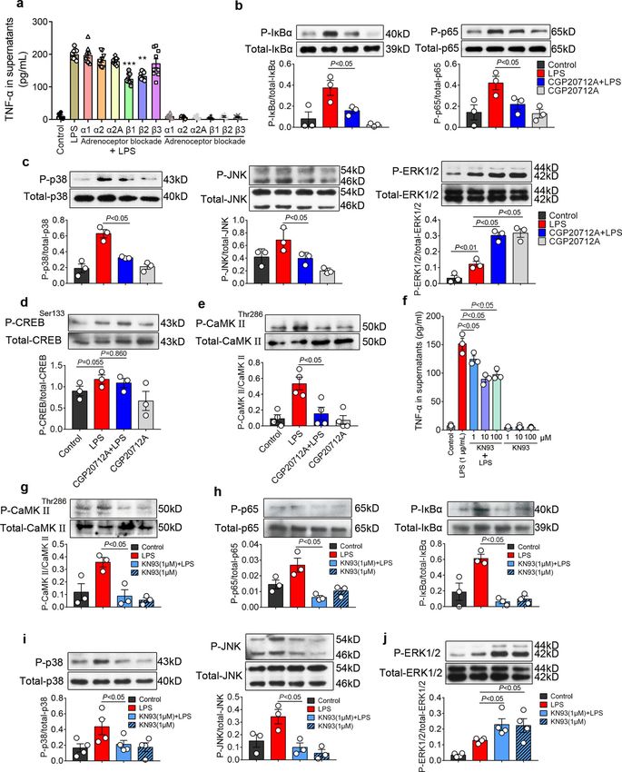
ARTICLE                                                             COMMUNICATIONS BIOLOGY | https://doi.org/10.1038/s42003-022-03007-6

M
           yocardial dysfunction is a frequent event that correlates       80) were in two distinct clusters (Fig. 1d). Therefore, ICA cells
           with the severity of sepsis, which accounts for the pri-        were not considered to be of the macrophage lineage. Since
           mary cause of death in intensive care units1,2. During          vimentins are type III intermediate filaments expressed especially
sepsis-induced myocardial dysfunction (SIMD), pathogen-                    in mesenchymal cells, co-staining of vimentin, cTn I, and TH in
associated molecular patterns (PAMPs), such as bacterial lipo-             co-culture of ICA cells and cardiomyocytes suggested that ICA
polysaccharide (LPS), interact with Toll-like receptor 4 (TLR4) on         cells are of mesenchymal cell origin (Fig. 1e). To validate the
immune and cardiac cells, activating NF-κB and mitogen-                    specificity of each staining, negative controls are provided in
activated protein kinase (MAPK) signaling cascades to produce              Fig. S2.
proinflammatory cytokines, including TNF-α, interleukin-1β and
interleukin-6. These cytokines act on endothelial cells, cardiac           Increased NE release from ICA cells promotes cardiomyocyte
fibroblasts, and cardiomyocytes to increase the production of               TNF-α production in response to LPS challenge. The primary
inflammatory mediators, which cause myocardial depression3–8.               NRVM culture containing ICA cells is defined as a co-culture of
Norepinephrine (NE), demonstrated to be associated with                    ICA cell and NRVM (NRVM ICA cell+). To analyze the role of
SIMD9,10, can activate β1-adrenergic receptor (AR) and promote             ICA cells, we used an established method of superparamagnetic
Ca2+/calmodulin-dependent protein kinase II (CaMKII) phos-                 iron oxide particles (SIOP) to obtain pure NRVM that contains
phorylation, IκBα phosphorylation, and TNF-α expression, all of            no ICA cells (NRVM ICA cell−) (Fig. 2a)24. LPS significantly raised
which contribute to cardiomyocyte apoptosis in septic mice11.              NE levels in the supernatants of NRVM ICA cell+ with either
Moreover, selective β1-AR blockade improves cardiac function               different doses of LPS (LPS 0.01 μg/mL, 0.1 μg/mL, and 1 μg/mL)
and survival in septic patients12,13.                                      or different treatment durations (LPS 0, 6, and 12 h) when
   The intrinsic cardiac adrenergic (ICA) cells, which express             compared with controls (Fig. 2b). During the Langendorff per-
tyrosine hydroxylase (TH) and dopamine-β-hydroxylase (DBH)                 fusion of adult rat hearts, LPS elicited negligible influences on NE
necessary for catecholamine biosynthesis, have been identified in           levels in perfusate, suggesting that circulating NE was not
the mammalian heart as an important origin of intrinsic cardiac            involved due to the lack of sympathetic nerves and the adrenal
catecholamines14,15. A growing body of literature has suggested            medulla in the Langendorff model (Fig. 2c). However, LPS
that the ICA system is a critical regulator of mammalian heart             administration significantly increased the level of NE in myo-
development, post-heart transplantation inotropic support, and             cardial homogenate (Fig. 2d). Immunostaining confirmed that
cardioprotection during ischemia/reperfusion15–21. However, the            macrophages were not present in the current cardiomyocyte
response of ICA cells in SIMD is unclear. Our previous study               culture system (Fig. 2e), and single-cell RNA sequencing
showed that blockade of β1-AR suppressed LPS-induced TNF-α                 demonstrated that Dbh was not induced in LPS-treated Cd68
production in primary neonatal rat cardiomyocytes cultured by              positive macrophages, which was expressed in ICA cells (Fig. 2f).
traditional methods in the absence of exogenous NE22, indicating           Therefore, the increase of NE level in NRVM ICA cell+ culture
that some β1-AR stimuli function in the culture. In addition, ICA          conditions and adult rat myocardial homogenate could be
cells were found to be present in the primary neonatal rat car-            attributed to the ICA cells, but not macrophages. Consequently,
diomyocyte culture using traditional methods14,23. In this con-            we found that blockade of β1-AR using CGP20712A, a selective
text, we postulated that LPS might stimulate ICA cells to                  β1-AR antagonist, dramatically suppressed LPS-induced TNF-α
synthesize NE that would promote LPS-induced cardiomyocyte                 production in NRVM ICA cell+, whilst removal of ICA cells
TNF-α production and myocardial dysfunction via β1-AR.                     abolished the impact of CGP20712A on TNF-α production
   In this study, we investigate the effect of LPS on NE production        (Fig. 2g). Moreover, the removal of ICA cells caused a lower level
in ICA cells and the role of ICA cell-derived NE in LPS-induced            of TNF-α in the supernatants of LPS-stimulated NRVM ICA cell−
myocardial dysfunction. We demonstrate that LPS increases NE               than that of NRVM ICA cell+ (Fig. 2h). These data support the
production in ICA cells that express TLR4, and identify ICA cell-          concept that LPS stimulates ICA cell-derived NE release, which
derived NE as a paracrine signal to enhance LPS-provoked                   promotes LPS-induced TNF-α production in cardiomyocytes.
proinflammatory response in cardiomyocytes and myocardial
dysfunction via β1-AR-CaMKII-NF-κB/MAPKs signaling
cascades.                                                                  LPS upregulates NE-producing enzyme expression in ICA cells.
                                                                           We next intended to elucidate how NE release is increased fol-
                                                                           lowing LPS treatment. Given TH and DBH are the key enzymes
Results                                                                    responsible for NE biosynthesis9, we evaluated the expression of
ICA cells are present in neonatal and adult rat hearts. Immu-              TH and DBH in ICA cells. Immuno-staining showed that LPS
noreactivity of TH, a key catecholamine-forming enzyme, in ICA             significantly stimulated TH expression in neonatal and adult rat
cells has been demonstrated previously (Fig. S1B)19, and the               ICA cells (Fig. 3a, b). In the NRVM ICA cell+ culture, LPS (1 μg/
established 3D structure of ICA cells illustrated its TH immu-             mL) stimulation for 6 h and 12 h dramatically increased TH and
noreactivity (Fig. S1C). We thus performed immunofluorescent                DBH mRNA expression, and different doses of LPS also sig-
staining of TH to identify ICA cells. As shown in Fig. 1a, in the          nificantly enhanced TH and DBH mRNA expression after 6 h
culture of cardiomyocytes isolated from neonatal rat hearts using          compared to controls (Fig. 3c). Consistently, a dramatic increase
a traditional enzymatic method24, the TH-positive ICA cells were           was observed in the protein expression of TH and DPH following
found to cluster nearby cardiac troponin I (cTn I)-positive neo-           LPS treatment (Fig. 3d, e). In Langendorff-perfused adult rat
natal rat ventricular myocytes (NRVM). In addition, these ICA              hearts, LPS increased TH and DBH expression significantly at
cells were also found in adult rat hearts by staining of TH and            both mRNA and protein levels compared to controls (Fig. 3f, g).
cTn I in tissue slices (Fig. 1b, white arrow). Macrophages were            These data show that LPS increases NE release by upregulating
reported as a source of catecholamines in response to acute                TH and DBH expression in ICA cells.
inflammatory injury25, but we demonstrated that ICA cells did
not express macrophage markers CD68 or F4/80 (Fig. 1c and                  LPS increases TH and DBH biosynthesis through TLR4-
Fig. S1D, E). Moreover, single-cell RNA sequencing revealed that           MyD88/TRIF-AP-1 signaling. We next attempted to pinpoint
ICA cells (present Dbh and Ddc coding for Dopa Decarboxylase)              the underlying mechanism of NE-producing enzyme upregula-
and macrophages (present Cd68, Msr1, and Adgre1 coding for F4/             tion in ICA cells stimulated by LPS. Since TLR4 is a well-known

2                        COMMUNICATIONS BIOLOGY | (2022)5:96 | https://doi.org/10.1038/s42003-022-03007-6 | www.nature.com/commsbio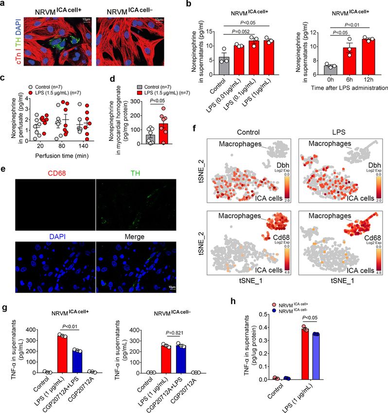
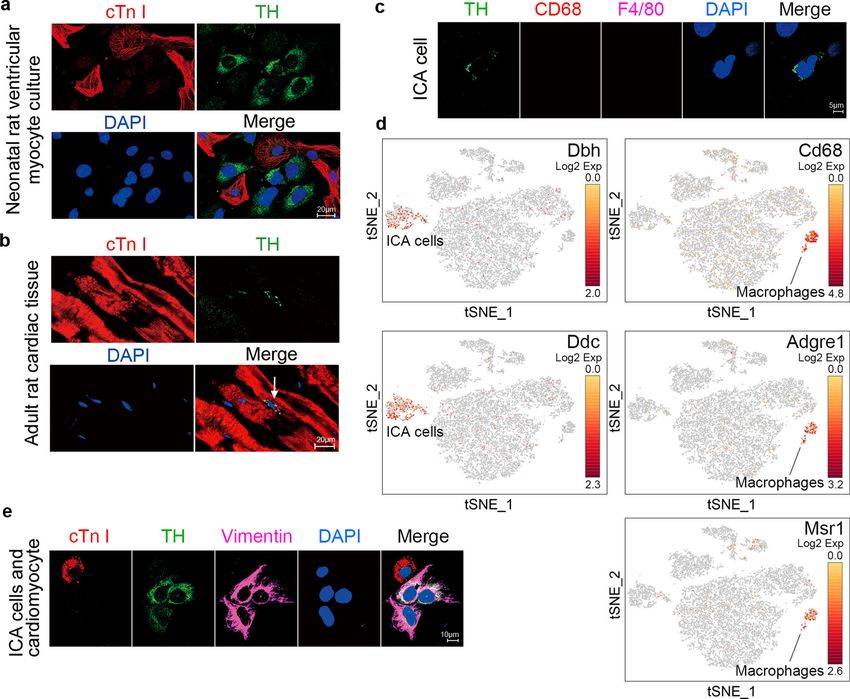
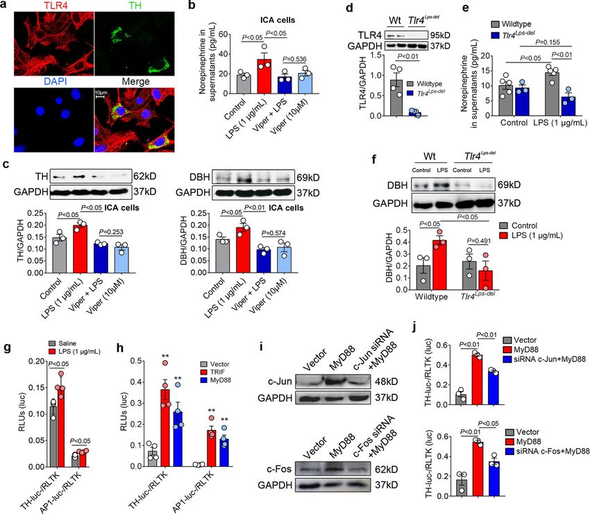
COMMUNICATIONS BIOLOGY | https://doi.org/10.1038/s42003-022-03007-6                                                                      ARTICLE

Fig. 1 Intrinsic cardiac adrenergic (ICA) cells are present in neonatal and adult rat hearts. a Immunofluorescent staining of primary cardiomyocytes and
ICA cells isolated from neonatal rat hearts using traditional method (n = 6). b Immunofluorescent staining of adult rat cardiac tissue slides (n = 3). cTn I
(cardiac troponin I): cardiomyocytes, red; TH (tyrosine hydroxylase): ICA cells, green (white arrow). c ICA cells identified using macrophage markers, TH:
ICA cell, green; CD68: marker of macrophage, red; F4/80: marker of macrophage, magenta (n = 6). d Single-cell RNA-sequencing analysis of cardiac cells
obtained using the Percol method. Note, both ICA cells and macrophages are identified as two different clusters. Dbh codes Dopamine Beta-Hydroxylase,
Ddc codes Dopa Decarboxylase, Cd68 codes CD68, Adgre1 codes F4/80, and Msr1 codes MSR1 (n = 24). e Immunostaining of ICA cells and cardiomyocyte,
cTn I: cardiomyocyte, red; TH: ICA cells, green; Vimentin: marker of mesenchymal cells, magenta (n = 6). Nuclei of cells in all groups were dyed using
DAPI: blue. The data represent two independent experiments, n represents the number of neonatal rats (a, c, and d) or adult rats (b).

receptor of LPS26, we thus hypothesized that TLR4 activation                   knocked down c-Jun and c-Fos using the siRNAs in HEK293/
mediates LPS-induced expression of TH and DBH in ICA cells.                    hTLR4-HA cells (Fig. 4i, Fig. S5). The activation of the TH
To this end, we employed immuno-staining to examine the                        promoter in response to MyD88 overexpression was dramatically
expression of TLR4 in ICA cells. Cardiomyocytes were included                  suppressed by c-Jun siRNA and c-Fos siRNA (Fig. 4j). In addi-
as a positive control on which TLR4 expression has been                        tion, single-cell RNA sequencing verified that LPS induced
demonstrated27. Immuno-staining of TLR4 in NRVM ICA cell+                      increased Jun expression in ICA cells (Fig. 5h). These results
and isolated ICA cells showed that ICA cells (as well as cardio-               reveal LPS induces NE enzyme biosynthesis via TLR4-MyD88/
myocytes) expressed TLR4 (Fig. 4a and Fig. S4A). Blockade of                   TRIF-AP1, which leads to activation of the TH promoter.
TLR4 signaling using the Viper peptide (a blocker of TLR4)28
markedly suppressed LPS-induced NE production as well as                       TLR4-expressing ICA cells do not produce TNF-α upon LPS
expression of TH and DBH in ICA cells (Fig. 4b, c). Consistently,              stimulation. In order to further clarify the role of ICA cells in
the DBH expression and NE production were not LPS inducible                    LPS-induced cardiac TNF-α production, we investigated whether
in TLR4-deficient (Tlr4Lps-del) NMVM ICA cell+, and lower than                  ICA cells are responsible for TNF-α production. To this end, we
those in LPS-treated wild-type controls (Fig. 4d–f). Furthermore,              performed co-immunofluorescence staining of TNF-α, TH, and
we cloned the rat TH promoter into a luciferase reporter                       cTn I in LPS-treated NRVM ICA cell+. Surprisingly, TNF-α was
(Fig. S4B–F), and found that LPS stimulation markedly activated                induced in cardiomyocytes but not in ICA cells (Fig. 5a). To
the TH promoter as well as the AP-1 promoter (Fig. 4g). Simi-                  validate this phenotype, we implored single-cell RNA sequencing
larly, overexpression of the TLR4 adaptors, MyD88 and TRIF,                    to confirm TNF-α expression by cardiomyocytes and ICA cells.
significantly activated both the TH and the AP-1 promoters                      The batch-to-batch variations between the control and LPS
(Fig. 4h and Fig. S4H). Of note, AP-1 is a heterodimer composed                groups were normalized to be comparable (Fig. 5c). Distinct
of proteins belonging to the c-Jun and c-Fos families that have                populations of cardiac cells were identified in different clusters on
specific binding sites on the TH promoter29. To see whether                     the t-SNE map (Fig. 5d). Unsupervised clustering revealed mac-
disruption of AP-1 binding impacts TH promoter activation, we                  rophages expressing Cd68 and ICA cells expressing Dbh in two

COMMUNICATIONS BIOLOGY | (2022)5:96 | https://doi.org/10.1038/s42003-022-03007-6 | www.nature.com/commsbio                                               3
ARTICLE                                                                 COMMUNICATIONS BIOLOGY | https://doi.org/10.1038/s42003-022-03007-6

Fig. 2 LPS-induced ICA cell-derived NE enhances cardiac TNF-α production. a Immunofluorescent staining of neonatal rat ventricular myocyte (NRVM)
co-culturing with ICA cells (NRVM ICA cell+) and NRVM without ICA cells (NRVM ICA cell−), cTn I: cardiomyocytes, red; TH: ICA cells, green; DAPI: nuclei,
blue (n = 12). b Norepinephrine (NE) level in supernatants of NRVM ICA cell+ stimulated with different doses of LPS for 6 h, and with 1 µg/mL LPS for
different durations (n = 36, each dot represents one independent repeat). c NE in perfusate from Langendorff-perfused adult rat hearts, control: K-H buffer.
d NE in myocardial homogenates from adult rat hearts Langendorff perfused for 140 min, control: K-H buffer (n = 7 rats per group in c and d, each dot
represents an adult rat). e Immunostaining of cells in current cardiomyocyte culture, CD68: marker of macrophage, red; TH: maker of ICA cells, green
(n = 6). f Single-cell RNA-sequencing analysis of cardiac cells obtained using the Percol method, containing both ICA cells and macrophages treated with
LPS. Dbh codes Dopamine Beta-Hydroxylase, Cd68 codes CD68 (n = 24). g TNF-α in supernatants 6 h post LPS-treated NRVM ICA cell+ and NRVM ICA cell-
pretreated with 2 µM CGP20712A for 30 min prior to LPS. h TNF-α in supernatants of NRVM ICA cell+ and NRVM ICA cell− stimulated with 1 µg/mL LPS for
6 h (n = 36, each dot represents one independent repeat in g and h). Data are presented as mean ± SEM, the P-values were assigned on each panel. b the
LPS dose data were analyzed using non-parametric Kruskal–Wallis test and Dunn’s method of comparison, the duration data and g and h were analyzed
using one-way ANOVA and Bonferroni multiple comparison test; c multiple t-test followed by the Holm-Sidak method; d independent Student’s t-test. The
in vitro data represent three independent experiments. n represents the number of neonatal rats (a, b, e–h) or adult rats (c, d).

separate clusters, supporting our immuno-staining results that                 were used as a positive control, resulting in upregulation of Tnf,
ICA cells were not of monocyte origin (Fig. 5d, e). Analysis of                Il1b, and Il6 gene expression (Fig. 5i). Mechanistically, analyses of
substructure in cluster 3 (ICA cells and cardiomyocytes) showed                DEGs showed significantly increased expressions of Nfkbia and
four distinct groups in which the new sub-cluster 3 (purple) was               Tnfaip6 in LPS-treated ICA cells compared with controls (Fig. 5j,
ICA cells expressing Dbh (Fig. 5f, g). Further, differentially                 k and Fig. S6C, D). These data suggest the lack of TNF-α pro-
expressed genes (DEGs) heat maps and clusters displayed an                     duction in ICA cells may be attributed to the upregulation of
upregulation of Dbh and Jun expression, but not Tnf expression                 Nfkbia and Tnfaip6, which are involved in p65 nuclear
in LPS-treated ICA cells (Fig. 5h). LPS-stimulated macrophages                 translocation30, and suppression of NF-κB activation31,

4                           COMMUNICATIONS BIOLOGY | (2022)5:96 | https://doi.org/10.1038/s42003-022-03007-6 | www.nature.com/commsbio
COMMUNICATIONS BIOLOGY | https://doi.org/10.1038/s42003-022-03007-6 ARTICLE
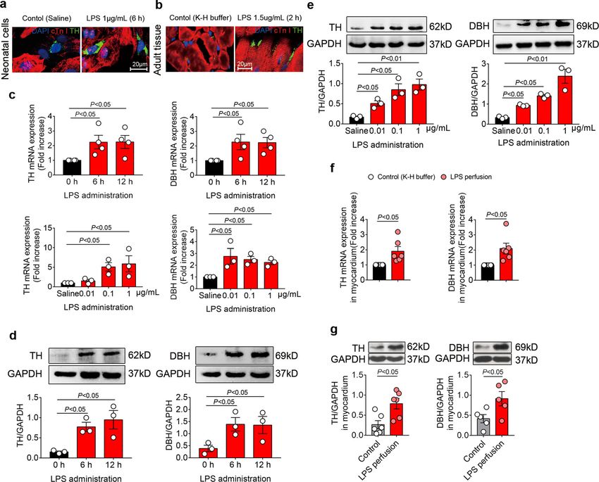
Fig. 3 LPS stimulation upregulates NE-producing enzymes in ICA cells. a and b TH expression in NRVM ICA cell+ treated with saline or LPS for 6 h (a,
n = 6) and adult rat hearts Lagendorff perfused for 2 h (b, n = 6 per group); cTn I: cardiomyocytes, red; TH: ICA cells, green; DAPI: nuclei, blue. c–e mRNA
and protein expression of TH and DBH relative to GAPDH in NRVM ICA cell+ stimulated with LPS at different time points and doses (n = 36, one-way
ANOVA and Bonferroni multiple comparison test, each dot represents one independent repeat). f and g mRNA and protein expression of TH and DBH
relative to GAPDH in myocardium from adult rat hearts Lagendorff perfused for 2 h, control: K-H buffer, LPS: 1.5 µg/mL in K-H buffer (n = 6 rats per group,
independent Student’s t-test, each dot represents a rat). Data are presented as mean ± SEM. P-values were assigned on each panel. n represents the
number of neonatal rats (a–e) or adult rats (f–g).
respectively. We, therefore, performed immunofluorescence increases IκBα phosphorylation and TNF-α expression in septic
staining of p65 localization to examine NF-κB activation in LPS- mouse hearts11. The levels of phosphorylated-p38 and
treated NRVM ICA cell+. Strikingly, nuclear translocation of p65 phosphorylated-JNK in the CGP20712A + LPS group were also
was significantly induced by LPS in non-ICA cells but not in ICA significantly decreased compared to the LPS alone group, while the
cells, compared to saline-treated controls (Fig. 5l). These findings ERK1/2 phosphorylation was increased by CGP20712A with or
suggest that ICA cells lack the capacity of producing TNF-α due without LPS (Fig. 6c). These results suggest that the NF-κB and
to impaired nuclear translocation of p65 by elevation of Nfkbia MAPK signaling pathways are involved in the process of ICA cell-
and Tnfaip6. derived NE promotion of cardiomyocyte TNF-α production. Since
protein kinase A (PKA) signaling has proven to be a major route for
channeling cardiac β1-AR signaling32, we thus asked if PKA med-
ICA cell-derived NE acts via β1-AR-CaMKII signaling to reg- iates induction of ICA cell-derived NE on NF-κB and MAPK
ulate NF-κB and MAPK pathways in cardiomyocyte. Catecho- pathway activation. Intriguingly, the phosphorylation of CREB-
lamines activate various adrenoceptors, to screen out the targets of Ser133, which indicates PKA activation33, showed no significant
ICA cell-derived NE in cardiomyocytes, different adrenoceptor alteration in either of the groups (Fig. 6d). As the other major
inhibitors were employed, in which only β1-AR or β2-AR blockade downstream element of the β1-AR pathway, CaMKII was reported
significantly reduced the LPS-induced TNF-α production in NRVM to mediate effects of β1-AR stimulation independent of PKA in
ICA cell+ (Fig. 6a). Administration of CGP20712A (2 µM), a β -AR
1
heart failure, cardiac contractility, and apoptosis34. A recent study
blocker, prior to LPS (1 μg/mL) treatment dramatically suppressed also showed that CaMKII plays an important role in cardiac con-
LPS-induced phosphorylation of IκBa and p65 (Fig. 6b), which is tractile dysfunction associated with sepsis35. We, therefore, exam-
consistent with previous work demonstrating activation of β1-AR ined the activation of CaMKII in this process. Indeed, the
COMMUNICATIONS BIOLOGY | (2022)5:96 | https://doi.org/10.1038/s42003-022-03007-6 | www.nature.com/commsbio 5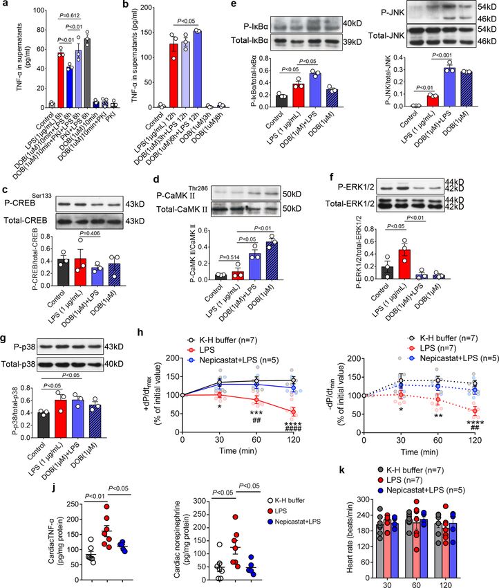
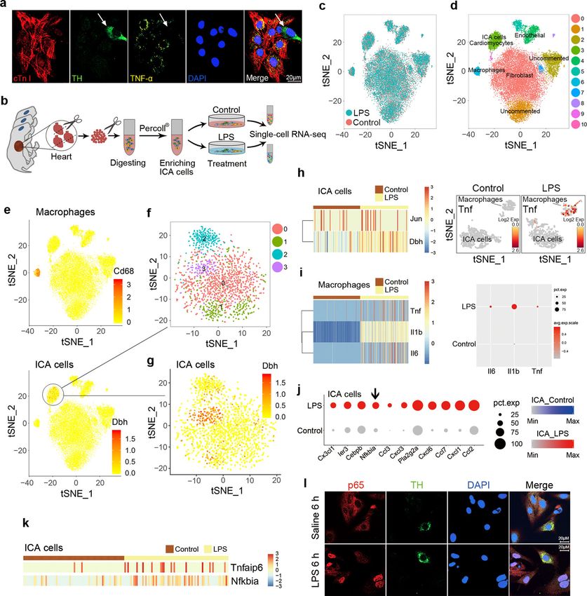
ARTICLE                                                                    COMMUNICATIONS BIOLOGY | https://doi.org/10.1038/s42003-022-03007-6

Fig. 4 LPS-TLR4/AP-1 signaling mediates NE-producing enzyme expression. a Co-immunofluorescent staining of TLR4 and TH in NRVM ICA cell+, TLR4:
toll-like receptor 4, red; TH: ICA cells, green; DAPI: nuclei, blue (n = 6). b and c NE production as well as TH and DBH expression in rat ICA cells treated
with Viper (TLR4 inhibitor, 10 μM) for 2 h prior to LPS for 6 h. (n = 48, each dot represents one independent repeat). d TLR4 expression in wildtype (WT)
and TLR4-deficient (Tlr4Lps-del) NMVM ICA cell+. e and f NE production and DBH expression in WT and Tlr4Lps-del NMVM ICA cell+ stimulated with LPS for
6 h, control: saline (n = 12, each dot represents a biological replicate in d–f). g Luciferase activity of AP-1-luc- and TH-promoter-luc- relative to RLTK-luc- in
HEK293/hTLR4 cells at 36 h after transfection and LPS stimulation. h Luciferase activity of AP-1-luc- and TH-promoter-luc- relative to RLTK-luc- in
HEK293/hTLR4 cells at 24 h after transfection with plasmids (0.1 μg/mL) encoding MyD88 and TRIF. i Expression of c-Jun and c-Fos in HEK293/hTLR4
cells at 24 h after transfection with MyD88 plasmid, c-Jun siRNA and c-Fos siRNA. j Luciferase activity of TH-promoter-luc- relative to RLTK-luc- in
HEK293/hTLR4 cells at 24 h after transfection with MyD88 plasmid, c-Jun siRNA, and c-Fos siRNA, (0.1 μg/mL for plasmid) (each dot represents one
independent repeat in (g–j)). Data are presented as mean ± SEM using one-way ANOVA and Bonferroni multiple comparison test, *P < 0.05, **P < 0.01,
most of the P-values were assigned on the figure. n represents the number of neonatal rats (a–c) or neonatal mice (d–f).

phosphorylation of CaMKIIThr286 was dramatically enhanced in                      (Fig. 6h, i), and increased ERK1/2 phosphorylation (Fig, 6j). These
LPS-stimulated NRVM ICA cell+ compared with controls, while                       results show that CaMKII blockade phenocopies the effects of β1-
CGP20712A significantly reduced CaMKIIThr286 phosphorylation                       AR blockade on LPS-treated NRVM ICA cell+, which suggests that
compared to the LPS group (Fig. 6e). These data hint that CaM-                    CaMKII is essential during β1-AR signal transduction activated by
KIIThr286 phosphorylation is critical in mediating the effects of β-              ICA cell-derived NE.
AR stimulation by ICA cell-derived NE. We next used KN93, a
selective inhibitor of CaMKII, to block CaMKII signaling. KN93
markedly decreased the levels of TNF-α in the supernatants of LPS-                Dobutamine recapitulates the contributions of ICA cells to
treated NRVM ICA cell+ in a dose-dependent manner (Fig. 6f). The                  cardiomyocyte TNF-α production upon LPS stimulation. The
phosphorylation of CaMKIIThr286 in the KN93 + LPS group was                       aforementioned data suggest that the NE from ICA cells con-
significantly reduced compared with LPS-treated NRVM ICA cell+                     tribute to the activation of NF-κB and MAPKs through β1-AR-
(Fig. 6g). Accordingly, KN93 dramatically decreased LPS-induced                   CaMKII signaling in cardiomyocytes. We next employed dobu-
phosphorylation of p65, IkBa, p38, and JNK in NRVM ICA cell+                      tamine (DOB), a selective β1-AR agonist analogous to NE36, to

6                            COMMUNICATIONS BIOLOGY | (2022)5:96 | https://doi.org/10.1038/s42003-022-03007-6 | www.nature.com/commsbio
COMMUNICATIONS BIOLOGY | https://doi.org/10.1038/s42003-022-03007-6                                                                            ARTICLE

Fig. 5 ICA cells do not produce TNF-α upon LPS stimulation. a Immunofluorescent-staining of TNF-α in NRVM ICA cell+ stimulated with 1 µg/mL LPS for
6 h, cTn I: cardiomyocytes, red; TH: ICA cell (white arrow), green; TNF-α, yellow; DAPI: nuclei, blue (n = 12 neonatal rats). b Schematic of single-cell RNA-
sequencing experimental strategy. c t-distributed stochastic neighbor embedding (t-SNE) projection of cardiac cells, batch effect analysis between control
and LPS groups, control: saline, 24,210 cells. d t-SNE map of cardiac cells, different colored clusters represent distinct populations, 24,210 cells. e ICA cells
expressing Dbh and macrophages expressing Cd68 are separated in two clusters, 24,210 cells. f and g Analysis of substructure in ICA and cardiomyocyte
cluster indicates subcluster 3 (Purple) is a ICA cell subpopulation, 1765 cells. h Expression of Dbh and Jun in ICA cells between control and LPS groups, and
Tnf gene expression in ICA cells and macrophages between control and LPS groups, 138 cells. i Expression of Tnf, Il6, and Il1b in macrophages between
control and LPS groups, 572 cells. j Top 11 differentially expressed genes (DEGs) which include Nfkbia in the ICA subcluster between control and LPS
groups, control: saline, 138 cells. k Expression of Tnfaip6 and Nfkbia in ICA cells between control and LPS groups, 138 cells. l Immunofluorescent-staining of
p65 localization in NRVM ICA cell+ stimulated with saline or 1 µg/mL LPS for 6 h, p65, red; TH: ICA cells, green; DAPI: nuclei, blue (n = 6 neonatal rats). For
single-cell transcriptomes (b–j), n = 24 neonatal rats. Gene-expression values represent mean of log; differential expression test: Wilcoxon rank sum test;
avg_logFC= log (mean(group1)/mean(group2)); adjusted p_value: Bonferroni Correction; min.pct ≥10%; avg_logFC ≥ 0.1.

recapitulate the contributions of ICA cells. We cultured NRVM                     14–22 amide (PKI) increased LPS-induced TNF-α production
without ICA cells (NRVM ICA cell−) and then treated with DOB                      and reversed the suppressive effect of DOB (Fig. 7a). Such
for different durations prior to LPS stimulation. Short-term (10-                 observations are in agreement with published data suggesting that
min) β1-AR stimulation with DOB reduced the LPS-induced                           short-term β1-adrenergic stimulation elicits a rapid increase of
TNF-α production in NRVM ICA cell−, while PKA inhibitor                           cellular cAMP activating PKA37, which inhibits LPS-induced

COMMUNICATIONS BIOLOGY | (2022)5:96 | https://doi.org/10.1038/s42003-022-03007-6 | www.nature.com/commsbio                                                     7
ARTICLE                                                               COMMUNICATIONS BIOLOGY | https://doi.org/10.1038/s42003-022-03007-6

Fig. 6 ICA cell-derived NE enhanced by LPS acts via β1-AR-CaMKII pathway to regulate NF-κB and MAPK signaling pathways. a Effect of different
adrenoceptor blockers on TNF-α production in NRVM ICA cell+ upon LPS stimulation. Each dot represents a biological replicate. b–e NRVM ICA cell+ were
stimulated with 2 µM CGP20712A (β1-AR antagonist) for 30 min prior to 1 µg/mL LPS for 30 min, b phosphorylation of IκBα and p65, c phosphorylation of
p38, JNK, and ERK1/2, d phosphorylation of CREB-Ser133, indicating PKA activation. e phosphorylation of CaMKII in NRVM ICA cell+. f TNF-α in
supernatants of NRVM ICA cell+ treated with KN93 in different doses for 30 min prior to 1 µg/mL LPS for 6 h. g–j Phosphorylation of CaMKII, p65, IκBα,
p38, JNK and ERK1/2 in NRVM ICA cell+ treated with 1 µM KN93 for 30 min prior to 1 µg/mL LPS for 30 min. Data are presented as mean ± SEM using one-
way ANOVA and Bonferroni multiple comparison test, P-values were assigned on each panel. n = 36 neonatal rats (a–j), data represent three or four
independent experiments.

8                          COMMUNICATIONS BIOLOGY | (2022)5:96 | https://doi.org/10.1038/s42003-022-03007-6 | www.nature.com/commsbio
COMMUNICATIONS BIOLOGY | https://doi.org/10.1038/s42003-022-03007-6                                                                       ARTICLE

Fig. 7 Dobutamine recapitulates the effects of ICA cell-derived NE, whereas blockade of ICA cell-derived NE prevents myocardial dysfunction induced
by LPS. a TNF-α in supernatants of NRVM without ICA cell (NRVM ICA cell−) treated with 1 µM dobutamine (DOB) for 10 min and/or 5 µM PKA inhibitor
14–22 amide (PKI) for 1 h prior to LPS 1 µg/mL for 6 h. b TNF-α in supernatants of NRVM ICA cell− treated with 1 µM DOB for 3 h or 6 h prior to 1 µg/mL LPS
for 12 h. c–g Phosphorylation of CREB-Ser133, CaMKII-Thr286, IκBα, JNK, ERK1/2 and p38 in NRVM ICA cell− treated with 1 µM DOB for 6 h prior to 1 µg/
mL LPS for 30 min. n = 36 neonatal rats, data represent three independent experiments (a–g). h, i and k Systolic (+dP/dtmax), diastolic (−dP/dtmin)
function and heart rate of Langendorff-perfused adult rat hearts, LPS: 1.5 µg/mL, Nepicastat: Dopamine-β-hydroxylase (DBH) inhibitor, 15 µg/mL. j TNF-α
and NE in myocardial homogenates from adult rat hearts Langendorff perfused for 120 min. n = 7 rats (K-H buffer), n = 7 rats (1.5 µg/mL LPS), n = 5 rats
(15 µg/mL Nepicastat + LPS) in (h–k), each dot represents an adult rat, the solid dots on lines in (h) and (i) represent the mean of each group at each time
point. Data are presented as mean ± SEM. a–g and j one-way ANOVA and Bonferroni multiple comparison test, h, i and k two-way repeated measures
ANOVA and Tukey’s multiple comparisons test. *P < 0.05, **P < 0.01, ***P < 0.001, ****P < 0.0001 in comparison of LPS vs. K-H buffer, ##P < 0.01, ####
P < 0.0001 in comparison of LPS vs. Nepicastat + LPS. P-values were assigned on the panels.

COMMUNICATIONS BIOLOGY | (2022)5:96 | https://doi.org/10.1038/s42003-022-03007-6 | www.nature.com/commsbio                                                9
ARTICLE                                                             COMMUNICATIONS BIOLOGY | https://doi.org/10.1038/s42003-022-03007-6

cardiac expression of TNF-α38. Of note, cardiomyocytes are                 pathophysiology          and        post-heart       transplantation
perpetually regulated by intrinsic cardiac adrenergic activities. We       support14–17,19,21,46,47. Stimulation of ICA cells enhances epi-
thus prolonged the treatment time of DOB, and found that β1-AR             nephrine release to reduce ischemia/reperfusion injury through
stimulation for 6 h significantly increased the level of TNF-α in           the δ-opioid-regulated cardioprotective adrenopeptidergic co-
supernatants of LPS-treated NRVM ICA cell− (Fig. 7b). Similar to           signaling pathway17,19,48. Although ICA cell density shows neg-
our observations regarding ICA cell-derived NE, DOB stimula-               ligible connection with reduced myocardial efficiency in failing
tion of NRVM ICA cell− for 6 h did not alter phosphorylation of            myocardium18, irregular stimulation of ICA cells raises cardiac
CREB-Ser133 (Fig. 7c), but did dramatically enhance the level of           NE levels49. Considering the increase of cardiac NE levels and β1-
phosphorylated-CaMKIIThr286 compared with control and LPS                  AR activation aggravate sepsis-induced cardiomyocyte apoptosis
groups (Fig. 7d). The phosphorylation of CaMKIIThr286 in LPS-              and myocardial injury8,11, it is thus plausible that ICA cells could
treated NRVM ICA cell− exhibited negligible differences compared           promote septic cardiomyopathy pathogenesis. Indeed, ICA cells
with controls, showing that there was no NE production in                  are not separated from cardiomyocytes in most of the current
NRVM ICA cell− culture due to the lack of ICA cells (Fig. 7d).             cardiac models using primary neonatal rat or mouse ventricular
Consistent with β1-AR blockade and CaMKII blockade in LPS-                 myocytes (NRVM or NMVM)50,51. Our previous study estab-
stimulated NRVM ICA cell+, DOB treatment of NRVM ICA cell−                 lished a method of superparamagnetic iron oxide particle (SIOP)
for 6 h prior to LPS markedly increased phosphorylation of IκBα            to purify NRVM from ICA cells and demonstrated that the ICA
and JNK, whereas a decreased level of phosphorylated-ERK1/2                cell-free NRVM showed lower calcium transient amplitude,
and comparable p38 phosphorylation when compared with the                  longer time to begin autonomous beating and less expression of
LPS alone group (Fig. 7e–g). Given that β2-AR activation sup-              natriuretic peptide A (Nppa) and -B (Nppb) (markers of stress
presses p38 MAPK phosphorylation39, the unchanged p38                      response) than those of NRVM mixed with ICA cells24. More-
phosphorylation could be due in part to the mild β2-AR agonist             over, we observed a reduced TNF-α production in ICA cell-free
activity of DOB40. Nonetheless, the data collectively shows DOB            NRVM compared to unpurified cardiomyocytes in response to
mimics ICA-cell derived NE that promotes TNF-α production in               LPS stimulation24. Therefore, it is important to distinguish and
LPS-stimulated cardiomyocytes via β1-AR-CaMKII-NF-κB/                      evaluate the function of ICA cells in cardiac studies.
MAPKs signaling cascades.                                                     In the current study, we defined ICA cell-derived NE as a
                                                                           contributor to myocardial dysfunction via activation of β1-AR
                                                                           signaling in cardiomyocytes upon LPS challenge. Our data
Blockade of ICA cell-derived NE prevents LPS-induced myo-
                                                                           demonstrated ICA cells as a source of increased NE in LPS-treated
cardial dysfunction. The above mentioned data suggest a critical
                                                                           neonatal and adult rat hearts. Although endothelial cells have been
role of ICA cells in facilitating LPS-induced myocardial dys-
                                                                           shown to produce catecholamines in response to ischemia52, the
function. We, therefore, attempted to assess the influence of ICA
                                                                           typical “cobblestone” monolayer pattern of morphology in endo-
cell-derived NE on cardiac function in vivo. Of note, circulating
                                                                           thelial cell culture largely differs from ICA cells53. In the Lan-
NE sourced from sympathetic nerves and adrenal medulla are
                                                                           gendorff perfusion experiments, the unchanged NE levels in the
significantly enhanced during sepsis26, however, the impact of
                                                                           perfusate from LPS-perfused hearts also indicate that the increased
ICA cell-derived NE on cardiac function is challenging to mea-
                                                                           NE levels in the hearts were not from vascular endothelial cells.
sure exclusively in vivo. Yet, the Langendorff perfusion system
                                                                           Utilization of a β1-AR blocker, CaMKII inhibitor, and β1-AR
can eliminate the influence of circulating NE, and has proven
                                                                           agonist showed that the NE from ICA cells can active cardiac β1-
invaluable and stalwart in studying pharmacological effects on
                                                                           AR-CaMKII signaling, which crosstalks with NF-κB and MAPK
myocardial function in the investigation of clinically relevant
                                                                           pathways to promote TNF-α production and myocardial dys-
disease states such as heart failure41,42. It was thus utilized to
                                                                           function during sepsis. This concept was further evidenced by the
investigate the impact of ICA cells on myocardial dysfunction
                                                                           administration of Nepicastat inhibiting NE production in the
induced by LPS in adult rats. The hearts in the LPS group
                                                                           ex vivo Langendorff model assessing septic heart function.
exhibited significantly worse phenotypes in both systolic (+dP/
                                                                              The early phase of sepsis has been characterized by high levels
dtmax) and diastolic (−dP/dtmin) functions than those in the K-H
                                                                           of circulating catecholamines, which boost the inflammatory
buffer group (control). Furthermore, blockade of ICA cell-derived
                                                                           response26, but the molecular mechanisms are multifactorial and
NE biosynthesis using Nepicastat, an inhibitor of DBH, markedly
                                                                           not yet fully understood. TLR4, mediating recognition of LPS, has
rescued both systolic and diastolic functions compared to the LPS
                                                                           been well-known as the first-line sentinel of host defense against
group (Fig. 7h, i). Nepicastat simultaneously abrogated the ele-
                                                                           bacteria in innate immunity26. Previous studies show that TLR4 is
vation of NE and TNF-α production in LPS-perfused hearts,
                                                                           present on immune cells and neurons4,54 which possess the
consistent with changes in systolic and diastolic functions
                                                                           capacity of catecholamine biosynthesis25,55. Whether TLR4 has
(Fig. 7j). The Langendorff-perfused hearts in different groups
                                                                           functions in the regulation of catecholamine secretion remains
showed a negligible alteration in heart rate (Fig. 7k). These
                                                                           elusive. We observed that TLR4 and its downstream MyD88/
findings demonstrate that blockade of ICA cell-derived NE
                                                                           TRIF-AP-1 signaling pathway mediate LPS-induced biosynthesis
reduces LPS-induced cardiac TNF-α production and protects
                                                                           of cellular NE through upregulation of NE-producing enzyme
against LPS-induced myocardial dysfunction.
                                                                           expression, assessed by the activation of TH-promoter. Con-
                                                                           sistently, a previous study showed that LPS continuously
Discussion                                                                 increased TH expression in mouse brains56, suggesting a pro-
Catecholamines are irreplaceable in both developing and adult              motive action to catecholamine biosynthesis for TLR4. Intrigu-
hearts. Targeted disruption in mice of the genes encoding cate-            ingly, although the activation of TLR4 triggers cytokine release
cholamine biosynthetic enzymes are embryonic lethal, likely due            including TNF-α26, this is not the case in ICA cells despite TLR4
to cardiac failure43,44. The developing heart initially relies on ICA      presence. This discrepancy could be due to the upregulation of
cells as the major source of catecholamines45. Published evidence          Nfkbia and Tnfaip6 expression that results in impaired p65
also shows that ICA cells synthesize cardiac intrinsic catechola-          nuclear translocation in LPS-treated ICA cells. This concept fits
mines, occurring independently of cardiac sympathetic nerves,              well with published evidence that the increase of either Nfkbia or
either in neonatal or adult hearts, thereby functioning as a critical      Tnfaip6 expression suppresses NF-κB activation30,31. These
and integral regulator in mammalian heart development, cardiac             findings hint towards a function of TLR4 signaling in hormone

10                       COMMUNICATIONS BIOLOGY | (2022)5:96 | https://doi.org/10.1038/s42003-022-03007-6 | www.nature.com/commsbio
COMMUNICATIONS BIOLOGY | https://doi.org/10.1038/s42003-022-03007-6 ARTICLE
regulation during sepsis. In addition, the marked upregulations of the US National Institutes of Health59 and approved by the Animal Care and Use
chemokine family gene expression (i.e., Cxc3cl1, ccl3, cxcl1, ccl2, Committee at Jinan University. Neonatal (1–3 days) and adult (8–10 weeks)
Sprague-Dawley rats were obtained from the laboratory animal center of Southern
etc.) in LPS-treated ICA cells indicate that ICA cells may also Medical University (Guangzhou, China). TLR4-deficient mice (Tlr4Lps-del, strain:
contribute to cell migration involved in homeostatic and C57BL/10ScNJNju) were purchased from Nanjing Biomedical Research Institute of
inflammatory processes. Nanjing University. For neonatal rats and mice, both male and female were used.
PKA and CaMKII are two major downstream elements of β1- For adult rats, male were used due to the impact of sex on septic cardiomyopathy60
AR that is critical for the regulation of cardiac function in chronic and the differences in TNF-α levels and vascular reactivity of female following the
administration of endotoxin61. HEK293/hTLR4-HA cells (InvivoGen) was a gift
heart failure37,57. In this study, we demonstrated an essential role from Guang Yang (Jinan University).
for CaMKII in the pathogenesis of LPS-induced myocardial
dysfunction. CaMKII activation was not observed in LPS-treated
Inhibitors. α1-AR antagonist: Prazosin (Sigma-Aldrich#7791); α2-AR antagonist:
NRVM without the presence ICA cells in culture, whereas PKA Yohimbine (Sigma-Aldrich #Y3125); α2A-AR antagonist: BRL 44408 (Sigma-
activation showed no change. Recent evidence shows that CaM- Aldrich #B4559); β1-AR antagonist: CGP20712A (Sigma-Aldrich #C231); β2-AR
KII oxidation in response to myocardial infarction contributes to antagonist: ICI-118 551 (Sigma-Aldrich #I127); β3-AR antagonist: SR59230A
NF-κB-dependent inflammatory transcription in (Sigma-Aldrich #S8688); TLR4 inhibitor: Viper peptide (Novus#NBP2-226244);
CaMKII inhibitor: KN-93 Phosphate (Selleckchem #S7423); DBH inhibitor:
cardiomyocytes58. Moreover, it is also reported that oxidation- Nepicastat (MCE MedChem Express#HY-13289); PKA Inhibitor: 14-22 Amide
activated CaMKII has a causal role in the contractile dysfunction (Calbiochem® #476485).
during sepsis35. Therefore, the present study demonstrates that
ICA cell-derived NE is another factor in activating cardiomyocyte Isolation of primary NRVM and ICA cells. Co-culture of NRVM and ICA cells
CaMKII via β1-AR to enhance LPS-induced cardiac inflamma- (NRVM ICA cell+), NRVM without ICA cell (NRVM ICA cell−) and ICA cells were
tion, and β1-AR-CaMKII activation is the principal mechanism isolated and purified using the published methods24. Briefly, the neonatal Sprague-
by which ICA cell-derived NE enhances cardiomyocyte NF-κB Dawley rats (1–3 days) were deeply anesthetized by inhalation of CO2, and then the
hearts were excised and transferred to pre-cold PBS. (1) NRVM ICA cell+ isolation:
and MAPK signaling to promote LPS-induced cardiomyocyte the heart tissues were digested 5–6 times using 0.125% trypsin without EDTA (pH
TNF-α production. 7.30–7.40) to harvest NRVM ICA cell+, and cultured in complete DMEM (10% FBS,
In summary, we provide evidence for a pro-inflammatory role 0.1 mM HEPES and 100U/ml Penicillin-Streptomycin) for downstream use
of ICA cells in LPS-induced myocardial dysfunction. Our data (Fig. S1A). (2) NRVM ICA cell− purification using superparamagnetic iron oxide
particles (SIOP) (BioMag#BM547): NRVM ICA cell+ suspensions were centrifuged
reveal that TLR4-MyD88/TRIF-AP-1 signaling is responsible for at 800 rpm, 4 °C for 7 min. The pellets were suspended in the pre-cold SIOP
LPS-induced biosynthesis of NE that serves as a paracrine signal solution (40uL SIOP: 4 ml PBS), and then applied magnet (Life technologies™) to
to aggravate LPS-induced myocardial TNF-α production and the side of the tube for 5-10 min to isolate NRVM ICA cell−, which contained no
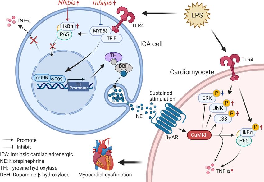
dysfunction via β1-AR-CaMKII signaling (Fig. 8). Considering ICA cells. The supernatant containing NRVM were centrifuged and the NRVM
the observation that blockade of ICA cell-derived NE prevents pellets were suspended in complete DMEM for culture. (3) ICA cell isolation: the
SIOP binding ICA cells obtained from the last step were washed with ice-cold PBS,
LPS-induced myocardial dysfunction in adult rat hearts, it seems centrifuged, and suspended in complete DMEM for further use.
plausible that ICA cells and CaMKII may be potential therapeutic
targets in the fight against SIMD.
Langendorff perfusion. Myocardial functions of the adult rats were measured
using a Langendorff perfusion system as we described previously62. Briefly, the
Sprague-Dawley rats (8–10 weeks) were heparinized (i.p. injection heparin, 2000
Methods U) for 15 min, and then deeply anesthetized with isoflurane inhalation (3% iso-
Animals and cell line. The experiments with animals were conducted in com- flurane in 100% oxygen at a flow rate of 1 L/min) using a face mask. The hearts
pliance with the Guide for the Care and Use of Laboratory Animals published by were isolated and the aortas were retrograde set up to a Langendorff perfusion
Fig. 8 Schematic of ICA cell contribution to septic cardiomyopathy. ICA cell produces no TNF-α due to the elevated Nfkbia and Tnfaip6 expression.
Increased NE release by LPS-TLR4 signaling activates β1-AR-CaMKII signaling in cardiomyocytes to regulate NF-κB and mitogen-activated protein kinase
pathways, subsequently aggravating myocardial TNF-α production and dysfunction.
COMMUNICATIONS BIOLOGY | (2022)5:96 | https://doi.org/10.1038/s42003-022-03007-6 | www.nature.com/commsbio 11ARTICLE COMMUNICATIONS BIOLOGY | https://doi.org/10.1038/s42003-022-03007-6
apparatus (Radnoti Langendorff system#120102EZ) with a recirculating mode RNA interference. RNA interference was designed to disrupt AP-1 binding in the
(volume of 50 mL) to perfuse at 10 mL/min with Krebs-Henseleit buffer (bubbled rat TH promoter region (Fig. S5A–C). c-Jun siRNA, c-Fos siRNA (Sequences refer
with 95% O2 and 5% CO2 gas mixture and maintained at 37 °C). Rat hearts were to Table S4), and control siRNA transfection were processed using Lipofecta-
divided into groups of K-H buffer (control) or LPS (Sigma-Aldrich, #L2880, mine®3000 Reagent (Invitrogen™#L3000001) and Opti-MEM (Gibco#31985-070)
Escherichia coli, 055: B5, 1.5 µg/mL). The hearts were then perfused for 140 min under a standard protocol.
with K-H buffer or LPS (1.5 µg/mL) using the recirculating mode, respectively.
Perfusion fluid was collected at different time points, and heart tissues were har-
Isolation of rat peritoneal macrophages. Rat peritoneal macrophages were iso-
vested at the end of perfusion. In separate experiments, adult rat hearts were
lated using a published method68. Adult rats (250–300 g) were euthanized with
arranged into groups of K-H buffer (control), LPS or LPS + Nepicastat. After a 30-
CO2 plus cervical dislocation, and then the abdomen was soaked with 70% alcohol
min equilibration perfusion in K-H buffer, a mixture of LPS (1.5 µg/mL) or/and
followed by making a small incision along the midline. 10 ml of DMEM were
Nepicastat (a selective DBH inhibitor63, 15 µg/mL) were perfused for 2 h. The
injected into each rat peritoneal cavity and gently massaged. About ~8 ml of fluid
physiological parameters of hearts were continuously recorded. The perfusate and was aspirated from the peritoneum per rat. The peritoneal cells were collected by
left ventricular tissues were harvested for TNF-α and NE concentration determi-
centrifuging for 10 min, 400 × g at 4 °C, and cultured for downstream use.
nation as well as immunofluorescence staining (Fig. S1F).
Statistics and reproducibility. All data were analyzed for statistical significance
10X genomics single-cell RNA-sequencing analysis. The strategy for single-cell with SPSS 13.0 software (SPSS Inc., Chicago, IL, USA). Two-tailed independent
RNA sequencing analysis of cardiac cells is shown in Fig. 5b. The neonatal Student’s t-test was used for two groups comparison. More than two groups were
Sprague-Dawley rats (1–3 days) were deeply anesthetized with CO2 and the hearts compared using one-way analysis of variance (ANOVA) followed by Bonferroni
were excised and washed in cold PBS to remove the blood. Heart tissues were multiple comparison test for normally distributed and equal variance data, or
minced into ~1 mm3 masses and digested using 0.125% trypsin. (1) Cells were nonparametric Kruskal–Wallis test and Dunn’s method of comparison for non-
prepared using a modified Percoll gradient procedure described previously to normal distributions. Data are presented as mean ± SEM, where n is the number of
enrich ICA cells64, of which details are described in Supplementary material. animals. P < 0.05 was considered statistically significant. ∗P < 0.05, ∗∗P < 0.01,
Immuno-staining of the cells was performed to examine the existence of ICA cells ∗∗∗P < 0.001. Statistical details, including sample sizes and experiment repeats, are
prior to single-cell RNA sequencing (Fig. S6A). (2) 10X genomics single-cell RNA reported in the figures and figure legends.
sequencing: the cells were collected after treatment and analyzed to determine the
viability of more than 90% by a cell count system. Individual samples were then Reporting summary. Further information on research design is available in the Nature
loaded on the 10X Genomics Chromium System. Cells counted in the system were
Research Reporting Summary linked to this article.
around 1.1–1.2 × 10⁴ per group (Fig. S6B). The libraries were prepared following
10X Genomics protocols and sequenced under standard procedure, followed by cell
lysis and barcoded reverse transcription of RNA. The library construction and Data availability
sequencing as well as computational analysis of data were performed at the The data generated in this study are included in the paper and Supplementary files, and
Guangzhou Saliai Stem cell science and technology company. Single-cell gene available from the corresponding author upon reasonable request. The single-cell RNA-
expression was visualized in a two-dimensional projection with t-distributed sto- seq data have been deposited in GEO (GSE189964). The uncropped gel and images for
chastic neighbor embedding (t-SNE) map, in which all cells were grouped into 10 western blots are provided in the Supplementary Information. The source data behind all
clusters (distinguished by their colors), and non-linear dimensional reduction was graphs and charts in the main manuscript are included in the Supplementary Data 1 file.
used (Fig. 5d)65. Analyses of batch effect correction and Differentially expressed The sequence of the recombinant pGL3-Rat-TH-Luci reporter plasmid is in the
genes (DEGs) were performed using the Seurat (version: 2.3.4) function, RunCCA Supplementary Data 2 file.
and FindClusters; Resolution for granularity: 0.5; Differential expression test:
Wilcoxon rank sum test; avg_logFC = log(mean(group1)/mean(group2)); adjusted
p_value: Bonferroni Correction; min.pct ≥10%; avg_logFC ≥ 0.1. Received: 6 April 2021; Accepted: 23 December 2021;
Immunofluorescence staining. The immunofluorescence staining (IF) was per-
formed according to our previous publication with minor modification22. Briefly,
cells or tissue slices were fixed with 4% paraformaldehyde for 15 min followed by
two washes with cold PBS and permeabilized with 0.25% Triton X-100 for 10 min. References
The specimens were then blocked at room temperature (RT) for 1 h and incubated
1. Aneman, A. & Vieillard-Baron, A. Cardiac dysfunction in sepsis. Intensive
with primary antibodies at 4 °C overnight. On the second day, the specimens were
Care Med. 42, 2073–2076 (2016).
washed three times with cold PBS and then incubated in the dark with secondary
2. Hollenberg, S. M. & Singer, M. Pathophysiology of sepsis-induced
antibodies at RT for 1 h, and then washed twice with cold PBS, followed by
cardiomyopathy. Nat. Rev. Cardiol. 18, 424–434 (2021).
counterstaining with DAPI solution in the dark at RT for 10 min. The specimens
3. Avlas, O., Fallach, R., Shainberg, A., Porat, E. & Hochhauser, E. Toll-like
were observed using a laser-scanning confocal microscope (Leica TCS SP8 X).
Details of buffer preparation and antibody dilution are listed in Table S2. receptor 4 stimulation initiates an inflammatory response that decreases
cardiomyocyte contractility. Antioxid. Redox Signal. 15, 1895–1909 (2011).
4. van der Poll, T., van de Veerdonk, F. L., Scicluna, B. P. & Netea, M. G. The
ELISA, Western blotting, and Quantitative RT-PCR assay. NE and TNF-α immunopathology of sepsis and potential therapeutic targets. Nat. Rev.
concentration in heart tissues, perfusate, and cell supernatants were determined Immunol. 17, 407–420 (2017).
using the NE research enzyme-linked immunosorbent assay (ELISA) kit 5. Lv, X. & Wang, H. Pathophysiology of sepsis-induced myocardial dysfunction.
(ALPCO#17-NORHU-E01-RES) and the TNF-α Quantikine ELISA kit (R&D Military Med. Res. 3, 30 (2016).
System#RTA00), respectively. The western blotting assays of proteins were per- 6. Han, C. et al. Acute inflammation stimulates a regenerative response in the
formed using standard protocol22. Antibodies for western blotting assays are neonatal mouse heart. Cell Res. 25, 1137–1151 (2015).
available in the Supplementary Methods. Quantitative RT-PCR assay of relative 7. Honda, T., He, Q., Wang, F. & Redington, A. N. Acute and chronic remote
mRNA expression was analyzed using standard real-time PCR protocol according ischemic conditioning attenuate septic cardiomyopathy, improve cardiac
to TAKARA qPCR kits. In brief, total RNA was reverse transcribed using a Pri- output, protect systemic organs, and improve mortality in a
meScriptTM RT Reagent Kit (TAKARA#RR047A). Real-time PCR were performed lipopolysaccharide-induced sepsis model. Basic Res. Cardiol. 114, 15 (2019).
with the SYBR Premix Ex Taq II (TAKARA#RR820A) in a LightCycler480 real- 8. Rudiger, A. & Singer, M. Mechanisms of sepsis-induced cardiac dysfunction.
time PCR system. Primer sequences for genes are shown in Table S3. Crit. Care Med. 35, 1599–1608 (2007).
9. Andreis, D. T. & Singer, M. Catecholamines for inflammatory shock: a Jekyll-
and-Hyde conundrum. Intensive Care Med. 42, 1387–1397 (2016).
Plasmids, molecular cloning, and reporter analysis. The pGL3-Basic Luciferase 10. Staedtke, V. et al. Disruption of a self-amplifying catecholamine loop reduces
Reporter vector (Promega), pcDNA3.1 plasmid, plasmids encoding human TRIF cytokine release syndrome. Nature 564, 273–277 (2018).
and MyD88, RLTK-Luci, and AP-1-Luci reporters were a gift from Fuping You 11. Wang, Y. et al. Beta(1)-adrenoceptor stimulation promotes LPS-induced
(Peking University)66,67. For reporter assays, a recombinant pGL3-Rat-TH-Luci cardiomyocyte apoptosis through activating PKA and enhancing CaMKII and
reporter was designed and cloned (Fig. S4B–D), and then verified by sequencing IkappaBalpha phosphorylation. Crit. Care 19, 76 (2015).
the relevant region (Supplementary Data 2, Fig. S4E, F). The EGFP plasmid was 12. Morelli, A. et al. Effect of heart rate control with esmolol on hemodynamic
used as transfection efficiency control (Fig. S4G). HEK293/hTLR4-HA cells seeded and clinical outcomes in patients with septic shock: a randomized clinical trial.
in 24-well plates were transfected with 50 ng of the luciferase reporter together with Jama 310, 1683–1691 (2013).
a total of 300 ng various expression plasmids or control plasmids. Quantification of 13. Kimmoun, A. et al. beta1-Adrenergic inhibition improves cardiac and vascular
dual-luciferase activity was performed using a GALEN dual-luciferase assay kit function in experimental septic shock. Crit. Care Med. 43, e332–e340 (2015).
(GALEN#GN201).
12 COMMUNICATIONS BIOLOGY | (2022)5:96 | https://doi.org/10.1038/s42003-022-03007-6 | www.nature.com/commsbioCOMMUNICATIONS BIOLOGY | https://doi.org/10.1038/s42003-022-03007-6 ARTICLE
14. Huang, M. H. et al. An intrinsic adrenergic system in mammalian heart. J. 41. Liao, R., Podesser, B. K. & Lim, C. C. The continuing evolution of the
Clin. Investig. 98, 1298–1303 (1996). Langendorff and ejecting murine heart: new advances in cardiac phenotyping.
15. Ebert, S. N. & Thompson, R. P. Embryonic epinephrine synthesis in the rat Am. J. Physiol. Heart Circ. 303, H156–H167 (2012).
heart before innervation: association with pacemaking and conduction tissue 42. Bell, R. M., Mocanu, M. M. & Yellon, D. M. Retrograde heart perfusion: the
development. Circ. Res. 88, 117–124 (2001). Langendorff technique of isolated heart perfusion. J. Mol. Cell Cardiol. 50,
16. Tamura, Y. et al. Neural crest-derived resident cardiac cells contribute to the 940–950 (2011).
restoration of adrenergic function of transplanted heart in rodent. Cardiovasc. 43. Zhou, Q. Y., Quaife, C. J. & Palmiter, R. D. Targeted disruption of the tyrosine
Res. 109, 350–357 (2016). hydroxylase gene reveals that catecholamines are required for mouse fetal
17. Huang, M. H. et al. Reducing ischaemia/reperfusion injury through delta- development. Nature 374, 640–643 (1995).
opioid-regulated intrinsic cardiac adrenergic cells: adrenopeptidergic co- 44. Thomas, S. A., Matsumoto, A. M. & Palmiter, R. D. Noradrenaline is essential
signalling. Cardiovasc. Res. 84, 452–460 (2009). for mouse fetal development. Nature 374, 643–646 (1995).
18. van Eif, V. W., Bogaards, S. J. & van der Laarse, W. J. Intrinsic cardiac 45. Fedele, L. & Brand, T. The intrinsic cardiac nervous system and its role in
adrenergic (ICA) cell density and MAO-A activity in failing rat hearts. J. cardiac pacemaking and conduction. J Cardiovasc. Dev. Dis. 7, 54 (2020).
Muscle Res. Cell Motility 35, 47–53 (2014). 46. Ebert, S. N. & Taylor, D. G. Catecholamines and development of cardiac
19. Huang, M. H. et al. Mediating delta-opioid-initiated heart protection via the pacemaking: an intrinsically intimate relationship. Cardiovasc. Res. 72,
beta2-adrenergic receptor: role of the intrinsic cardiac adrenergic cell. Am. J. 364–374 (2006).
Physiol. Heart Circ. 293, H376–H384 (2007). 47. Nguyen, V. T. et al. Delta-opioid augments cardiac contraction through beta-
20. Mahmoud, A. I. & Lee, R. T. Adrenergic function restoration in the adrenergic and CGRP-receptor co-signaling. Peptides 33, 77–82 (2012).
transplanted heart: a role for neural crest-derived cells. Cardiovasc. Res. 109, 48. Huang, M. H., Poh, K. K., Tan, H. C., Welt, F. G. & Lui, C. Y. Therapeutic
348–349 (2016). synergy and complementarity for ischemia/reperfusion injury: beta1-
21. Huang, M. H. et al. Neuroendocrine properties of intrinsic cardiac adrenergic adrenergic blockade and phosphodiesterase-3 inhibition. Int. J. Cardiol. 214,
cells in fetal rat heart. Am. J. Physiol. Heart Circ. 288, H497–H503 (2005). 374–380 (2016).
22. Yu, X. et al. adrenoceptor activation by norepinephrine inhibits LPS-induced 49. Saygili, E. et al. Irregular electrical activation of intrinsic cardiac adrenergic
cardiomyocyte TNF-α production via modulating ERK1/2 and NF-κB cells increases catecholamine-synthesizing enzymes. Biochem. Biophys. Res.
pathway. J. Cell. Mol. Med. 18, 263–273 (2014). Commun. 413, 432–435 (2011).
23. Natarajan, A. R., Rong, Q., Katchman, A. N. & Ebert, S. N. Intrinsic cardiac 50. Sreejit, P., Kumar, S. & Verma, R. S. An improved protocol for primary
catecholamines help maintain beating activity in neonatal rat cardiomyocyte culture of cardiomyocyte from neonatal mice. In Vitro Cell. Dev. Biol. -
cultures. Pediatr. Res. 56, 411–417 (2004). Animal 44, 45–50 (2008).
24. Yang, D. et al. A new method for neonatal rat ventricular myocyte purification 51. Zhao, J. et al. The different response of cardiomyocytes and cardiac fibroblasts
using superparamagnetic iron oxide particles. Int. J. Cardiol. 270, 293–301 to mitochondria inhibition and the underlying role of STAT3. Basic Res.
(2018). Cardiol. 114, 12 (2019).
25. Flierl, M. A. et al. Phagocyte-derived catecholamines enhance acute 52. Sorriento, D. et al. Endothelial cells are able to synthesize and release
inflammatory injury. Nature 449, 721–725 (2007). catecholamines both in vitro and in vivo. Hypertension 60, 129–136 (2012).
26. Rittirsch, D., Flierl, M. A. & Ward, P. A. Harmful molecular mechanisms in 53. Jiménez, N., Krouwer, V. J. D. & Post, J. A. A new, rapid and reproducible method
sepsis. Nat. Rev. Immunol. 8, 776–787 (2008). to obtain high quality endothelium in vitro. Cytotechnology 65, 1–14 (2013).
27. Frantz, S. et al. Toll4 (TLR4) expression in cardiac myocytes in normal and 54. Zhao, M., Zhou, A., Xu, L. & Zhang, X. The role of TLR4-mediated PTEN/
failing myocardium. J. Clin. Investig. 104, 271–280 (1999). PI3K/AKT/NF-κB signaling pathway in neuroinflammation in hippocampal
28. Lysakova-Devine, T. et al. Viral inhibitory peptide of TLR4, a peptide derived neurons. Neuroscience 269, 93–101 (2014).
from vaccinia protein A46, specifically inhibits TLR4 by directly targeting 55. Huang, H. P. et al. Physiology of quantal norepinephrine release from
MyD88 adaptor-like and TRIF-related adaptor molecule. J. Immunol. 185, somatodendritic sites of neurons in locus coeruleus. Front. Mol. Neurosci. 5,
4261–4271 (2010). 29 (2012).
29. Nagamoto-Combs, K., Piech, K. M., Best, J. A., Sun, B. & Tank, A. W. 56. Girard-Joyal, O. & Ismail, N. Effect of LPS treatment on tyrosine hydroxylase
Tyrosine hydroxylase gene promoter activity is regulated by both cyclic AMP- expression and Parkinson-like behaviors. Hormones Behavior 89, 1–12 (2017).
responsive element and AP1 sites following calcium influx. Evidence for cyclic 57. Zhu, W. Z. et al. Linkage of beta1-adrenergic stimulation to apoptotic heart
amp-responsive element binding protein-independent regulation. J. Biol. cell death through protein kinase A-independent activation of Ca2+/
Chem. 272, 6051–6058 (1997). calmodulin kinase II. J. Clin. Investig. 111, 617–625 (2003).
30. Anest, V. et al. A nucleosomal function for IkappaB kinase-alpha in NF- 58. Singh, M. V. et al. MyD88 mediated inflammatory signaling leads to CaMKII
kappaB-dependent gene expression. Nature 423, 659–663 (2003). oxidation, cardiac hypertrophy and death after myocardial infarction. J. Mol.
31. Mittal, M. et al. TNFα-stimulated gene-6 (TSG6) activates macrophage Cell. Cardiol. 52, 1135–1144 (2012).
phenotype transition to prevent inflammatory lung injury. Proc. Natl Acad. 59. National Research Council Committee for the Update of the Guide for the, C.
Sci. USA 113, E8151–E8158 (2016). & Use of Laboratory, A. The National Academies Collection: Reports funded
32. Whelan, R. S., Konstantinidis, K., Xiao, R. P. & Kitsis, R. N. Cardiomyocyte by National Institutes of Health. in Guide for the Care and Use of Laboratory
life-death decisions in response to chronic beta-adrenergic signaling. Circ. Res. Animals (National Academies Press (US) Copyright © 2011, National
112, 408–410 (2013). Academy of Sciences., Washington (DC), 2011).
33. Gonzalez, G. A. & Montminy, M. R. Cyclic AMP stimulates somatostatin gene 60. Choudhry, M. A., Bland, K. I. & Chaudry, I. H. Gender and susceptibility to
transcription by phosphorylation of CREB at serine 133. Cell 59, 675–680 sepsis following trauma. Endocrine, Metabolic Immune Disorders Drug Targets
(1989). 6, 127–135 (2006).
34. Grimm, M. & Brown, J. H. β-Adrenergic receptor signaling in the heart: role 61. van Eijk, L. T. et al. Gender differences in the innate immune response and
of CaMKII. J. Mol. Cell. Cardiol. 48, 322–330 (2010). vascular reactivity following the administration of endotoxin to human
35. Sepúlveda, M. et al. Calcium/calmodulin protein kinase II-dependent volunteers. Crit. Care Med. 35, 1464–1469 (2007).
ryanodine receptor phosphorylation mediates cardiac contractile dysfunction 62. Yu, X. et al. alpha2A-adrenergic blockade attenuates septic cardiomyopathy by
associated with sepsis. Crit. Care Med. 45, e399–e408 (2017). increasing cardiac norepinephrine concentration and inhibiting cardiac
36. Stapel, B. et al. Low STAT3 expression sensitizes to toxic effects of beta- endothelial activation. Sci. Rep. 8, 5478 (2018).
adrenergic receptor stimulation in peripartum cardiomyopathy. Eur. Heart J. 63. Stanley, W. C. et al. Catecholamine modulatory effects of nepicastat (RS-
38, 349–361 (2017). 25560-197), a novel, potent and selective inhibitor of dopamine-beta-
37. Wang, W. et al. Sustained beta1-adrenergic stimulation modulates cardiac hydroxylase. Br. J. Pharmacol. 121, 1803–1809 (1997).
contractility by Ca2+/calmodulin kinase signaling pathway. Circ. Res. 95, 64. Golden, H. B. et al. Isolation of cardiac myocytes and fibroblasts from
798–806 (2004). neonatal rat pups. Methods Mol. Biol. 843, 205–214 (2012).
38. Wagner, D. R. et al. Adenosine inhibits lipopolysaccharide-induced cardiac 65. Dimitriadis, G., Neto, J. P. & Kampff, A. R. t-SNE visualization of large-scale
expression of tumor necrosis factor-alpha. Circ. Res. 82, 47–56 (1998). neural recordings. Neural Comput. 30, 1750–1774 (2018).
39. Zhang, F. F. et al. Stimulation of spinal dorsal horn beta2-adrenergic receptor 66. You, F. et al. ELF4 is critical for induction of type I interferon and the host
ameliorates neuropathic mechanical hypersensitivity through a reduction of antiviral response. Nat. Immunol. 14, 1237–1246 (2013).
phosphorylation of microglial p38 MAP kinase and astrocytic c-jun N- 67. Hu, Y., O’Boyle, K., Auer, J. & Raju, S. Multiple UBXN family members
terminal kinase. Neurochem. Int. 101, 144–155 (2016). inhibit retrovirus and lentivirus production and canonical NFκΒ signaling by
40. Tibayan, F. A., Chesnutt, A. N., Folkesson, H. G., Eandi, J. & Matthay, M. stabilizing IκBα. PLoS Pathogens 13, e1006187 (2017).
A. Dobutamine increases alveolar liquid clearance in ventilated rats by 68. Zhang, X., Goncalves, R. & Mosser, D. M. The isolation and characterization
beta-2 receptor stimulation. Am. J. Resp. Crit. Care Med. 156, 438–444 of murine macrophages. Curr. Protocols Immunol. Chapter 14, Unit 14.11
(1997). (2008).
COMMUNICATIONS BIOLOGY | (2022)5:96 | https://doi.org/10.1038/s42003-022-03007-6 | www.nature.com/commsbio 13You can also read