PTK7-Targeting CAR T-Cells for the Treatment of Lung Cancer and Other Malignancies
←
→
Page content transcription
If your browser does not render page correctly, please read the page content below
ORIGINAL RESEARCH
                                                                                                                                                published: 12 August 2021
                                                                                                                                         doi: 10.3389/fimmu.2021.665970

                                          PTK7-Targeting CAR T-Cells
                                          for the Treatment of Lung Cancer
                                          and Other Malignancies
                                          Yamin Jie 1, Guijun Liu 2, Lina Feng 3, Ying Li 4, Mingyan E 3, Liangliang Wu 5, Yinyin Li 6,
                                          Guanghua Rong 6, Yongwu Li 7, Huafeng Wei 5 and Anxin Gu 3*
                                          1 Department of Radiation Oncology, The Fourth Affiliated Hospital of Harbin Medical University, Harbin, China, 2 The Second

                                          Affiliated Hospital of Heilongjiang University of Chinese Medicine, Harbin, China, 3 Department of Radiation Oncology, Harbin
                                          Medical University Cancer Hospital, Harbin, China, 4 Institute of Hard Tissue Development and Regeneration, The Second
                                          Affiliated Hospital of Harbin Medical University, Harbin, China, 5 Key Lab of Cancer Center, General Hospital of Chinese PLA &
                                          Beijing Key Laboratory of Cell Engineering & Antibody, Beijing, China, 6 Liver Cancer Unit, Department of Liver Disease, The Fifth
                                          Medical Center of PLA General Hospital, Beijing, China, 7 Department of Radiology, The Fifth Medical Center of PLA General
                                          Hospital, Beijing, China

                                          In spite of impressive success in treating hematologic malignancies, adoptive therapy with
                                          chimeric antigen receptor modified T cells (CAR T) has not yet been effective in solid tumors,
                           Edited by:
                    Cristina Maccalli,
                                          where identification of suitable tumor-specific antigens remains a major obstacle for CAR T-
               Sidra Medicine, Qatar      cell therapy due to the “on target off tumor” toxicity. Protein tyrosine kinase 7 (PTK7) is a
                     Reviewed by:         member of the Wnt-related pseudokinases and identified as a highly expressed antigen
                  Yongsheng Wang,
                                          enriched in cancer stem cells (CSCs) from multiple solid tumors, including but not limited to
           Sichuan University, China
                      Degang Song,        triple-negative breast cancer, non-small-cell lung cancer, and ovarian cancer, suggesting it
      Janssen Pharmaceuticals, Inc.,      may serve as a promising tumor-specific target for CAR T-cell therapy. In this study, we
                       United States
                    John P. Murad,
                                          constructed three different PTK7-specific CAR (PTK7-CAR1/2/3), each comprising a
         City of Hope, United States      humanized PTK7-specific single-chain variable fragment (scFv), hinge and
                 *Correspondence:         transmembrane (TM) regions of the human CD8a molecule, 4-1BB intracellular co-
                          Anxin Gu
                                          stimulatory domain (BB-ICD), and CD3z intracellular domain (CD3z-ICD) sequence, and
              guanxin@hrbmu.edu.cn
                                          then prepared the CAR T cells by lentivirus-mediated transduction of human activated T
                  Specialty section:      cells accordingly, and we sequentially evaluated their antigen-specific recognition and killing
        This article was submitted to     activity in vitro and in vivo. T cells transduced with all three PTK7-CAR candidates exhibited
                     Cancer Immunity
                 and Immunotherapy,       antigen-specific cytokine production and potent cytotoxicity against naturally expressing
              a section of the journal    PTK7-positive tumor cells of multiple cancer types without mediating cytotoxicity of a panel
             Frontiers in Immunology
                                          of normal primary human cells; meanwhile, in vitro recursive cytotoxicity assays
        Received: 09 February 2021
           Accepted: 13 July 2021
                                          demonstrated that only PTK7-CAR2 modified T cells retained effective through multiple
         Published: 12 August 2021        rounds of tumor challenge. Using in vivo xenograft models of lung cancers with different
                            Citation:     expression levels of PTK7, systemic delivery of PTK7-CAR2 modified T cells significantly
     Jie Y, Liu G, Feng L, Li Y, E M,     prevented tumor growth and prolonged overall survival of mice. Altogether, our results
 Wu L, Li Y, Rong G, Li Y, Wei H and
  Gu A (2021) PTK7-Targeting CAR          support PTK7 as a therapeutic target suitable for CAR T-cell therapy that could be applied
   T-Cells for the Treatment of Lung      for lung cancers and many other solid cancers with PTK7 overexpression.
    Cancer and Other Malignancies.
         Front. Immunol. 12:665970.       Keywords: chimeric antigen receptor (CAR), cancer stem cells (CSCs), lung cancer 4, adoptive cell therapy (ACT),
  doi: 10.3389/fimmu.2021.665970          tumor-initiating cells (TICs), PTK7

Frontiers in Immunology | www.frontiersin.org                                      1                                          August 2021 | Volume 12 | Article 665970
Jie et al.                                                                                                                PTK7 CAR T-Cells for Lung Cancer

INTRODUCTION                                                                          efficacy (1, 8, 9). In addition, due to the extreme heterogeneous
                                                                                      antigen expression and highly genomic instability in solid
Chimeric antigen receptor (CAR)-modified T-cell (CAR T-cell)                           tumors, tumor cells are prone to produce antigen-loss variants
therapy is an innovative immunotherapeutic approach that                              under the immune selection pressure from CAR T-cell therapy,
vigorously rejuvenates the long-term pursuit adoptive cell                            leading to immune escape (8). Therefore, the identification of
transfer (ACT) for cancer immunotherapy (1, 2). Typical                               new target antigens that are not easy to generate immune escape
synthetic CAR comprises of single-chain variable fragment                             is still a key issue for the successful treatment of solid tumors
(scFv) of a monoclonal antibody (mAb), hinge/spacer and                               with CAR T cells.
transmembrane (TM), and co-stimulatory and activating                                     PTK7, also known as colon carcinoma kinase 4 (CCK-4), is a
signaling domains from one or two co-stimulatory molecules                            member of the pseudokinase family of receptor tyrosine kinases
and CD3z chain of the T cell receptor (TCR) complex                                   (RTKs) that have an intracellular catalytically inactive tyrosine
respectively (1, 3). CAR modification confers T cells with “de                         kinase-like domain (10, 11). PTK7 is expressed during
novo” defined antigen specificities independently of both the                           embryogenesis but absent from normal vital adult tissues, apart
natural TCR and major histocompatibility complex (MHC)                                from a subset of immature CD4+ recent thymic emigrants
restriction, which not only overcomes the downregulation of                           (RTEs) and plasmacytoid dendritic cells (pDCs) and low-level
Human Leucocyte Antigen (HLA, human MHC) molecules                                    expression on some normal tissues (10–12). Genetic and
frequently observed in cancer cells, but also widens the                              biochemical studies have demonstrated an involvement of
repertoire of actionable targets due to scFv-mediated antigen                         PTK7 in non-canonical Wnt signaling via interacting with
recognition of non-protein epitopes, thus greatly expanding the                       Wnt ligands such asROR2, Wnt5a, or Wnt3a (13, 14). PTK7 is
potentials of ACT for cancer immunotherapy (1, 3, 4). CAR T-                          strongly associated with planar cell polarity (PCP) regulation as
cell therapy targeting CD19 antigen has achieved a remarkable                         PTK7-deficient embryos exhibit severe developmental defects in
therapeutic efficacy in treating relapse or refractory B-cell                          PCP (15, 16). In addition, evidence is also present for context-
malignancies, culminating in the regulatory approval of two                           dependent roles of PTK7 in the vascular endothelial growth
CAR T-cell products for patients with certain leukemia and                            factor (VEGF), semaphorin/plexin, and canonical Wnt signaling
lymphoma (1, 2); in addition, CAR T cells targeting other                             pathways (11). Oncogenic functions of PTK7 have been
antigens, such as BCMA and CD22, have also exhibited a                                documented in several hematological and solid tumors (10, 11).
promising therapeutic potential in treating some type of                              Recent studies showed that PTK7 is overexpressed in triple-
intractable leukemia and multiple myeloma (5–7). These                                negative breast cancer (TNBC), non-small-cell lung cancer
results have demonstrated that CAR T cells can be artificially                         (NSCLC), ovarian cancer (OVCA), cervical cancer, esophageal
generated with desirable characteristics to induce durable and                        squamous cell carcinoma (ESCC), and hepatocellular carcinoma
complete responses in cancer patients even with highly                                (HCC) and enriched in tumor-initiating cells (TICs) from TNBC,
refractory disease.                                                                   OVCA, and NSCLC patient-derived xenografts (PDXs), and its
    Despite great success in treating hematological malignancy,                       overexpression is associated with poor survival in NSCLC,
CAR T-cell therapy in solid tumor is still in its infancy with scant                  cervical cancer, ESCC, and HCC (12, 17–22). Bie J et al. found
objective response seen (4). Among various factors constraining                       that PTK7 was dramatically upregulated in the ESCC tissues and
the efficacy of CAR T-cell therapy in solid tumor, a major                             cancer stem cell (CSC)-like cells and its knockdown reduced
obstacle is the lack of appropriate tumor antigens suitable for                       sphere formation, promoted apoptosis, and suppressed invasive
CAR-T targeting (4, 8). At present, the majority of CAR T-cell                        behavior of tumor cells (17). Chen et al. conducted a large-scale
targets in solid tumors are overexpressed tumor-associated                            meta-analysis to search the genes specifically overexpressed in
antigens (TAA) with lower-level expression in normal tissues                          lung adenocarcinoma where PTK7 was identified to be the one of
as compared to tumor tissues, such as HER2, GPC-3, EGFR,                              overexpressed six genes confirmed by IHC analysis in primary
mesothelin, PSMA, and IL13Ra2, which greatly limits the                               adenocarcinoma samples. Functional investigation revealed that
maximum safety dosage in order to avoid on-target off-tumor                           PTK7 knockdown decreased cell viability and increased
side effect and consequently results in unsatisfactory clinical                       apoptosis in lung adenocarcinoma cell lines. More importantly,
                                                                                      a PTK7-targeting antibody-drug conjugate (ADC) induced
Abbreviations: CAR, chimeric antigen receptor; CSCs, cancer stem cells; scFv,         sustained tumor regressions in lung and breast tumor
single-chain variable fragment; ACT, adoptive cell therapy; TM, transmembrane;        xenograft models (12); furthermore, recent studies have
TCR, T cell receptor; MHC, major histocompatibility complex; HLA, Human               documented the success and feasibility of PTK7-based tumor-
Leucocyte Antigen; TAA, tumor-associated antigens; CCK-4, colon carcinoma             targeting strategies by using PTK7-specific antibodies or
kinase 4; RTKs, receptor tyrosine kinases; PCP, planar cell polarity; VEGF,
vascular endothelial growth factor; TNBC, triple-negative breast cancer; NSCLC,
                                                                                      aptamers for in vivo imaging or drug delivery (23–25). These
non-small-cell lung cancer; OVCA, ovarian cancer; ESCC, cervical cancer,              pioneering studies strongly support the potential of the PTK7 as
esophageal squamous cell carcinoma; HCC, hepatocellular carcinoma; TICs,              an attractive candidate for CAR T-cell therapy that could be
enriched in tumor-initiating cells; PDXs, patient-derived xenografts; ADC,            broadly applied.
antibody-drug conjugate; FBS, fetal bovine serum; HUVECs, human umbilical
                                                                                          In this study, we developed an alternative approach of
vein endothelial cells; IHC, immunohistochemistry; tEGFR, truncated EGFR;
ROR1, Receptor tyrosine kinase-like orphan receptor 1; EpCAM, epithelial cell         exploiting PTK7 as a target for CAR T-cell therapy. The
adhesion molecule.                                                                    rationale is based in part upon the hypothesis that PTK7

Frontiers in Immunology | www.frontiersin.org                                     2                                August 2021 | Volume 12 | Article 665970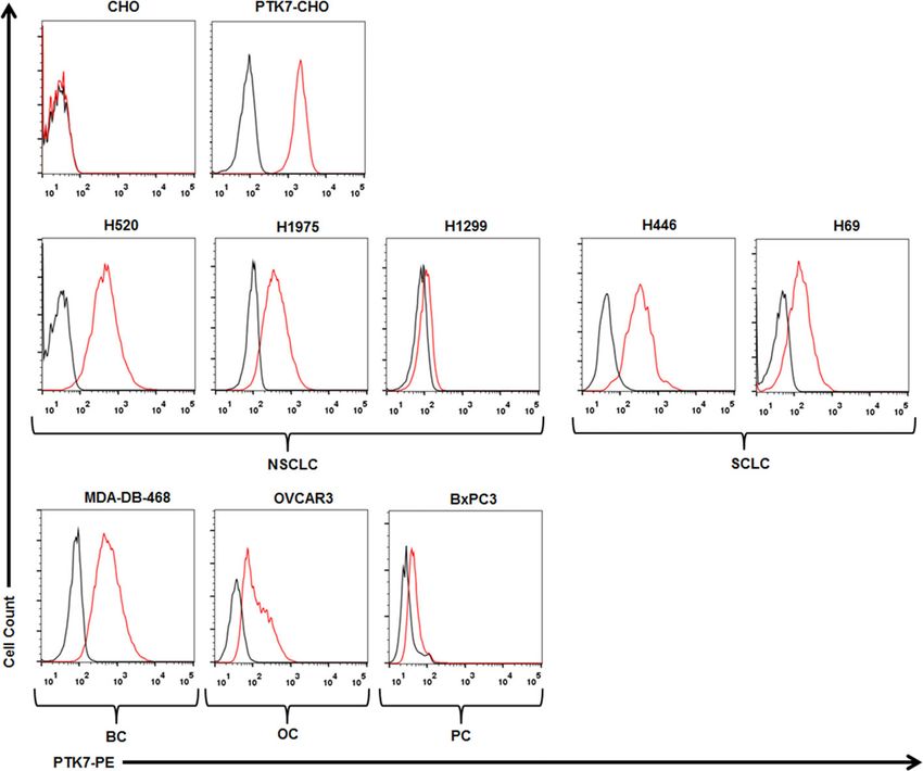
Jie et al.                                                                                                 PTK7 CAR T-Cells for Lung Cancer

expression is enriched on TIC/CSC-like cells, and targeting            PGK promoter with human EF1a promoter from pWPXLd
antigens with enriched expression in TIC/CSC-like cells would          vector (Plasmid #12258, Addgene). As a negative control,
achieve a long-term antitumor effect (26). Given the predicted         lentiviral vector encoding truncated tEGFR was constructed.
potential and safety of PTK7 as an immunotherapy target, we
sought to develop PTK7-specific CAR T-cell therapy for lung             Lentivirus Production
cancer and to evaluate its efficacy and safety in in vitro and          High-titer replication-incompetent lentiviruses were produced
in vivo preclinical models.                                            and concentrated as described previously (29). Briefly, HEK-
                                                                       293 T cells were transfected with pVSV-G (VSV glycoprotein
                                                                       expression plasmid), pRSV-Rev (Rev expression plasmid),
MATERIALS AND METHODS                                                  pMDLg/p.RRE (Gag/Pol expression plasmid), and pLVEF
                                                                       transfer plasmid using polyethylenimine (PEI, Sigma). The
Cell Lines                                                             viral supernatant was harvested at 24 and 48 h after
Human NSLCL cell lines H520, H1975, and H1299, SCLC cell               transfection and concentrated by using Lenti-X Concentrator
lines H446 and H69, pancreatic cancer cell line BxPC3, breast          (Clontech) in accordance with the manufacturer’s instructions.
cancer cell line MDA-DB-468, ovarian cancer cell line OVCAR3,
CHO, and HEK-293 T cells were purchased from American Type             CAR T-Cell Production
Culture Collection (ATCC) and maintained in DMEM medium                Human PBMCs were obtained from healthy donors under
(Thermo Fisher Scientific) supplemented with 10% heat-                  protocols approved by the Institutional Review Board of
inactivated fetal bovine serum (FBS), 2 mM glutamine, and 1%           Harbin Medical University and isolated by density gradient
penicillin/streptomycin (all from Thermo Fisher Scientific); and        centrifugation over Ficoll-Paque (GE Healthcare). Freshly
all cell lines were cultured at 37°C in a humidified chamber with       isolated PBMCs were then activated with antihuman CD3/
5% CO2. Stably transfected PTK7-CHO cell line was constructed          CD28 Dynabeads (Thermo Fisher Scientific) at a 3:1 ratio for
by infecting parental CHO cells with lentiviral supernatants           48 h followed by two sequential transductions with lentiviruses
containing PTK7 gene (#HG19399-UT, Sino Biological) and                on RetroNectin-coated non-tissue treated plates and maintained
sorting for PTK7 expression by using MoFloTM XDP cell                  in culture in RPMI-1640 (Thermo Fisher Scientific)
sorting system (Beckman Coulter). These cell lines were also           supplemented with 10% FBS (Thermo Fisher Scientific) and
infected with the lentiviral supernatants containing Luciferase-       recombinant human IL-2 (300 U/ml). Fresh media containing
IRES-GFP (GL) and were then sorted for GFP expression to               cytokine were replenished every other day to maintain T-cell
obtain GL-expressing cell lines. Human primary normal                  concentration at 0.5×10⁶ cells/ml. Five days after transduction,
epithelial cell lines (Mammary, Small Airway, and Renal                the CD3/CD28 Dynabeads were removed from the culture by
Epithelial Cells) and human umbilical vein endothelial cells           magnetic separation, and CAR T cells were propagated for 14
(HUVECs) were obtained from PriCells (Wuhan, China) and                days in total before using for functional assays. To track T cell
cultured according to the supplier’s instructions.                     numbers over time, viable cells were counted using trypan blue.

PTK7-CAR Construction                                                  Flow Cytometry
Sequences of three humanized mouse antihuman PTK7                      PTK7 expression on tumor cells was detected by mouse
antibodies (Hu23, Hu24, and Hu58 with the affinity of 3.9, 1.2,         monoclonal anti-PTK7 antibody (clone OTI2E7, Invitrogen)
and 2.1 nM, respectively) were obtained from a US patent               and goat anti-mouse IgG-phycoerythrin (PE)-conjugated
(US20150315293A1). The variable region sequences of heavy              antibody (Jackson ImmunoResearch). CAR expression on 293T
(VH) and light chain (VL) of these antibodies were used to             cells was detected by APC-conjugated rabbit monoclonal
design scFv with the sequence of VH-(G4S)3 Linker-VL. PTK7-            anti-EGFR antibody (Clone E01, Sino Biological) and
CARs containing scFv from Hu23, Hu24, and Hu58 were                    biotin-conjugated goat antihuman IgGF(ab′)2 (Jackson
designated as PTK7-CAR1, PTK7-CAR2, and PTK7-                          ImmunoResearch) and streptavidin-PE (BioLegend). CAR
CAR3, respectively. From the 5′-end to 3′-end, each CAR is             expression on T cells was detected by BV510-conjugated CD3
comprised of the human CD8a signal peptide sequence, PTK7-             (clone UCHT1), APC-Cy7-conjugated CD4 (clone OKT4), and
scFv, hinge and TM regions of the human CD8a molecule, 4-1BB           FITC-conjugated rabbit monoclonal anti-EGFR antibody (Clone
intracellular domain sequence (BB-ICD), and CD3z intracellular         E01, Sino Biological) and biotin-conjugated goat antihuman
domain sequence (CD3z-ICD) as previously described (27).               IgGF(ab′)2 and streptavidin-APC (BioLegend). The phenotype
Following CAR, a truncated tEGFR sequence is included via              and effector molecule expression on CAR T cells were detected
T2A ribosomal skipping sequence in the construct to allow for          with a panel of monoclonal antihuman antibodies as follows:
potential enrichment, tracking, and depletion if needed of             BV510-conjugated CD3 (clone UCHT1), BV421-conjugated
transduced T cells (28). DNA encoding the CARs was codon-              CD4 (clone OKT4), APC-Cy7-conjugated CD8 (clone SK1),
optimized and synthesized by General Biosystems (Anhui, China)         FITC-conjugated rabbit monoclonal anti-EGFR antibody,
with appropriate restriction sites. The CAR sequences were then        APC-conjugated CD45RO (clone UCHL1), PE-conjugated
cloned into third-generation self-inactivated lentiviral vector        CCR7 (clone G043H7), PE-conjugated TIM-3 (clone F38-2E2),
pLVEF derived from pRRLSIN.cPPT.PGK-GFP.WPRE vector                    APC-conjugated PD-1 (clone EH12.2H7), and PE-conjugated
(Plasmid #12252, Addgene) with replacing its original human            Granzyme B (clone GB11, all from BioLegend). CAR T cells in

Frontiers in Immunology | www.frontiersin.org                      3                                August 2021 | Volume 12 | Article 665970
Jie et al.                                                                                                         PTK7 CAR T-Cells for Lung Cancer

peripheral blood from tumor-bearing tumor were detected by                    of each round, a duplicate well was harvested for counting of
BV510-conjugated CD3 and APC-conjugated rabbit anti-EGFR                      residual tumor cells (GFP+) and CAR T cells (CD3+EGFR+) and
antibody. In most assays, cells were stained with Zombie Aqua™                other phenotypic analysis (granzyme B, PD-1, TIM-3) of CAR T
Fixable Viability Kit (BioLegend) to exclude dead cells from                  cells by flow cytometry.
analysis. Flow cytometry data were acquired with a
FACSCantoTM system (BD Biosciences) using DIVA software                       In Vivo Tumor Models
according to the manufacturers’ instructions.                                 All animal experiments were conducted under a protocol
                                                                              approved by the Institutional Animal Care and Use Committee
Cytokine Release Assays                                                       of the Harbin Medical University. Six- to 8-week-old B-NSG
Control or PTK7-CAR T cells (1×10⁵ cells/100 ml media) were                   mice (NOD-Prkdcscid Il2rgtm1/Bcgen) were obtained from
co-cultured with an equal number of target cells for 24 h, after              Biocytogen Co., Ltd (Beijing, China) and maintained on a 12 h
which cell-free supernatants were harvested for testing IL-2 and              light-dark cycle in a temperature-controlled high-barrier facility
IFN-g secretion by ELISA kits (R&D Systems) according to the                  with free access to food and water and treated under specific
manufacturer’s instructions.                                                  pathogen-free conditions at the Animal Centre of the Harbin
                                                                              Medical University. The tumor xenograft model was established
Proliferation Assay                                                           by subcutaneous (s.c.) inoculation with 3 × 10⁵ H520 or H69
Control or CAR T cells were first labeled with 5 mM fluorescent                 tumor cells suspended in 100 µl PBS. After 7 days, when the
dye carboxyfluorescein diacetate succinimidyl ester (CFSE;                     tumor was consistently palpable (50–100 mm3), mice were
Invitrogen) according to the manufacturer’s instructions, and                 randomized into three groups (three to five mice per group)
then co-cultured with tumor cells at an effector-to-target ratio of           and intravenously (i.v.) injected with 5 × 10⁶control or PTK7-
1:1. CFSE dilution was measured on gated T cells on day 3 using               CAR T cells suspended in 100 µl PBS and repeated once 1 week
flow cytometry.                                                                later. Mice were weekly monitored for tumor growth by using a
                                                                              caliper for 60 days, and then euthanized by cervical dislocation
In Vitro Killing Assays                                                       with blood and tumor harvested for analysis when they seemed
For tumor cell killing assays, GL-expressing target cells (1×10⁴              moribund or their tumors reached 15 mm in diameter. Tumor
cells/100 ml media) were co-cultured with control or PTK7-CAR                 volume (V) was calculated according to the following formula: V
T cells at the varying effector-to-target ratios in triplicate wells of       (mm3) = 0.5 × length × width2.
white 96-well plates. In some assays, it was conducted in the
presence of soluble PTK7 protein (OriGene). Target cell viability
was monitored 18 h later by using Bright-Glo™ Luciferase Assay                Immunohistochemistry (IHC)
System (Promega) according to the manufacturer’s instructions.                Tumor tissues were fixed with formalin and embedded in paraffin.
The percent lysis (%) was calculated by using the following                   Then, 4 mm thick sections were deparaffinized with xylene and
equation: 1-[bioluminescence value in sample well (target cells +             rehydrated in decreasing concentrations of ethanol. After heat-
CAR T cells)/maximum bioluminescence value (target                            induced antigen retrieval, slides were then blocked by 3% BSA and
cells alone)].                                                                stained with rabbit monoclonal antihuman CD3ϵ antibody (clone
    For human primary normal cell killing assays, target cells                SP162, Abcam) or rabbit polyclonal anti-PTK7 antibody
were first labeled with 5 mM fluorescent dye CFSE according to                  (Invitrogen) in the blocking solution overnight at 4°C.
the manufacturer’s instructions, and then co-cultured with                    Commercially available normal human tissue microarray (TMA;
control or PTK7-CAR T cells at the indicated effector-to-target               Shanghai Outdo Biotec) including 20 normal tissues (two to four
ratios in triplicates. After 18 h incubation at 37°C, mixed cells             sections per tissue) were retrieved by EDTA solution (Solarbio)
were harvested and stained with 7-AAD and then subjected to                   and then stained with the same polyclonal anti-PTK7 antibody
flow cytometric analysis to quantify remaining live (7-AAD                     (1:200 dilution at 4°C overnight). Slides were then rinsed with
negative) target cells. The cytotoxicity was calculated as                    Tris-HCl/0.05% Tween-20 buffer and visualized with a
100%—the percentage of alive target cells/alive target cells in               horseradish peroxidase (HRP)-conjugated anti-rabbit EnVision+
control wells without effectors.                                              Kit (Dako). PBS substituted for the primary antibody was used as
                                                                              the negative control.
In Vitro Recursive Cytotoxicity Assays
GL-expressing tumor cells (1×10⁵ cells/500 ml CAR-T culture                   Statistical Analysis
media) were seeded in 24-well tissue culture plates. After                    Statistical analyses were performed with GraphPad Prism
overnight plating, 2.5 × 10⁴ (effector-to-target ratio of 1:4)                software (version 7.0). Differences in groups were determined
CART cells in 500 ml media were added to the monolayer of                     by two-way ANOVA with Tukey’s multiple comparison test with
tumor cells (round 1). Three days later, when most of the target              P 0.05; *p < 0.05; **p < 0.01; ***p <
was repeated one more time, if applicable (round 3). At the end               0.001, or ****p < 0.0001.

Frontiers in Immunology | www.frontiersin.org                             4                                 August 2021 | Volume 12 | Article 665970
Jie et al. PTK7 CAR T-Cells for Lung Cancer
RESULTS each of the CARs and cloned into a self-inactivating lentiviral
vector. Then 293T cells were infected with CAR-encoding
Generation of PTK7-CAR T Cells replication-incompetent lentiviruses where CAR and tEGFR
To assess the suitability of PTK7 as a target for CAR T cells, we displayed a linear co-expression pattern, indicating that tEGFR
designed three CARs (PTK7-CAR1, PTK7-CAR2, and PTK7- is a reliable marker for PTK7-CAR expression (Figure 1B).
CAR3) each containing ascFv derived from one of three PBMC from healthy donors were then transduced with the
humanized antihuman PTK7 monoclonal antibodies lentiviruses following anti-CD3/CD28 bead stimulation, and
(Figure 1A). The PTK7-specific scFv was fused to CD8a hinge both CAR and tEGFR expressions were determined by FACS
and transmembrane domain with intracellular 4-1BB (CD137) analysis 5–7 days after transduction. We observed a similar linear
co-stimulatory and CD3z activating signaling domains in co-expression pattern of both CAR and tEGFR in each CAR-
tandem. To facilitate the detection of transduced T cells, a transduced T cells with CAR transduction efficiency
truncated EGFR (tEGFR) tag was included via T2A ribosomal approximately 40–60% and 20–30% in CD4+ and CD8+ T
skipping sequence. Expression of tEGFR alone served as a cells, respectively (Figures 1C and S1A). Although there was a
negative control. We synthesized full-length DNA encoding similar CAR expression positivity among three PTK7-CAR
A
B
C
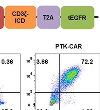
FIGURE 1 | PTK7-CAR generation, cell-surface expression, and transduction of human T cells. (A) PTK7-CAR was generated by fusing PTK7-specific scFv to the co-
stimulatory signaling domain of the 4-1BB (BB-ICD) and activating signaling domain of CD3z (CD3z-ICD), a T2A ribosomal skipping sequence, and tEGFR was included for
the detection of CAR-modified T cells. (B) 293T cells transfected with control or PTK7-CAR constructs express both CAR and the marker gene tEGFR. (C) PTK7-CAR
expression on transduced human CD4+ and CD8+ T cells was approximately 40–60% and 20–30%, respectively, as determined by tEGFR and CAR co-staining.
Frontiers in Immunology | www.frontiersin.org 5 August 2021 | Volume 12 | Article 665970
Jie et al. PTK7 CAR T-Cells for Lung Cancer
candidates, we consistently observed a high CAR expression per PTK7 representative of multiple cancer types, including NSCLC
cell in the PTK7-CAR2 T cells (Figure S1B). Phenotypic analysis (H520, H1975, H1299), SCLC (H446, H69), pancreatic (BxPC3),
showed that PTK7-CAR T cells contained central memory, breast (MDA-DB-468), and ovarian (OVCAR3) cancer
effector memory, and T stem cell memory, without significant (Figure 2). Similarly, PTK7-CAR T cells produced a large
differences among three candidates (Figure S1C). In addition, no amount of IFN-g and IL-2, which is positively associated with
difference in T-cell expansion without antigen stimulation was the expression level of PTK7 on respective tumor cells
seen in vitro among control and those candidates (Figure S1D). (Figure 3A). Notably, PTK7-CAR2 T cells had a trend of
producing a higher level of cytokines especially responding to
PTK7-CAR T Cells Secrete Effector stimulation by tumor cell lines expressing lower level of PTK7
Cytokines and Proliferate After Exposure (H1299 and BxPC3 cells), consistent with the higher level of
to PTK7-Expressingtumor Cells CAR expression per cell in this construct (Figure S1B).
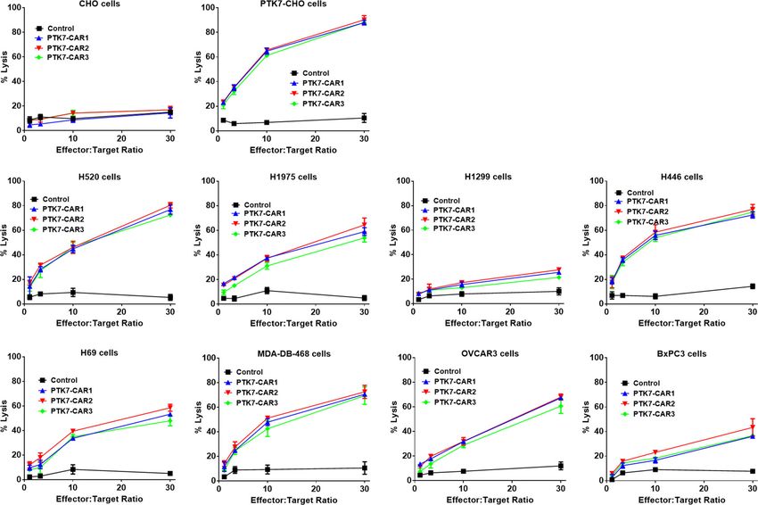
To test specific recognition by PTK7-CART cells, we initially We also evaluated the antigen-specific proliferation of PTK7-
exploited PTK7-negative parental CHO cells and stably CAR T cells in response to PTK7-expressing cells. T-cell
transfected PTK7-expressing PTK7-CHO cells (Figure 2). proliferation was dependent on the expression level of PTK7 on
PTK7-CAR T cells and control T cells of three donors were target cells, and tumor cells with a high level of PTK7 expression
co-cultured with CHO or PTK7-CHO cells, and effector cytokine induced more vigorous T-cell proliferation than that with a lower
IFN-g and IL-2 release in the supernatants were evaluated after level of PTK7 expression (Figure 3B). Again, PTK7-CAR2 T cells
24 h (Figure 3A). PTK7-CAR T cells secreted significant exhibited a trend of more potent proliferation when stimulated with
amounts of IFN-g and IL-2 after exposure to PTK7-CHO cells tumor cells expressing a lower level of PTK7.
compared with control T cells; however, parental CHO cells did
not stimulate PTK7-CAR T cells to produce effector cytokines, PTK7-CAR T Cells Specifically Kill PTK7-
indicating that cytokine production requires both the expression Expressing Tumor Cells and Retain Effector
of PTK7 on target cells and PTK7-CAR expression on Function Upon Recursive Target Exposure
transduced T cells. We confirmed the above findings using a We next evaluated the specific killing of PTK7-positive tumor cells
panel of tumor cell lines naturally expressing the varying levels of by PTK7-CAR T cells in both short-term (18 h) and recursive
FIGURE 2 | PTK7 is overexpressed on several tumor cell lines. CHO and PTK7-CHO cells served as negative and positive controls, respectively. PTK7
overexpression was observed on NSCLC (H520, H1975, H1299), SCLC (H446, H69), MDA-DB-468 breast cancer (BC), BxPC3 pancreatic cancer (PC), OVCAR3
ovarian cancer (OC) cells. Black and red lines denote the control (secondary antibody alone) and PTK7 staining, respectively.
Frontiers in Immunology | www.frontiersin.org 6 August 2021 | Volume 12 | Article 665970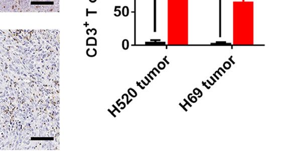
Jie et al.                                                                                                                        PTK7 CAR T-Cells for Lung Cancer

                              A

                              B

  FIGURE 3 | PTK7-CAR T cells release IFN-g and IL-2 and proliferate in response to PTK7-positive target cells. (A) Control or PTK7-CAR T cells from healthy donors
  (n = 3) were co-cultured with CHO and PTK7-CHO and various PTK7-expressing tumor cell lines for 24 h before performing IFN-g and IL-2 ELISA. Mean and SEM
  are shown. (B) T cells were labeled with CFSE and co-cultured for 3 days with CHO, PTK7-CHO, H520, H1975, or H1299 cells in the absence of exogenous IL-2,
  and CFSE dilution was analyzed by flow cytometry. A representative histogram from three independent assays is shown.

long-term (three rounds with each round of 3 days) cytotoxicity                       other two PTK7-CAR T cells failed to control tumor cell growth
assays. In the short-term assays, PTK7-CAR T cells exhibited a                        after the first or second round of challenge (Figure 5B). In parallel,
robust dose-dependent cytotoxicity against PTK7-expressing                            PTK7-CAR2 T cells had a better persistence after each round of
PTK7-CHO cells and tumor cells but not parental CHO cells                             challenge (Figure 5C). PTK7-CAR2 T cells also exhibited superior
(Figure 4). Noticeably, PTK7-CAR2 T cells demonstrated a                              effector function at the individual cell level as evidenced by higher
comparatively higher degree of cytotoxicity against tumor cells                       levels of lytic enzyme granzyme B expression and reduced
expressing the lower level of PTK7 (H69, BxPC3, and H1299 cells).                     expression of the exhaustion markers PD-1 and lower
As PTK7 has be reported to be shed from tumor cells in a soluble                      percentage of PD-1+TIM-3+ cells as compared to the other two
form (12), we also evaluated the effect of soluble PTK7 on the                        PTK7-CAR T cells (Figures 5D–F and S3).
cytotoxicity of PTK7-CAR T cells, which showed it minimally
impacted the tumor killing of these cells (Figure S2). Maintenance                    PTK7-CAR T Cells Mediate Antitumor
of specific cytotoxicity and proliferative response exposure to                        Activity Against Established Lung
continuous antigen stimulation has been described to be                               Cancer Xenografts
associated with preferential antitumor activity (30, 31). To                          In view of the in vitro preferential target-specific recognition and
mimic that context in vitro, we performed the recursive long-                         cytotoxicity of PTK7-CAR2 T cells as well as the fact that the
term cytotoxicity assay where CAR T cells were exposed to                             antibody from which the scFv used by PTK7-CAR2 is derived
recursive target cells at a certain ratio, and tumor cell killing and                 has been tested in the clinical trial (12), we evaluated the in vivo
T cell proliferation served as readouts after each round                              antitumor activity of these candidate CAR T cells in the
(Figure 5A). We observed that PTK7-CAR2 T cells retained                              xenograft tumor models established from two lung cancer cell
effective through three rounds of tumor challenge, whereas the                        lines with distinct antigen expression: H520 and H69 cells with

Frontiers in Immunology | www.frontiersin.org                                     7                                       August 2021 | Volume 12 | Article 665970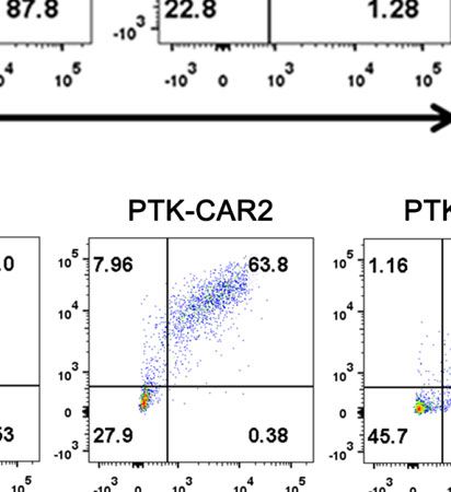
Jie et al.                                                                                                                          PTK7 CAR T-Cells for Lung Cancer

  FIGURE 4 | PTK7-CAR T cells kill PTK7-positive tumor cell lines. GL-expressing tumor target cells were co-cultured with control or PTK7-CAR T cells at the varying
  effector-to-target ratios in triplicate wells of white 96-well plates. Target cell viability was monitored 18 h later by using Bright-Glo™ Luciferase Assay System
  according to the manufacturer’s instructions. The percent lysis (%) was calculated by using the following equation: 1-[bioluminescence value in sample well (target
  cells + CAR T cells)/maximum bioluminescence value (target cells alone)]. Shown are means ± SEM of % cell killing in triplicate wells.

high or moderate level of PTK7 expression respectively as                              infiltration in tumor xenografts was determined by IHC
determined by flow cytometry and IHC staining of cell line-                             staining at the endpoint of the experiment, and mice treated
derived xenografts (Figures 2 and S4). NSG mice (n = 3–5/                              with PTK7-CAR2 T cells exhibited a prominent accumulation of
group) were s.c. inoculated with H520 or H69 tumor cells. Seven                        T cells within tumor tissues compared to mice treated with
days later mice started to receive two injection of control or                         control T cells (Figure 6D).
PTK7-CAR2 T cells 1 week apart, and tumor growth was                                       Importantly, there was no overt evidence of adverse reaction
monitored by measuring tumor size. Three independent                                   associated with the infusion of PTK7-CAR2 T cells to mice, as
experiments with T cells from different donors showed that                             measured by body weight loss and physical signs of toxicity in
administration of PTK7-CAR2 T cells greatly inhibited tumor                            above animal studies performed (Figure S6).
growth and significantly prolonged the overall survival of mice
bearing H520 (p
Jie et al.                                                                                                                           PTK7 CAR T-Cells for Lung Cancer

                               A

                               B

                               C

                               D                              E                              F

  FIGURE 5 | PTK7-CAR2 T cells retain effector function upon recursive target exposure. (A) Schematics of the long-term cytotoxicity assay. (B) Counts of H520 and
  H446 target cells after each round of recursive co-culture (rounds 1–3, R1–R3) with control or PTK7-CAR T cells. (C) Counts of control or PTK7-CAR T cells after
  each round of recursive co-culture with target cells. (D) Intracellular staining for granzyme B of control or PTK7-CAR T cells at the end of round 1 and 3 co-culture
  with H520 tumor cells. (E) PD-1 expression in control or PTK7-CAR T cells after rounds 1 and 3 of recursive co-culture with H520 tumor cells. (F) Percentage of PD-
  1+TIM-3+ cells in control or PTK7-CAR T cells after rounds 1 and 3 of recursive co-culture with H520 tumor cells. Data are shown as mean ± SEM (n = 3). *P < 0.05,
  **P < 0.01, ***P < 0.001, and ****P < 0.0001, determined by repeated-measures two-way ANOVA with Tukey’s post hoc test.

highest expression was observed in the stomach with moderate to                        control T cells, PTK7-CAR2 T cells did not exhibit more potent
strong cytoplasmic staining of gastric epithelium, colon with weak                     killing against this limited panel of normal human primary cells,
cytoplasmic staining of epithelium, and kidney with weak                               except for low-level cytotoxicity of HUVECs that was only
cytoplasmic staining of tubule epithelial cells. Since on-target off-                  observed at the highest effector-to-target ratio tested (Figure 7).
tumor toxicity is a key limiting factor when developing novel CAR                      As not all human tissues with PTK7 expression are represented,
T therapies, we roughly address this concern using a panel of                          these studies are limited but can serve as an initial screen for off-
primary human normal cell lines with low-level expression of                           tumor activity.
PTK7 (Figure S8). Control or PTK7-CAR2 T cells were co-
cultured with the primary human normal epithelial cell lines
from the mammary gland (Mammary Epithelial Cells, MECs),                               DISCUSSION
lung (Small Airway Epithelial Cells, SAECs), and kidney (Renal
Epithelial Cells, RECs) and human umbilical vein endothelial cells                     Here, we described the generation and antitumor efficacy of
(HUVECs), and cytotoxicity assays were performed. Compared to                          second-generation PTK7-targeting CAR T cells with 4-1BB

Frontiers in Immunology | www.frontiersin.org                                      9                                        August 2021 | Volume 12 | Article 665970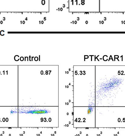
Jie et al.                                                                                                                        PTK7 CAR T-Cells for Lung Cancer

                                A

                                B

                                C

                                D

  FIGURE 6 | Systemic treatment with PTK7-CAR2 T cells leads to tumor growth control and increased survival of mice in both human tumor xenograft models.
  (A) NSG mice were s.c. implanted with H520 or H69 tumor cells, after 7 days, received two intravenous infusion of control or PTK7-CAR2 T cells (5 × 10⁶ cells in
  100 µl PBS) week apart and tumor growth quantified by measuring tumor size. Data are shown as mean ± SEM (n = 5 mice per group). *P < 0.05, ***P < 0.001, and
  ****P < 0.0001, determined by repeated-measures two-way ANOVA with Tukey’s post hoc test. (B) Kaplan–Meier survival curves summarizing three independent
  experiments (n = 11 mice per group). ***P < 0.001, and ****P < 0.0001 determined by log-rank test. (C) Frequency of human CD3+tEGFR+ CAR T cells in the
  peripheral blood collected 10 days after T cell infusion or at the end of experiment. Data are shown as mean ± SEM (n = 4 mice per group). **P < 0.01 and
  ***P < 0.001, determined by repeated-measures two-way ANOVA with Tukey’s post hoc test. (D) Representative IHC images and quantification of T-cell infiltration in
  tumor tissues (n = 3) from treated mice harvested at the end of experiment. Scale bars, 100 µm. Data are shown as mean ± SEM (n = 3 mice per group). ***P < 0.001,
  determined by repeated-measures two-way ANOVA with Tukey’s post hoc test.

Frontiers in Immunology | www.frontiersin.org                                    10                                       August 2021 | Volume 12 | Article 665970
Jie et al. PTK7 CAR T-Cells for Lung Cancer FIGURE 7 | PTK7-CAR2 T cells do not mediate detectable on-target off-tumor toxicity. Control or PTK7-CAR T cells were tested reactivity against a panel of primary human normal epithelial cells or HUVECs in the cytotoxicity assays at the indicated effector-to-target ratios. Shown are mean ± SEM of % cell killing in triplicate wells. **P < 0.01, determined by repeated-measures two-way ANOVA with Tukey’s post hoc test. intracellular co-stimulatory signaling domain and demonstrated studies will be needed to evaluate the antitumor efficacy of PTK7- antigen-specific cytokine production and cytotoxicity against CAR T cells in a more clinically relevant setting such as using multiple PTK7-positive tool cells and naturally expressing PDXs and patient-derived cancer cell lines, our data support human tumor cells in vitro; more importantly, in vitro recursive PTK7-CAR T cells as a viable therapeutic option for lung tumor challenge assays pointed to a preferred candidate (PTK7- cancers and many other solid cancers with PTK7 overexpression CAR2) out of three CAR constructs in terms of repetitive target given that it is impractical to develop blocking antibodies or small- cell killing, CAR T-cell expansion and exhaustion–associated molecule inhibitors as typically done with receptor tyrosine phenotypes, which was previously reported to be associated with kinases due to PTK7’s lack of catalytic activity. in vivo antitumor effect of CAR T cells (32). Using in vivo lung Several reports have identified PTK7 as a potential antigen cancer cell line–derived xenograft models, we showed that PTK7- target in solid tumors. Previous studies documented that PTK7 is CAR2 T cells significantly inhibited tumor growth and prolonged overexpressed in multiple types of solid cancer, and more overall survival of tumor-bearing mice. The reason why PTK7- significantly, its expression is enriched in TICs/CSCs from CAR2 exhibited a better functionality remains to be explored; PDXs or cancer cell lines. As TICs/CSCs with unlimited self- however, we consistently observed a high CAR expression per cell renewal capacity and differentiation potential have been broadly in the PTK7-CAR2 T cells (Figure S1B), which may underlie the considered to be source to tumor recurrence, metastasis, and fact that PTK7-CAR2 T cells had a better response to stimulation therapeutic resistance, it is reasonable to hypothesize that a by tumor cell lines expressing lower levels of PTK7. It is possible durable antitumor efficacy would be achieved if specifically that the unique scFv sequence in this construct makes this CAR targeting TIC/CSCs by immunotherapeutic modalities, more easy expression or more stable on the CAR-T cell surface, including CAR T-cell therapy. In fact, CAR T cells targeting enabling CAR-T cells a better response in recursive target several biomarkers of TICs/CSCs, including CD133, CD24, exposure. We did not evaluate PTK7-CARs integrating CD28 Receptor tyrosine kinase-like orphan receptor 1 (ROR1), or the co-stimulation as CD28-containing CAR T cells have undesirable epithelial cell adhesion molecule (EpCAM), have been developed increases in T cell exhaustion markers, limited persistence, and and exhibited the excellent antitumor effects in preclinical increased possibility of recognizing normal cells with very low models (34–40); more importantly, CD133-targeting CAR T levels of antigen as previously reported (30, 33). Although further cells alone or in combination have demonstrated antitumor Frontiers in Immunology | www.frontiersin.org 11 August 2021 | Volume 12 | Article 665970
Jie et al.                                                                                                      PTK7 CAR T-Cells for Lung Cancer

activity in treating patients with CD133-postive metastatic               PTK-CARs. Positively, Damelin et al. showed that a PTK7-
malignancies with controllable toxicities in clinical trials (41,         targeting ADC did not exhibit target-dependent toxicity in any
42). Intriguingly, both PTK7 and ROR1 belong to Wnt ligand                of the tissues examined, including those with PTK7 expression
binding receptors with important roles in the non-canonical               (12). As the scFv we used to generate PTK7-CAR2 is derived
Wnt signaling (10). ROR1 exhibits high and homogeneous cell               from the same antibody (Hu24) of that PTK7-ADC, the non-
surface expression in many epithelial tumors with expression              clinical safety profile of PTK7-ADC in that study provides some
profile similar to PTK7, and targeting ROR1 with CAR T-cell                evidence of safety and potential toxicity estimate of PTK7-CAR2
therapy improved survival in xenograft models of ROR1+ human              T cells in vivo. Caution still should be taken when translating this
tumors with treating lung and breast cancer in an ongoing                 PTK7-CAR T cells into clinic considering different mechanisms
clinical trial (NCT02706392) (38, 40). Thus, our result                   of action and target recognition sensitivity (potency) between
documenting a potent antitumor effect of PTK7-CAR T cells                 ADC and CAR T cells directing the same targets. In this regard,
adds PTK7 to the kind list of ROR1, which, as a member of Wnt             CAR-T cells targeting epithelial cell adhesion molecule
signaling-related pseudokinases, had a characteristic enriched            (EpCAM), a tumor-associated antigen overtly presented on the
expression in TIC/CSCs and is suitable as a potential therapeutic         cell surface of various carcinomas, is a typical precedent.
target for cancer immunotherapy.                                          Although anti-human EpCAM CAR-T cells unable of
    On-target off-tumor effect is a major concern when                    recognizing mouse EpCAM eradicated established tumor
developing CAR T-cell therapy targeting less tolerable TAAs               xenografts without toxicities in the immunodeficient animal
for solid tumors (8). On-target toxicities have been observed in          models, anti-mouse EpCAM CAR-T cells induced severe
clinical trials with CAR T cells specific for antigens that are            pulmonary immunopathology in theimmunocompetent mice
shared on some normal tissues (43, 44), and a critical issue to be        due to CAR-T recognition of basal EpCAM expression in
addressed is whether targeting PTK7 will be safe. Damelin et al.          normal lung (49). In addition, we may learn from the
have shown that PTK7 is absent in vital organs, but they detected         experience of targeting ROR1 by CAR T cells as these two
a low level of expression in esophagus, urinary bladder, kidney,          molecules have similar expression profiles in both normal and
mammary gland, lung, ovary, uterus, and digestive tract with              tumor tissues (10, 50). Although ROR1-CAR T cells (derived
more prominent expression in stromal part (12); in addition,              from R12- and 2A-scFv) without cross-reactivity with murine
previous studies detected PTK7 expression in human                        ROR1 exhibited no evident toxicity in NSG mice tumor model,
hematopoietic progenitors committed to myeloid and T                      murine ROR1-specific CAR T cells (derived from R11-scFv)
lymphoid lineages, illustrating the potential for toxicity to             induced lethal bone marrow failure due to recognition of ROR1+
normal cells (45–48). Roughly consistent with these previous              stromal cells, which can be rescued by the logic-Gated strategy of
studies, we also did not detect PTK7 expression in normal                 CAR construction (38, 40). Thus, the same configuration should
human major organs; however, focal, weak to moderate PTK7-                be considered when similar results are seen in future
positive staining was observed in the cytoplasm of some normal            investigations of PTK7-CAR T-cell’s toxicity profiles;
human tissues, including digestive tracts (stomach, esophagus,            alternatively, tuning scFv affinity and/or concomitantly
colon) and kidney. We further evaluated the activity of PTK7-             integrating different co-stimulatory domains may ameliorate
CAR T cells in vitro against a normal cell panel that included            the potential concern of on-target off-tumor effect as typical
mammary, lung, kidney epithelial cells, and HUVECs where a                representation for CAR T-cell therapy targeting a range of
low level of lysis against HUVECs was detected only at the                different antigens including but not limited to HER2, EGFR,
highest effector-to-target ratio tested, which is consistent with         CD38 (8, 51, 52). Given the above inherent risks, multiple
comparatively higher PTK7 expression on these cells as                    inducible safety controls that can be built into or applied in
determined by FACS. As CAR-T cells will first accumulate in                conjunction with CAR-T cells should be considered, such as
the lung through blood vessels, a very high local concentration           inducible caspase 9 (iCasp9) or tEGFR tag as we integrated in
may be achieved when CAR-T cells are intravenously infused,               CAR design where tEGFR-expressing CAR-T can be depleted by
leading to the blood vessel in the lung attacked by CAR-T cells           commercially available antibody cetuximab in case of emergent
and consequently on-target off-tumor toxicity. In addition, if            side effects (53). In sum, further thorough investigations are
applied for lung cancer treatment, a high level of cytokines              definitely needed to fully explore the potential toxicities of PTK7-
released from the on-target on-tumor recognition may                      CAR T cells before translating into clinic by using PTK7-CARs
constitute another important concern as this may induce                   with cross-reactivity in mouse and even non-human
pulmonary edema, which may be lethal if not diagnosed and                 primate models.
treated timely. Although mouse and human PTK7 protein has
90.93% homology in total sequence with 92.98% homology in the
extracellular domain, which means that tumor-bearing mouse                CONCLUSION
model should be suitable for the evaluation of on-target toxicity,
however, we cannot evaluate the potential toxicity profiles of             Here we describe a PTK7 targeting strategy that is based upon
PTK-CAR T cells in current murine tumor models due to lack of             CAR T-cell engineering. This synthetic biology approach
cross-reactivity with murine counterpart of humanized                     overcomes the issues related with PTK7 being pseudokinase
antihuman PTK7 antibodies used to construct scFv part of our              unsuited for developing antibody and small-molecule

Frontiers in Immunology | www.frontiersin.org                        12                                  August 2021 | Volume 12 | Article 665970
Jie et al. PTK7 CAR T-Cells for Lung Cancer
inhibitors as therapeutic agents, and is supported by the effector SUPPLEMENTARY MATERIAL
functions of modified T-cell in order to deliver PTK7-specific
cytotoxicity. In summary, the data presented herein serve as an The Supplementary Material for this article can be found online
initial step for future clinical development of PTK7-CAR T-cell at: https://www.frontiersin.org/articles/10.3389/fimmu.2021.
therapy safely and efficiently treating PTK7-expressing lung 665970/full#supplementary-material
cancer and other malignancies.
Supplementary Figure 1 | (A) The percentage of CAR expression on T cells
transduced with control or PTK-CAR lentiviruses. Data are shown as mean ± SEM
(n = 5 donors). (B) The intensity of CAR expression in both CD4 and CD8 T cells
DATA AVAILABILITY STATEMENT from 3 different CAR constructs. (C) CAR T-cell memory phenotypic analysis in both
CD4+ and CD8+ cells based on CD45RO and CCR7 expression as follows: Tn/
The original contributions presented in the study are included in Tscm naive/stem cell memory (CD45RO-/CCR7+), Tem central memory
(CD45RO+/CCR7+), Tem effector memory (CD45RO+/CCR7-), Teff effector cells
the article/Supplementary Material. Further inquiries can be (CD45RO-/CCR7-) (n = 5 donors). (D) The in vitro expansion curve of control or
directed to the corresponding author. PTK-CAR T cells. Data are normalized on the starting input T cell number and
shown as mean ± SEM of triplicates from one representative donor. *P < 0.05,
***P < 0.001, determined by two-way ANOVA with Turkey’s post hoc test.
ETHICS STATEMENT
Supplementary Figure 2 | Soluble PTK7 antigen does not impact tumor killing of
PTK7-CAR T cells in vitro. PTK7-CAR T cell-mediated tumor killing of H520 (A) or
This study was approved by ethical committees of the Harbin
H1975 (B) cells at the effector-to-target ratio of 10 in the presence or absence of 10
Medical University Cancer Hospital. µg/mL of soluble purified PTK7 antigen in the short-term cytotoxicity assay.
Supplementary Figure 3 | (A) Representative plots of flow cytometric analysis of
intracellular granzyme B (GZMB) expression in control or PTK7-CAR T cells at the
AUTHOR CONTRIBUTIONS end of round 1 and 3 co-culture with H520 tumor cells. (B) Representative plots of
flow cytometric analysis of PD-1 and TIM-3 expression on control or PTK7-CAR T
Conception and design of studies: YJ, GR, HW, AG. Acquisition, cells at the end of round 1 and 3 co-culture with H520 tumor cells.
analysis and interpretation: YJ, GL, LF, YL, ME, LW, XL, YYL,
YWL, HW, AG. Drafting article: YJ, AG. Critical review and Supplementary Figure 4 | Immunohistochemistry of PTK7 in tumor xenografts.
discussion: GR, HW. All authors contributed to the article and Immunohistochemistry with polyclonal anti-PTK7 antibody was performed on
formalin-fixed, processed, and paraffin-embedded (FFPE) tumor tissues from H520
approved the submitted version.
(A) or H69 (B) xenografts. Scale bar, 100 mm.
Supplementary Figure 5 | Representative plots of flow cytometric analysis of
FUNDING human CD3+tEGFR+ CAR T cells in the peripheral blood collected 10 days (D10)
after T cell infusion or at the end of experiment (End) from the H520 (A) or H69 (B)
This work was supported by the grants from the National Science xenograft tumor models.
and Technology Major Project (2017ZX10203206), the National Supplementary Figure 6 | Body weights were measured before tumor injection,
Natural Science Foundation of China (81572461, 81672274, and before CAR T-cell injection, and 7 and 28 days after CAR T-cell injection and
81372528), the Science Foundation of the Postdoctoral compared with PBS or control T-cell treated NSG mice. Lines indicate means ±
Department of Heilongjiang Province (No. LBH-Z16114), and SEM (n = 5 mice).
the Harbin Medical University Science Foundation of Innovative
Supplementary Figure 7 | Representative micrographs of PTK7 expression in
Science Research (2017LCZX107). indicated normal human organs assessed by staining with the rabbit polyclonal anti-
PTK7 antibody (Invitrogen) at the final concentration of 1 µg/ml. Micrographs are
representative of at least 2-3 sections per tissue. Magnification, x 40.
ACKNOWLEDGMENTS
Supplementary Figure 8 | PTK7 expression on the primary human normal
epithelial cell lines from mammary gland (Mammary Epithelial Cells, MECs), lung
The authors gratefully acknowledge Rui Chen for generously
(Small Airway Epithelial Cells, SAECs), and kidney (Renal Epithelial Cells, RECs) and
providing the lentiviral transfer plasmids (Luciferase/GFP) that human umbilical vein endothelial cells (HUVECs) determined by FACS analysis.
were used for cytotoxicity assays. The authors thank Lingxiong Black and red line denote the control (secondary antibody alone) and PTK7 staining
Wang and Chunxi Liu for excellent technical assistance. respectively.
3. Maus MV, Fraietta JA, Levine BL, Kalos M, Zhao Y, June CH. Adoptive
REFERENCES Immunotherapy for Cancer or Viruses. Annu Rev Immunol (2014) 32:189–
225. doi: 10.1146/annurev-immunol-032713-120136
1. Guedan S, Ruella M, June CH. Emerging Cellular Therapies for Cancer. Annu 4. Fuca G, Reppel L, Landoni E, Savoldo B, Dotti G. Enhancing Chimeric
Rev Immunol (2019) 37:145–71. doi: 10.1146/annurev-immunol-042718- Antigen Receptor T Cell Efficacy in Solid Tumors. Clin Cancer Res (2020)
041407 26:2444–51. doi: 10.1158/1078-0432.CCR-19-1835
2. Singh AK, McGuirk JP. CAR T Cells: Continuation in a Revolution of 5. Raje N, Berdeja J, Lin Y, Siegel D, Jagannath S, Madduri D, et al. Anti-BCMA
Immunotherapy. Lancet Oncol (2020) 21:e168–78. doi: 10.1016/S1470-2045 CAR T-Cell Therapy bb2121 in Relapsed or Refractory Multiple Myeloma.
(19)30823-X N Engl J Med (2019) 380:1726–37. doi: 10.1056/NEJMoa1817226
Frontiers in Immunology | www.frontiersin.org 13 August 2021 | Volume 12 | Article 665970Jie et al. PTK7 CAR T-Cells for Lung Cancer
6. Xu J, Chen LJ, Yang SS, Sun Y, Wu W, Liu YF, et al. Exploratory Trial of a 26. Prager BC, Xie Q, Bao S, Rich JN. Cancer Stem Cells: The Architects of the
Biepitopic CAR T-Targeting B Cell Maturation Antigen in Relapsed/ Tumor Ecosystem. Cell Stem Cell (2019) 24:41–53. doi: 10.1016/j.stem.
Refractory Multiple Myeloma. Proc Natl Acad Sci USA (2019) 116:9543–51. 2018.12.009
doi: 10.1073/pnas.1819745116 27. Milone MC, Fish JD, Carpenito C, Carroll RG, Binder GK, Teachey D, et al.
7. Fry TJ, Shah NN, Orentas RJ, Stetler-Stevenson M, Yuan CM, Ramakrishna S, Chimeric Receptors Containing CD137 Signal Transduction Domains
et al. CD22-Targeted CAR T Cells Induce Remission in B-ALL That Is Naive or Mediate Enhanced Survival of T Cells and Increased Antileukemic Efficacy
Resistant to CD19-Targeted CAR Immunotherapy. Nat Med (2018) 24:20–8. In Vivo. Mol Ther (2009) 17:1453–64. doi: 10.1038/mt.2009.83
doi: 10.1038/nm.4441 28. Wang X, Chang WC, Wong CW, Colcher D, Sherman M, Ostberg JR, et al. A
8. Rafiq S, Hackett CS, Brentjens RJ. Engineering Strategies to Overcome the Transgene-Encoded Cell Surface Polypeptide for Selection, In Vivo Tracking,
Current Roadblocks in CAR T Cell Therapy. Nat Rev Clin Oncol (2020) and Ablation of Engineered Cells. Blood (2011) 118:1255–63. doi: 10.1182/
17:147–67. doi: 10.1038/s41571-019-0297-y blood-2011-02-337360
9. MacKay M, Afshinnekoo E, Rub J, Hassan C, Khunte M, Baskaran N, et al. 29. Kuroda H, Kutner RH, Bazan NG, Reiser J. Simplified Lentivirus Vector
The Therapeutic Landscape for Cells Engineered With Chimeric Antigen Production in Protein-Free Media Using Polyethylenimine-Mediated
Receptors. Nat Biotechnol (2020) 38:233–44. doi: 10.1038/s41587-019-0329-2 Transfection. J Virol Methods (2009) 157:113–21. doi: 10.1016/j.jviromet.
10. Karvonen H, Perttila R, Niininen W, Barker H, Ungureanu D. Targeting Wnt 2008.11.021
Signaling Pseudokinases in Hematological Cancers. Eur J Haematol (2018) 30. Kawalekar OU, O’Connor RS, Fraietta JA, Guo L, McGettigan SE, Posey AD
101:457–65. doi: 10.1111/ejh.13137 Jr, et al. Distinct Signaling of Coreceptors Regulates Specific Metabolism
11. Peradziryi H, Tolwinski NS, Borchers A. The Many Roles of PTK7: A Pathways and Impacts Memory Development in CAR T Cells. Immunity
Versatile Regulator of Cell-Cell Communication. Arch Biochem Biophys (2016) 44:380–90. doi: 10.1016/j.immuni.2016.01.021
(2012) 524:71–6. doi: 10.1016/j.abb.2011.12.019 31. Zhao Z, Condomines M, van der Stegen SJC, Perna F, Kloss CC, Gunset G,
12. Damelin M, Bankovich A, Bernstein J, Lucas J, Chen L, Williams S, et al. A et al. Structural Design of Engineered Costimulation Determines Tumor
PTK7-Targeted Antibody-Drug Conjugate Reduces Tumor-Initiating Cells Rejection Kinetics and Persistence of CAR T Cells. Cancer Cell (2015)
and Induces Sustained Tumor Regressions. Sci Transl Med (2017) 9:eaag2611. 28:415–28. doi: 10.1016/j.ccell.2015.09.004
doi: 10.1126/scitranslmed.aag2611 32. Wang D, Aguilar B, Starr R, Alizadeh D, Brito A, Sarkissian A, et al.
13. Bin-Nun N, Lichtig H, Malyarova A, Levy M, Elias S, Frank D. PTK7 Glioblastoma-Targeted CD4+ CAR T Cells Mediate Superior Antitumor
Modulates Wnt Signaling Activity via LRP6. Development (2014) 141:410– Activity. JCI Insight (2018) 3:e99048. doi: 10.1172/jci.insight.99048
21. doi: 10.1242/dev.095984 33. Long AH, Haso WM, Shern JF, Wanhainen KM, Murgai M, Ingaramo M,
14. Hayes M, Naito M, Daulat A, Angers S, Ciruna B. Ptk7 Promotes non- et al. 4-1BB Costimulation Ameliorates T Cell Exhaustion Induced by Tonic
Canonical Wnt/PCP-Mediated Morphogenesis and Inhibits Wnt/Beta- Signaling of Chimeric Antigen Receptors. Nat Med (2015) 21:581–90. doi:
Catenin-Dependent Cell Fate Decisions During Vertebrate Development. 10.1038/nm.3838
Development (2013) 140:1807–18. doi: 10.1242/dev.090183 34. Zhu X, Prasad S, Gaedicke S, Hettich M, Firat E, Niedermann G. Patient-
15. Martinez S, Scerbo P, Giordano M, Daulat AM, Lhoumeau AC, Thome V, Derived Glioblastoma Stem Cells Are Killed by CD133-Specific CAR T Cells
et al. The PTK7 and ROR2 Protein Receptors Interact in the Vertebrate WNT/ But Induce the T Cell Aging Marker CD57. Oncotarget (2015) 6:171–84. doi:
Planar Cell Polarity (PCP) Pathway. J Biol Chem (2015) 290:30562–72. doi: 10.18632/oncotarget.2767
10.1074/jbc.M115.697615 35. Berger C, Sommermeyer D, Hudecek M, Berger M, Balakrishnan A,
16. Lu X, Borchers AG, Jolicoeur C, Rayburn H, Baker JC, Tessier-Lavigne M. Paszkiewicz PJ, et al. Safety of Targeting ROR1 in Primates With Chimeric
PTK7/CCK-4 Is a Novel Regulator of Planar Cell Polarity in Vertebrates. Antigen Receptor-Modified T Cells. Cancer Immunol Res (2015) 3:206–16.
Nature (2004) 430:93–8. doi: 10.1038/nature02677 doi: 10.1158/2326-6066.CIR-14-0163
17. Bie J, Hu X, Yang M, Shi X, Zhang X, Wang Z. PTK7 Promotes the Malignant 36. Klapdor R, Wang S, Morgan M, Dork T, Hacker U, Hillemanns P, et al.
Properties of Cancer Stem-Like Cells in Esophageal Squamous Cell Lines. Characterization of a Novel Third-Generation Anti-CD24-CAR Against
Hum Cell (2020) 33:356–65. doi: 10.1007/s13577-019-00309-6 Ovarian Cancer. Int J Mol Sci (2019) 20:660. doi: 10.3390/ijms20030660
18. Zou RC, Liang Y, Li LL, Tang JZ, Yang YP, Geng YC, et al. Bioinformatics 37. Maliar A, Servais C, Waks T, Chmielewski M, Lavy R, Altevogt P, et al.
Analysis Identifies Protein Tyrosine Kinase 7 (PTK7) as a Potential Prognostic Redirected T Cells That Target Pancreatic Adenocarcinoma Antigens
and Therapeutic Biomarker in Stages I to IV Hepatocellular Carcinoma. Med Eliminate Tumors and Metastases in Mice. Gastroenterology (2012)
Sci Monit (2019) 25:8618–27. doi: 10.12659/MSM.917142 143:1375–84.e1375. doi: 10.1053/j.gastro.2012.07.017
19. Meng L, Sefah K, O’Donoghue MB, Zhu G, Shangguan D, Noorali A, et al. Silencing 38. Srivastava S, Salter AI, Liggitt D, Yechan-Gunja S, Sarvothama M, Cooper K, et al.
of PTK7 in Colon Cancer Cells: Caspase-10-Dependent Apoptosis via Mitochondrial Logic-Gated ROR1 Chimeric Antigen Receptor Expression Rescues T Cell-
Pathway. PloS One (2010) 5:e14018. doi: 10.1371/journal.pone.0014018 Mediated Toxicity to Normal Tissues and Enables Selective Tumor Targeting.
20. Sun JJ, Li HL, Guo SJ, Ma H, Liu SJ, Liu D, et al. The Increased PTK7 Cancer Cell (2019) 35:489–503.e488. doi: 10.1016/j.ccell.2019.02.003
Expression Is a Malignant Factor in Cervical Cancer. Dis Markers (2019) 39. Zhang BL, Li D, Gong YL, Huang Y, Qin DY, Jiang L, et al. Preclinical
2019:5380197. doi: 10.1155/2019/5380197 Evaluation of Chimeric Antigen Receptor-Modified T Cells Specific to
21. Shin WS, Kwon J, Lee HW, Kang MC, Na HW, Lee ST, et al. Oncogenic Role Epithelial Cell Adhesion Molecule for Treating Colorectal Cancer. Hum
of Protein Tyrosine Kinase 7 in Esophageal Squamous Cell Carcinoma. Gene Ther (2019) 30:402–12. doi: 10.1089/hum.2018.229
Cancer Sci (2013) 104:1120–6. doi: 10.1111/cas.12194 40. Hudecek M, Lupo-Stanghellini MT, Kosasih PL, Sommermeyer D, Jensen MC,
22. Chen R, Khatri P, Mazur PK, Polin M, Zheng Y, Vaka D, et al. A Meta- Rader C, et al. Receptor Affinity and Extracellular Domain Modifications Affect
Analysis of Lung Cancer Gene Expression Identifies PTK7 as a Survival Gene Tumor Recognition by ROR1-Specific Chimeric Antigen Receptor T Cells.
in Lung Adenocarcinoma. Cancer Res (2014) 74:2892–902. doi: 10.1158/0008- Clin Cancer Res (2013) 19:3153–64. doi: 10.1158/1078-0432.CCR-13-0330
5472.CAN-13-2775 41. Wang Y, Chen M, Wu Z, Tong C, Dai H, Guo Y, et al. CD133-Directed CAR T
23. Ding D, Yang C, Lv C, Li J, Tan W. Improving Tumor Accumulation of Cells for Advanced Metastasis Malignancies: A Phase I Trial.
Aptamers by Prolonged Blood Circulation. Anal Chem (2020) 92:4108–14. Oncoimmunology (2018) 7:e1440169. doi: 10.1080/2162402X.2018.1440169
doi: 10.1021/acs.analchem.9b05878 42. Feng KC, Guo YL, Liu Y, Dai HR, Wang Y, Lv HY, et al. Cocktail Treatment
24. Zhang J, Zhao X, Yang C, Huang Z, Shi M, Pan S, et al. A Versatile Polyion With EGFR-Specific and CD133-Specific Chimeric Antigen Receptor-
Complex Can Intelligently Respond to a Tumor Microenvironment to Modified T Cells in a Patient With Advanced Cholangiocarcinoma.
Eliminate Tumor Stem Cells for Enhanced Lung Cancer Targeted Therapy. J Hematol Oncol (2017) 10:4. doi: 10.1186/s13045-016-0378-7
Biomater Sci (2019) 7:3751–63. doi: 10.1039/C9BM00812H 43. Morgan RA, Yang JC, Kitano M, Dudley ME, Laurencot CM, Rosenberg SA.
25. Yan H, Ren W, Liu S, Yu Y. Two-Photon Imaging of Aptamer-Functionalized Case Report of a Serious Adverse Event Following the Administration of T
Copolymer/TPdye Fluorescent Organic Dots Targeted to Cancer Cells. Anal Cells Transduced With a Chimeric Antigen Receptor Recognizing ERBB2.
Chim Acta (2020) 1106:199–206. doi: 10.1016/j.aca.2020.02.001 Mol Ther (2010) 18:843–51. doi: 10.1038/mt.2010.24
Frontiers in Immunology | www.frontiersin.org 14 August 2021 | Volume 12 | Article 665970You can also read