Chemosensory signal transduction in Caenorhabditis elegans
←
→
Page content transcription
If your browser does not render page correctly, please read the page content below
2
                                                                                                                           GENETICS, 2021, 217(3), iyab004
                                                                                                                               DOI: 10.1093/genetics/iyab004
                                                                                                                Advance Access Publication Date: 9 March 2021
                                                                                                                                                WormBook

Chemosensory signal transduction in Caenorhabditis
elegans
Denise M. Ferkey,1,*,† Piali Sengupta,2,*,† and Noelle D. L’Etoile                   3, ,†
                                                                                      *

                                                                                                                                                                Downloaded from https://academic.oup.com/genetics/article/217/3/iyab004/6162992 by guest on 11 September 2021
1
  Department of Biological Sciences, University at Buffalo, The State University of New York, Buffalo, NY 14260, USA
2
  Department of Biology, Brandeis University, Waltham, MA 02454, USA
3
  Department of Cell and Tissue Biology, University of California, San Francisco, CA 94143, USA
†
  These authors contributed equally to this work.

*Corresponding author: dmferkey@buffalo.edu (D.M.F.); sengupta@brandeis.edu (P.S.); noelle.letoile@ucsf.edu (N.D.L’E)

Abstract
Chemosensory neurons translate perception of external chemical cues, including odorants, tastants, and pheromones, into information that
drives attraction or avoidance motor programs. In the laboratory, robust behavioral assays, coupled with powerful genetic, molecular and
optical tools, have made Caenorhabditis elegans an ideal experimental system in which to dissect the contributions of individual genes
and neurons to ethologically relevant chemosensory behaviors. Here, we review current knowledge of the neurons, signal transduction
molecules and regulatory mechanisms that underlie the response of C. elegans to chemicals, including pheromones. The majority of identi-
fied molecules and pathways share remarkable homology with sensory mechanisms in other organisms. With the development of new tools
and technologies, we anticipate that continued study of chemosensory signal transduction and processing in C. elegans will yield addi-
tional new insights into the mechanisms by which this animal is able to detect and discriminate among thousands of chemical cues with a
limited sensory neuron repertoire.

Keywords: WormBook; chemosensation; signal transduction; taste; odorant; pheromone; C. elegans; olfaction; GPCR; sensory; signal-
ing; gustation

Introduction                                                                         Attractants and repellents
The first comprehensive review of C. elegans chemosensation was                      To effectively utilize C. elegans as a model system to study sen-
published in WormBook in 2006 (Bargmann 2006). This review                           sory neurobiological principles, systematic screens of worm
summarized our understanding of chemosensation in the nema-                          responses to individual chemicals have been conducted over the
tode at that time, beginning with work initiated in the 1970s                        years, beginning in the 1970s (Dusenbery 1973, 1974, 1975; Ward
when C. elegans was first being developed as a laboratory model                      1973). Table 1 provides a nonexhaustive list of water-soluble and
system. In the 15 years since its publication, the number of labs                    volatile compounds that have been demonstrated to attract or re-
studying chemosensation has grown considerably, along with                           pel wild-type animals (defined here as the Bristol N2 strain) in
our understanding of C. elegans nervous system function.                             the laboratory. Furthermore, C. elegans can discriminate between
    In this study, we focus specifically on behavioral responses of                  many of these compounds (Chou et al. 1996; L’Etoile and
C. elegans to attractants and repellents, chemosensory neuron                        Bargmann 2000). As in other animals, the behavioral responses of
physiology, and chemosensory signal transduction molecules                           C. elegans to a specific chemical can depend on its concentration.
and pathways. We also briefly discuss behavioral plasticity, but                     For instance, a subset of the chemical cues that are attractive at
only in the context of intracellular regulation of signaling cas-                    low concentrations can elicit avoidance responses at high con-
cades. By necessity, several salient topics have been omitted, in-                   centrations (Table 1).
cluding gas sensation, neuromodulation, and the mechanisms by                           Beyond having a catalog of the compounds that C. elegans can
which chemical information is processed and relayed to other                         respond to, an understanding of what each compound might rep-
neurons within sensory circuits (e.g., downstream interneurons).                     resent to the nematode in the wild provides context for its neuro-
The ability of several C. elegans sensory neurons to detect multi-                   anatomy, physiology, and sensory integration. In its natural
ple classes of stimuli (polymodality) also is not explicitly covered,                habitat, C. elegans is typically associated with microbe-rich or-
but this ability suggests that, given a limited number of neurons,                   ganic matter such as rotting fruit and vegetable matter (and also
polymodality may be necessary to achieve maximum functional-                         slugs) (Frézal and Félix 2015). A wide variety of bacterial strains
ity. Other nonchemosensory functions of a subset of these neu-                       have been found along with C. elegans in the wild, and several
rons are described elsewhere (Goodman and Sengupta 2019).                            nonpathogenic nutritious bacterial strains (Alcaligenes sp. JUb4,

Received: September 25, 2020. Accepted: January 05, 2021
C The Author(s) 2021. Published by Oxford University Press on behalf of Genetics Society of America.
V
This is an Open Access article distributed under the terms of the Creative Commons Attribution License (http://creativecommons.org/licenses/by/4.0/), which
permits unrestricted reuse, distribution, and reproduction in any medium, provided the original work is properly cited.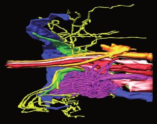
2 | GENETICS, 2021, Vol. 217, No. 3
Table 1 A nonexhaustive list of compounds that attract or repel wild-type animals in the laboratory and the neurons demonstrated to
detect them
Chemical stimulus Neuron(s) Soluble (S) or Volatile (V) Reference(s)
Attractants
Cyclic nucleotides ASE (ADF, ASG, ASI) S (Ward 1973)
cAMP (Bargmann and Horvitz 1991)
cGMP
Cations ASEL (ADF, ASG, ASI) S (Ward 1973)
Naþ ASER (ASEL) (Dusenbery 1974)
Kþ (Bargmann and Horvitz 1991)
(Pierce-Shimomura et al. 2001)
(Ortiz et al. 2009)
Anions ASER (ADF, ASG, ASI) S (Ward 1973)
Downloaded from https://academic.oup.com/genetics/article/217/3/iyab004/6162992 by guest on 11 September 2021
Cl (Dusenbery 1974)
(Bargmann and Horvitz 1991)
(Pierce-Shimomura et al. 2001)
Basic pH ASEL S (Ward 1973)
(Dusenbery 1974)
(Murayama et al. 2013)
Amino acids ASE (ASG, ASI, ASK) S (Ward 1973)
Lysine (Bargmann and Horvitz 1991)
Histidine (Ortiz et al. 2009)
Cysteine
Methionine
Biotin ASE (ADF, ASG, ASI) S (Bargmann and Horvitz 1991)
Pyrazine AWA V (Bargmann et al. 1993)
Diacetyl (low) AWA V (Bargmann et al. 1993)
Diacetyl (intermediate)a AWA, AWC V (Chou et al. 2001)
2,4,5-Trimethylthiazole (low) AWA, AWC V (Bargmann et al. 1993)
Butyric acidb AWA (AWC ?) V (Choi et al. 2018)
Isobutyric acid AWA (AWC ?) V (Choi et al. 2018)
Benzyl proprionate AWA, AWC V (Choi et al. 2018)
Benzaldehyde (low) AWC (AWA) V (Bargmann et al. 1993)
(Leinwand et al. 2015)
Isoamyl alcohol (low) AWC (AWA) V (Bargmann et al. 1993)
2-Butanone AWCON V (Bargmann et al. 1993)
(Wes and Bargmann 2001)
Acetone AWCON V (Bargmann et al. 1993)
(Worthy et al. 2018)
Dimethylthiazole AWC V (Bargmann et al. 1993)
(Choi et al. 2018)
1-Methylpyrrole AWC V (Choi et al. 2018)
1-Pentanol AWC V (Bargmann et al. 1993)
(Choi et al. 2018)
2-Cyclohexylethanol AWC V (Choi et al. 2018)
2-Ethoxythiazole AWC V (Bargmann et al. 1993)
(Choi et al. 2018)
2-Isobutylthiazole AWC (AWA ?) V (Bargmann et al. 1993)
(Choi et al. 2018)
2-Methylpyrazine AWC (AWA ?) V (Choi et al. 2018)
4-Chlorobenzyl mercaptan AWC (AWA ?) V (Choi et al. 2018)
Benzyl mercaptan AWC (AWA ?) V (Choi et al. 2018)
2-Heptanone AWCON V (Bargmann et al. 1993)
(Zhang et al. 2016)
2,3-Pentanedione (low) AWCOFF V (Chou et al. 2001)
(Wes and Bargmann 2001)
2,3-Pentanedione (intermediate) c AWA, AWC V (Chou et al. 2001)
Repellents (avoidance)
Acidic pH ASH, ADF, ASK, ASE S (Dusenbery 1974)
(Sambongi et al. 2000)
Basic pH (>10.5) ASH S (Sassa et al. 2013)
Copper ASH, ADL, ASE S (Bargmann et al. 1990)c
(Sambongi et al. 1999)
Cadmium ASH, ADL, ASE S (Sambongi et al. 1999)
SDS ASH (ASK, ASI, ASJ) S (Bargmann et al. 1990)c
PHA, PHB (antagonistic) (Hilliard et al. 2002)
(LIU et al. 2018)
Bitters quinine ASH (ASK) S (Hilliard et al. 2004)
Diacetyl (high) ASH V (Yoshida et al. 2012)
(Taniguchi et al. 2014)
2,4,5-Trimethylthiazole (high) V (Bargmann et al. 1993)
(Yoshida et al. 2012)
Benzaldehyde (high) ASH (AWB) V (Bargmann et al. 1993)
(continued)D. M. Ferkey, P. Sengupta, and N. D. L’Etoile | 3
Table 1 (continued)
Chemical stimulus Neuron(s) Soluble (S) or Volatile (V) Reference(s)
(Troemel et al. 1995)
(Luo et al. 2008)
(Yoshida et al. 2012)
Isoamyl alcohol (high) ASH (ADL, AWB) V (Luo et al. 2008)
(Yoshida et al. 2012)
Alcohols ASH (ADL, AWB—off food) V (Bargmann et al. 1993)
1-Octanol (100%) ASH (Troemel et al. 1995)
1-Octanol (30%) (Troemel et al. 1997)
(Chao et al. 2004)
Ketones AWB (ASH) V (Bargmann et al. 1993)
2-Nonanone (Troemel et al. 1997)
(Tanimoto et al. 2017)
Downloaded from https://academic.oup.com/genetics/article/217/3/iyab004/6162992 by guest on 11 September 2021
Serrawettin W2 AWB S (Pradel et al. 2007)
Phenazine-1-carboxamide ASJ S (Meisel et al. 2014)
Pyochelin ASJ S (Meisel et al. 2014)
Dodecanoic acid ASH (ADL ?, ADF ?) S (Tran et al. 2017)
PHA PHB
The references include those first reporting behavioral response to the chemicals, as well as those demonstrating the neurons involved in the response. The roles of
most neurons were shown by cell ablation, although some were revealed via genetic mutation or calcium imaging. Neurons with a more minor role are indicated by
a smaller font. Question marks indicate neurons with a possible role in detecting a stimulus.
a
Chou et al. (2001) refers to 1:10 dilutions of diacetyl and 2,3-pentanedione as “high” concentration. We have indicated them here as “intermediate” to
distinguish it from undiluted diacetyl and 2,3-pentanedione, which animals avoid (Yoshida et al. 2012).
b
Butyric acid was previously reported to be a neutral compound (Bargmann et al. 1993).
c
J. Thomas unpublished, cited in Bargmann et al. (1990).
Providenica sp. JUb5, Providencia sp. JUb39, and Flavobacteria sp. odors. However, following pathogenic infection, animals learn to
JUb43) release the “fruity” smelling attractive volatiles isoamyl al- avoid these odors (Zhang et al. 2005). This plasticity provides a
cohol, ethyl isobutyrate, and ethyl isovalerate (Samuel et al. 2016; model for learning and vertical transmission of pathogenic bacte-
Schulenburg and Félix 2017; Worthy et al. 2018a). The well- rial memory (Moore et al. 2019). C. elegans is also able to associate
studied attractant diacetyl is also released from a Lactobacillus chemicals with food or starvation and exhibit attraction or repul-
species that was found in rotting citrus (yazu) fruit that also con- sion, respectively, to these conditioned chemicals (for examples,
tained C. elegans (Choi et al. 2016). While the natural prey of C. ele- see Colbert and Bargmann 1997; Torayama et al. 2007; Kunitomo
gans have not been definitively identified (Schulenburg and Félix et al. 2013; Luo et al. 2014).
2017), the volatile chemicals emitted by these bacteria likely pro- In addition to compounds produced by potentially pathogenic
vide long-range attractive cues for seeking food. organisms or predators, C. elegans also avoids many compounds
Not all soil microbes are beneficial for C. elegans, and there are that are generally considered harmful at high concentrations
nematocidal fungi and bacteria that exude chemical cues that (Table 1). These include heavy metals (e.g., copper, and cad-
C. elegans avoids. For example, the pathogenic bacteria Serratia mium), and plant alkaloids or derivatives (e.g., quinine) that are
marcescens releases the cyclic lipodepsipentapeptide compound perceived as bitter by humans and are toxic for most animals
serrawettin W2 (Pradel et al. 2007), the pathogen Pseudomonas aer- (Sambongi et al. 1999; Hilliard et al. 2004). Taken together, the
uginosa emits phenazine-1-carboxamide (PCN) and the sidero- complex natural environment of C. elegans necessitates that
phore pyochelin (Meisel et al. 2014), and the nematocidal bacteria these animals be able to sense and respond robustly and sensi-
Streptomyces secretes dodecanoic acid (Tran et al. 2017)—all of tively to a range of chemical cues for optimal survival and repro-
which repel C. elegans. Aversive odorants such as 1-octanol and 2- duction.
nonanone may also indicate the presence of fungi or pathogenic
bacteria (Kaminski et al. 1974; Sharpell 1985). C. elegans may also
use chemical cues to be alerted to the presence of hungry nema- Assessing behavioral and neuronal
tode predators such as Pristionchus pacificus that release soluble responses
repellent sulfolipids when they are starved (Liu et al. 2018b). Behavioral strategies underlying C. elegans chemotaxis have been
Interestingly, some nematocidal predators exploit innate at- identified by studying animal movement in controlled spatial
tractive responses of C. elegans to specific compounds by releas- and temporal chemical gradients (Pierce-Shimomura et al. 1999,
ing attractive chemicals. For example, at least one nematode- 2005; Iino and Yoshida 2009; Broekmans et al. 2016). The behav-
trapping fungus (Arthrobotrys oligospora) appears to lure its prey ioral strategies used by C. elegans to migrate toward or away from
by releasing attractive volatile compounds that might mimic favorable (attraction) and noxious (avoidance) chemical cues, re-
food and pheromone cues (Hsueh et al. 2017). In addition, the spectively, are described in the Appendix. Here, we briefly outline
pathogenic bacterium B. nematocida B16 secretes an attractive the most common tools and paradigms for assessing behavioral
odor bouquet that includes benzaldehyde and 2-heptanone responses, and refer the reader to the Behavior methods chapter
(among others) that lures nematodes to their death via a “Trojan (Hart 2006) for more detailed descriptions of chemosensory
horse” mechanism (Niu et al. 2010). assays. Interested readers may also wish to consult these reviews
Innate responses of C. elegans to chemicals can be modified by for additional relevant information: (de Bono and Maricq 2005;
experience. The attractive chemicals butanone and acetone are Bargmann 2006; Bergamasco and Bazzicalupo 2006; Sengupta
emitted by the pathogenic bacteria S. marcescens and P. aeruginosa 2007; Hart and Chao 2010; Lockery 2011; Hobert 2013; Walker
(Worthy et al. 2018b), and inexperienced worms seek out these et al. 2017; Metaxakis et al. 2018).4 | GENETICS, 2021, Vol. 217, No. 3
Population assays provide good platforms to rapidly screen for Microfluidics-based assays have been very useful for simulta-
mutations that disrupt sensory function, as well as to catalog neous recording of behavior and neuronal activity in real time
chemicals that elicit behavioral responses. Typically, population (Albrecht and Bargmann 2011; Larsch et al. 2013). Briefly, animals
assays are performed on agar-filled Petri dishes, with a gradient are placed into a microfluidic device made of PDMS bonded to a
emanating from a point source of a stimulus (Bargmann and coverslip and shaped into an arena within which the animals’ be-
Horvitz 1991a; Bargmann et al. 1993). Uniform concentrations of havior can be observed. Within the arena, PDMS posts are ar-
soluble and/or volatile chemicals within quadrants of a Petri dish ranged to provide an artificial “dirt” substrate that the animals
are also used to assess preferences (Wicks et al. 2000; Frøkjær- can push against as they swim (Lockery et al. 2008). Ports flow
Jensen et al. 2008; Lee et al. 2009). These approaches can be high buffer and stimulus such that they produce a laminar stream,
throughput, and allow the assessment of responses of tens to allowing different spatiotemporal stimulus presentations. Using
hundreds of animals in a single assay. The output behavior is ei- two cameras, one with a low and the other a high magnification
ther scored as an endpoint assay (often reported as a chemotaxis objective, both locomotion and neuronal activity (e.g., calcium
Downloaded from https://academic.oup.com/genetics/article/217/3/iyab004/6162992 by guest on 11 September 2021
index) or tracked and assessed while the behavior is ongoing, transients) can be monitored simultaneously (Larsch et al. 2013;
thereby allowing a description of how an animal alters its loco- Levy and Bargmann 2020).
motor behavioral strategies to respond to a stimulus over time
(Brown et al. 2013; Husson et al. 2013; Tanimoto et al. 2017). The
responses of single animals can also be assessed and have been Neurons and their contributions to
used to quantitate avoidance behaviors. These assays typically chemosensation
measure the time for an individual animal to reverse from the There are 32 presumed chemosensory neurons in the hermaph-
aversive stimulus or report the percentage of animals that re- rodite C. elegans nervous system. They are housed within the
spond by reversing within a given timeframe (Troemel et al. 1995; head amphid and inner labial organs, as well as the tail phasmid
Hart et al. 1999; Hilliard et al. 2002). organs, and are directly or indirectly exposed to the environment
Changes in intracellular calcium levels are generally ac- (Ward et al. 1975; Ware et al. 1975; Perkins et al. 1986; White et al.
cepted as a useful readout for sensory neuron activity and are 1986; Bargmann 2006; Inglis et al. 2006). An additional pair of
the most accessible surrogate for electrophysiological experi- amphid neurons (AFD) is thermosensory (Goodman and
ments in C. elegans. However, when interpreting calcium imag- Sengupta 2019). Male-specific chemosensory neurons are de-
ing data, as described below, it is important to note that there scribed elsewhere (Barr et al. 2018). The functions of the eleven
may be scenarios in which calcium signaling does not directly pairs of amphid and two pairs of phasmid neurons have been ex-
correlate with neuronal depolarization (Zahratka et al. 2015). To tensively characterized in the context of chemosensation, and
report changes in calcium, calmodulin-based fluorescent pro- are the focus here. The ADL, ADF, ASE, ASG, ASH, ASI, ASJ, and
teins have been used, including FRET-based “cameleon” ASK neurons have simple, rod-like ciliated sensory endings that
(Miyawaki et al. 1997; Kerr et al. 2000; Suzuki et al. 2003; Fukuto terminate within a channel formed by glial cells associated with
et al. 2004; Hilliard et al. 2005) and single emission circularly per- the amphid sensilla. These neurons primarily detect soluble
mutated GFP proteins (GCaMP and its variants) (Romoser et al. ligands, although ASH and ADL can also detect volatile ligands
1997; Dana et al. 2019). An inverse-type reporter was also re- (Table 1). The AWA, AWB, and AWC amphid neurons embedded
cently developed to more reliably quantify a drop in calcium fol- within the sheath glial cells also have ciliated sensory endings
lowing stimulation (Hara-Kuge et al. 2018). Importantly, one that are more complex, and these neurons appear to detect pri-
needs to be aware that if the reporter sequesters calcium, neu- marily volatile chemicals (Table 1). For a high-resolution ultra-
rotransmission can be disrupted (Ferkey et al. 2007). Other read- structural analysis of the anterior endings of sensory neurons
outs for neuronal activity/regulation include cyclic nucleotides, (and glia) see Doroquez et al. (2014) and Figure 1. The PHA and
and cGMP levels can also be recorded (Couto et al. 2013; Shidara PHB nociceptive neurons in the phasmid sensilla have ciliated
et al. 2017; Woldemariam et al. 2019). However, there may be endings that terminate in the animal’s tail.
subcellular differences in calcium or cyclic nucleotides, includ- The majority of examined chemosensory neurons exhibit one
ing plasma membrane versus the cell body, as well as differen- of three distinct modes of response to chemical cues: (1) ON
ces in the cilia, dendrite, cell body, and axon to be considered (S. responses are increases in cytoplasmic calcium presumably due to
Woldemariam and N. L’Etoile, unpublished observations) depolarization that occurs when the concentration of the chemi-
(Shidara et al. 2017). Strains that express GCaMP in the nuclei of cal cue increases; (2) OFF responses are increases in cytoplasmic cal-
each neuron have been used to image the entire neural network cium that occur when the concentration of the chemical cue
in real time (Kato et al. 2015). decreases; (3) ON/OFF (biphasic) responses are increases in cytoplas-
In addition to changes in calcium levels, opening of other mic calcium that occur in response to both the onset and offset
nonspecific cation channels may contribute to membrane depo- (presentation and removal) of the chemical cue (Figure 2). In this
larization, and this needs to be considered. Thus, electrophysio- section, we briefly discuss the response physiology, including cal-
logical recordings provide the highest absolute and time-resolved cium responses and electrophysiological potentials when known,
insights into neuronal activity. Although technically difficult, this of the amphid and phasmid chemosensory neurons. A detailed
method has been used to provide high time resolution insights description of signal transduction molecules follows below.
(Goodman et al. 1998, 2012) that include the finding that RMD
(Mellem et al. 2008), AWA (Liu et al. 2018a), ASEL (Shindou et al. ASH
2019), and other neurons (Faumont et al. 2012) fire action poten- The ASH sensory neurons are the main nociceptors in C. elegans.
tials and/or exhibit regenerative plateau potentials. To fill the These neurons are considered to be “polymodal” because they de-
gap between calcium imaging and electrophysiological record- tect a wide range of aversive stimuli, including both chemical
ings, genetically encoded voltage sensory hold promise and port- and mechanical cues, similar to nociceptors in systems ranging
ing such sensors as the ASAP3 from mice could pave the way from other invertebrates such as Drosophila (Tracey et al. 2003;
(Villette et al. 2019). Zhong et al. 2010; Im and Galko 2012; Johnson and Carder 2012) toD. M. Ferkey, P. Sengupta, and N. D. L’Etoile | 5
Downloaded from https://academic.oup.com/genetics/article/217/3/iyab004/6162992 by guest on 11 September 2021
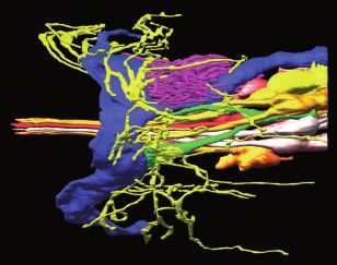
Figure 1 Cilia of amphid sensory neurons. (A and A0 ) 3 D reconstruction
model of the sensory endings of 12 amphid neuronal cilia on the right
side. Complex sensory endings of the winged cilia of AWA, AWB, and
AWC and microvilli of the AFD neurons are shown in (B). Single (ASH,
ASG, ASE, ASI, ASJ, and ASK) and double rod-shaped (ADF and ADL)
channel cilia are shown in (C) and (D), respectively. Individual amphid
neurons are color coded as indicated. Scale bar: 1 mm. Adapted from
Doroquez et al. (2014).
vertebrates (Besson and Chaouch 1987; Treede 1999; Lee et al.
2005). Examples of ASH-detected chemical stimuli are included
in (Table 1), and include high concentrations of several odorants
that are normally attractive at lower concentrations.
The ASH sensory neurons exhibit a phasic ON response when
presented with aversive chemical stimuli; for examples see
(Fukuto et al. 2004; Hilliard et al. 2005; Mills et al. 2012; Tanimoto
et al. 2017; Liu et al. 2018b). However, although the ON response
appears to be the general rule for ASH, there are also experimen-
tal paradigms where an OFF (Thiele et al. 2009) or biphasic (ON
and OFF) (Chronis et al. 2007; Kato et al. 2014; Wang et al. 2015) re-
sponse has been observed. Analysis of ASH temporal filter prop-
erties suggests that these nociceptors integrate noxious cues over Figure 2 Calcium responses in sensory neurons. Sensory neurons can
show a phasic increase in calcium levels upon presentation of stimulus
seconds to rapidly reach the activation threshold for avoidance
(ON response), an increase in calcium upon removal of stimulus (OFF
behavior (Kato et al. 2014). ASH calcium signaling in response to response), or an increase in calcium upon both the application and again
chemosensory stimuli, and the effects of genetic mutations on it, with the subsequent removal of stimulus (ON/OFF or biphasic response).
are discussed extensively in the signal transduction section be-
low. See also (Mirzakhalili et al. 2018) for additional computa- responses (Zahratka et al. 2015; Williams et al. 2018). These data
tional modeling of ASH signaling. have been interpreted to indicate that 5-HT enhances ASH excit-
While neuronal calcium flux is widely considered an indirect ability by suppressing a calcium-dependent inhibitory feedback
measure of neuronal activity, calcium transient amplitudes loop (Williams et al. 2018). Thus, calcium signals and depolariza-
within the soma may not always be predictive of neuronal depo- tion may not always be directly correlated.
larization and synaptic signaling. For example, exposure to
1-octanol leads to ASH depolarization (Zahratka et al. 2015). But,
surprisingly, while the neuromodulator serotonin (5-HT) potenti- ADL
ates ASH depolarization and ASH-mediated avoidance of 1-octa- In addition to their major role in pheromone detection
nol, it actually decreases 1-octanol-evoked ASH calcium (Pheromone), the ADL neurons play a minor role in chemical6 | GENETICS, 2021, Vol. 217, No. 3
avoidance such that their contribution to chemical detection is et al. 2018a; Dobosiewicz et al. 2019; Table 1). However, there are
often revealed only when they are ablated in combination with sex differences in attraction to some odorants, including diacetyl
other sensory neurons. Single and multineuron ablation experi- (Lee and Portman 2007; White et al. 2007; Ryan et al. 2014; Barr
ments have revealed a role for ADL in detecting several aversive et al. 2018).
stimuli (Table 1). In addition, ADL displays an ON response to re- AWA is an ON neuron that shows an elevated calcium levels
pellent P. pacificus predator cue (Liu et al. 2018b). However, al- in response to increases in diacetyl, pyrazine, 2-methylpyrazine,
though neuronal ablation studies implicate ADL in 1-octanol 2,4,5-trimethylthiazole and hexyl acetate (Shinkai et al. 2011;
avoidance (Troemel et al. 1995, 1997; Chao et al. 2004), ADL does Larsch et al. 2013, 2015; Zaslaver et al. 2015; Itskovits et al. 2018;
not shows a change in calcium levels following 1-octanol expo- Liu et al. 2018a; Dobosiewicz et al. 2019). This neuron pair also
sure (Mills et al. 2012). It is possible that ADL does not respond di- shows an increase in calcium in response to the addition of E. coli
rectly to 1-octanol, or perhaps ablation of ASH causes supernatant, and a decrease in calcium upon its removal
compensatory changes in ADL and AWB (see below) responsive- (Zaslaver et al. 2015). Activated AWA neurons signal to first order
Downloaded from https://academic.oup.com/genetics/article/217/3/iyab004/6162992 by guest on 11 September 2021
ness (Mills et al. 2012). interneurons such as AIA that reduce turning probability, thereby
elongating runs when an animal heads up the concentration gra-
AWB dient of an attractive chemical (Larsch et al. 2015).
The AWB neurons detect volatile aversive chemicals. They are As a food sensor, AWA’s ability to detect volatiles in gradients
the primary mediators of 2-nonanone avoidance (Troemel et al. that span large concentration ranges is likely to be important for
1997) and play a minor role in the avoidance response to several an animal’s survival. Indeed, the response properties of AWA en-
other odorants (Table 1). Calcium imaging experiments revealed able animals to respond to odorants over a 100,000-fold range of
that AWB can respond to distinct stimuli in a variety of ways. For concentrations (e.g., from as low as 11 nM up to 115 mM diacetyl)
example, while they showed an ON response when presented (Bargmann et al. 1993; Larsch et al. 2013). Calcium imaging
with 50 mM NaCl (Zaslaver et al. 2015), these neurons are acti- showed that the AWA neurons themselves respond reliably over
vated upon removal of 2-nonanone (OFF response) (Ha et al. 2010; the same wide span of concentrations (Larsch et al. 2013, 2015),
Tanimoto et al. 2017). Similarly, AWB showed an OFF response with oscillatory responses whose maxima remained constant
upon removal of high-isoamyl alcohol (Yoshida et al. 2012) or re- and did not scale with the concentration of the odor the worm
moval of an Escherichia coli supernatant (Zaslaver et al. 2015). was exposed to (Larsch et al. 2015; Itskovits et al. 2018). Responses
They also showed an unexpected ON/OFF biphasic response to a of these neurons to diacetyl sensitize rapidly at high concentra-
low concentration of isoamyl alcohol (104), which may be re- tions, thereby allowing the AWA neurons to retain response sen-
lated to their possible (very minor) contribution to chemotaxis to- sitivity over a wide dynamic range. AWA responses also adapt to
ward this odorant (Yoshida et al. 2012). Similar to ADL (above), the rate of change in concentration rather than to the absolute
1-octanol exposure/removal did not elicit AWB calcium transi- concentration, which allows the animal to seek out odor concen-
ents (Mills et al. 2012), although ablation studies suggest a minor trations that change most rapidly, thus allowing them to progress
role for AWB in 1-octanol avoidance (Troemel et al. 1997; Chao along the shortest route to an odor source (Itskovits et al. 2018).
et al. 2004). Interestingly, the oscillations of the left and right AWA neurons
were anti-correlated, but between the two they exhibited calcium
ASK transients at each upstep of odor (Itskovits et al. 2018).
The ASK neuron pair was first shown to play a minor role in che- Electrophysiological recordings provided additional insights
motaxis toward the amino acid lysine (Bargmann and Horvitz into how AWA may respond to odors over a broad dynamic range.
1991a). Although it is unusual for a C. elegans sensory neuron to These studies indicated that AWA fires bursts of 5–20 spikes in
detect both attractive and aversive stimuli, ASK also contributes about 15% of trials, and these have some of the hallmarks of an
to the avoidance of several soluble stimuli, including SDS action potential (Liu et al. 2018a); they are self-limiting, rising
(Table 1). Interestingly, ASK showed an OFF response to lysine, sharply then falling to a steady baseline, and they regenerate to
but an ON response to SDS (Wakabayashi et al. 2009). Because recur as a train of spikes (Bean 2007). By imaging GCaMP while
ASK activation promotes reversals (Wakabayashi et al. 2004; Gray injecting current, an algorithm was trained to use the electro-
et al. 2005), suppression of calcium signaling by a chemoattrac- physiological recording to detect spikes within the GCaMP traces.
tant and activation by a chemorepellent could both contribute to Applying this algorithm to GCaMP traces obtained when the
appropriate behavioral responses and locomotion strategies in AWA neurons were responding to intermediate concentrations of
complex chemosensory environments. For example, calcium im- diacetyl uncovered spiking calcium signals; changes in diacetyl
aging revealed that inhibition of ASK by the addition of diacetyl concentration elicited a similar spiking regime as seen with cur-
contributes to the disinhibition of the downstream interneuron rent injections (Liu et al. 2018a).
AIA, allowing AIA to more reliably respond to diacetyl-evoked de- Electrophysiological investigations of AWA also revealed
polarization of AWA (Dobosiewicz et al. 2019). While the applica- aspects of their responses that indicate how the neurons allow
tion of E. coli supernatant decreases ASK calcium levels, an animals to ignore noise, either in the environment or generated
elevation (OFF response) was seen upon its removal (Zaslaver by the animal’s movement. The time threshold for AWA activa-
et al. 2015). Similarly, an OFF response was also observed with re- tion was long, about 300 ms, such that only stimuli that lasted
moval of large (but not small) concentrations of suspended bacte- for longer than a third of a second were able to trigger spiking
ria (Calhoun et al. 2015). ASK also contributes to pheromone (Liu et al. 2018a). This time lag was also sufficient to filter out
detection (Pheromone). changes in concentration that would be generated by the typical
frequency of head swings generated by self-movement. This abil-
AWA ity to filter out noise could be attributed to as yet unidentified po-
The AWA olfactory neuron pair senses bacterially produced vola- tassium channels that increase the resistance of the AWA
tile cues to direct animals toward potential food sources membrane and keep small fluctuating stimuli from depolarizing
(Bargmann et al. 1993; Larsch et al. 2013; Choi et al. 2016; Worthy the cell (Liu et al. 2018a).D. M. Ferkey, P. Sengupta, and N. D. L’Etoile | 7
The calcium spikes generated by the AWA neurons adapt to Itskovits et al. 2018; Dobosiewicz et al. 2019; Levy and Bargmann
the magnitude of the change in odor concentration over time (Liu 2020). When the responses of AWC and AWA are modeled to-
et al. 2018a). Thus, turns should decrease as a function of an in- gether, they predict that animals are able to climb less continu-
crease in odor concentration. However, because AWA activity is ous gradients more efficiently (Itskovits et al. 2018; Dobosiewicz
discontinuous, rather than directing uninterrupted runs, a de- et al. 2019). Furthermore, in contrast to a salt gradient, animals in
crease in AWA activity is predicted to allow turns to emerge even an odor gradient (isoamyl alcohol) run faster up than down the
as an animal climbs a gradient (Itskovits et al. 2018). Thus, to gradient (Albrecht and Bargmann 2011). This also biases their
model robust climbing of a gradient at higher odor concentra- movement toward the peak of the odor stimulus.
tions, the spiking ON neuron pair had to be complemented with Levels of calcium and cGMP, the primary second messenger in
OFF neurons that had graded responses (Itskovits et al. 2018). The AWC sensory signaling (see below), both initially decrease in the
AWC neurons, with their response to intermediate concentra- cilia and dendrites in response to onset of odor presentation. But,
tions of diacetyl, may fulfill this role (Dobosiewicz et al. 2019). in the cell bodies, although calcium decreases, cGMP increases
Downloaded from https://academic.oup.com/genetics/article/217/3/iyab004/6162992 by guest on 11 September 2021
with odor onset (Shidara et al. 2017; Figure 3). How the cGMP sign
AWC is inverted between the cilia and the cell body is unclear, as is the
Many attractive odors are sensed by the paired AWC neurons physiological purpose of this inversion.
(Table 1), which along with the AWA neurons are the main olfac-
tory neurons in C. elegans (Bargmann et al. 1993). The two AWC ASI
neurons are not symmetric, as they express different G protein- The ASI sensory neurons play an important role in inhibiting en-
coupled receptors (GPCRs) (Troemel et al. 1997; Bauer Huang et al. try into the alternative stress-resistant dauer stage under
2007; Vidal et al. 2018) and respond to different odorants nondauer-inducing conditions (Bargmann and Horvitz 1991b;
(Table 1). Odorant bouquets from nutritive bacteria have been Schackwitz et al. 1996), and are the only source of DAF-7/TGF-b in
found to include known AWC-detected attractive volatiles C. elegans grown under standard conditions (Ren et al. 1996)
(Worthy et al. 2018a). Some attractive chemicals are also released (Pheromone). The ASIs also play a minor role in chemotaxis to
by nematophagus fungi (Hsueh et al. 2017) and pathogenic bacte- water-soluble stimuli (Table 1), but their contribution is only
ria (Worthy et al. 2018b), which may coopt AWC-mediated attrac- revealed when ASE (major) and other sensory neurons (minor)
tion to lure C. elegans (Zhang et al. 2016). These normally are ablated (Bargmann and Horvitz 1991a; Kaufman et al. 2005).
attractive odors can become repulsive when worms are sickened Ablation studies also showed a role for the ASI neurons in avoid-
or starved in their presence (Tsunozaki et al. 2008; Jin et al. 2016; ance of worm extract (Zhou et al. 2017), SDS and P. pacificus preda-
Kaletsky et al. 2018). The AWC neurons still sense these chemi- tor cue (Liu et al. 2018b). They also promote P. aeruginosa
cals under these conditions, but they instead direct repulsion avoidance, although it is not clear whether this is via direct detec-
(Tsunozaki et al. 2008; Jin et al. 2016). tion of pathogen-released chemical cues (Cao et al. 2017).
Calcium imaging showed that both AWC neurons are OFF Calcium imaging experiments revealed that the ASI displays an
neurons (Chalasani et al. 2007, 2010). They are tonically active in ON response to HB101 E. coli bacteria (Gallagher et al. 2013), OP50
buffer, showing low but constant activity that is silenced upon
odor addition. Conversely, when odor (or E. coli supernatant) is
withdrawn, both neurons show a sharp rise in calcium
(Chalasani et al. 2007, 2010; Kato et al. 2014; Calhoun et al. 2015;
Zaslaver et al. 2015; Cho et al. 2016; Hsueh et al. 2017; Hara-Kuge
et al. 2018). The AWC neurons induce turns when they are active
and forward runs when they are silent (Gray et al. 2005; Larsch
et al. 2013; Gordus et al. 2015; Itskovits et al. 2018; Dobosiewicz
et al. 2019), thereby directing runs up an attractive odor gradient.
The AWC calcium response to both odor exposure and re-
moval is rapid (less than a second), robust and reproducible (Kato
et al. 2014). Modeling showed that the speed of the response is
sufficiently rapid, relative to head swings, to allow animals to
track an odor gradient using the klinotaxis strategy (Izquierdo
and Lockery 2010) (see Appendix), and this was experimentally
verified using sensory signal transduction mutants (Kato et al.
2014). In addition, the response to a decrease in odor is graded
such that it scales with both the amount of odor prior to the de-
crease and to the change in odor concentration (Cho et al. 2016).
That is, the odor concentration is integrated over time to set the
neuron’s response threshold such that odor decreases that fall
below the set point (Levy and Bargmann 2020) enable reliable gra-
dient tracking.
The AWC neurons respond to some of the same odors as the Figure 3 Second messenger levels in cilia versus soma. When odorant is
AWA neurons, including diacetyl and isoamyl alcohol (Chou et al. removed from AWC, calcium levels increase in the cilia and the cell
2001; Larsch et al. 2015; Itskovits et al. 2018; Worthy et al. 2018a). body, while cGMP levels increase slightly in the cilia but fall in the cell
Interestingly, although AWA shows an oscillatory response to body. Likewise, when salt is removed from ASER, calcium levels increase
in the cilia and cell body, and cGMP levels fall in the cell body.
gradients of these odors, AWC responds with graded responses
Preliminary data indicate that cGMP levels rise in the ASER cilia when
such that the AWC calcium signal is directly proportional to the salt is removed (S. Woldemariam and N. L’Etoile, personal
change in stimulating odor concentration (Cho et al. 2016; communication).8 | GENETICS, 2021, Vol. 217, No. 3
E. coli bacteria (Calhoun et al. 2015) and supernatant (Zaslaver ASJ
et al. 2015), and Luria Broth (LB) (Gallagher et al. 2013; Davis et al. The major role of the ASJ neurons is to regulate dauer entry and
2018), suggesting a role in food sensation. The activation of ASI exit (Pheromone). ASJ promotes dauer formation, such that killing
by external nutrients promotes satiety quiescence (You et al. these neurons significantly impaired the ability of wild-type ani-
2008; Gallagher et al. 2013). In addition, the aversive stimulus mals to form dauers when exposed to dauer pheromone
CuSO4 elicits an OFF response in ASI that allows them to modu- (Schackwitz et al. 1996). ASJ also promotes dauer recovery, and
late copper nociception in a reciprocal inhibition circuit with the when the ASJ neurons are ablated animals permanently arrest in
primary copper detectors, the ASH neurons (Guo et al. 2015). P. the dauer stage (Bargmann and Horvitz 1991b). In addition to
pacificus predator cue also elicits an OFF calcium response in ASI these roles in the regulation of the dauer state, the ASJ neurons
(Liu et al. 2018b). mediate avoidance of P. aeruginosa, most likely by detecting both
secondary metabolites (Meisel et al. 2014) and nitric oxide (Hao
et al. 2018) produced by these bacteria (Table 1). They also con-
Downloaded from https://academic.oup.com/genetics/article/217/3/iyab004/6162992 by guest on 11 September 2021
ADF tribute to the avoidance of SDS and P. pacificus predator cue (Liu
The ADF neurons are the only serotonergic sensory neurons in et al. 2018b). Calcium imaging experiments revealed that the ap-
the hermaphrodite (Sze et al. 2000) and appear to be tonically ac- plication of the P. aeruginosa secondary metabolite PCN led to an
tive (Thiele et al. 2009). Thus, they are uniquely positioned to re- increase in ASJ calcium levels (Meisel et al. 2014), as did presenta-
spond to environmental cues and modulate chemosensory tion of 50 mM NaCl, pH 5 or E. coli supernatant (Zaslaver et al.
behavioral responses. In the larva, they inhibit entry into the 2015). Alternatively, an OFF response was seen upon removal of
dauer stage under nondauer-inducing conditions (Bargmann and P. pacificus predator cue (Liu et al. 2018b). ASJ may also play a very
Horvitz 1991b; Schackwitz et al. 1996) (Pheromone). In adults, un- minor role in chemotaxis to some water-soluble stimuli
der “normoxic” conditions the ADF neurons (along with ASG and (Bargmann and Horvitz 1991a; Kaufman et al. 2005).
ASI) also play a minor role in chemotaxis to water-soluble stimuli
(Table 1), but their contribution is only revealed when ASE (ma- ASE
jor) and other sensory neurons (minor) are ablated (Bargmann
The left and right ASE neurons signal to both shared and distinct
and Horvitz 1991a). However, under hypoxic conditions (e.g.,
interneurons (Cook et al. 2019) (see also http://wormwiring.org)
those created by high bacterial metabolism in enclosed spaces)
and they respond to different chemicals (Table 1). The left and
the role of ADF (and ASG) in salt chemotaxis may be enhanced
right ASE neurons also express different genes, including receptor
due to the upregulation of 5-HT in these neurons (Pocock and
guanylyl cyclases (rGCs) that may be tuned to detect these dis-
Hobert 2010). Calcium imaging experiments have revealed that
tinct stimuli (Chang et al. 2003; Ortiz et al. 2009; Smith et al. 2013).
the ADF neurons show an ON response to E. coli supernatant
In addition to this profound difference in sensory function, the
(Zaslaver et al. 2015), and they respond directly to repellent levels
two neurons differ in size (subtly) and electrophysiological prop-
(1/100) of isoamyl alcohol and indirectly to copper (Shao et al.
erties (Pierce-Shimomura et al. 2001; Goldsmith et al. 2010).
2019). ADF activation by these stimuli in turn inhibits the ASH
The left and right ASE neurons also differ in their contribution
nociceptors to modulate aversive chemosensory responses (Shao
to the locomotor strategies utilized during salt chemotaxis. ASEL
et al. 2019). The ADF neurons also show a calcium ON response to
responds to an increase in cations and its activity correlates with
NaCl upsteps, although their activation may not be the result of
runs up the gradient, while ASER responds to decreases in anions
direct stimulation in this context; ADF may be postsynaptic to a
by initiating pirouettes and decreasing run length (Figure A1).
salt-sensitive neuron(s) (Thiele et al. 2009).
Calcium imaging studies (Pierce-Shimomura et al. 2001; Suzuki
et al. 2008; Kunitomo et al. 2013; Luo et al. 2014; Wang et al. 2017;
Lim et al. 2018; Shindou et al. 2019) and electrophysiology
ASG (Shindou et al. 2019) corroborate the finding that ASEL is an ON
The ASGs play a minor role in inhibiting entry into the dauer cell that depolarizes and increases intracellular calcium in re-
stage under nondauer-inducing conditions (Bargmann and sponse to increases in salt concentration (upsteps), while ASER is
Horvitz 1991b; Schackwitz et al. 1996). In addition, under ambient an OFF cell that depolarizes and increases intracellular calcium
(“normoxic”) oxygen conditions, the ASG neurons (along with with decreases in salt concentration (downsteps). ASEL and ASER
ADF and ASI) play a minor role in chemotaxis to water-soluble respond to changes in salt with a transient influx of calcium that
stimuli (Table 1), but their contribution is only revealed when marks the onset of the change (salt up or down, respectively)
ASE (major) and other sensory neurons (minor) are ablated (Suzuki et al. 2008; Oda et al. 2011; Luo et al. 2014; Lim et al. 2018;
(Bargmann and Horvitz 1991a). However, under hypoxic condi- Shindou et al. 2019). This combination of ON and OFF sensory
tions the role of ASG (and ADF) in salt chemotaxis may be en- cells underlies the ability of animals to reliably track a smooth
hanced due to the upregulation of 5-HT biosynthesis in these gradient, composed of dissolved ion pairs, to its source (Pierce-
neurons (Pocock and Hobert 2010). Surprisingly, in contrast to the Shimomura et al. 1999, 2001; Suzuki et al. 2008; Iino and Yoshida
cell ablation results, calcium imaging (under normoxic condi- 2009; Izquierdo et al. 2015).
tions) did not reveal ASG calcium transients in response to either Electrical responses to current injection reveal that ASEL and
NaCl upsteps or downsteps (Thiele et al. 2009; Jang et al. 2019). ASER signal in a nonlinear regenerative manner (Goodman et al.
However, the ASG neurons do show spontaneous calcium fluxes 1998; Shindou et al. 2019) generating plateau potentials (Lockery
independent of salt stimulation, and both the frequency and av- and Goodman 2009). Responses to current injection depend on
erage size of the activity peaks were higher after salt conditioning extracellular sodium and calcium in concert, but are robust to re-
under starvation conditions (Jang et al. 2019). Thus, via their con- moval of either alone (Shindou et al. 2019). This observation sug-
tribution to switching an animal’s navigation direction relative to gests that voltage- and/or calcium-dependent channels underpin
a salt gradient, ASG activity may help animals to avoid salt con- nonlinear regenerative signaling. Salt upsteps also evoke plateau
centrations associated with starvation (Jang et al. 2019). potentials in ASEL and the probability of triggering this responseD. M. Ferkey, P. Sengupta, and N. D. L’Etoile | 9
is proportional to the change in salt concentration (Shindou et al. appears to be cell autonomous, the OFF response was abolished
2019), providing a mechanism by which ASEL detects and signals in unc-31 mutant animals lacking neuropeptidergic signaling,
the changes in external salt concentration that drive chemotaxis. suggesting that PHA/PHB may be, in part, postsynaptically acti-
Additional channels are likely to act in concert with the voltage- vated by copper removal via neuropeptides (Zou et al. 2017). No
gated calcium channel (VGCC) EGL-19 to allow triggering of neu- calcium transients were observed in response to quinine or acidic
rotransmission. pH (Zou et al. 2017).
Within ASEL, the salt upstep signal is seen as an influx of cal-
cium in sensory cilium, dendrites, soma and axons (Lim et al.
et al. 2018; Shindou et al. 2019). As described further below, cGMP Chemosensory signal transduction
is the primary second messenger in salt sensory transduction. molecules
The cGMP signal at the sensory cilia is translated into changes in
Below we describe current knowledge about the signaling mole-
intracellular calcium dynamics and further amplified via VGCCs
cules that transduce chemosensory information within the sen-
Downloaded from https://academic.oup.com/genetics/article/217/3/iyab004/6162992 by guest on 11 September 2021
(Shindou et al. 2019). Interestingly, although calcium levels in-
sory neurons. We also refer the reader to Hobert (2013) for a
crease in the ASEL soma as a result of a salt upstep, cGMP levels
broader description of the gene families that function in the C.
decrease (Woldemariam et al. 2019). Similarly, ASER somal cal-
elegans nervous system. While many gene families with neuronal
cium rises and cGMP falls in response to a salt downstep
functions appear to be expanded in C. elegans, a notable exception
(Woldemariam et al. 2019; Figure 3). However, the mechanism un-
is the absence of voltage-gated sodium channels (Bargmann
derlying the opposite calcium and cGMP changes in the soma of
1998; Hobert 2013). See Figure 4 for a summary of the signal
these neurons is currently unclear.
transduction pathways that function specifically within the ASH,
The ASE neurons also allow an animal to tune its response
AWA, AWC, and ASE neurons.
to salt such that it will become attracted to the salt concentra-
tion associated with food experience (Kunitomo et al. 2013; Luo
et al. 2014). Imaging ASEL and ASER calcium levels as the ani- G protein-coupled receptors (GPCRs)
mal is exposed to abrupt downsteps (Kunitomo et al. 2013) or is The first expression analysis of putative C. elegans chemosensory
traversing a more natural gradient (Luo et al. 2014) revealed GPCRs was undertaken over 20 years ago (Troemel et al. 1995).
that ASER changes the dynamics of its responses to decreases This foundational study, utilizing the partial genome sequence
and increases in salt as a function of the salt concentration at available, initially identified 41 potential C. elegans chemorecep-
cultivation. ASER is most active in response to decreases in salt tor genes that fell into six families (sra, srb, srg, srd, sre, and sro)
when the animal is below this set point, driving the animal to based on sequence similarity with one another (Troemel et al.
higher salt by increasing turning (Kunitomo et al. 2013). But, 1995). As completion of the full-genome sequence, a total of ap-
when the animal is at or above the set point and tracks to a proximately 1,300 genes and 400 pseudogenes have been identi-
lower salt concentration, similar downsteps in salt evoke fied, and they are now classified into 19 families (15 of these
smaller (Kunitomo et al. 2013) and more complex (Luo et al. comprise three major superfamilies: sra, str, srg) (Robertson and
2014) calcium transients. Thomas 2006; Thomas and Robertson 2008). Chemosensory
GPCR genes are now known to be the largest gene family in C. ele-
PHA/PHB gans, comprising 8.5% of all its genes (Thomas and Robertson
The PHA and PHB neurons are located in the phasmid sensory 2008). We refer the reader to the primary literature for a more
organs of the tail of C. elegans, and their role in chemosensation thorough analysis of these gene families and their evolution
was first shown in 2002 (Hilliard et al. 2002). Although ablation of (Troemel et al. 1995; Robertson 1998, 2000, 2001; Chen et al. 2005;
PHA and PHB did not affect SDS avoidance, their ablation in com- Thomas et al. 2005; Thomas and Robertson 2008; Nagarathnam
bination with ASH (or ASH and ASK) leads to a stronger avoid- et al. 2012; Krishnan et al. 2014).
ance response than ablation of ASH alone (or ASH and ASK) GFP-based expression analysis of a subset of the first identi-
(Hilliard et al. 2002). This suggested that PHA/PHB antagonize SDS fied putative receptor genes revealed that many were expressed
avoidance that is mediated by the amphid neurons (Table 1), and in only a small subset of chemosensory neurons (Troemel et al.
that the decision to initiate backward locomotion (reversal) is 1995). In addition, this work established that a single type of che-
based on the integration of sensory information from the head mosensory neuron can express multiple chemoreceptor genes
and the tail (Hilliard et al. 2002; Oren-Suissa et al. 2016). Shared (Troemel et al. 1995). This observation has been corroborated
connections with command interneurons in hermaphrodites fur- multiple times, through studies of individual receptors and sen-
ther support this model (White et al. 1986) (and wormwiring.org). sory neurons, and more recently by a large-scale study that ex-
In addition, PHA and PHB also mediate avoidance of dodecanoic amined the expression pattern of 244 rhodopsin-like (class A) C.
acid presented to the tail (Tran et al. 2017). elegans chemoreceptors (Vidal et al. 2018). A small number of C.
Calcium imaging experiments have shown that PHA and PHB elegans chemosensory GPCRs show left/right asymmetric gene ex-
act as polymodal nociceptors, with an ON response to SDS, aver- pression, but this asymmetry has so far only been observed for
sive odors (1-octanol), high isoamyl alcohol, alkaline pH (12), high the AWC sensory neuron pair (Troemel et al. 1999; Bauer Huang
osmolarity and harsh touch (Zou et al. 2017). For each of these et al. 2007; Vidal et al. 2018). Consistent with the original findings
stimuli, the responses of PHA and PHB were similar (Zou et al. (Troemel et al. 1995), some of the putative chemoreceptors were
2017). cGMP imaging of PHB also indicated that SDS triggers an also found to be expressed in interneurons and motor neurons,
increase in cGMP (Woldemariam et al. 2019), which could drive and sometimes even in nonneuronal cells (Vidal et al. 2018).
the opening of cyclic nucleotide-gated (CNG) channels that func- Thus, it is possible that some receptors may sense internal cues
tion in the phasmids (Hilliard et al. 2002). In contrast, the applica- in addition to environmental stimuli. Complementing GFP-based
tion of copper decreased calcium levels in PHA/PHB, while copper studies with single cell transcriptional profiling (Hammarlund
removal led to an increase in calcium levels (OFF response) (Zou et al. 2018) should provide additional insights into the receptor
et al. 2017). However, while the decrease in calcium signaling code of individual cells.10 | GENETICS, 2021, Vol. 217, No. 3
Downloaded from https://academic.oup.com/genetics/article/217/3/iyab004/6162992 by guest on 11 September 2021
Figure 4 Signal transduction pathways in the ASH, AWA, AWC, and ASE sensory neurons. Simplified models of the potential signal transduction
pathways for these representative neurons are shown. See text within the Signal Transduction section for additional details. (A) ASH: Odorant or
tastant binding to a GPCR initiates G protein-coupled signaling that likely leads to the generation of PUFAs that activate TRPV channels. Stimuli may
also activate other classes of receptors or channels directly. The resulting membrane depolarization activates voltage-gated calcium channels (VGCCs).
In a regulatory feedback loop, ASH excitability may be dampened by a calcium-activated potassium channel. Signaling can also be downregulated at
the level of GPCRs (via phosphorylation by GRK-2) or at the level of G proteins (by RGS proteins). (B) AWA: AWA signaling is initiated by odorant binding
to a GPCR that initiates G protein-coupled signaling that likely leads to the generation of PUFAs that activate TRPV channels. The resulting membraneD. M. Ferkey, P. Sengupta, and N. D. L’Etoile | 11
In 1996, as the result of behavioral screens for C. elegans family (Zwaal et al. 1997; Roayaie et al. 1998; Jansen et al. 1999;
mutants with specific olfactory defects (odorant-response Lans et al. 2004). Antibody staining revealed that while some Ga
mutants), ODR-10 became the first odorant receptor in any or- subunits (ODR-3 and GPA-13) localize primarily to the sensory cil-
ganism to be paired with its chemical ligand, diacetyl (Sengupta ium of the neurons in which they are expressed, others (GPA-2,
et al. 1996). Consistent with a role in detecting environmental GPA-3, and GPA-5) localize to cilia, cell bodies and axons (Roayaie
stimuli, ODR-10 is localized to the AWA sensory cilia (Sengupta et al. 1998; Lans et al. 2004). Interestingly, GPA-6 was not found in
et al. 1996), and ODR-10 expression conferred diacetyl responsive- sensory cilia, but instead was seen in cell bodies and axons (Lans
ness to other nondiacetyl-sensing neurons and to human HEK293 et al. 2004). Thus, while some Gas may be dedicated to transduc-
cells in culture (Zhang et al. 1997). Over the years, many groups ing signals from chemosensory GPCRs that detect environmental
have attempted to pair additional putative C. elegans chemore- stimuli, others may also interact with GPCRs that respond to in-
ceptors with their relevant ligands. However, these efforts have ternal signals (e.g., neurotransmitters or neuropeptides).
yielded only limited success. This may be due to redundancy Consistent with localization of ODR-3 in the cilia of the AWA,
Downloaded from https://academic.oup.com/genetics/article/217/3/iyab004/6162992 by guest on 11 September 2021
among the chemoreceptor genes that sense a particular stimu- AWB, AWC, ASH, and ADF head sensory neurons, odr-3 mutant
lus, or could suggest that GPCR heteromers are the primary animals are highly defective for response to most AWA, AWC,
receptors for most chemical stimuli sensed by C. elegans. The and ASH-detected stimuli (Bargmann et al. 1993; Roayaie et al.
large size of the C. elegans chemoreceptor gene family also makes 1998; Yoshida et al. 2012), and partly defective for response to 2-
large-scale candidate gene approaches to de-orphanizing recep- nonanone (AWB) and quinine (ASH) (Troemel et al. 1997; Hilliard
tors challenging. To date, only six C. elegans (nonpheromone) che- et al. 2004). The overall relative severity of the odr-3 mutants sug-
mosensory receptors have been paired with a chemical ligand gests that ODR-3 is the primary stimulatory Ga protein that acts
(Table 2). Some GPCRs have also been characterized to be phero- downstream of chemosensory receptors in multiple sensory neu-
mone receptors, and these are described separately below rons. However, somewhat surprisingly, ODR-3 may also play an
(Pheromone). inhibitory role in AWB, affecting the time-differential property
for sensory input (Tanimoto et al. 2017).
G proteins ODR-3 also transmits sensory information to influence the be-
Heterotrimeric G proteins (comprised of Ga, Gb, and Gc subunits) havioral strategies (see Appendix) used during odor tracking.
transduce the signals from the transmembrane chemosensory Contributing to their defect in isoamyl alcohol chemotaxis, odr-3
GPCRs to different pathways in different sensory neurons [e.g., mutant animals were shown to be defective in klinotaxis
see CNG and TRP channels, below]. Briefly, in the classical G pro- throughout a 60-minutes chemotaxis assay using 102 isoamyl
tein pathway, when ligand binds to a GPCR a conformational alcohol (Yoshida et al. 2012). A defect in klinokinesis (turning) was
change in the receptor allows it to act as a guanine nucleotide ex- not observed until after 30 minutes at this concentration, sug-
change factor (GEF) to facilitate the exchange of GDP for GTP on gesting that other Ga proteins might contribute to proper klinoki-
Ga. Ga-GTP and Gbc can then activate distinct effectors within nesis during the early time period (Yoshida et al. 2012). Although
the cell (McCudden et al. 2005; Weis and Kobilka 2018). The C. ele- both wild-type and odr-3 animals suppress turning when moving
gans genome encodes 21 Ga, two Gb and two Gc subunits. The toward isoamyl alcohol and increase turning when moving away
complete family of C. elegans G proteins, and their roles in diverse from the odor (klinokinesis), odr-3 mutants curve in the wrong di-
processes, have been reviewed previously (Bastiani and Mendel rection when moving away from the odor source (Kato et al.
2006). Here, we focus specifically on the role of G proteins in che- 2014). This may be due to altered “active sensing” during forward
mosensory signaling, excluding pheromone responses. locomotion (Kato et al. 2014). When animals are in a spatial gradi-
ent, head swings should result in an oscillation in the odor con-
Ga subunits centration at the tip of the animal’s nose that guides steering as
part of the klinotaxis strategy. However, dynamic analysis of
C. elegans has one clear ortholog of each Ga subunit family: GSA-1
AWC signaling in response to pulses of isoamyl alcohol showed
(Gs), GOA-1 (Gi/o), EGL-30 (Gq), and GPA-12 (G12) (Lochrie et al.
that, in addition to being diminished, the calcium fluxes lag be-
1991; Brundage et al. 1996; Park et al. 1997; Jansen et al. 1999). The
hind odor presentation in odr-3 mutants (Kato et al. 2014). This
remaining 17 C. elegans Ga subunits (ODR-3, GPA-1 to GPA-11, and
suggests that ODR-3 normally accelerates the AWC response to
GPA-13 to GPA-17) are somewhat more similar to the Gi/o family,
short pulses of stimulus, thereby allowing these neurons to ac-
but are sufficiently divergent that they are usually referred to as
tively sense changes in the odor gradient as the animal swings its
nematode-specific (Roayaie et al. 1998; Jansen et al. 1999; Jovelin
head (Kato et al. 2014).
et al. 2003; O’Halloran et al. 2006). Consistent with a role in sen-
Because odr-3 mutant animals do retain at least a residual be-
sory signaling, 14 of these (ODR-3, GPA-1, GPA-2, GPA-3, GPA-4,
havioral response to most stimuli tested, it suggests a role for ad-
GPA-5, GPA-6, GPA-8, GPA-9, GPA-10, GPA-11, GPA-13, GPA-14,
ditional Ga proteins in chemosensory signaling (Bargmann et al.
and GPA-15) are expressed in subsets of chemosensory neurons,
1993; Troemel et al. 1997; Roayaie et al. 1998; Jansen et al. 1999;
with individual neurons expressing multiple members of this
Figure 4 Continued depolarization can trigger an all or none feed-forward action potential that is generated by opening of the VGCC EGL-19. The
amplified voltage change opens voltage-gated potassium channels that subsequently dampen signaling. Signaling is also downregulated by GRK-2 and
arrestin, and by an RGS protein. (C) AWC: In the presence of odorant, AWC is silenced. Odorant binding to a GPCR might activate a Ga that inhibits cGMP
formation by guanylyl cyclases. The CNG channels may also be inhibited by EGL-4 and possibly by an unidentified protein “X.” Once odor is removed,
opening of the CNG channels leads to membrane depolarization that activates VGCCs. Negative regulation of the AWC response occurs via GRK-2 and
arrestin, and by an RGS protein. The cGMP-dependent protein kinase EGL-4 likely phosphorylates CNG channels during the adaptation response. (D)
ASEL: Signaling is initiated when salt binds to the extracellular domain of the rGC and the intracellular cyclase domains dimerize to cyclize GTP into
cGMP. The cGMP produced binds to and opens CNG channels. Membrane depolarization activates VGCCs. EGL-4 is required for calcium signals in
response to salt, but its targets (besides TAX-2), and role are unknown. (E) ASER: Salt binding to the extracellular domain of the rGC inhibits cyclase
activity and signaling is silenced. Signaling is initiated when salt is removed and the rGC cyclase domains dimerize to cyclize GTP into cGMP, which
opens the CNG channel. Membrane depolarization activates a VGCC. Via an unknown mechanism, EGL-4 is required for the calcium flux in ASER.You can also read