Gasdermin D in peripheral myeloid cells drives neuroinflammation in experimental autoimmune encephalomyelitis
←
→
Page content transcription
If your browser does not render page correctly, please read the page content below
Gasdermin D in peripheral myeloid cells drives neuroinflammation in
experimental autoimmune encephalomyelitis
Li, S., Wu, Y., Yang, D., Wu, C., Ma, C., Liu, X., Moynagh, P. N., Wang, B., Hu, G., & Yang, S. (2019).
Gasdermin D in peripheral myeloid cells drives neuroinflammation in experimental autoimmune
encephalomyelitis. Journal of Experimental Medicine, 216(11), 2562. https://doi.org/10.1084/jem.20190377

Published in:
Journal of Experimental Medicine

Document Version:
Publisher's PDF, also known as Version of record

Queen's University Belfast - Research Portal:
Link to publication record in Queen's University Belfast Research Portal

Publisher rights
© 2019 Crown copyright. The government of Australia, Canada, or the UK ("the Crown") owns the copyright interests of authors who are
government employees. The Crown Copyright is not transferable.
https://creativecommons.org/licenses/by/4.0/
This article is available under a Creative Commons License (Attribution 4.0 International, as described at
https://creativecommons.org/licenses/by/4.0/).
General rights
Copyright for the publications made accessible via the Queen's University Belfast Research Portal is retained by the author(s) and / or other
copyright owners and it is a condition of accessing these publications that users recognise and abide by the legal requirements associated
with these rights.

Take down policy
The Research Portal is Queen's institutional repository that provides access to Queen's research output. Every effort has been made to
ensure that content in the Research Portal does not infringe any person's rights, or applicable UK laws. If you discover content in the
Research Portal that you believe breaches copyright or violates any law, please contact openaccess@qub.ac.uk.

Download date:13. Nov. 2021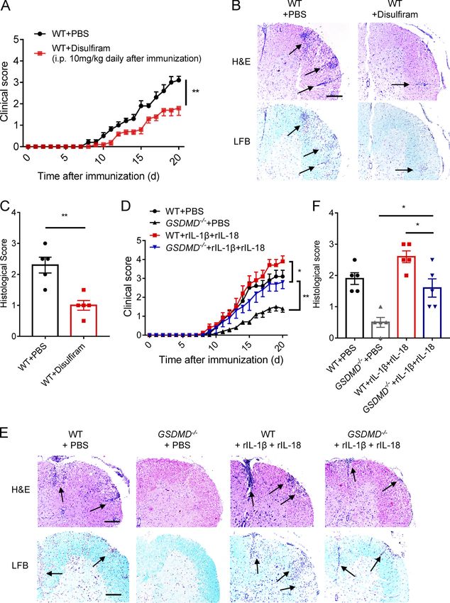
Published Online: 29 August, 2019 | Supp Info: http://doi.org/10.1084/jem.20190377
Downloaded from jem.rupress.org on October 30, 2019
ARTICLE
Gasdermin D in peripheral myeloid cells drives
neuroinflammation in experimental autoimmune
encephalomyelitis
Sheng Li1*, Yuqing Wu1*, Dongxue Yang1, Chunyan Wu1, Chunmei Ma1, Xue Liu1, Paul N. Moynagh2,3, Bingwei Wang4, Gang Hu4, and Shuo Yang1
The NLRP3 inflammasome is critical for EAE pathogenesis; however, the role of gasdermin D (GSDMD), a newly identified
pyroptosis executioner downstream of NLRP3 inflammasome, in EAE has not been well defined. Here, we observed that the
levels of GSDMD protein were greatly enhanced in the CNS of EAE mice, especially near the areas surrounding blood vessels.
GSDMD was required for the pathogenesis of EAE, and GSDMD deficiency in peripheral myeloid cells impaired the infiltration
of immune cells into the CNS, leading to the suppression of neuroinflammation and demyelination. Furthermore, the loss of
GSDMD reduced the activation and differentiation of T cell in the secondary lymphoid organs and prevented T cell infiltration
into CNS of EAE. The administration of inflammasome-related cytokines partially rescued the impairment of pathogenesis of
EAE in GSDMD KO mice. Collectively, these findings provide the first demonstration of GSDMD in peripheral myeloid cells
driving neuroinflammation during EAE pathogenesis.
Introduction
Multiple sclerosis (MS) is an incurable and progressive 2011). Additionally, myeloid cells produce cytokines and che-
inflammatory disease of the central nervous system (CNS) with mokines that respectively facilitate the activation and differen-
neuropathological features, including inflammatory demyelina- tiation of naive CD4+ T cells toward T helper type 1 (Th1) and
tion, chronic axonal damage, and neurodegeneration (Frohman Th17 cell lineages, and the translocation of effector T cells into
et al., 2006). However, the etiology of MS is not well understood, CNS, all of which contributes to the progression of EAE. Fur-
with the underlying basis to pathogenesis being ill-defined. thermore, the peripheral myeloid cells themselves can be re-
Experimental autoimmune encephalomyelitis (EAE) is the cruited into CNS and together with CNS-resident innate
most commonly used animal model for studying MS since it immune cells, such as microglia and astrocyte, facilitate neu-
shares neuropathological features with MS and can help to de- roinflammation to cause neuron damage in EAE (Greter et al.,
fine the contribution of immune events to the development of 2005; Goverman, 2009; Chastain et al., 2011; Duffy et al., 2014).
MS (Ransohoff, 2012). EAE is mediated by myelin-specific Inflammasomes are cytosolic multiprotein complexes that
T cells, which are activated in the peripheral lymphoid organs are vital players of innate immunity and especially in the initi-
and then migrate into CNS by crossing the blood–brain barrier ation of inflammatory responses in myeloid cells (Martinon
to mediate inflammatory responses, resulting in demyelination et al., 2002). Upon recognition of stimuli, inflammasome-
and neurodegeneration (Stromnes et al., 2008). Moreover, much associated sensor proteins, together with apoptosis-associated
evidence suggests that innate immune cells play critical roles speck-like protein containing CARD (ASC) and pro-inflammatory
during the process of EAE. Myeloid cells, such as dendritic cells cysteinyl aspartate–specific proteinase (caspases; caspase-
(DCs) and macrophages, serve as APCs that mediate the cellular 1 and -11), assemble into an oligomeric complex leading to cas-
immune response by processing and presenting myelin antigens pase autoactivation, which controls the cleavage of pro–IL-1β and
for recognition by T cells (Heppner et al., 2005; Chastain et al., pro–IL-18 precursors into their mature forms and also induces a
.............................................................................................................................................................................
1
Department of Immunology, Key Laboratory of Immunological Environment and Disease, State Key Laboratory of Reproductive Medicine, Nanjing Medical University,
Nanjing, China; 2Maynooth University Human Health Research Institute, Department of Biology, National University of Ireland Maynooth, Maynooth, Ireland; 3Wellcome-
Wolfson Institute for Experimental Medicine, Queen’s University Belfast, Belfast, UK; 4Department of Pharmacology, Nanjing University of Chinese Medicine, Nanjing,
China.
*S. Li and Y. Wu contributed equally to this paper; Correspondence to Shuo Yang: shuoyang@njmu.edu.cn; Gang Hu: ghu@njmu.edu.cn; Bingwei Wang: bingweiwang@
njucm.edu.cn.
© 2019 Crown copyright. The government of Australia, Canada, or the UK ("the Crown") owns the copyright interests of authors who are government employees. The
Crown Copyright is not transferable. This article is available under a Creative Commons License (Attribution 4.0 International, as described at https://creativecommons.org/
licenses/by/4.0/).
Rockefeller University Press https://doi.org/10.1084/jem.20190377 1
J. Exp. Med. 2019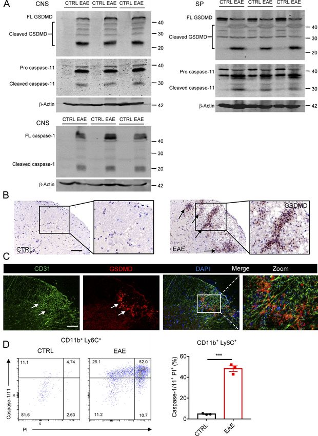
type of proinflammatory programmed cell death termed py-                pyroptosis mediated by GSDMD is indeed involved in the dis-
roptosis (Rathinam et al., 2012; Latz et al., 2013; Jorgensen and       ease. Immunohistochemical analysis further confirmed the
Miao, 2015). Notably, several independent studies showed that           high-level expression of GSDMD in EAE CNS, and it was espe-
the expression of caspase-1, IL-1β, and IL-18 is elevated in the        cially concentrated in lesion areas of spinal cord that were also
peripheral blood mononuclear cells and serum of MS patients             characterized by strong cell infiltration. Interestingly, the posi-
(Huang et al., 2004). Furthermore, increasing recent evidence           tive staining of GSDMD was barely detectable in the CNS
indicate that the NOD-like receptor protein 3 (NLRP3) in-               of control mice (Fig. 1 B). In addition, most of the cells highly
flammasome in APCs plays a critical role in the pathogenesis            expressing GSDMD and caspase-11 were detected in close
of EAE model by mediating chemotactic immune cell recruit-              proximity to the expanded capillaries at the edge of lesions in the
ment to the CNS (Gris et al., 2010; Inoue et al., 2012). Addi-          spinal cord, as evidenced by closely adjacent expression of the
tionally, a recent study reported that Th17 cell–intrinsic ASC          endothelial marker CD31 (Fig. 1 C and Fig. S1 B), suggesting that
can promote the process of EAE by inducing IL-1β production             these GSDMD-positive cells may represent cells recruited from
via ASC–NLRP3–caspase-8 inflammasome axis (Martin et al.,               the periphery. Moreover, FACS revealed a marked increase of
2016). These studies highlight an important role for in-                caspase-1/11+ propidium iodide (PI)+ cells gated on the CD11b+
flammasome in MS and EAE pathogenicity.                                 Ly6C+ (infiltrated peripheral monocytes/macrophages) in the
   Gasdermin D (GSDMD) is a recently identified pore-forming            CNS of mice during EAE compared with control mice (Fig. 1 D).
protein, which mediates pyroptosis (Kayagaki et al., 2015; Shi          Collectively, these data suggest that GSDMD-mediated py-
et al., 2015). Proinflammatory caspases (caspase-1 and -4/5 in          roptosis is associated with EAE development, and its activation
humans and caspase-1 and -11 in mice) can cleave GSDMD at its           in CNS may be derived from infiltrating peripheral cells.
central linker domain, thereby releasing the autoinhibition by
C-terminal GSDMD domain on the N-terminal GSDMD domain,                 GSDMD is essential for EAE induction
causing N-terminal GSDMD domain fragments to translocate to             To directly evaluate the requirement of GSDMD for EAE pro-
the plasma membrane, where they oligomerize to form mem-                gression, we compared EAE in GSDMD KO mice and WT mice.
brane pores, leading to lytic cell death or the secretion of pro-       We found that GSDMD−/− mice were more resistant to EAE and
cessed IL-1β and IL-18 in living cells through pores (Ding et al.,      developed less severe disease and clinical scores compared with
2016; Liu et al., 2016; Evavold et al., 2018). Recent studies           WT mice (Fig. 2 A). Histopathological analysis (H&E staining
demonstrate that GSDMD plays a critical role in septic shock in         and Luxol fast blue [LFB] staining) showed fewer infiltrating
response to bacterial infection, and in inflammasome-driven             inflammatory cells and less demyelination in the spinal cord of
autoinflammatory diseases such as familial Mediterranean fe-            GSDMD−/− mice relative to their WT counterparts (Fig. 2 B).
ver and neonatal-onset multisystem inflammatory disease                 Myelin basic protein (MBP) is a protein that contributes to the
(Kanneganti et al., 2018; Xiao et al., 2018). Interestingly, a recent   process of myelination of nerves and maintenance of neural
study reported that GSDMD accumulation was observed in mi-              structure and is also as a marker of the myelin sheath of oligo-
croglia and oligodendrocytes of CNS in both MS and EAE                  dendrocytes, with its loss being a major pathological feature of
(McKenzie et al., 2018). However, to date there have been no            MS (Bradl and Lassmann, 2010). Immunofluorescence analysis
reports on a pathophysiological role for GSDMD in neuro-                showed regions of considerable diminution MBP expression at
inflammation or EAE pathogenesis.                                       the peak of EAE disease in WT mice, but these were not apparent
   We now provide the first insight into the physiological              in GSDMD−/− mice (Fig. 2 C). Moreover, GSDMD−/− mice had
function of GSDMD in EAE pathogenesis and describe the reg-             significantly reduced numbers of activated macrophages, mi-
ulatory mechanism of GSDMD-mediated pyroptosis in the de-               croglia, and astrocytes in EAE spinal cord compared with WT
velopment of neuroinflammation during EAE. We discover that             mice as determined by expression of the microglial and astro-
myeloid cell–intrinsic GSDMD is crucial for EAE pathogenesis by         cyte cell markers, ionized calcium-binding adapter molecule
mediating the activation and differentiation of T cells in the          1 (IBA1), and glial fibrillary acidic protein (GFAP), respectively
secondary lymphoid organs, which facilitates T cell–mediated            (Fig. S1, C and D). This indicates that GSDMD deficiency is as-
neuroinflammation and demyelination in the CNS.                         sociated with less severe neuroinflammation in EAE. Further-
                                                                        more, fewer CD4+ T cells were detected at the edge of spinal
                                                                        cords of GSDMD−/− mice relative to WT counterparts, further
Results                                                                 suggesting the impairment of recruitment of immune cells in
GSDMD-mediated pyroptosis is activated in the CNS of EAE                GSDMD−/− mice during EAE (Fig. 2 D). Consistent with im-
To investigate whether GSDMD contributes to the pathogenesis            munostaining analysis, FACS revealed a marked reduction of
in MS disease, we first tested the level of its expression in CNS,      T cells (CD45+, CD4+, and CD8+), myeloid cells, and activated
spleen, and LNs of an EAE mice model, which was induced                 microglia cells (CD45+, CD11b+) in the CNS of GSDMD−/− mice
by administration of myelin oligodendrocyte glycoprotein                during EAE compared with WT mice (Fig. 2, E and F). With
(MOG35–55) peptide and pertussis toxin. Immunoblotting                  respect to the population of CNS infiltrating CD4+ T cells, the
analysis demonstrated greatly increased expression and cleav-           percentages and absolute numbers of both Th1 and Th17 cells
age of GSDMD and caspase-1/11 in the CNS and peripheral                 were greatly reduced in GSDMD−/− mice (Fig. 2, G and H). Al-
lymphoid organs of EAE-induced mice at the peak stage relative          together, these data suggest that GSDMD is essential for EAE
to untreated mice (Fig. 1 A and Fig. S1 A), suggesting that the         pathogenesis.

Li et al.                                                                                                   Journal of Experimental Medicine    2
Gasdermin D drives EAE in peripheral myeloid cells                                                       https://doi.org/10.1084/jem.20190377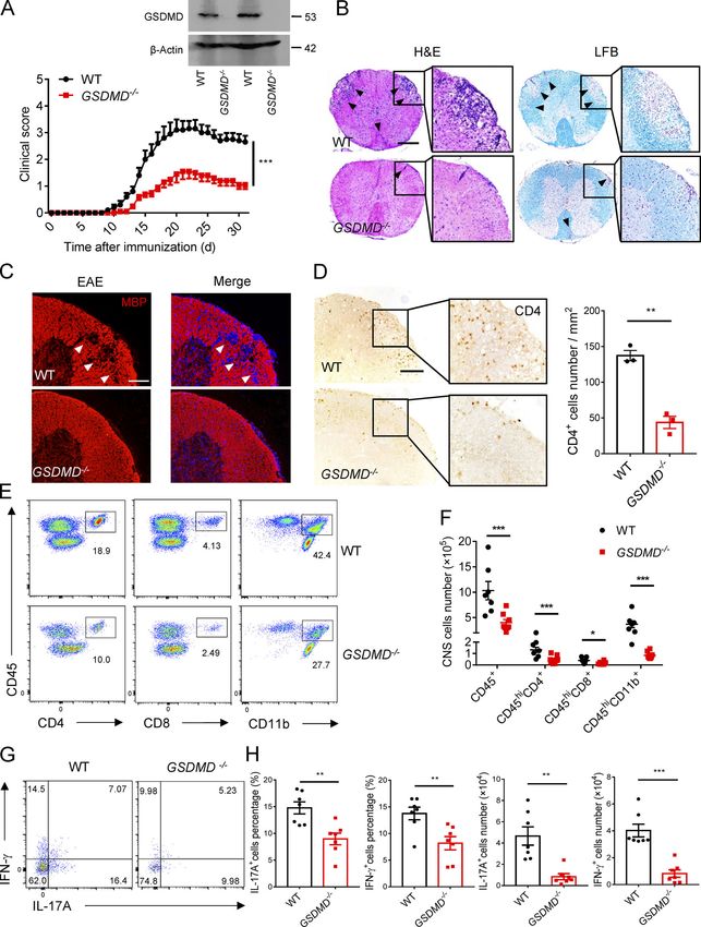
Figure 1. GSDMD is strongly expressed and processed in CNS during EAE. (A) Immunoblot analysis of full-length (FL) and cleaved GSDMD, caspase-11 and
caspase-1 in the spinal cords and spleen (SP) from three pairs of control (CTRL) or EAE-induced WT mice at day 18 after immunization. (B) Immunohisto-
chemistry images showing GSDMD expression in the spinal cord of control or EAE-induced WT mice at day 18 after immunization. Scale bar, 100 µm.
(C) Immunofluorescent labeling of CD31 (green), GSDMD (red), and DAPI (blue) demonstrates the angiogenesis and the distribution of GSDMD (indicated by
arrows) in the lesion area of spinal cord of EAE-induced WT mice at day 18 after immunization. Scale bar, 100 µm. (D) Flow-cytometric analysis of caspase-1/
11+PI+ cells from CD11b+ Ly6C+ cells infiltrated to the spinal cord and brain of CTRL and MOG35-55-immunized WT mice at day 18 after immunization (n = 3 mice
per group). Data are presented as a representative plot (left) and quantified percentages (right). Data are representative of three independent experiments. ***,
P < 0.001. Error bars show means ± SEM. Unpaired t test for D.

Li et al.                                                                                                                   Journal of Experimental Medicine        3
Gasdermin D drives EAE in peripheral myeloid cells                                                                       https://doi.org/10.1084/jem.20190377

Figure 2. GSDMD-deficient mice are resistant to EAE pathogenesis and neuroinflammation. (A) Mean clinical scores of age-matched female WT and
GSDMD−/− mice subjected to MOG35-55-induced EAE (n = 10 mice per group); immunoblot analysis of GSDMD expression in spleen from indicated mice. (B) H&E
staining and LFB staining of spinal cord sections from EAE-induced WT and GSDMD−/− mice showing inflammatory cell infiltration and demyelination,
respectively (arrowheads). Scale bar, 500 µm. (C) Immunofluorescent labeling of MBP (red) and DAPI (blue) visualizing the impairment of myelination
(arrowheads) of EAE-induced WT and GSDMD−/− mice, respectively. Scale bar, 100 µm. (D) Immunohistochemistry analysis for infiltrating CD4+ T cells in the
spinal cord of EAE-induced WT and GSDMD−/− mice (representative images on left and quantified cell numbers on right, n = 3 mice per group), respectively.
Scale bar, 100 µm. (E and F) Flow-cytometric analysis of immune cells (including CD45+CD4+ T cells, CD45+CD8+ T cells, and CD45+CD11b+ monocytes)
infiltrated to the spinal cord and brain of MOG35-55-immunized WT and GSDMD−/− mice at day 18 after immunization (n = 7 for WTs, n = 8 for GSDMD−/− mice).
Data are presented as a representative plot (E) and summary graph of the absolute cell numbers (F). (G and H) Flow-cytometric analysis of Th1 (IFN-γ+) and
Th17 (IL-17A+) cells from CD4+ T cells infiltrated to the spinal cord and brain of MOG35-55-immunized WT and GSDMD−/− mice at day 18 after immunization (n = 7
mice per group). Data are presented as a representative plot (G), quantified percentage, and absolute cell numbers (H). Data are pooled from three independent
experiments. *, P < 0.05; **, P < 0.01; ***, P < 0.001. Error bars show means ± SEM. Unpaired t test for A, D, and H, and multiple unpaired t test for F.
Li et al.                                                                                                                 Journal of Experimental Medicine       4
Gasdermin D drives EAE in peripheral myeloid cells                                                                     https://doi.org/10.1084/jem.20190377
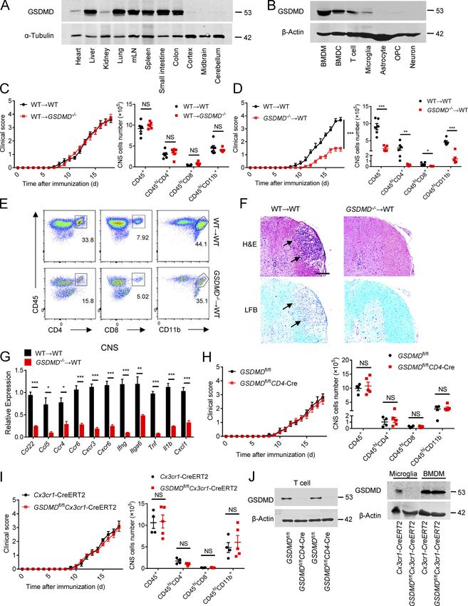
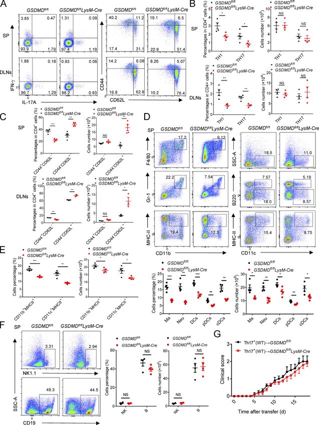
GSDMD deficiency in peripheral myeloid cells suppresses             that deletion of GSDMD in peripheral myeloid cells might be
neuroinflammation and pathogenesis of EAE                           responsible for the suppression of the pathogenesis and neuro-
To determine whether GSDMD deficiency in peripheral cells or        inflammation of EAE.
CNS-resident cells contributes to the suppression of EAE, we
measured the expression of GSDMD in various tissues, but it was     GSDMD deficiency impairs the priming and differentiation of
strikingly low in CNS of homeostatic state (Fig. 3 A). Moreover,    T cell by myeloid cells and increases T cell accumulation in
compared with peripheral cells (including bone marrow–              peripheral lymphoid organs during EAE
derived macrophages [BMDMs], bone marrow–derived DCs, and           Antigen-activated T cells are key effector cells in the patho-
T cells), microglia displayed very low expression of GSDMD with     genesis of EAE, which undergo activation in secondary lymphoid
barely detectable expression other CNS cells including as-          organs and then migrate into CNS due to the actions of cytokines
trocytes, oligodendrocyte progenitor cells, and neurons (Fig. 3     and chemokines produced by APCs (Goverman, 2009). Given the
B). Next, we generated bone marrow chimeric mice by adop-           phenotype observed above, we hypothesized that GSDMD in
tively transferring WT bone marrow cells into lethally irradiated   peripheral myeloid cells may be important for T cell response in
WT or GSDMD−/− recipient mice. Comparable EAE clinical scores       peripheral lymphoid organs during EAE. Thus, decreased infil-
were observed between these two recipients (Fig. 3 C), which        tration of CD4+ T cells in the CNS of GSDMD−/− mice may be
also had comparable levels of infiltrated immune cells in the       caused by decreased cellularity in the periphery. Unexpectedly,
CNS (Fig. 3 C). However, a reverse bone marrow transfer ex-         GSDMD-deficient mice show enlargement of spleen and drain-
periment in which lethally irradiated WT recipient mice were        ing LNs (DLNs) at the peak stage of EAE (Fig. 4, A and B), in-
reconstituted with bone marrow cells isolated from WT or            dicating the potential accumulation of T cells in peripheral
GSDMD−/− mice showed that WT mice reconstituted with the            lymphoid organs after immunization. Most notably, while the
GSDMD deficiency bone marrow were more refractory to EAE            absolute numbers of CD4+ T cells (Fig. 4 C) were increased in
induction than WT donors (Fig. 3 D). Additionally, the recruit-     the secondary lymphoid organs of GSDMD−/− mice after EAE, the
ment of peripheral immune cells into the CNS was reduced in         percentages of Th1 and Th17 cells were reduced, but their
GSDMD−/− donors compared with WT donors (Fig. 3, D and E).          numbers were comparable in WT and GSDMD−/− mice (Fig. 4, D
H&E and LFB (Fig. 3 F) staining also confirmed that fewer in-       and E). We next analyzed the naive CD62+CD44−CD4+ T cells
filtrating inflammatory cells and decreased demyelination were      (Th0) that have not been activated by APCs during EAE. Inter-
apparent in the spinal cord of GSDMD−/− donor mice. Notably,        estingly, the numbers of naive T cell in spleen and LNs were
the CNS of WT recipients reconstituted with the GSDMD−/− bone       much higher in GSDMD−/− mice after EAE than WT mice (Fig. 4,
marrow had significantly decreased expression of several in-        D and F), which might be responsible for the observed increase
flammatory chemokines and cytokines known to mediate im-            of CD4+ T cells in the secondary lymphoid organs of GSDMD−/−
mune cells recruitment and neuroinflammation after the              mice. Given that we observed significantly reduced percentages
induction of EAE (Fig. 3 G). Thus, these results suggest that       of Th1 and Th17 cells in the secondary lymphoid organs of
GSDMD functions in peripheral cells to promote EAE patho-           GSDMD−/− mice during EAE, we suggest that GSDMD plays an
genesis in the CNS.                                                 important role in regulating T cell priming and differentiation in
    We next examined if this role for GSDMD in peripheral cells     the peripheral lymphoid organs during EAE. This was directly
was intrinsic to peripheral T cells by crossing the GSDMDfl/fl      addressed by performing in vitro T cell proliferation and dif-
mice with CD4-Cre mice to generate T cell conditional GSDMD         ferentiation assays that revealed GSDMD−/− DCs to be greatly
KO mice. GSDMDfl/flCD4-Cre and littermate control GSDMDfl/fl        impaired in driving EAE MOG-induced T cell activation and
mice developed similar clinical symptoms and immune cells           differentiation relative to WT DCs (Fig. 4, G–I). Since the num-
infiltration after immunization (Fig. S2 and Fig. 3, H and J),      ber of Th1 and Th17 cells in the secondary lymphoid organs was
suggesting that GSDMD in T cells has no role in driving EAE-        unaltered in GSDMD−/− mice after EAE and the infiltration of Th1
associated pathogenesis. However, peripheral myeloid cells (DCs     and Th17 cells in the CNS was decreased (Fig. 2, G and H), we
and macrophages) and CNS-resident microglia also contribute to      speculate that GSDMD deficiency also affects T cell migration
neuroinflammation and pathogenesis in EAE (Duffy et al., 2014).     into CNS. Additionally, we evaluated the profiles of T cells in
To further explore the potential contribution of GSDMD in mi-       thymus, spleen, and LN of GSDMD−/− mice in homeostatic state.
croglia to EAE pathogenesis, we crossed GSDMDfl/fl mice with        There were no significant differences observed between WT and
Cx3cr1-CreERT2 mice to generate microglia conditional GSDMD         GSDMD−/− mice in the development of T cells and the profiles of
KO mice. Tamoxifen was administered to these mice to specif-        naive T cell, effective T cell, and T regulatory (T reg) cell (Fig.
ically delete GSDMD in microglia. This experimental design is       S3). Therefore, these data suggest that GSDMD deficiency sup-
based on a previous report showing that short-lived blood           presses the priming and differentiation of T cells after the in-
monocytes are renewed within 6 wk after tamoxifen adminis-          duction of EAE, meanwhile increasing T cell accumulation in
tration, whereas GSDMD is specifically deleted in long-lived        peripheral lymphoid organs during disease.
microglia of GSDMDfl/flCx3cr1-CreERT2 mice (Yona et al., 2013).         To further address whether GSDMD functions on the myeloid
GSDMD deficiency in microglia had no effect on the EAE phe-         cells to regulate T cell responses, we next investigated the
notype (Fig. 3, I and J), and this is consistent with the bone      composition of myeloid cells in the spleen during EAE. FACS
marrow transfer experiments that concluded a lack of role for       revealed a marked reduction in the percentages and absolute
GSDMD in CNS resident cells. Together, these data demonstrate       numbers of macrophages (CD11b + F4/80 + ), neutrophils

Li et al.                                                                                               Journal of Experimental Medicine    5
Gasdermin D drives EAE in peripheral myeloid cells                                                   https://doi.org/10.1084/jem.20190377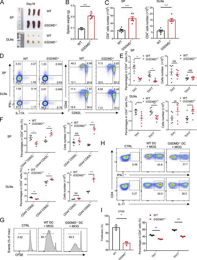
Figure 3. GSDMD deletion in peripheral cells attenuates the development of EAE. (A) Immunoblot analysis of the protein expression of GSDMD in
different tissues (heart, liver, kidney, lung, mesenteric LNs [mLN], spleen, small intestine, colon, cortex, midbrain and cerebellum) from WT mice. (B) Im-
munoblot analysis of the protein expression of GSDMD in peripheral cells (BMDMs, bone marrow–derived DCs [BMDCs], and T cells) and CNS resident cells
(microglia, astrocyte, oligodendrocyte progenitor cell [OPC], and neuron) from WT mice. (C) Mean clinical scores after EAE induction and summary graph of
CNS-infiltrating immune cells at day 18 after immunization in WT and GSDMD−/− mice (n = 6 mice per group) adoptively transferred with WT bone marrow
cells. (D) Mean clinical scores after EAE induction and summary graph of CNS-infiltrating immune cells at day 18 after immunization in WT mice (n = 5 or 6 mice

Li et al.                                                                                                                  Journal of Experimental Medicine       6
Gasdermin D drives EAE in peripheral myeloid cells                                                                      https://doi.org/10.1084/jem.20190377

per group) adoptively transferred with WT or GSDMD−/− bone marrow cells. (E) Representative plot from flow-cytometric analysis of immune cells infiltrated to
the CNS (spinal cord and brain) of the mice in D. (F) H&E staining and LFB staining of spinal cord sections harvested from WT mice adoptively transferred with
WT or GSDMD−/− bone marrow cells showing inflammatory cell infiltration and demyelination, respectively (arrows). Scale bar, 200 µm. (G) Quantitative PCR
analysis of the relative mRNA expression of proinflammatory cytokines and chemokines in the spinal cord of WT mice (n = 4 mice per group) adoptively
transferred with WT or GSDMD−/− bone marrow cells at the EAE peak. Data were normalized to a reference gene, Hprt. (H) Mean clinical scores after EAE
induction and summary graph of CNS-infiltrating immune cells at day 18 after immunization in GSDMDfl/flCD4-Cre and littermate control GSDMDfl/fl mice (n = 4
or 5 mice per group). (I) Mean clinical scores after EAE induction and summary graph of CNS-infiltrating immune cells at day 18 after immunization in
GSDMDfl/flCx3cr1-CreERT2 and Cx3cr1-CreERT2 mice (n = 4 or 5 mice per group). (J) Immunoblot analysis of GSDMD expression in T cells from indicated mice,
and in microglia and BMDMs from indicated mice at 6 wk after administration of tamoxifen. Data are pooled from three independent experiments. *, P < 0.05;
**, P < 0.01; ***, P < 0.001. Error bars show means ± SEM. Unpaired t test for C, D, H, and I, and multiple unpaired t test for C, D, and G–I.

(CD11b+Gr-1+), and DCs (CD11c+, including classical DCs [cDCs;                  deficiency greatly impaired the expression of Cxcr6, Ccl5, TNF-α,
CD11c+B220−], and plasmacytoid DCs [pDCs; CD11c+B220+]) in                      and CD3e expression on Th1 and Th17 cells during EAE (Fig. 6, E and
the spleen of GSDMD−/− mice during EAE relative to WT mice                      F). Thus, these data further demonstrate that the expression of
(Fig. 5 A). Importantly, the percentages and absolute numbers                   genes involved in T cell activation, differentiation, and migration in
of MHCII-expressing cells (CD11b+MHCII+ and CD11c+MHCII+)                       CD11b+ and CD4+ cells of peripheral lymphoid organs during EAE is
in spleen were greatly reduced in EAE GSDMD−/− mice (Fig. 5                     significantly impaired by the deficiency of GSDMD.
A). In addition, GSDMD deficiency did not affect the number of
B and natural killer (NK) cells in the spleen during EAE (Fig. 5                GSDMD in myeloid cells is required for the activation of CD4+
B). Moreover, we didn’t find any differences in the frequency                   T cells during EAE
and numbers of lymphocytic and myeloid cells in bone marrow,                    To further test the impact of GSDMD deficiency on T cell
blood, and spleens of GSDMD−/− mice relative to WT controls                     function during EAE, CD4+ T or Th17 cells isolated from spleens
under homeostatic conditions (Fig. S4). Thus, all these results                 of WT and GSDMD−/− mice at the time of EAE onset (day 12) were
further strengthen the conclusion of a key role for GSDMD in                    i.v. transferred to Rag1−/− or C57BL/6 WT recipients (Fig. 7 A).
T cell priming by myeloid cells in the secondary lymphoid or-                   CD4+ T cells from immunized GSDMD−/− mice failed to induce
gans after EAE.                                                                 passive EAE in Rag1−/− or WT recipients, but CD4+ T cells ob-
                                                                                tained from WT immunized mice promoted mild EAE (Fig. 7 B).
GSDMD deficiency impairs the expression of genes required                       This indicated that GSDMD deficiency results in functional im-
for T cell response in CD11b+ cells and CD4+ T cells of                         pairment of T cells during EAE induction. Next, to further in-
peripheral lymphoid organs during EAE                                           vestigate whether GSDMD deficiency affects T cell migration,
It is clear from the above results that peripheral myeloid cell–                we directly transferred CD4+ T cells into the brain of WT mice by
intrinsic GSDMD functions in peripheral lymphoid organs to                      intracerebroventricular (i.c.v.) injection in order to bypass the
regulate T cell response during EAE. To further dissect the                     migration of T cells from peripheral to CNS. Again, CD4+ T cells
mechanistic role of GSDMD in myeloid cells that regulates T cell                obtained from immunized GSDMD−/− mice developed lower
responses, we performed RNA sequencing analysis using CD11b+                    levels of EAE pathogenesis compared with immunized WT mice
cells sorted from spleens of WT and GSDMD−/− mice at the onset                  (Fig. 7 C), suggesting that the critical function of GSDMD may
stage of EAE. Kyoto Encyclopedia of Genes and Genomes (KEGG)                    reside in regulating the activation and/or differentiation of
analysis showed the top pathways down-regulated in GSDMD−/−                     T cells during EAE.
CD11b+ cells mainly included Th cell differentiation, antigen                       Given that we have earlier shown that GSDMD in peripheral
processing and presentation, cell adhesion molecules, and cy-                   myeloid cells is responsible for the neuroinflammation and
tokine signaling (Fig. 6 A). Consistently, the heatmap and real-                pathogenesis of EAE, we next examined if peripheral myeloid
time PCR analysis displayed significantly decreased expression                  cell–intrinsic GSDMD plays a function in T cell activation during
in several genes associated with T cell response and migration,                 EAE. To this end, we generated myeloid cell conditional GSDMD-KO
such as Il1b, Ifng, Tnf, Osm, H2-Q10, H2-Ob, Itga6, and Ccl5 in                 mice by crossing the GSDMDfl/fl mice with LysM-Cre mice. Since
GSDMD−/− cells (Fig. 6, C and D). To further study the impact of                these conditional KO mice lack GSDMD expression in myeloid
GSDMD deficiency on T cell responses and determine whether                      cells and we have already excluded the effect of GSDMD in
T cells in GSDMD−/− mice display an alteration in gene expres-                  microglia using Cx3cr1-CreERT2 mice (Fig. 3, I and J), the LysM-
sion during EAE, we also performed RNA sequencing (RNA-seq)                     Cre mice allowed for direct investigation of the role of GSDMD
and examined the related genes expression in CD4+ T cells sorted                in peripheral myeloid cells in T cell activation after EAE in-
from spleen. The top down-regulated pathways in GSDMD−/− T cells                duction. Notably, the EAE clinical scores and infiltration of
after EAE induction were involved with Th cell differentiation,                 immune cells into CNS were considerably diminished in
T cell receptor signaling pathway, and cytokine–chemokine sig-                  GSDMDfl/flLysM-Cre mice relative to GSDMDfl/fl mice (Fig. 7, D
naling according to KEGG analysis (Fig. 6 B). Several genes associ-             and E). Moreover, T cell proliferation was significantly impaired
ated with T cell activation and migration, such as Il2, Ifng, Tnf, Cd3e,        in the CNS and spleens of GSDMDfl/flLysM-Cre mice, as shown by
Ccl5, Ccl22, Ccr4, Cxcr3, and Cxcr6, were down-regulated in GSDMD−/−            reduced staining of the proliferation marker CD4+Ki67+ at the
T cells at the onset of EAE as indicated in the heatmap and real-time           EAE peak stage (Fig. 7, F and G). Additionally, we observed a
PCR analysis (Fig. 6, C and D). Moreover, FACS revealed GSDMD                   significant increase in the number of CD4 T cells in the spleen

Li et al.                                                                                                                 Journal of Experimental Medicine       7
Gasdermin D drives EAE in peripheral myeloid cells                                                                     https://doi.org/10.1084/jem.20190377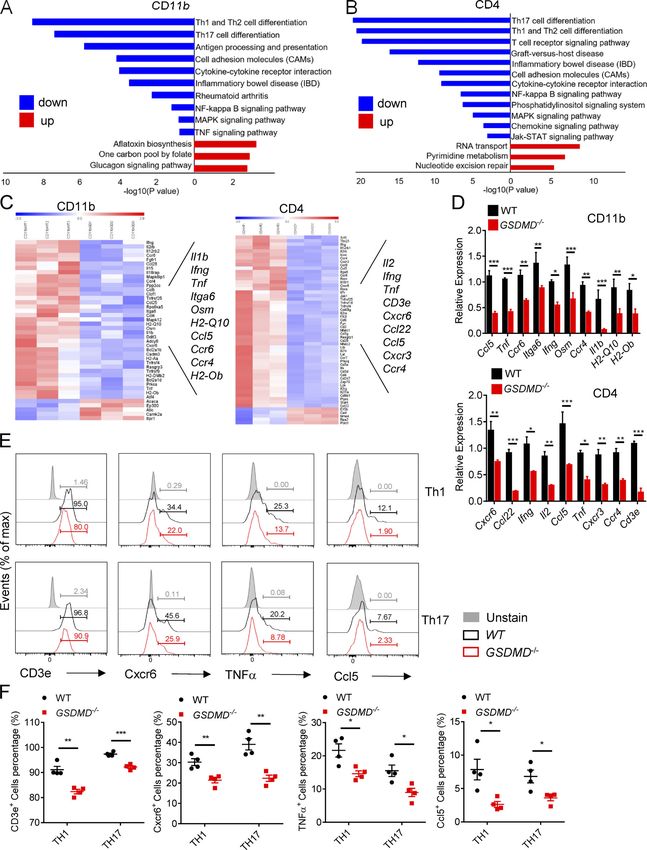
Li et al.                                               Journal of Experimental Medicine    8
Gasdermin D drives EAE in peripheral myeloid cells   https://doi.org/10.1084/jem.20190377
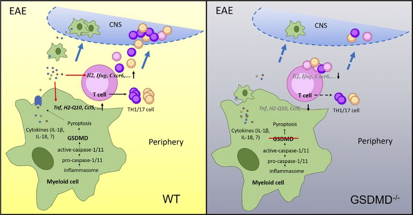
Figure 4. The differentiation and egress of T cells in peripheral lymphoid organs during EAE are impaired in GSDMD−/− mice. (A and B) Spleens and
DLNs harvested from three pairs of WTs and GSDMD−/− mice at day 18 after EAE induction (A) and quantified spleen (SP) weights (n = 5 mice per group; B).
(C) Quantified absolute cell numbers of CD4+ T cells in spleen and DLNs from WTs and GSDMD−/− mice at day 18 after EAE induction (n = 6 mice per group).
(D–F) Flow-cytometric analysis of Th1 (IFN-γ+) and Th17 (IL-17A+) cells from CD4+ T cells in the spleen and DLNs of WT and GSDMD−/− mice (n = 6 mice per
group) at day 18 after EAE induction, and effective T cell (CD44+CD62L−) and naive T cell (CD44−CD62L+) cells from CD4+ T cells in the spleen and DLNs of WT
and GSDMD−/− mice (n = 4 mice per group) at day 18 after EAE induction. Data are presented as a representative plot (D), and quantified percentages and
absolute cell numbers (E and F). (G–I) Flow-cytometric analysis of CFSE-labeled cells, Th1 (IFN-γ+) cells, and Th17 (IL-17A+) cells from CD4+ T cells at day 5 after
in vitro cocultured with DCs (n = 3 per group). Data are presented as a representative plot (G and H) and quantified percentages (I). Data are pooled from three
independent experiments (A–E) or from two independent experiments (F–I). *, P < 0.05; **, P < 0.01; ***, P < 0.001. Error bars show means ± SEM. Unpaired
t test for B, C, and I, and multiple unpaired t test for E, F, and I. CTRL, control; max, maximum.

and DLNs of GSDMDf/f LysM-Cre mice during EAE relative to                          pyroptosis or forming pores in living cells (Shi et al., 2015;
littermate controls (Fig. 7 H), but a marked reduction in the                      Evavold et al., 2018). To understand whether the impairment of
percentages of Th1 and Th17 cells (Fig. 8, A and B). Moreover,                     EAE pathogenesis in GSDMD-deficient mice is completely de-
the number of naive CD62+CD44−CD4+(Th0) T cells in spleen                          pendent on the loss of inflammasome-related cytokines, we as-
and LNs was much higher in GSDMDf/f LysM-Cre mice after EAE                        sessed the effects of IL-1β and IL-18 on EAE progression in vivo
than tissues from controls (Fig. 8, A and C). In addition, the                     by i.v. administration of their recombinant proteins in WT and
percentages and numbers of macrophages, neutrophils, DCs,                          GSDMD−/− mice immunized with MOG. Although administration
and MHCII-expressing cells were greatly reduced in the spleen                      of recombinant IL-1β (rIL-1β) and rIL-18 in GSDMD−/− mice de-
of GSDMDf/f LysM-Cre mice during EAE (Fig. 8, D and E). We                         veloped more severe EAE as demonstrated by higher clinical and
didn’t find any differences in the frequency and numbers of                        histopathological scores compared with their PBS-control mice,
lymphocytic and myeloid cells in bone marrow, blood, and                           the disease severity of GSDMD−/− mice still was milder than WTs
spleens of GSDMDf/f LysM-Cre mice relative to littermate con-                      even after administration with rIL-1β and rIL-18 (Fig. 9, D–F). It
trols (Fig. S5). Furthermore, GSDMD deficiency in myeloid cells                    is hardly surprising that this exogenous supplementation ex-
did not affect the number of B and NK cells in the spleen during                   acerbates pathology in WTs since the exogenous levels of these
EAE (Fig. 8 F). Thus, all of these data strongly suggest that                      cytokines likely greatly exceed the endogenous levels under
GSDMD in peripheral myeloid cells is essential for T cell acti-                    conditions of EAE, and maximal pathogenesis is limited by the
vation and ensuing pathogenesis in EAE. To further exclude an                      endogenous levels of IL-1β and IL-18. Importantly, the exacer-
effect of GSDMD deficiency in microglia, we next performed                         bation of EAE severity by these cytokines in GSDMD KO mice
adoptive transfer studies in GSDMDf/f LysM-Cre mice and their                      did not completely reach the severity observed in WT PBS
littermate controls by adoptive transfer of Th17 cells from im-                    control mice. Thus, these results indicate that administration of
munized WT mice. We found that MOG-reactive Th17 cells in-                         IL-1β and IL-18 can only partially rescue the impairment of EAE
duced comparable EAE clinical scores between GSDMDf/f LysM-                        pathogenesis in GSDMD−/− mice, which suggests that the path-
Cre mice and controls (Fig. 8 G), which further suggests that                      ogenic role of GSDMD may also be dependent on the release of
GSDMD in peripheral myeloid cells but not in CNS resident                          other factors in response to the pyroptotic actions of GSDMD.
microglia is essential for pathogenesis in EAE.

Pharmacological inhibition of GSDMD attenuates EAE, and                            Discussion
inflammasome-related cytokines promote EAE pathogenesis                            GSDMD protein, a newly identified pyroptosis executioner
in GSDMD-deficient mice                                                            downstream of inflammasome activation, has been reported to
Given that GSDMD-deficient mice show impaired pathogenesis,                        accumulate in the CNS of MS (McKenzie et al., 2018), but it is not
we were keen to assess if pharmacological inhibition of GSDMD                      clear what the physiological role and mechanism of GSDMD is in
could phenocopy the genetic models. We thus directly assessed                      the EAE pathogenesis. In this study, we found that the ablation
the effects of pyroptosis inhibition on EAE progression in vivo                    of GSDMD in peripheral myeloid cells reduced the infiltration of
by i.p. administration with disulfiram, a recently described                       immune cells into the CNS and conferred robust protection from
potent inhibitor of GSDMD (Hu et al., 2018). We found that                         EAE development in mice immunized with MOG35-55. In addi-
disulfiram treatment protected against EAE development and                         tion, GSDMD deficiency impaired the activation and differenti-
greatly attenuated the clinical and histopathological scores                       ation of T cell in the secondary lymphoid organs, thus preventing
(Fig. 9, A–C). Thus, this result further indicates an important                    T cell–mediated neuroinflammation in EAE.
role of GSDMD in the pathogenesis of EAE and suggests a po-                           While the recent study suggested GSDMD-mediated py-
tential therapeutic use of targeting GSDMD-mediated pyroptosis                     roptosis appeared in the CNS of patients with MS and in EAE
in MS treatment. Different reports have shown that IL-1β and                       mice (McKenzie et al., 2018), the issues of the relative contri-
IL-18, the downstream cytokines of inflammasome activation,                        bution of peripheral versus CNS GSDMD to disease pathology
have high-level expression in MS patients and promote T cell                       were unknown. The conclusion that GSDMD-mediated py-
differentiation, survival, and migration during EAE (Lalor et al.,                 roptosis involves microglia during EAE progression cannot be
2011; Inoue et al., 2012; Martin et al., 2016). Moreover, GSDMD                    addressed only by GSDMD immunoreactivity in Iba-1 immuno-
can facilitate the secretion of IL-1β and IL-18 through cell lysis by              positive cells in that study because of Iba-1 expression in both

Li et al.                                                                                                                     Journal of Experimental Medicine         9
Gasdermin D drives EAE in peripheral myeloid cells                                                                         https://doi.org/10.1084/jem.20190377
Figure 5. Myeloid cells in peripheral lymphoid organs during EAE are impaired in GSDMD−/− mice. (A) Flow-cytometric analysis of macrophages
(Ma; CD11b+F4/80+), neutrophils (Neu; CD11b+Gr-1+), DCs (CD11c+, including cDCs [CD11c+B220−] and pDCs [CD11c+B220+]), and MHCII-expressing cells
(CD11b+MHCII+ and CD11c+MHCII+) in the spleen (SP) of WT and GSDMD−/− mice (n = 4 mice per group) at day 18 after EAE induction. Data are presented as a
representative plot, quantified percentages, and absolute cell numbers. (B) Flow-cytometric analysis of NK cells (NK1.1+) and B cells (CD19+) in the spleen of
WT and GSDMD−/− mice (n = 4 mice per group) at day 18 after EAE induction. Data are presented as a representative plot, quantified percentages, and absolute
cell numbers. Data are pooled from two independent experiments. *, P < 0.05; **, P < 0.01; ***, P < 0.001. Error bars show means ± SEM. Multiple unpaired
t test for A and B. SSC-A, side scatter–area.

Li et al.                                                                                                                 Journal of Experimental Medicine       10
Gasdermin D drives EAE in peripheral myeloid cells                                                                     https://doi.org/10.1084/jem.20190377
Figure 6. GSDMD deficiency attenuates the expression of genes required for T cell response in CD11b+ cell and CD4+ T cell of peripheral lymphoid
organs during EAE. (A and B) KEGG analysis shows the most significantly enriched signaling pathways in CD11b+ macrophages (A) and CD4+ T cells (B) sorted
from spleens of WT and GSDMD−/− mice at day 12 after EAE induction. (C) The heatmaps of genes with adjusted P value
Figure 7. GSDMD in peripheral myeloid cells is essential for the activation of CD4+ T Cells during EAE. (A) Schematic representation of the experiments
in B, C, and Fig. 8 G. CD4+ T cells were obtained from spleens of WT or GSDMD−/− mice at day 12 after EAE induction, then transferred into Rag1−/− mice by i.v.
injection or into WT mice by i.c.v. injection. DLN cells were obtained from WT or GSDMD−/− mice at day 12 after EAE induction, then cultured in vitro with

Li et al.                                                                                                                  Journal of Experimental Medicine       12
Gasdermin D drives EAE in peripheral myeloid cells                                                                      https://doi.org/10.1084/jem.20190377
MOG35-55 and IL-23 under Th17 cell–polarizing conditions before transfer into sublethal irradiated WTs or GSDMDfl/fl and GSDMDfl/flLysM-Cre mice, respectively.
(B and C) Mean clinical scores after CD4 T cell and Th17 cell transfer by i.v. (B) and CD4 T cell transfer by i.c.v. (C; n = 5 mice per group). (D) Mean clinical scores
after EAE induction in GSDMDfl/flLysM-Cre and littermate control GSDMDfl/fl mice (n = 5 mice per group); immunoblot analysis of GSDMD expression in BMDMs
from indicated mice. (E) Summary graph of CNS-infiltrating immune cells at day 18 after immunization in GSDMDfl/flLysM-Cre and littermate GSDMDfl/fl mice
(n = 5 mice per group). (F) Flow-cytometric analysis of KI67+ cells among CD4+ T cells in the CNS and spleens (SP) of GSDMDfl/flLysM-Cre and littermate
GSDMDfl/fl mice at day 18 after immunization (n = 5 mice per group). (G) Immunofluorescent labeling of CD3 (red) and KI67 (green) demonstrate the pro-
liferation of T cells in the spinal cord of indicated mice at day 18 after immunization. Data are presented as a representative image (left) and quantified
percentage (right). Scale bar, 50 µm. (H) Quantified absolute cell numbers of CD4+ T cell in spleen and DLNs from GSDMDfl/flLysM-Cre and littermate control
GSDMDfl/fl mice at day 18 after EAE induction (n = 4 mice per group). Data are pooled from three independent experiments (A–G) or from two independent
experiments (H). *, P < 0.05; **, P < 0.01; ***, P < 0.001. Error bars show means ± SEM. Unpaired t test for B–D and F–H, and multiple unpaired t test for E.

peripheral and CNS macrophages (Müller et al., 2015). In fact,                       GSDMD−/− cells majorly include Th1 and Th17 cell differentia-
both peripheral and CNS macrophages contribute to EAE pro-                           tion, T cell signaling pathway, cell adhesion molecules, and
gression (Bauer et al., 1995; Xiao et al., 2013; Duffy et al., 2014).                cytokine–chemokine signaling. Significant reductions in mRNA
Here we found that GSDMD in peripheral cells is indispensable                        levels of Il2, Ifng, Tnf, Cd3e, Ccl5, Ccl22, Ccr4, Cxcr3, and Cxcr6 were
for EAE induction by using the bone marrow chimeric EAE                              identified in splenic CD4+ T cells from GSDMD−/− mice. IL-2 and
models in which the bone marrow from GSDMD−/− mice exhibits                          CD3e are necessary for the expansion and survive of reactive
impaired capability to induce neuroinflammation and patho-                           T cells (Tamura and Nariuchi, 1992; Liao et al., 2013). TNF-α and
genesis of EAE. Moreover, GSDMD-specific deletion in microglia                       IFN-γ can drive and amplify T cell response and inflammation
by using the GSDMDfl/fl Cx3cr1-CreERT2 mice does not suppress                        during EAE pathogenesis (Raphael et al., 2015). Ccl5, Ccl22, Ccr4,
CNS autoimmune inflammation after EAE induction, suggesting                          Cxcr3, and Cxcr6 are migration-related molecules, which are
that GSDMD in microglia does not play a major role in regulating                     required for T cells to enhance their migration ability to the CNS
EAE pathogenesis. Notably, we observed an abundance of                               during the development of EAE (dos Santos et al., 2005; Inoue
GSDMD immunoreactivity near the areas surrounding blood                              et al., 2012). Therefore, GSDMD-mediated pyroptosis up-
vessels of spinal cords within EAE, indicating that peripheral                       regulates the expression of genes required for T cell response
cells might be recruited and undergo pyroptosis in the diseased                      in the secondary lymphoid organs, which are involved in de-
brain. Additionally, the studies in GSDMDf/f LysM-Cre mice also                      velopment of EAE and probably in MS as well.
showed that GSDMD deficiency in myeloid cells attenuated the                            Although RNA-seq results indicated that GSDMD deficiency
progression of EAE. Therefore, our studies provide the first                         might affect T cell migration into CNS, direct injection of CD4+
demonstration of a peripheral myeloid–intrinsic role for GSDMD                       T cell from immunized GSDMD−/− mice injection into the CNS
in driving neuroinflammation in EAE.                                                 induced much milder EAE compared with that induced by WT
    Our RNA-seq analysis revealed that the major immune                              CD4+ T cells, suggesting that T cell activation is likely to be the
pathways down-regulated in CD11b+ cells from immunized                               substantive pathway affected by GSDMD deficiency during EAE.
GSDMD−/− mice were associated with T cell response, including                        Consistent with this, our studies in GSDMDf/f LysM-Cre mice
Th1 and Th17 cell differentiation, antigen processing and pre-                       further showed that the protective effect of GSDMD deficiency
sentation, cell adhesion molecules, and cytokine signaling,                          in myeloid cells was associated with the attenuation of T cell
which indicated that GSDMD-sufficient APCs are essential for                         activation and proliferation during EAE. We demonstrate that
priming T cell immune response. The heatmap results summa-                           the administration of IL-1β and IL-18 only partially rescued the
rized that GSDMD deficiency severely impaired the expression                         impairment of EAE pathogenesis in GSDMD−/− mice. Notably, a
of several genes for T cell activation and differentiation, such as                  recent study showed that GSDMD deficiency promotes cyclic
Tnf, Ifng, Il1b, H2-Q10, and H2-Ob. TNF-α, IFN-γ, and IL-1β are                      GMP-AMP synthase–dependent IFN production by reducing
key pro-inflammatory cytokines for the expansion and differ-                         intracellular potassium (K+) efflux through GSDMD-formed
entiation of reactive T cells in the secondary lymphoid organs,                      pores (Banerjee et al., 2018). IFN-β administration is a first-
and an abundance of previously published reports demonstrated                        line disease-modifying therapy for MS and thought to control
their critical roles in EAE development (Hirota et al., 2011;                        CNS inflammation and neurodegeneration (Banerjee et al.,
Raphael et al., 2015). H2-Q10 and H2-Ob are certain MHC                              2018). Thus, whether the effects of GSDMD on IFN production
haplotypes, and the importance of specific MHC molecules in                          are involved in GSDMD-mediated EAE progression needs to be
determining EAE susceptibility has been well established                             further investigated in the future.
(Weissert et al., 1998; Irla et al., 2010). In addition, the signifi-                   In summary, our study is the first to report the physiological
cantly reduced expression of genes for chemotaxis and adhesion,                      role of GSDMD as a key mediator in autoimmune inflammation
including Ccl5, Ccr4, Ccr6, and Itga6, was observed in splenic                       and pathogenesis during EAE. We propose a model in which
CD11b+ cells from immunized GSDMD−/− mice. There have been a                         peripheral myeloid cell–intrinsic GSDMD mediates pyroptosis to
number of reports that chemotaxis molecules that enhance im-                         instigate inflammation and promote the activation and differ-
mune cell migration are strongly connected with EAE progres-                         entiation of T cells in the secondary lymphoid organs, thus
sion (Inoue et al., 2012; Poppensieker et al., 2012; Cheng and                       driving T cell–mediated neuroinflammation and demyelination
Chen, 2014). For CD4+ T cell from EAE mice, KEGG pathway                             in the CNS of EAE (Fig. 10). The development of therapeutic
analysis showed that the biological terms down-regulated in                          strategies to specifically target GSDMD-mediated pyroptosis

Li et al.                                                                                                                        Journal of Experimental Medicine          13
Gasdermin D drives EAE in peripheral myeloid cells                                                                            https://doi.org/10.1084/jem.20190377
Li et al. Journal of Experimental Medicine 14 Gasdermin D drives EAE in peripheral myeloid cells https://doi.org/10.1084/jem.20190377
Figure 8. GSDMD in peripheral myeloid cells is essential for T cell priming by myeloid cells during EAE. (A–C) Flow-cytometric analysis of Th1 (IFN-γ+)
and Th17 (IL-17A+) cells, effective T cells (CD44+CD62L−), and naive T cells (CD44−CD62L+) from CD4+ T cells in the spleen (SP) and DLNs of GSDMDfl/flLysM-Cre
and littermate control GSDMDfl/fl mice (n = 4 mice per group) at day 18 after EAE induction. Data are presented as a representative plot (A), and quantified
percentages and absolute cell numbers (B and C). (D and E) Flow-cytometric analysis of macrophages (Ma; CD11b+F4/80+), neutrophils (Neu; CD11b+Gr-1+),
DCs (CD11c+, including cDCs [CD11c+B220−] and pDCs [CD11c+B220+]), and MHCII-expressing cells (CD11b+MHCII+ and CD11c+MHCII+) in the spleen of
GSDMDfl/flLysM-Cre and littermate control GSDMDfl/fl mice (n = 4 mice per group) at day 18 after EAE induction. Data are presented as a representative plot (D),
and quantified percentages and absolute cell numbers (D and E). (F) Flow-cytometric analysis of NK cells (NK1.1+) and B cells (CD19+) in the spleen of GSDMDfl/fl
and GSDMDfl/flLysM-Cre mice (n = 4 mice per group) at day 18 after EAE induction. Data are presented as a representative plot, quantified percentages and
absolute cell numbers. (G) Mean clinical scores after Th17-cell transferring by i.v. to indicated mice (n = 5 mice per group). Data are pooled from two in-
dependent experiments. *, P < 0.05; **, P < 0.01; ***, P < 0.001. Error bars show means ± SEM. Unpaired t test for G and multiple unpaired t test for B, C, E, and
F. SSC-A, side scatter–area.

might be useful for the inhibition of CNS inflammation and                        study. All mice were kept in a barrier facility, and all animal
MS treatment.                                                                     experiments were conducted in accordance with the procedure
                                                                                  approved by the Ethical Review Committee for Laboratory An-
                                                                                  imal Welfare of Nanjing Medical University.
Materials and methods
Mice                                                                              Antibodies and reagents
Female mice with the C57BL/6 background were used in this                         Antibodies to GSDMD (ab219800, ab209845) and CD31
study. The Gsdmd−/− mice were kindly provided by Dr. Feng                         (ab24590) were from Abcam. Anti–caspase-11 (17D9, C1354),
Shao (National Institute of Biological Sciences, Beijing, China).                 anti–α-tubulin (T9026), anti–β-actin (A1978), and anti-GFAP
The Gsdmdflox/flox mice was generated using conditional gene                      (G3893) were from Sigma-Aldrich. Anti–caspase-11 (NB120-
targeting methods by Cyagen Biosciences. To generate this                         10454) was from Novus. Anti–caspase-1 (AG-20B-0042-C100)
mouse, the strategy of gene targeting and construction of the                     was from Adipogen. Anti-Iba1 (019–19741) was from Wako. Anti-
conditional targeting vector were established to delete the exon                  CD45-FITC (30-F11,11-0451-82), anti-CD8a-PE (53–6.7,12-0081-
3 genomic region of Gsdmd gene by flanking loxP sites and a Neo                   83), anti-CD11b-APC (M1/70,17-0112-82), anti-CD11b-FITC (M1/
cassette. C57BL/6 mouse embryonic stem (ES) cells were elec-                      70,11-0112-82), anti-F4/80-APC (BM8,17-4801-82), anti-Gr-1-
troporated with linearized Gsdmd targeting vector, and screen-                    PerCP-Cyanine5.5 (RB6-8C5,45-5931-80), anti-CD11c-PE
ing of the G418 resistant colonies was performed according to                     (N418,12-0114-81), anti-B220-PerCP-Cyanine5.5 (RA3-6B2,5-
routine protocol. The homologous recombined ES cell clones                        0452-80), anti-NK1.1-PE-Cyanine7 (PKB6,25-5941-82), anti-IL17-
were identified by PCR and confirmed by Southern blot analysis.                   PE (eBio18B7,12-7177-81), anti-IFN-γ-PerCP-Cyanine5.5
The Neo selection cassette on the targeting construct was ca-                     (XMG1.2,85-45-7311-82), anti-Hu/MoCD44-FITC (IM7,11-0441-
pable of removing itself after ES cell targeting without the need                 82), anti-Foxp3-APC (FJK-16s,17-5773-80A), anti-CD62L-APC
to breed to Flp deleter mice by the TetraOne ES Cell technique.                   (MEL-14,17-0621-82), anti-CD25-PE (PC61.5,12-0251-81B),
Tail DNAs were genotyped using the primer sets specific to the                    anti-CD19-APC (eBio1D3,17-0193-82), and anti-CD3e-FITC
floxed regions of Gsdmdflox/+ mice. Primers for 59 floxed allele                  (145-2C11,11-0031-81) were from eBioscience. Anti-CD4-APC-
were as follows: primers P1 (59-GGACCCTGAGAGAAAGACATA                             Cy7(GK1.5,100414), anti-Ki-67-PE (11F6,151209), anti-Cxcr6-Briliant
CCCAT-39) and P2 (59-GTTGGGATGTTGGGATGGAACTCC-39);                                Violet 421 (SA051D1,151109), anti-TNF-α-PE (MP6-XT22,506306),
WT mice results in generation of a 148-bp fragment with a 261-                    and anti-CCL5-PE (2E9/CCL5,149104) were from BioLegend. FAM-
bp fragment corresponding to the floxed allele–positive mice.                     FLICA Caspase-1 Assay Kit (98) was from ImmunoChemistry.
Primers for 39 floxed allele were as follows: primers P3 (59-CTC                  Anti-IL4 (11B11, 16–7041-85), anti–IFN-γ (RA-6A2,16-7312-85),
TACTCCTCTGGTCCTATTTCC-39) and P4 (59- CACAGCACTACG                                anti-IL12/IL23p40 (C17.8,16-7123-81), mouse IL-6 recombi-
TTCCATCGGT-39). The 364-bp fragment for the WT allele and                         nant protein (14–8061-62), mouse IL-12 p70 recombinant
the 420-bp fragment for the floxed allele were reconfirmed for                    protein (14–8121-62), and mouse TGF β 1 recombinant protein
heterozygous Gsdmd flox/+ mice. Gsdmd floxed mice were crossed                    (14–8342-62) were from Invitrogen. Pertussis toxin (180) was
with lysozyme M-Cre mice (LysM-Cre; The Jackson Laboratory) to                    from List Biological Laboratories. Mycobaterium tuberculosis H37Ra
generate myeloid cell–conditional Gsdmd KO mice (Gsdmdflox/flox                   (231141) was from BD. Incomplete Freund’s adjuvant (F5506) was
LysM-Cre) or with CD4-Cre mice (The Jackson Laboratory) to                        from Sigma-Aldrich. MOG35-55 peptide (residues 35–55, Met-
produce T cell–conditional Gsdmd KO mice (Gsdmdflox/flox CD4-                     Glu-Val-Gly-Trp-Tyr-Arg-Ser-Pro-Phe-Ser-Arg-Val-Val-His-Leu-
Cre). For the microglia-conditional Gsdmd KO mice, we crossed                     Tyr-Arg-Asn-Gly-Lys) was synthesized by Sangon Biotech.
Gsdmd floxed mice with Cx3cr1-CreERT2-EYFP mice (The Jackson
Laboratory) to generate Gsdmdflox/flox Cx3cr1-CreERT2-EYFP                        Induction and assessment of EAE
mice, and then the mice were i.p injected with 3 mg tamoxifen                     To induce EAE, MOG35-55 peptide (200 μg per mouse) was
(T5648; Sigma-Aldrich) dissolved in 200 µl corn oil (C8267;                       emulsified with CFA (50 μl per mouse, including 4 mg/ml M.
Sigma-Aldrich) for 5 d consecutively to induce the expression of                  tuberculosis H37Ra) and 50 μl incomplete Freund’s adjuvant.
Cre recombinase. After 6 wk, the tamoxifen-induced mice were                      Pertussis toxin (250 ng per mouse) was applied intravenously on
microglia–conditional Gsdmd KO mice and used for the EAE                          days 0 and 2 after immunization. Mice were assessed daily for

Li et al.                                                                                                                    Journal of Experimental Medicine        15
Gasdermin D drives EAE in peripheral myeloid cells                                                                        https://doi.org/10.1084/jem.20190377
Figure 9. Pharmacological inhibition of GSDMD attenuates EAE, and inflammasome-related cytokines promote EAE pathogenesis in GSDMD- deficient mice. (A) Mean clinical scores of WT mice injected (i.p.) with disulfiram or PBS daily after EAE induction, respectively (n = 5 mice per group). (B and C) Representative images (B) and histological scores (C) of H&E staining and LFB staining of spinal cord sections from mice in A. Inflammatory cell infiltration and demyelination indicated by arrow. Scale bar, 200 µm. (D) Mean clinical scores of WTs and GSDMD−/− mice which were i.v. injected with rIL- 1β and rIL-18 combination or PBS at day 4, 8, 12, and 16 after EAE induction, respectively (n = 5 mice per group). (E and F) Representative images (E) and histological scores (F) of H&E staining and LFB staining of spinal cord sections from mice in D. Inflammatory cell infiltration and demyelination indicated by arrow. Scale bars, 200 µm. Histological scores in C and F: 0, no inflammatory cell infiltration and no demyelination; 1, slight inflammatory cell infiltration or demyelination observed; 2, moderate inflammatory cell infiltration or demyelination in several spots; 3, substantial inflammatory cell infiltration and large area of demyelination. Data are pooled from three independent experiments (D–F) or from two independent experiments (A–C). *, P < 0.05; **, P < 0.01. Error bars show means ± SEM. Unpaired t test for A, C, D, and F. Li et al. Journal of Experimental Medicine 16 Gasdermin D drives EAE in peripheral myeloid cells https://doi.org/10.1084/jem.20190377
You can also read