Glycan chip based on structure-switchable DNA linker for on-chip biosynthesis of cancer-associated complex glycans
←
→
Page content transcription
If your browser does not render page correctly, please read the page content below

ARTICLE
                  https://doi.org/10.1038/s41467-021-21538-0                 OPEN

                  Glycan chip based on structure-switchable DNA
                  linker for on-chip biosynthesis of cancer-associated
                  complex glycans
                  Hye Ryoung Heo              1,2,   Kye Il Joo      2,   Jeong Hyun Seo           3,   Chang Sup Kim           4✉    & Hyung Joon Cha      2✉
1234567890():,;

                  On-chip glycan biosynthesis is an effective strategy for preparing useful complex glycan
                  sources and for preparing glycan-involved applications simultaneously. However, current
                  methods have some limitations when analyzing biosynthesized glycans and optimizing
                  enzymatic reactions, which could result in undefined glycan structures on a surface, leading
                  to unequal and unreliable results. In this work, a glycan chip is developed by introducing a pH-
                  responsive i-motif DNA linker to control the immobilization and isolation of glycans on chip
                  surfaces in a pH-dependent manner. On-chip enzymatic glycosylations are optimized for
                  uniform biosynthesis of cancer-associated Globo H hexasaccharide and its related complex
                  glycans through stepwise quantitative analyses of isolated products from the surface. Suc-
                  cessful interaction analyses of the anti-Globo H antibody and MCF-7 breast cancer cells with
                  on-chip biosynthesized Globo H-related glycans demonstrate the feasibility of the structure-
                  switchable DNA linker-based glycan chip platform for on-chip complex glycan biosynthesis
                  and glycan-involved applications.

                  1 School of Interdisciplinary Bioscience and Bioengineering, Pohang University of Science and Technology, Pohang, Republic of Korea. 2 Department of

                  Chemical Engineering, Pohang University of Science and Technology, Pohang, Republic of Korea. 3 School of Chemical Engineering, Yeungnam University,
                  Gyeongsan, Republic of Korea. 4 School of Chemistry and Biochemistry, Yeungnam University, Gyeongsan, Republic of Korea. ✉email: cskim1409@ynu.ac.kr;
                  hjcha@postech.ac.kr

                  NATURE COMMUNICATIONS | (2021)12:1395 | https://doi.org/10.1038/s41467-021-21538-0 | www.nature.com/naturecommunications                            1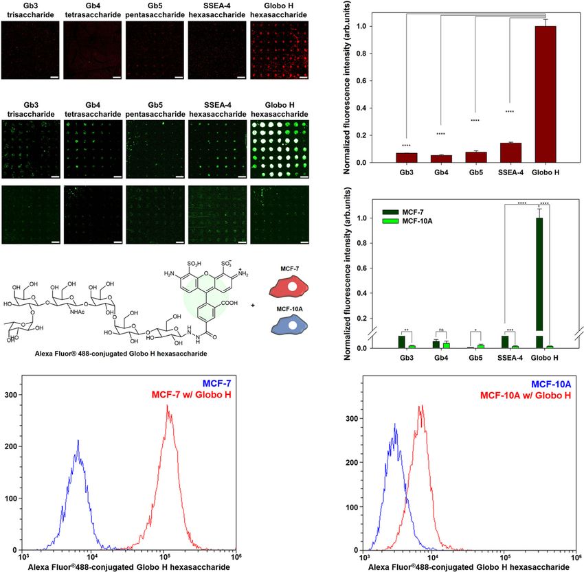
ARTICLE                                                                 NATURE COMMUNICATIONS | https://doi.org/10.1038/s41467-021-21538-0

G
         lycan–protein interactions play an important role in a              allowing better presentation of glycans and glycoclusters on the
         variety of biological processes, including angiogenesis,            surface21–23,27. They also permit tailoring of spatial
         stem cell development, immune responses, and neuronal               arrangements21,22,27. This performance leads to the glycoside
development1,2. Alterations in glycosylation patterns have also              cluster effect21–23,25,27,28, which is a key factor for glycan–protein
been shown to regulate the development and progression of                    interactions24,26,29,30. In addition, DNA linkers were used as
cancer3. Therefore, it is important to understand these interac-             identifiers to code individual glycans binding to glycan-binding
tions to analyze the mechanisms of glycan-mediated biological                proteins and cells by conjugating individual oligonucleotides
processes and to develop therapeutic agents to treat glycan-                 according to glycans31. With these merits, a DNA-based glycan
related diseases.                                                            chip coupled with mass spectrometry showed the possibility of
   Glycan chips have been developed in response to the essential             on-chip glycan biosynthesis by analyzing the activities of glycan-
need for high-throughput analysis of glycan-involved interac-                processing enzymes on the chip24.
tions. Currently, glycan chips have been employed for screening                 In the present work, we develop a glycan chip platform for the
therapeutic agents and profiling glycan-involved interactions,                on-chip enzymatic synthesis of complex glycans based on pH-
including glycan–lectin, glycan–cytokine, glycan–antibody, and               responsive i-motif DNA as a linker material for immobilizing
glycan–virus/bacteria interactions4–8. However, studies on                   glycans on solid supports. The i-motif DNA containing stretches
glycan-related biological processes using glycan chips are still at          of cytosine residues forms a stable four-stranded helical second-
an early stage relative to our knowledge of the biological functions         ary structure (quadruplex) at acidic pH32–35. The pH-responsive
of proteins and genes. To construct glycan chips, most homo-                 structural switch makes it possible to reversibly immobilize and
geneous glycans are commonly provided by either chemical                     isolate glycans on a solid surface. As shown in Fig. 1a, conjugates
synthesis or natural purification using multiphase chromato-                  of glycans and complementary single-stranded oligonucleotides
graphy. These methods have several limitations, including being              can hybridize to i-motif DNAs that are immobilized on the chip
labor-intensive, costly, and time-consuming processes, the                   surface under slightly basic conditions. As the pH is lowered, i-
requirement for protection and deprotection steps, and the dif-              motif DNA tends to strongly form a quadruplex structure,
ficulty in controlling the stereochemistry of glycosidic linkages,            resulting in the denaturation of the DNA double helix and
resulting in poor purity and low stepwise yield9–11. Limited access          thereby enabling the isolation of glycan-oligonucleotide con-
to glycan libraries with diverse structures restricts extensive stu-         jugates from the surface. This property would make it possible to
dies on their roles in vivo using glycan chips. In addition, current         optimize on-chip enzymatic glycosylation by analyzing isolated
glycan chip platforms have used a method of conjugating che-                 glycan-oligonucleotide conjugates using liquid chromatography,
moenzymatically synthesized glycans with linkers to immobilize               resulting in the synthesis of structurally defined complex glycans
them on the surface4–8. This method would be unsuitable for                  on chip. Therefore, our proposed glycan chip platform can
complex glycans because these compounds have a significantly                  improve the limitations of current platforms by immobilizing
low synthesis yield compared to simple glycans, and conjugation              structurally simple disaccharides with high synthesis yields and
with linkers has the limitations of being capable of eliminating             then synthesizing complex glycans on the chip using glycosyl-
labile sialic acid and resulting in significant loss12–14.                    transferases under optimized conditions.
   Considering that on-chip syntheses of oligonucleotides and                   To determine the feasibility of on-chip enzymatic glycosylation
peptides have been successfully utilized for genomics and pro-               using the structure-switchable DNA-based glycan chip platform,
teomics15–17, on-chip enzymatic glycan synthesis is an attractive            Globo H series (Table 1) are selected as target complex glycans,
tool for glycomics. This method has several merits over con-                 which are aberrantly overexpressed on human tumor cells and
ventional methods, including a low-cost and simple process                   known to be involved in tumor progression36,37. Five Globo H-
without any additional protection/deprotection, purification, and             related complex glycans (from trisaccharide to hexasaccharide)
immobilization steps, the use of small amounts of expensive                  are successfully synthesized from chip surface-immobilized lac-
glycan-processing enzymes and nucleotide sugar donors, the                   tose (disaccharide) using the pH-responsive i-motif DNA linker
synthesis of many glycosidic linkages in a straightforward man-              under optimized conditions of stepwise enzymatic glycosylation
ner, and the direct application of synthesized glycans for gly-              reactions. We hypothesize that specific binding proteins for gly-
comics. However, only a few glycan chips have been generated by              cans can be present on breast cancer cells for tumor progression
on-chip enzymatic synthesis18–20. Previously, we demonstrated                and metastasis when considering that globoseries glyco-
the on-chip enzymatic synthesis of GM1 pentasaccharide-related               sphingolipids Gb5, SSEA-4, and Globo H are specifically over-
complex glycans19. Although the feasibility of on-chip complex               expressed on cancer cells38–40. We analyze interactions between
glycan biosynthesis was confirmed, the current platforms have a               breast cancer cells and on-chip-biosynthesized Globo H-related
key technical barrier. Quantitative information on biosynthesized            complex glycans to examine the glycan-binding specificity. This
complex glycans on the surface is unavailable because it is                  analysis can show the potential of our DNA-based glycan chip
impossible to isolate and analyze immobilized glycans on the                 platform combined with on-chip enzymatic glycosylation to
current platforms. In addition, these studies analyzed on-chip               prepare complex glycan sources and to simultaneously analyze
enzymatic glycosylation reactions by only interacting with                   glycan-related biological interactions.
fluorescence dye-labeled lectins. It is impossible to calculate the
enzymatic glycosylation efficiency from the fluorescence intensity             Results
value of lectin bound on the chip. These drawbacks make it                   Preparation of DNA linker for glycan chip. i-motif DNAs with
difficult to control and optimize stepwise glycosylation reactions            four stretches of a series of cytosines (CCCs) in sequence were
using the current platforms, resulting in structurally undefined              modified with thiolated T10 at 5′ to immobilize them onto gold-
glycans on the chip, which leads to unequal and unreliable results.          coated glass slides (Supplementary Table 1). The T10 sequence
Therefore, an advanced strategy is required for on-chip enzymatic            was introduced to increase the structural stability of i-motif DNA
glycosylation-based glycan chips.                                            and to prevent fluorescence quenching by maintaining a constant
   A DNA hybridization-based immobilization method has been                  distance from the gold surface. Complementary single-stranded
used to immobilize glycans for fabrication of glycan chips21–26.             DNA (ssDNA) with three mismatches was designed for
Double-stranded DNA (dsDNA) linkers act as rigid arms,                       glycan-oligonucleotide conjugates (Supplementary Table 1). The

2                NATURE COMMUNICATIONS | (2021)12:1395 | https://doi.org/10.1038/s41467-021-21538-0 | www.nature.com/naturecommunications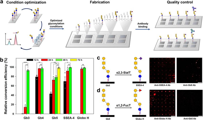
NATURE COMMUNICATIONS | https://doi.org/10.1038/s41467-021-21538-0                                                                           ARTICLE

Fig. 1 Structure-switchable DNA-based glycan chip platform. a Schematic illustration of a structure-switchable DNA-based glycan chip platform using a pH-
responsive i-motif DNA linker. b Schematic illustration and scanned raw image for hybridization of complementary single-stranded oligonucleotides with
surface-immobilized i-motif DNAs under basic conditions (pH 9.0). c–g Schematic illustrations and scanned raw images for hybridization and denaturation of
lactose-oligonucleotide conjugates by pH-responsive structural change in surface-immobilized i-motif DNAs. c The first round hybridization of lactose-
oligonucleotide conjugates with surface-immobilized i-motif DNAs under basic conditions (pH 9.0). d The first round of denaturation of lactose-oligonucleotide
conjugates from surface-immobilized i-motif DNAs under acidic conditions (pH 4.5). e The second round of hybridization of isolated lactose-oligonucleotide
conjugates with surface-immobilized i-motif DNAs under basic conditions. f The second round of denaturation of lactose-oligonucleotide conjugates from
surface-immobilized i-motif DNAs under acidic conditions. g The third round of hybridization of isolated lactose-oligonucleotide conjugates with surface-
immobilized i-motif DNAs under basic conditions. The hybridized lactose-oligonucleotide conjugates were detected by using biotinylated RCA120 lectin and
Alexa Fluor® 647-conjugated streptavidin. Scale bar is 800 μm. Symbols: blue circle, Glc; yellow circle, Gal.

 Table 1 Glycans used in this work and their sequences.

        Glycan                Saccharide                      Sequence                                                                  Symbola

                              Hexasaccharide
        Globo H                                               Fucα1-2Galβ1-3GalNAcβ1-3Galα1-4Galβ1-4Glc
                              (globohexaose)
                              Hexasaccharide
        SSEA 4                                                Neu5Acα2-3Galβ1-3GalNAcβ1-3Galα1-4Galβ1-4Glc
                              (globohexaose)
                              Pentasaccharide
        Gb5                                                   Galβ1-3GalNAcβ1-3Galα1-4Galβ1-4Glc
                              (globopentaose)
                              Tetrasaccharide
        Gb4                                                   GalNAcβ1-3Galα1-4Galβ1-4Glc
                              (globotetraose)
                              Trisaccharide
        Gb3                                                   Galα1-4Galβ1-4Glc
                              (globotriaose)

        Lactose               Disaccharide                    Galβ1-4Glc
 a   , glucose (Glc);   , galactose (Gal);   , N-acetylgalactosamine (GalNAc);   , N-acetylneuraminic acid (Neu5Ac);   , fucose (Fuc)

NATURE COMMUNICATIONS | (2021)12:1395 | https://doi.org/10.1038/s41467-021-21538-0 | www.nature.com/naturecommunications                                   3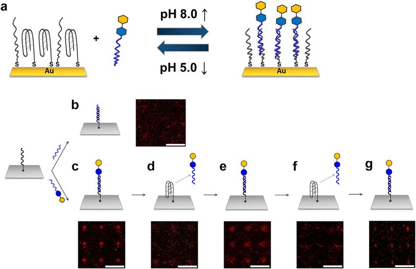
ARTICLE                                                                 NATURE COMMUNICATIONS | https://doi.org/10.1038/s41467-021-21538-0

three mismatches enable prevention of the formation of the G-                On-chip enzymatic synthesis of cancer-associated complex
quadruplex structure of the ssDNA itself and facilitate the                  glycans. The hybridized form should be structurally stable under
denaturation of hybridized dsDNA41. Lactose-oligonucleotide                  neutral pH conditions in which on-chip enzymatic glycosylation
conjugates were synthesized using a thiol-ene photochemical                  reactions are performed. Thus, prior to on-chip glycan bio-
reaction in which 5′ thiol-modified complementary ssDNA was                   synthesis, we checked the stability of the hybridized form in pH
covalently linked to allyl lactose under UV light (Supplementary             7.0 solution according to incubation time. After treatment with
Fig. 1). The synthesized lactose-oligonucleotide conjugates were             complementary ssDNA on the i-motif DNA-immobilized chip,
purified using high-pressure liquid chromatography (Supple-                   the chip was incubated with a pH 7.0 solution for 24–72 h, and
mentary Fig. 2) and confirmed by 1H nuclear magnetic resonance                then doxorubicin, which has intrinsic fluorescence, was added to
(NMR) and mass spectrometry (MS) analyses (Supplementary                     the chip. There was no change in fluorescence intensity until 72 h
Figs. 3–9).                                                                  of incubation time (Supplementary Fig. 11), indicating that
                                                                             hybridization is structurally stable under on-chip enzymatic gly-
                                                                             cosylation conditions, which is supported by a previous study
Development of a glycan chip platform based on a pH-                         showing that i-motif DNA has a linear structure in solution above
responsive DNA linker. Under acidic conditions, i-motif DNA                  pH 6.432.
exists as a four-stranded quadruplex structure via intramolecular               To substantiate the feasibility of the DNA-based glycan chip
base pairing between cytosine (C) and protonated cytosine (C+)               platform for on-chip biosynthesis of complex glycans, enzymatic
in its sequence42. The folded structure can prevent the dense                glycosylations were performed on a chip surface using several
immobilization of i-motif DNAs on the surface, providing suffi-               glycosyltransferases. Each glycosylation product was analyzed by
cient surface space for reversible structural changes in i-motif             using fluorescent dye-labeled lectins. All enzymatic reactions were
DNA. First, 70 μM i-motif DNAs dissolved in phosphate-buffered               performed for 48 h at 37 °C. First, a solution of Tris-HCl (pH 7.0)
saline (PBS) solution (pH 4.5) were robotically spotted onto a               containing α-1,4-galactosyltransferase (LgtC), UDP-Gal, and
gold chip and incubated overnight in a humidified chamber. i-                 MgCl2 was applied to the lactose disaccharide-immobilized
motif DNAs were attached onto a gold surface via gold–thiol                  surface. The α-Gal-specific Griffonia simplicifolia isolectin B4
interactions43. After immobilization of i-motif DNAs, the chip               (GS-IB4) bound to the glycosylated products on the LgtC
was blocked with poly(ethylene glycol) methyl ether thiol in PBS             enzyme-treated surface, indicating that Gb3 trisaccharides were
(pH 4.5). The lactose-oligonucleotide conjugates were incubated              successfully biosynthesized (Fig. 2a). Next, the synthesized Gb3
onto an i-motif DNA-immobilized surface that was pretreated                  trisaccharide-immobilized surface was treated with a Tris-HCl
with PBS buffer (pH 9.0). DNA hybridization-based lactose                    (pH 7.0) solution containing β-1,3-N-acetylgalactosaminyltrans-
immobilization was confirmed through interaction analysis of                  ferase (LgtD), UDP-N-acetylgalactosamine (UDP-GalNAc), and
Ricinus communis agglutinin I (RCA120) lectin, which specifically             MgCl2. The soybean agglutinin (SBA) lectin interacted with the
binds to terminal galactose (Gal). While the complementary                   products synthesized by the LgtD enzyme on the chip surface,
ssDNA-treated surface did not show strong fluorescence intensity              while the LgtD-untreated surface showed no fluorescence
(Fig. 1b), the lactose-oligonucleotide conjugate-treated surface did         (Fig. 2b). This result indicated that GalNAc residues were
(Fig. 1c). To check whether immobilization and isolation of                  successfully transferred onto immobilized Gb3 trisaccharide by
lactose-oligonucleotide conjugates could be controlled by rever-             LgtD. A Tris-HCl (pH 7.0) solution containing UDP-Gal, LgtD,
sible structural changes of i-motif DNAs on the surface, the                 and MgCl2 was incubated on a Gb4 tetrasaccharide-immobilized
fabricated chip was sequentially treated with pH 4.5 and pH 9.0              surface. RCA120 lectin bound to the microspots where Gb4
PBS buffers. When the chip was incubated with RCA120 after                   tetrasaccharides reacted with LgtD on the surface, indicating that
treatment with the pH 4.5 solution, no fluorescence was observed              Gb5 pentasaccharide was enzymatically synthesized from Gb4
(Fig. 1d, f). To further validate the separation of glycan-                  tetrasaccharide (Fig. 2c). Then, a solution of α-2,3-sialyltransfer-
oligosaccharide conjugates at acidic pH, single-stranded oligo-              ase (α2,3-SialT) was added to the Gb5 pentasaccharide-
nucleotides conjugated with Alexa Fluor® 647 at the 5′ end were              immobilized surface to synthesize SSEA-4 hexasaccharide. The
used as a model. There was barely any fluorescence even at high               α2,3-linked N-acetylneuraminic acid (Neu5Ac)-specific Maackia
concentrations of i-motif DNA when treated with acidic solution              amurensis lectin II (MAL II) interacted with the synthesized
(pH 4.5) after incubating Alexa Fluor® 647-conjugated com-                   SSEA-4 hexasaccharide on the surface, while the α2,3-SialT-
plementary oligonucleotides on the chip (Supplementary Fig. 10).             untreated surface did not (Fig. 2d). This result indicated that
Because Alexa Fluor® 647 dye is pH-resistant from pH 4 to pH                 Neu5Ac was transferred to immobilized Gb5 pentasaccharide by
1044, the fluorescence change seemed to be due to the separation              α2,3-SialT. Finally, Globo H hexasaccharide was synthesized by
of the dye-conjugated oligonucleotides, not to the inactivation of           incubating a solution of Tris-HCl (pH 7.0) containing α-1,2-
the dye in acidic conditions. These results indicated that lactose-          fucosyltransferase (α1,2-FucT), GDP-fucose (GDP-Fuc), and
oligonucleotide conjugates were completely separated from the                MgCl2 onto a Gb5 pentasaccharide-immobilized surface. How-
chip surface by forming a quadruplex shape of i-motif DNA                    ever, the α-L-Fuc-specific Lotus tetragonolobus (LTL) lectin
under acidic conditions. When the isolated lactose-                          poorly interacted with the synthesized Globo H hexasaccharide
oligonucleotide conjugates dissolved in pH 9.0 buffer were                   on the surface (Fig. 2e). This result might be due to the
added again on the chip immobilized with i-motif DNAs, the                   intrinsically poor binding affinity of LTL to large glycans
lactose-oligonucleotide conjugates were successfully rehybridized            containing Fucα1-2Galβ1-3(4)GlcNAc(GalNAc)45.
(Fig. 1e, g). These results showed that the pH-dependent struc-
tural change of i-motif DNAs caused the glycan-oligonucleotide
conjugates to be separated from the surface and reimmobilized on             Optimization of on-chip glycan biosynthesis reactions. To
the surface. Consequently, a glycan chip platform based on the               optimize the on-chip enzymatic glycosylations, each reaction was
reversible structural change of i-motif DNAs in a pH-dependent               performed by changing the reaction times in the presence of
manner was successfully developed to quantitatively analyze                  sufficient amounts of glycosyltransferases and glycan donors. The
complex glycans biosynthesized on the surface and to directly                biosynthesized products were recovered from the chip surface by
provide complex glycans for glycan-related applications.                     DNA denaturation through pH-dependent structural change of

4                NATURE COMMUNICATIONS | (2021)12:1395 | https://doi.org/10.1038/s41467-021-21538-0 | www.nature.com/naturecommunications
NATURE COMMUNICATIONS | https://doi.org/10.1038/s41467-021-21538-0 ARTICLE the i-motif DNA and quantitatively analyzed using liquid chro- surface (Fig. 3b and Supplementary Fig. 12). For Gb4 tetra- matography to determine the conversion efficiency for the bio- saccharide synthesis, Gb3 trisaccharide-immobilized surfaces synthesized glycans (Fig. 3a). The LgtC enzyme was treated on a (LgtC reaction for 48 h at 37 °C) were reacted with the LgtD lactose disaccharide-immobilized surface for 12, 24, and 48 h at enzyme. Approximately 80% of Gb3 trisaccharide was rapidly 37 °C. Initially, Gb3 trisaccharides were enzymatically synthesized converted to Gb4 tetrasaccharide by the LgtD reaction within slowly at a conversion efficiency of ~14%, but after 48 h, all lac- 12 h. Moreover, the conversion was almost complete within 24 h tose disaccharides were converted to Gb3 trisaccharides on the (Fig. 3b and Supplementary Fig. 13). Compared with Gb3 NATURE COMMUNICATIONS | (2021)12:1395 | https://doi.org/10.1038/s41467-021-21538-0 | www.nature.com/naturecommunications 5
ARTICLE                                                                        NATURE COMMUNICATIONS | https://doi.org/10.1038/s41467-021-21538-0

Fig. 2 Enzymatic glycosylation on structure-switchable DNA-based glycan chip. Scanned raw images and quantitative intensity plots for on-chip
biosynthesized (a) Gb3 trisaccharide, (b) Gb4 tetrasaccharide, (c) Gb5 pentasaccharide, (d) SSEA-4 hexasaccharide, and (e) Globo H hexasaccharide.
Synthesized complex glycans were detected by using biotinylated lectins and Alexa Fluor® 647-conjugated streptavidin. Each value presents the mean ± SEM
from forty-nine independent spots excluding the highest and lowest signals. Statistical significance was assessed using Student’s unpaired t test (****p < 0.0001).
GS-IB4 Griffonia simplicifolia isolectin B4, SBA soybean agglutinin lectin, RCA120 Ricinus communis agglutinin I lectin, MAL II Maackia amurensis lectin II, LTL Lotus
tetragonolobus lectin, LgtC α-1,4-galactosyltransferase, LgtD β-1,3-N-acetylgalactosaminyltransferase/β-1,3-galactosyltransferase, α2,3-SialT α-2,3-
sialyltransferase, α1,2-FucT α-1,2-fucosyltransferase. Symbols: blue circle, Glc; yellow circle, Gal; yellow square, GalNAc; red triangle, Fuc; purple square, Neu5Ac.
Scale bar is 800 μm. Source data are provided as a Source Data file.

Fig. 3 Structure-switchable DNA-based glycan chip combined with on-chip glycan biosynthesis. a The workflow for the construction of a structure-
switchable DNA-based glycan chip via on-chip complex glycan biosynthesis under optimized conditions. On-chip enzymatic glycosylation conditions were
optimized by analyzing biosynthesized glycans isolated from the chip in acidic pH (4.5) through Bio-LC. To fabricate a DNA-based glycan chip composed of
Globo H and its related structures, a lactose-oligonucleotide conjugate-immobilized surface was divided into five blocks, followed by treatment with
glycosyltransferases under optimized conditions. For quality control, the DNA-based glycan chips were incubated with antibodies against starting materials
and products. b Quantitative analyses of relative conversion efficiencies for on-chip enzymatic syntheses of Globo H hexasaccharide and related structures
in Supplementary Figs. 6–10. Each value presents the mean ± SEM from forty-nine independent spots excluding the highest and lowest signals. Statistical
significance was assessed using Student’s unpaired t test (*p < 0.05; **p < 0.01; ***p < 0.001; ****p < 0.0001). Schematic illustrations and scanned raw
images for on-chip enzymatic glycosylation of (c) SSEA-4 hexasaccharide and (d) Globo H hexasaccharide under optimized conditions (Scale bar: 800
μm). Synthesized complex glycans were detected by using DyLight 650-conjugated monoclonal antibodies or monoclonal antibodies with Alexa Fluor®
647-conjugated polyclonal secondary antibodies. Anti-Gb5 Ab DyLight 650-conjugated anti-Gb5 monoclonal antibody, Anti-SSEA-4 Ab DyLight 650-
conjugated anti-SSEA-4 monoclonal antibody, Anti-Globo H Ab Anti-Globo H monoclonal antibody (VK9), α2,3-SialT α-2,3-sialyltransferase, α1,2-FucT α-
1,2-fucosyltransferase. Symbols: blue circle, Glc; yellow circle, Gal; yellow square, GalNAc; red triangle, Fuc; purple square, Neu5Ac. Source data are
provided as a Source Data file.

trisaccharide, Gb4 tetrasaccharide was biosynthesized on the                        conversion efficiency of ~43% for 12 h (Fig. 3b and Supplemen-
surface more quickly due to the higher catalytic activity of LgtD                   tary Fig. 14). Even after 48 h, the conversion efficiency of Gb4
than of LgtC46. In the same way, a solution of LgtD containing                      tetrasaccharide to Gb5 pentasaccharide was ~80%. Thus, an
UDP-Gal was added on the prepared Gb4 tetrasaccharide-                              enzymatic reaction time of 72 h was required for complete bio-
immobilized surface to synthesize Gb5 pentasaccharide. The                          synthesis of Gb5 pentasaccharide from Gb4 tetrasaccharide using
LgtD enzyme exhibits β-1,3-N-acetylgalactosaminyltransferase                        the same amount of LgtD. This result might be explained because
activity when Gb3 trisaccharide and UDP-GalNAc are used as an                       LgtD has much lower activity with Gb4 tetrasaccharide than with
acceptor and a donor, respectively44. The enzyme also shows β-                      Gb3 trisaccharide as an acceptor43. Finally, biosynthesis of Globo
1,3-galactosyltransferase activity when we use Gb4 tetra-                           H hexasaccharide and SSEA-4 hexasaccharide was conducted on
saccharide as an acceptor and UDP-Gal as a donor47. Unlike the                      a Gb5 pentasaccharide-immobilized surface using α1,2-FucT and
biosynthesis of Gb4 tetrasaccharide using the LgtD enzyme, Gb5                      α2,3-SialT, respectively. The Gb5 pentasaccharide-immobilized
pentasaccharide was slowly synthesized on the surface at a                          surface was prepared by serial enzymatic reactions under the

6                    NATURE COMMUNICATIONS | (2021)12:1395 | https://doi.org/10.1038/s41467-021-21538-0 | www.nature.com/naturecommunications
NATURE COMMUNICATIONS | https://doi.org/10.1038/s41467-021-21538-0                                                                  ARTICLE

optimized conditions. The results showed that Gb5 penta-                        Interaction analysis of MCF-7 breast cancer cells with on-chip
saccharides were almost completely converted to Globo H and                     biosynthesized Globo H-related complex glycans. Screening the
SSEA-4 hexasaccharides on the chip surface within 24 h, which                   glycan-binding specificities of cancer cells enables the discovery of
might be due to the high catalytic activities of both enzymes                   glycan-based target probes and suggests a direction for cancer cell
(Fig. 3b and Supplementary Figs. 15, 16)48–50. To check whether                 targeting for effective diagnostics and therapy. To examine the
there are minor unreacted starting glycans on the chip after                    feasibility of a DNA-based glycan chip combined with on-chip
enzymatic reactions under optimized conditions, we analyzed                     glycan biosynthesis for this screening application, we analyzed
antibody binding to spots where immobilized glycans reacted                     glycan binding of MCF-7 breast cancer cells on the developed
with glycosyltransferases (Fig. 3a). Due to the absence of com-                 Globo H series glycan chip composed of Globo H hexasaccharide
mercially available anti-lactose and anti-Gb4 antibodies, it was                and its related complex glycans. The Globo H series glycan chip
impossible to confirm that all glycosylation processes were                      was constructed as described above. All living cells (4 × 10⁵ cells/
completed. However, using anti-Gb5, anti-SSEA-4, and anti-                      mL) stained with calcein-AM dye were applied on the glycan
Globo H antibodies, we confirmed that there was no fluorescence                   chip. MCF-7 breast cancer cells strongly recognized Globo H
when the antibodies against starting glycans were treated on the                hexasaccharide on the chip, while MCF-10A normal breast cells
chip after on-chip enzymatic syntheses of Gb4 tetrasaccharide,                  did not (Fig. 4c, d). This was also supported by flow cytometry
SSEA-4 hexasaccharide, and Globo H hexasaccharide (Fig. 3c, d                   analyses of MCF-7 cancer and MCF-10A normal cells for Globo
and Supplementary Fig. 17). These results clearly indicated that                H hexasaccharide binding (Fig. 4e–g). Except for that of Globo H
the on-chip enzymatic glycosylations completely proceeded under                 hexasaccharide, the binding affinities of both cells for other bio-
the conditions used, along with high reproducibility (relative                  synthesized complex glycans were similar. These results indicated
standard deviation of 0.9–3.4%).                                                that Globo H hexasaccharide-binding proteins can be present on
                                                                                MCF-7 breast cancer cells, which supports a previous study that
                                                                                identified the major binding protein for Globo H hexasaccharide
Interaction analysis of anti-Globo H antibody with on-chip
                                                                                on the cancer cell membrane54. In addition, quantitative binding
biosynthesized Globo H-related complex glycans. To prepare a
                                                                                analysis was performed using a cell mixture with different ratios
Globo H-related glycan chip and apply it for interaction analysis
                                                                                of MCF-7 cancer cells to MCF-10A normal cells. The glycan chip
with antibody, we performed sequential syntheses of Gb3 tri-
                                                                                exhibited stronger fluorescence intensity according with an
saccharide, Gb4 tetrasaccharide, Gb5 pentasaccharide, Globo H
                                                                                increased ratio of MCF-7 cells to MCF-10A cells (Supplementary
hexasaccharide, and SSEA-4 hexasaccharide on a lactose
                                                                                Fig. 23).
disaccharide-immobilized chip using glycosyltransferases under
                                                                                   In this work, we clearly demonstrated that the structure-
their optimized conditions. Briefly, the lactose disaccharide-
                                                                                switchable DNA-based glycan chip platform simultaneously
immobilized surface was divided into five blocks, and these blocks
                                                                                enables the biosynthesis of complex glycans and the analysis of
were treated with glycosyltransferase(s) and sugar nucleotide(s) to
                                                                                glycan-involved applications. For its practical application, it is
individually biosynthesize Globo H hexasaccharide and its related
                                                                                necessary to prepare a large-scale glycan chip with a large number
glycans (Fig. 3a). The relative glycan-binding specificity of a
                                                                                of on-chip biosynthesized complex glycans. Thus, we are working
mouse IgG anti-Globo H monoclonal antibody (VK9) was
                                                                                on further studies to devise a solution for synthesizing a large
investigated for on-chip biosynthesized Globo H-related complex
                                                                                number of glycans on the chip via combination with a
glycans. It has been established that the VK9 antibody has a high
                                                                                microfluidics system containing multichannels. The introduction
binding affinity to Globo H hexasaccharide without any cross-
                                                                                of a microfluidics system has the advantages of providing a large
reactivity to other Globo H analogs51,52. Scanned fluorescence
                                                                                number of glycans on the chip by biosynthesizing different
imaging showed that VK9 had a much higher binding affinity to
                                                                                glycans for each channel, efficiently reusing glycan-processing
biosynthesized Globo H hexasaccharide than to the other syn-
                                                                                enzymes, and minimally using expensive reagents (e.g., nucleotide
thesized glycans (Fig. 4a, b), consistent with a previously reported
                                                                                sugars). The structure-switchable DNA-based glycan chip
binding specificity of VK952. The difference in fluorescence
                                                                                combined with a microfluidics system makes it possible to
intensities of Globo H and SSEA-4 hexasaccharides showed that
                                                                                synthesize a variety of complex glycans from simple glycans
the Fuc moiety plays a significant role in VK9 binding, which is
                                                                                immobilized on the chip by adjusting the combination of glycan-
supported by previous reports51,52. These results confirmed that
                                                                                processing enzymes according to channels. Given the optimized
the Globo H hexasaccharide series was successfully biosynthe-
                                                                                reaction conditions for various enzymes, users can easily fabricate
sized on the lactose disaccharide-immobilized surface.
                                                                                glycan chips composed of diverse complex glycans without
                                                                                further purification and conjugation with a specific linker. Our
Interaction analysis of cholera toxin B subunit with on-chip                    proposed platform would be advantageous in that it allows users
biosynthesized GM1-related complex glycans. To further vali-                    to fabricate customized glycan chips (microarrays) by directly
date the feasibility of the structure-switchable DNA-based glycan               synthesizing various glycans on the chip through a combination
chip for on-chip complex glycan biosynthesis, GM1 penta-                        of glycan-processing enzymes. However, this technology is still in
saccharide and its related glycans were also synthesized on the                 a relatively early stage of development, and there may be
lactose disaccharide-immobilized surface using glycosyl-                        questions about whether the final products are in structurally
transferases under their optimized conditions (Supplementary                    defined forms. Thus, further work will be performed to make our
Table 2 and Supplementary Figs. 18–20). Unlike GM3 tri-                         proposed platform a (semi)preparative scale capable of carrying
saccharide and GM2 tetrasaccharide, GM1 pentasaccharide was                     out full structural characterization of the final glycan products.
biosynthesized at low efficiency. This result was due to the low                 We anticipate that the improved platform would enable practical
catalytic activity of the CgtB enzyme (17 U/L), which was con-                  application of glycan chips with on-chip biosynthesized complex
sistent with a previous study53. We analyzed the interaction of the             glycans.
cholera toxin B subunit and GM1 pentasaccharide-related com-                       In particular, the structure-switchable DNA-based glycan chip
plex glycans biosynthesized on the chip surface (Supplementary                  platform might be used to analyze the activities of various glycan-
Fig. 21). This result showed that the intrinsic selectivity of the              processing enzymes in a label-free manner on a single chip
cholera toxin B subunit to the on-chip biosynthesized glycans was               because immobilized glycans can be individually separated by
consistent with previous studies5,19.                                           structural changes in pH-responsive i-motif DNA. We anticipate

NATURE COMMUNICATIONS | (2021)12:1395 | https://doi.org/10.1038/s41467-021-21538-0 | www.nature.com/naturecommunications                          7
ARTICLE NATURE COMMUNICATIONS | https://doi.org/10.1038/s41467-021-21538-0 Fig. 4 Applications of structure-switchable DNA-based glycan chips. a, b Analysis of the glycan-binding specificity of the VK9 antibody on the glycan chip. a Scanned raw images and (b) quantitative fluorescence intensity plot for the binding of VK9 with biosynthesized Gb3 trisaccharide, Gb4 tetrasaccharide, Gb5 pentasaccharide, SSEA-4 hexasaccharide, and Globo H hexasaccharide (Scale bar: 800 μm). c, d Analysis of glycan-binding specificity of MCF-7 breast cancer cells on the glycan chip. c Scanned raw images and (d) quantitative fluorescence intensity plot for the binding of MCF-7 breast cancer and MCF-10A normal breast cells with on-chip biosynthesized Gb3 trisaccharide, Gb4 tetrasaccharide, Gb5 pentasaccharide, SSEA-4 hexasaccharide, and Globo H hexasaccharide (Scale bar: 800 μm). Each value presents the mean ± SEM from forty-nine independent spots excluding the highest and lowest signals. Statistical significance was assessed using Student’s unpaired t test (*p < 0.05; **p < 0.01; ***p < 0.001; ****p < 0.0001). e Schematic presentation for analyzing the binding of MCF-7 breast cancer and MCF-10A normal breast cells to Globo H hexasaccharide using flow cytometry. FACS analyses for the binding of (f) MCF-7 breast cancer and (g) MCF-10A normal breast cells with Globo H hexasaccharide. Source data are provided as a Source Data file. that the number of available glycan-processing enzymes is H hexasaccharide and its related complex glycans were success- increased by efficiently screening their activities using our fully biosynthesized from lactose disaccharide on the surface developed glycan chip platform. using several glycosyltransferases under the optimized conditions. In summary, we developed a glycan chip platform based on an This platform was strongly confirmed by additional on-chip i-motif DNA linker with a pH-responsive structural change for biosynthesized GM1 pentasaccharide and its related complex effective on-chip enzymatic glycosylation of complex glycans. The glycans. The constructed glycan chip containing Globo H structural change enabled us to reversibly control the immobi- hexasaccharide and its related complex glycans was applied to lization and separation of the biosynthesized complex glycans on analyze the glycan-binding specificities of antibodies and breast a surface. This approach also enabled optimization of the on-chip cancer cells, clearly demonstrating the feasibility of a DNA-based enzymatic glycosylation reaction conditions through quantitative glycan chip with on-chip glycan biosynthesis for glycan- analyses of the biosynthesized glycans, which can overcome the biomolecule and glycan-cell interaction analyses. Therefore, we limitations of previous on-chip glycan synthesis methods. Globo anticipate that the developed structural switchable DNA-based 8 NATURE COMMUNICATIONS | (2021)12:1395 | https://doi.org/10.1038/s41467-021-21538-0 | www.nature.com/naturecommunications
NATURE COMMUNICATIONS | https://doi.org/10.1038/s41467-021-21538-0                                                                                      ARTICLE

glycan chip platform will present directions for the efficient on-                     a thickness of 100 nm. The film thickness was monitored using a quartz crystal
chip biosynthesis of complex glycans and the realization of                           microbalance (QCM; Inficon, Bad Ragaz, Switzerland). After the completion of
                                                                                      deposition, gold-coated glass slides were rinsed with acetone and methanol.
diverse glycan-related applications.                                                      A step-by-step protocol describing the fabrication and application of glycan
                                                                                      chip is available via Protocol Exchange58. Single-stranded i-motif DNA
Methods                                                                               (Supplementary Table 1) was dissolved in printing buffer (100 mM sodium
Materials. Poly(ethylene glycol) methyl ether thiol (average Mn 800), 1,4-dithio-     phosphate, 10% (v/v) N,N-dimethylformamide, and 1 M NaCl; pH 4.5) to a final
threitol, 4-dimethylaminopyridine, 2,2-dimethyoxy-2-phenylacetophenone                concentration of 70 μM. The solution was spotted on each gold slide (100 nm Au
(DMPA), uridine 5′-diphospho-N-acetylgalactosamine (UDP-GalNAc) disodium              and 10 nm Ti adhesion layer) using a Microssys 5100 microarrayer (Cartesian
salt, uridine 5′-diphosphogalactose (UDP-Gal) disodium salt, biotinylated Griffonia   Technologies, Ann Arbor, MI, USA) with a Chip Maker 2 pin (Telecom
simplicifolia isolectin B4 (GS-IB4), and Envi-Carb SPE columns were purchased         International, Sunnyvale, CA, USA). Array was designed using AxSys software v 1.
from Sigma-Aldrich (St. Louis, MO, USA). Cytidine-5′-monophospho-N-acet-              79. 4. 0. After incubating for 12 h under 75% humidity, the slides were treated with
ylneuraminic acid (CMP-Neu5Ac) sodium salt and Pasteurella multocida α-2,3-           blocking solution (1 mM poly(ethylene glycol) methyl ether thiol in PBS buffer; pH
sialyltransferase (α2,3-SialT) were obtained from GeneChem Inc. (Daejeon, Korea).     4.5) for 1 h to block nonspecific interactions. Next, the slides were removed from
Guanosine 5′-diphospho-β-L-fucose disodium salt (GDP-Fuc), Neisseria meningi-         the blocking solution and rinsed with washing buffer (100 mM sodium phosphate
tides β-1,3-N-acetylgalactosaminyltransferase (LgtD), Neisseria meningitides α-1,4-   and 1 M NaCl; pH 4.5) and deionized water. The slides were dried through
galactosyltransferase (LgtC), and Helicobacter mustelae α-1,2-fucosyltransferase      centrifugation at 213 × g for 3 min. Glycan immobilization was conducted by
(α1,2-FucT) were purchased from Chemily Glycoscience (Atlanta, GA, USA). 1-O-         reacting the i-motif DNA-immobilized chip with 40 μL of hybridization solution
Allyl-D-lactose was purchased from Carbosynth (Newbury, Berkshire, UK). PBS           (100 mM sodium phosphate and 1 M NaCl; pH 9.0) containing 1 nmol lactose-
buffers (100 mM sodium phosphate and 1 M NaCl; pH 4.5 and pH 9.0) were                oligonucleotide conjugates for 3 h. Next, the DNA-based glycan chip was washed
prepared with ultrapure Milli-Q water (resistance > 18 MΩ cm). All thiol-modified      with 1X saline-sodium citrate (SSC) solution (150 mM NaCl and 15 mM sodium
oligonucleotides were purchased from Integrated DNA Technologies, Inc. (Cor-          citrate; pH 7.0) with 0.2% (w/v) sodium dodecyl sulfate (SDS), 0.1X SSC solution
alville, IA, USA). Biotinylated Ricinus communis agglutinin I (RCA120), biotiny-      with 0.2% (w/v) SDS, 0.1X SSC solution, and deionized water for 1 min each. After
lated soybean agglutinin (SBA), biotinylated Lotus tetragonolobus lectin (LTL), and   drying, the slides were stored at room temperature under vacuum until further use.
biotinylated Maackia amurensis lectin II (MAL II) were purchased from Vector
Laboratories (Burlingame, CA, USA). Alexa Fluor® 647-conjugated streptavidin,         Condition optimization for on-chip enzymatic synthesis of Globo H series.
Geneframe (25 μL, 1.0 × 1.0 cm), and Calcein-AM were purchased from Thermo            Commercially available glycosyltransferases were used to biosynthesize Globo H
Fisher Scientific (Waltham, MA, USA). DyLight 650-conjugated anti-Gb5 mono-            hexasaccharide and its related glycans46. Each enzymatic glycosylation reaction was
clonal antibody, DyLight 650-conjugated anti-SSEA-4 monoclonal antibody, and          optimized by adjusting reaction times under certain concentrations of enzymes (4
anti-Globo H monoclonal antibody (VK9) were purchased from Invitrogen                 mU) and nucleotide donors (10 mM) to synthesize Gb3 trisaccharide, Gb4 tetra-
(Carlsbad, CA, USA). Anti-Gb3 monoclonal antibody was purchased from Tokyo            saccharide, Gb5 pentasaccharide, SSEA-4 hexasaccharide, and Globo H hex-
Chemical Industry Co., Ltd. (Tokyo, Japan). Alexa Fluor® 647-conjugated goat          asaccharide from surface-immobilized lactose disaccharide in consecutive order. The
anti-mouse IgG H&L was purchased from Abcam (Cambridge, MA, USA). MCF-7               slide was incubated with the enzyme solution at 37 °C for 12, 24, 48, and 72 h in a
cells (ATCC® HTB-22™) and MCF-10A cells (ATCC® CRL-10317) were purchased              humid chamber. After the reaction, the slide was washed once with washing buffer I
from ATCC (Manassas, VA, USA). HyClone™ Dulbecco’s modified Eagle’s medium             (137 mM NaCl, 2.7 mM KCl, 4.3 mM Na2HPO4, 1.4 mM KH2PO4, and 0.5% (v/v)
(DMEM), Illustra™ NAP-10 Column, HyClone™ Dulbecco’s phosphate-buffered               Tween 20; pH 7.5) and two times with washing buffer II (137 mM NaCl, 2.7 mM KCl,
saline (DPBS) solution, HyClone™ fetal bovine serum (FBS), and HyClone™               4.3 mM Na2HPO4, and 1.4 mM KH2PO4; pH 7.5) and dried by centrifugation at
penicillin–streptomycin 100X solution were purchased from GE Healthcare Life          213 × g for 3 min. PBS buffer (pH 4.5) was dropped onto the chip where enzymatic
Sciences (Chicago, IL, USA). The Mammary Epithelial Cell Growth Medium                glycosylation was carried out. After incubating for 2 h, the solutions were collected,
Bulletkit was purchased from Lonza (Basel, Switzerland).                              desalted using an NAP-10 column, and evaporated. The products were analyzed by
                                                                                      liquid chromatography (ICS-5000; Thermo Fisher Scientific) using a CarboPac PA100
Synthesis of glycan-oligonucleotide conjugates. Lactose-oligonucleotide con-          column (4 mm × 250 mm; Dionex, Sunnyvale, CA, USA), isocratic elution mode with
jugates were synthesized by conjugating allyl lactose with thiol-modified oligonu-     100 mM sodium hydroxide, and an Ag/AgCl reference electrode for electrochemical
cleotides via photochemical reaction using a photoinitiator55. Single-stranded        detection. Data were acquired using the Chromeleon software v 7. 2. SR4. Because the
oligonucleotides (Supplementary Table 1) pretreated with 1,4-dithiothreitol (100      Bio-LC used did not have a thermostat, an air conditioner was used to maintain a
nmol, 1 equiv.), 1-O-allyl-D-lactose (100 nmol, 1 equiv.), and photoinitiator (10     constant column temperature. The eluted sample was purified by solid-phase
nmol, DMPA) were mixed in 1 mL of deionized water. The mixture was stirred            extraction chromatography59. The Envi-Carb SPE column was equilibrated in a 15
under UV light (365 nm) using a UVP Blak-Ray® XX-15 L UV bench lamp (15 W;            mL conical tube using 80% (v/v) acetonitrile in 0.1% (v/v) trifluoroacetic acid and
Analytik Jena, Upland, CA, USA) for 2 h. After the reaction, the mixture was          ultrapure water and then spun at 60 × g for 50 s. A 1 mL sample was added to the
analyzed by normal-phase high-performance liquid chromatography (HPLC; Gil-           Envi-Carb column. The column was washed with 2 mL of ultrapure water, 2 mL of
son, Middleton, WI, USA) using an LC-321 and OD-300 column (4.6 mm × 250              25% (v/v) acetonitrile, 1 mL of ultrapure water, and 2 mL of 10 mM triethylammo-
mm; PerkinElmer, Waltham, MA, USA). The sample was eluted using a linear              nium acetate (pH 7.0) sequentially. The final product was eluted with 2 mL of 25% (v/
gradient of acetonitrile (25–100% (v/v)) in 0.1 M triethylammonium acetate (pH        v) acetonitrile in 50 mM triethylammonium acetate (pH 7.0) and dried to remove the
7.0) and detected at 260 nm with a diode array detector (UV/Vis-151 detector;         solvent. In addition, the slides were incubated with complexes of biotinylated GS-IB4,
Gilson). Data were acquired using the TRILUTION® LC software v 2 .1.                  RCA120, SBA, LTL, and MAL II labeled by streptavidin-Alexa Fluor® 647 to assess the
                                                                                      products of enzymatic reactions.
Nuclear magnetic resonance (NMR) analysis of glycan-oligonucleotide con-
jugates. 1H NMR spectra (solvent D2O) of glycan-oligonucleotide conjugates were       On-chip enzymatic synthesis of Globo H hexasaccharide series from surface-
acquired using an NMR spectrometer (500 MHz; Bruker, Karlsruhe, Germany) and          immobilized lactose. Commercially available glycosyltransferases were used for
TopSpin software v 3.6.2. Data are reported as follows: chemical shifts (δ ppm),      on-chip glycosylation of complex glycans46. The lactose-immobilized surface was
multiplicity (s = singlet, d = doublet, q = quartet, m = multiplet), and coupling     divided into five different blocks using Geneframe®. For synthesis of Gb3 tri-
constants (Hz).                                                                       saccharide, a 25 μL solution of LgtC (4 mU), UDP-Gal (10 mM), Tris-HCl (100
                                                                                      mM; pH 7.0), and MgCl2 (10 mM) was dropped into all five blocks, and the slide
                                                                                      was incubated at 37 °C for 48 h in a humidified chamber. For synthesis of Gb4
Matrix-assisted laser desorption/ionization time-of-flight (MALDI-TOF)
                                                                                      tetrasaccharide from Gb3 trisaccharide, a 25 μL solution of LgtD (4 mU), UDP-
mass spectrometry (MS) analyses of glycan-oligonucleotide conjugates. The
                                                                                      GalNAc (10 mM), Tris-HCl (100 mM; pH 7.0), and MgCl2 (10 mM) was dropped
MALDI-TOF spectrum was measured on an AXIMA LNR MALDI-TOF MS
                                                                                      into four blocks of Gb3 trisaccharide-synthesized five blocks, and the slide was
(Shimadzu, Kyoto, Japan) using 3-hydroxypyridine-2-carboxylic acid (3-HPA) as a
                                                                                      incubated at 37 °C for 48 h in a humidity chamber. For galactosylation (synthesis of
matrix (50 mg/mL in deionized water). The dried sample was mixed with 10 μL of
                                                                                      Gb5 pentasaccharide) of Gb4 tetrasaccharide, a 25 μL solution of LgtD (4 mU),
matrix solution directly on the MALDI target, followed by vacuum drying.
                                                                                      UDP-Gal (10 mM), Tris-HCl (100 mM; pH 7.0), and MgCl2 (10 mM) was dropped
                                                                                      into three of four Gb4 tetrasaccharide-synthesized blocks, and the slide was
Fabrication of the DNA-based glycan chip platform. Glass slides (76 × 26 × 1          incubated at 37 °C for 72 h in a humidified chamber. For fucosylation (synthesis of
mm; Marienfeld GmbH & Co. KG, Lauda-Königshofen, Germany) were coated                 Globo H hexasaccharide) of Gb5 pentasaccharide, a 25 μL solution of α1,2-FucT (4
with ~100 nm thick gold and an ~10 nm titanium adhesive layer56,57. Glass slides      mU), GDP-Fucose (10 mM), Tris-HCl (100 mM; pH 7.0), and MgCl2 (10 mM) was
were cleaned by ultrasonication in trichloroethylene, acetone, isopropyl alcohol,     dropped into one of three Gb5 pentasaccharide-synthesized blocks, and the slide
and pure water and dried by centrifugation. Titanium and gold films were formed        was incubated at 37 °C for 48 h in a humidified chamber. For the synthesis of
on glass slides with a deposition of 1 Å/s and a chamber pressure of 3 × 10⁻⁶ mbar    SSEA-4 hexasaccharide, a 25 μL solution of α2,3-SialT (4 mU), CMP-Neu5Ac
by an E-beam evaporator (KVE-4000; Korea Vacuum Tech, Gimpo, Korea). Pre-             (10 mM), Tris-HCl (100 mM; pH 7.5), and MgCl2 (20 mM) was dropped into one
pared slide substrates were placed on the sample holder disc, and a titanium          of three Gb5 pentasaccharide-synthesized blocks, and the slide was incubated at
adhesion layer of 10 nm thickness was first deposited, followed by a gold layer with   37 °C for 48 h in a humidified chamber. After each reaction, the slides were washed

NATURE COMMUNICATIONS | (2021)12:1395 | https://doi.org/10.1038/s41467-021-21538-0 | www.nature.com/naturecommunications                                                  9
ARTICLE NATURE COMMUNICATIONS | https://doi.org/10.1038/s41467-021-21538-0
once with washing buffer I and twice with washing buffer II. The slides were then 2. Rudd, P. M., Elliott, T., Cresswell, P., Wilson, I. A. & Dwek, R. A.
dried by centrifugation at 213 × g for 3 min. To determine whether there were Glycosylation and the immune system. Science 291, 2370–2376 (2001).
unreacted starting glycans on the chip after each enzymatic reaction, antibody- 3. Pinho, S. S. & Reis, C. A. Glycosylation in cancer: mechanisms and clinical
binding analyses were performed using anti-ganglioside antibodies. Monoclonal implications. Nat. Rev. Cancer. 15, 540–555 (2015).
antibodies and polyclonal secondary antibodies used in antibody-binding analyses 4. Liang, P. H., Wu, C. Y., Greenberg, W. A. & Wong, C. H. Glycan arrays:
were prepared by diluting to a concentration of 25 and 50 μg/mL in PBS buffer, biological and medical applications. Curr. Opin. Chem. Biol. 12, 86–92 (2008).
respectively. Finally, the prepared glycan chips were applied to analyze the glycan- 5. Kim, C. S., Seo, J. H. & Cha, H. J. Functional interaction analysis of GM1-
binding specificity of breast cancer cells as well as the interaction between the anti- related carbohydrates and vibrio cholerae toxins using carbohydrate chip.
Globo H antibody and Globo H glycan series. A laser scanner (GenePix® 4100 A; Anal. Chem. 84, 6884–6890 (2012).
Molecular devices, Sunnyvale, CA, USA) was used for image acquisition and data 6. Huang, C. Y. et al. Carbohydrate chip for profiling the antibodies interacting
were acquired using the GenePix Pro 7 Software. with Globo H tumor antigen. Proc. Natl Acad. Sci. U.S.A. 103, 15–20 (2006).
7. Hyun, J. Y., Pai, J. & Shin, I. The glycan chip story from construction to
Fluorescence-activated cell sorting (FACS) analysis. MCF-7 breast cancer cells applications. Acc. Chem. Res. 50, 1069–1078 (2017).
were cultured in DMEM (high glucose) supplemented with 10% (v/v) heat- 8. Seo, J. H., Kim, C. S., Hwang, B. H. & Cha, H. J. A functional carbohydrate
inactivated FBS, 100 U/mL penicillin, and 100 μg/mL streptomycin. MCF-10A chip platform for analysis of carbohydrate-protein interaction.
normal breast cells were cultured with Mammary Epithelial Basal Medium, which Nanotechnology 21, 215101 (2010).
contains bovine pituitary extract, hydrocortisone, human epidermal growth factor, 9. Valk-Weeber, R. L., Dijkhuizen, L. & van Leeuwen, S. S. Large-scale
insulin, gentamicin, and amphotericin-B. Both cell lines were incubated at 37 °C in quantitative isolation of pure protein N-linked glycans. Carbohydr. Res. 479,
a humidified atmosphere of 5% CO2 and 95% air, and they were subcultured every 13–22 (2019).
3 days. After incubation, the cells were detached and centrifuged. 10. Verostek, M. F., Lubowski, C. & Trimble, R. B. Selective organic precipitation/
To prepare dye-conjugated Globo H hexasaccharide, Alexa Fluor® 488 extraction of released N-glycans following large-scale enzymatic
hydrazide (1.75 μmol, 1 equiv.) and Globo H hexasaccharide (0.88 μmol, 0.5 equiv.) deglycosylation of glycoproteins. Anal. Biochem. 278, 111–122 (2000).
were mixed in 1 mL of 100 mM PBS buffer (pH 7.0)60. The mixture was incubated 11. Boltje, T. J., Buskas, T. & Boons, G. J. Opportunities and challenges in
at 37 °C for 6 h in a humidity chamber. After the reaction, the mixture was synthetic oligosaccharide and glycoconjugate research. Nat. Chem. 1, 611–622
analyzed by liquid chromatography–mass spectrometry (LC–MS; Waters, Milford, (2009).
MA, USA) (Supplementary Fig. 22). 12. Esposito, D., Hurevich, M., Castagner, B., Wang, C. –C. & Seeberger, P. H.
To analyze cell-Globo H hexasaccharide interactions on the cell surface, both cell Automated synthesis of sialylated oligosaccharide. Beilstein J. Org. Chem. 8,
lines were treated with Alexa Fluor® 488-conjugated Globo H hexasaccharide in 1601–1609 (2012).
culture medium at 37 °C for 1 h in a humidified atmosphere of 5% CO2 and 95% air. 13. Fair, R. J., Hahm, H. S. & Seeberger, P. H. Combination of automated solid-
Each solution was centrifuged to remove the remaining dye-conjugated Globo H phase and enzymatic oligosaccharide synthesis provides access to α(2,3)-
hexasaccharide. After washing with culture medium and DPBS, glycan-treated and
sialylated glycans. Chem. Commun. 51, 6183–6185 (2015).
nontreated cells resuspended in DPBS were placed into the wells of a noncoated 96-
14. Song, X. et al. Oxidative release of natural glycans for functional glycomics.
well plate. These cells were sorted by FACS (Beckman Coulter, Brea, CA, USA). Data
Nat. Methods 13, 528–536 (2016).
were acquired and analyzed by using CytExpert software v 2.3.0 (Beckman Coulter).
15. Quan, J. Y. et al. Parallel on-chip gene synthesis and application to
optimization of protein expression. Nat. Biotechnol. 29, 449–452 (2011).
Interaction analysis of MCF-7 breast cancer and MCF-10A normal breast cells 16. Pellois, J. P. et al. Individually addressable parallel peptide synthesis on
on the chip. MCF-7 breast cancer and MCF-10A normal breast cells were cultured microchips. Nat. Biotechnol. 20, 922–926 (2002).
as described above. To analyze cell–glycan interactions on the chip, cells were 17. Beyer, M. et al. Combinatorial synthesis of peptide arrays onto a microchip.
stained by referring to a previously reported method61. Both cell lines (4 × 10⁵ cell/ Science 318, 1888–1888 (2007).
mL, the number of cells was counted by using a C-chipTM) were treated with 4 nM 18. Park, S., Lee, M. R., Pyo, S. J. & Shin, I. Carbohydrate chips for studying high-
calcein-AM in DPBS for 15 min. The solution was centrifuged to remove the throughput carbohydrate-protein interactions. J. Am. Chem. Soc. 126,
remaining dye. After washing with DPBS and culture medium, dye-treated cells 4812–4819 (2004).
resuspended in cell culture medium were applied onto the glycan chip at 37 °C for 19. Kim, C. S., Heo, H. R., Seo, J. H. & Cha, H. J. On-chip biosynthesis of GM1
1 h in a humidified atmosphere of 5% CO2 and 95% air. To remove unbound cells, pentasaccharide-related complex glycans. Chem. Commun. 55, 71–74
the chip was washed once with cell culture medium and twice with DPBS. (2019).
To quantitatively analyze the binding of MCF-7 cancer cells to Globo H 20. Serna, S., Etxebarria, J., Ruiz, N., Martin-Lomas, M. & Reichardt, N. C.
hexasaccharide on the glycan chip, the number ratio of MCF-7 cancer cells was
Construction of N-glycan chips by using modular synthesis and on-chip
adjusted by mixing with MCF-10A normal cells. Dye-treated cells resuspended in
nanoscale enzymatic glycosylation. Chem. Eur. J. 16, 13163–13175 (2010).
cell culture medium were mixed in three ratios (MCF-7 cells accounted for 100%,
21. Chevolot, Y. et al. DNA directed immobilization glycocluster array:
50%, and 10%) and applied onto the glycan chip.
applications and perspectives. Curr. Opin. Chem. Biol. 18, 46–54 (2014).
22. Chevolot, Y. et al. DNA-based carbohydrate biochips: a platform for surface
Statistical analysis and reproducibility. All values of quantitative data are pre- glyco-engineering. Angew. Chem. Int. Ed. 46, 2398–2402 (2007).
sented as mean ± standard error of the means (SEM) in each legend. Experiments 23. Zhang, J. et al. DNA-directed immobilization of glycomimetics for glycoarrays
were performed with at least independent times yielding similar results and sta- application: comparision with covalent immobilization, and development of
tistical significance was analyzed using Student’s unpaired t test. Statistical analyses an on-chip IC50 measurement assay. Biosens. Bioelectron. 24, 2515–2521
and quantitative plots were performed using Microsoft Excel 2016 and SigmaPlot v (2009).
10.0. *p < 0.05; **p < 0.01; ***p < 0.001; ****p < 0.0001 were considered 24. van Munster, J. M. et al. Application of carbohydrate arrays coupled with mass
significant. spectrometry to detect activity of plant-polysaccharide degradative enzymes
from the fungus Aspergillus niger. Sci. Rep. 7, 43117 (2017).
Reporting summary. Further information on research design is available in the Nature 25. Morvan, F., Vidal, S., Souteyrand, E., Chevolot, Y. & Vasseur, J.-J. DNA
Research Reporting Summary linked to this article. glycoclusters and DNA-based carbohydrate microarrays: From design to
applications. RSC Adv. 2, 12043–12068 (2012).
26. Gerland, B. et al. Synthesis of a library of fucosylated glycoclusters and
Data availability determination of their binding toward Pseudomonas aeruginosa lectin B (PA-
All data that support the findings of this study are available within the paper and IIL) using a DNA-based carbohydrate chip. Bioconjug. Chem 23, 1534–1547
Supplementary Information Files and uploaded to Figshare (https://doi.org/10.6084/m9. (2012).
figshare.13634771). Source data are provided with this paper. 27. Spinelli, N., Defrancq, E. & Morvan, F. Glycoclusters on oligonucleotide and
PNA scaffolds: synthesis and applications. Chem. Soc. Rev. 42, 4557–4573
Received: 16 March 2020; Accepted: 29 January 2021; (2013).
28. Mende, M. et al. Multivalent glycan arrays. Faraday Discuss. 219, 9–32 (2019).
29. Wittmann, V. & Pieters, R. J. Bridging lectin binding sites by multivalent
carbohydrates. Chem. Soc. Rev. 42, 4492–4503 (2013).
30. Briard, J. G., Jiang, H., Moremen, K. W., Macauley, M. S. & Wu, P. Cell-based
glycan arrays for probing glycan-glycan binding protein interactions. Nat.
Commun. 9, 880 (2018).
References 31. Yan, M. et al. Next-generation glycan microarray enabled by DNA-coded
1. Haltiwanger, R. S. & Lowe, J. B. Role of glycosylation in development. Annu.
glycan library and next-generation sequencing technology. Anal. Chem. 91,
Rev. Biochem. 73, 491–537 (2004).
9221–9228 (2019).
10 NATURE COMMUNICATIONS | (2021)12:1395 | https://doi.org/10.1038/s41467-021-21538-0 | www.nature.com/naturecommunicationsYou can also read


