Ichthyoses-A Clinical and Pathological Spectrum from Heterogeneous Cornification Disorders to Inflammation - MDPI
←
→
Page content transcription
If your browser does not render page correctly, please read the page content below
Review
Ichthyoses—A Clinical and Pathological Spectrum from
Heterogeneous Cornification Disorders to Inflammation
Dieter Metze *, Heiko Traupe and Kira Süßmuth

                                          Klinik für Hautkrankheiten, Universitätsklinik Münster, 48149 Münster, Germany;
                                          traupeh@ukmuenster.de (H.T.); Kira.Suessmuth@ukmuenster.de (K.S.)
                                          * Correspondence: metzed@uni-muenster.de

                                          Abstract: Ichthyoses are inborn keratinization disorders affecting the skin only (non-syndromic) or
                                          are associated with diseases of internal organs (syndromic). In newborns, they can be life-threatening.
                                          The identification of the gene defects resulted in reclassification and a better understanding of the
                                          pathophysiology. Histopathologic patterns include orthohyperkeratosis with a reduced or well-
                                          developed stratum granulosum, hyperkeratosis with ortho- and parakeratosis with preserved or
                                          prominent stratum granulosum, and epidermolytic ichthyosis. Another pattern features “perinu-
                                          clear vacuoles and binucleated keratinocytes”, which is associated with keratin mutations. Some
                                          ichthyoses are histologically defined by psoriasis-like features, and distinct subtypes show follicular
                                          hyperkeratosis. In addition to histological and immunohistochemical methods, these patterns allow
                                          a better histopathologic diagnosis.

                                   Keywords: ichthyosis; hereditary keratinization disorders; dermatopathology; pattern analysis; im-
Citation: Metze, D.; Traupe, H.;          munohistochemistry
Süßmuth, K. Ichthyoses—A Clinical
and Pathological Spectrum from
Heterogeneous Cornification
Disorders to Inflammation.                1. Target Readership
Dermatopathology 2021, 8, 107–123.             The article was written for dermatologists and pathologists interested in genoder-
https://doi.org/10.3390/                  matoses and dermatohistology, especially the diagnosis of ichthyoses. It is supposed to
dermatopathology8020017
                                          help to diagnose different types of ichthyoses when genetic analyses are not available or
                                          before genetic testing.
Academic Editors: Sylvie Fraitag and
Gürkan Kaya                               2. Introduction
                                                Ichthyoses are hereditary keratinization disorders defined by universal scaling occur-
Received: 6 April 2021
Accepted: 28 April 2021
                                          ring over the entire body. Some forms manifest at birth (“congenital” forms), others during
Published: 7 May 2021
                                          the first year of life (“vulgar” forms) (Table 1) [1,2].
                                                An accurate and rapid diagnosis of a hereditary keratinization disorder is important
Publisher’s Note: MDPI stays neutral
                                          to identify associated diseases of internal organs in syndromic forms (Table 1), to initiate
with regard to jurisdictional claims in
                                          genetic counseling, and to start potential therapies [1,2].
published maps and institutional affil-         The histopathology of ichthyoses is mentioned in publications and book chapters [3]
iations.                                  but has not been systematically studied and is often considered “nonspecific.” Traditionally,
                                          therefore, the clinical picture, family history, and occasionally laboratory chemistry and
                                          electron microscopic studies have been crucial for diagnosis. Only the identification of
                                          the genetic causes has led to an understanding of the molecular mechanisms, as well as
Copyright: © 2021 by the authors.
                                          to the reclassification of these genodermatoses (Table 1) [4,5]. Assistance is provided by
Licensee MDPI, Basel, Switzerland.
                                          special networks (www.netzwerk-ichthyose.de (accessed on 3 May 2021)) and patient
This article is an open access article
                                          support-groups (https://www.ichthyose.de/ (accessed on 3 May 2021)).
distributed under the terms and                 In parallel, the pathological changes of the skin biopsies were characterized in more
conditions of the Creative Commons        detail. Certain histological patterns could be defined. They include the following criteria:
Attribution (CC BY) license (https://     hyperkeratosis with/without parakeratosis, expression/absence of the stratum granu-
creativecommons.org/licenses/by/          losum, atrophy/hyperplasia (acanthosis) of the epidermis, vacuolization/eosinophilic
4.0/).                                    granules in keratinocytes, or hyperkeratosis and a degree of development of hair follicles.

Dermatopathology 2021, 8, 107–123. https://doi.org/10.3390/dermatopathology8020017               https://www.mdpi.com/journal/dermatopathology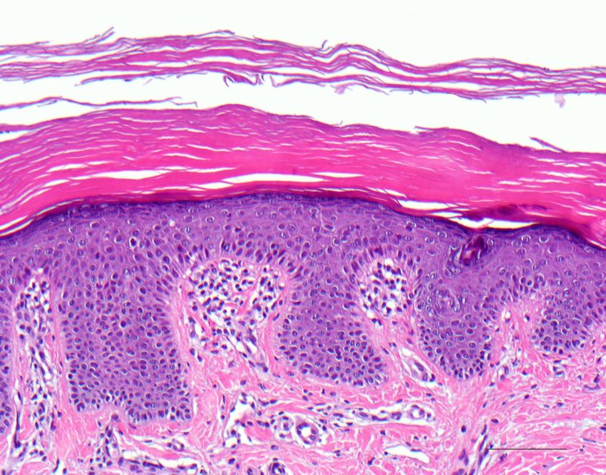
Dermatopathology 2021, 8                                                                                              108

                           Complementary histo- and immunohistochemical methods allow for, in part, a precise
                           diagnosis, but at least a limitation of differential diagnoses, which can then be further
                           clarified by targeted mutation analyses [6,7].

                           Table 1. Clinical classification of ichthyoses.

                            Vulgar ichthyosis, isolated
                            Ichthyosis vulgaris
                            X-linked recessive ichthyosis
                            Vulgar ichthyosis, syndromic
                            Refsum syndrome
                            Multiple sulfatase deficiency
                            Congenital ichthyosis, isolated
                            Keratinopathic ichthyosis
                            Autosomal recessive congenital ichthyosis (ARCI)
                            Harlequin ichthyosis (subtype of ARCI)
                            Autosomal dominant lamellar ichthyosis
                            Congenital reticular ichthyosiform erythroderma (CRIE, Confetti ichthyosis)
                            Ichthyosis hystrix type Curth–Macklin
                            Peeling skin disease
                            Erythrokeratodermia
                            and others
                            Congenital ichthyosis, syndromic
                            HID/KID syndrome
                            Netherton syndrome
                            CHILD syndrome
                            SAM syndrome
                            Conradi–Hünermann–Happle syndrome
                            Sjögren–Larsson syndrome
                            Chanarin–Dorfmann syndrome
                            Trichothiodystrophy
                            IFAP syndrome
                            and others

                                In the following, the dermatopathological diagnosis is presented on the basis of some
                           frequent, but also rare syndromic and life-threatening ichthyoses, which can be performed
                           quickly, easily, and economically on sample biopsies of the skin. Hereditary keratinization
                           disorders are also discussed. They often show a highly inflammatory, psoriasis-like picture
                           and are therefore often misdiagnosed (Table 2) [6,8].

                           Table 2. Ichthyoses with a psoriasis-like picture.

                            Ichthyoses with Psoriasis-Like Picture
                            Netherton syndrome
                            Peeling skin disease
                            CHILD syndrome
                            Severe dermatitis, multiple allergies, metabolic wasting syndrome (SAM syndrome)
                            Anular epidermolytic ichthyosis

                           3. Ichthyosis Vulgaris
                                 The autosomal semidominant inherited ichthyosis vulgaris is the most frequent
                           ichthyosis (prevalence from 1:100 to 1:250) [9]. It usually develops in the course of the
                           first year of life and manifests with dry skin or light gray fine scales (Figure 1) as well as
                           palmoplantar hyperlinearity. The disorder is caused by loss-of-function mutations in the
                           filaggrin gene (Table 3). Filaggrin is expressed in the keratohyalin granules and crosslinks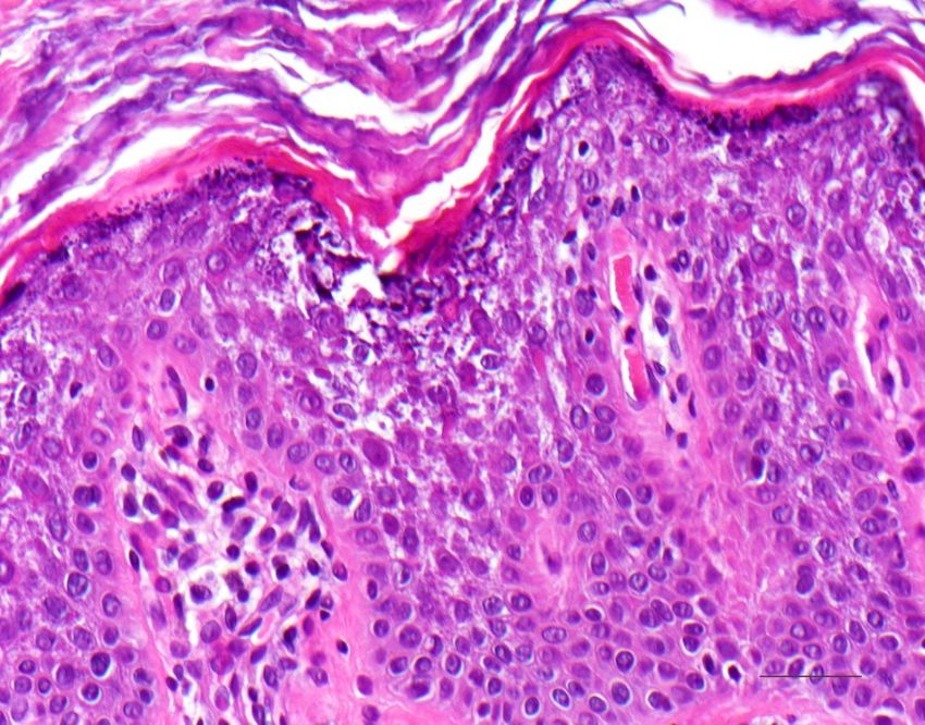
The autosomal semidominant inherited ichthyosis vulgaris is the mos
                            thyosis (prevalence from 1:100 to 1:250) [9]. It usually develops in the cou
                            year of life and manifests with dry skin or light gray fine scales (Figure
Dermatopathology 2021, 8                                                                                                  109
                            palmoplantar hyperlinearity. The disorder is caused by loss-of-function                               m
                            filaggrin gene (Table 3). Filaggrin is expressed in the keratohyalin granules
                            the keratin filaments in the horny layer. A deficiency of filaggrin predisp
                           the keratin filaments in the horny layer. A deficiency of filaggrin predisposes to atopic der-
                            dermatitis
                           matitis and/or and/or      allergic rhinoconjunctivitis
                                            allergic rhinoconjunctivitis                     [8,10].
                                                                         [8,10]. Interestingly,         Interestingly,
                                                                                                filaggrin                  filaggrin
                                                                                                          mutations can also
                            also
                           be     be observed
                               observed  in X-linkedinrecessive
                                                       X-linked    recessive
                                                                ichthyosis      ichthyosis
                                                                            underlying   steroidunderlying      steroid[10].
                                                                                                   sulfatase deficiency  sulfatase

                           Figure
                           Figure     1. Ichthyosis
                                  1. Ichthyosis       vulgaris.
                                                vulgaris. Fine greyFine  grey
                                                                   scaling      scaling
                                                                           on the        on the
                                                                                  extremities.    extremities.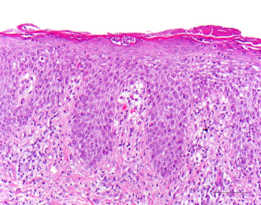
Dermatopathology 2021, 8 110
Table 3. Different types of ichthyosis with gene mutation and mode of inheritance.
Gene
Ichthyosis
(mode of inheritance)
FLG (filaggrin)
Common Ichthyoses Ichthyosis Vulgaris
(autosomal semidominant)
STS (steroid sulfatase)
X-Linked Ichthyosis
(X-linked recessive)
ABCA12 (ATP Binding Cassette Subfamily A
Harlequin Ichthyosis Member 12)
(autosomal recessive)
TGM1 (transglutaminase−1);
ALOX12B (Arachidonate 12-Lipoxygenase,
12R Type); ALOXE3 (Arachidonate
Lamellar Ichthyosis, Congenital Ichthyosiform
Non-Syndromic Lipoxygenase 3); CYP4F22 (Cytochrome P450
Erythroderma
Ichthyoses Family 4 Subfamily F Member 22); NIPAL4
(Ichthyin) and others
ARCI and (autosomal recessive)
Keratinopathic
Ichthyoses TGM1
Bathing Suit Ichthyosis
(autosomal recessive)
KRT1 (keratin 1);
KRT10 (keratin 10)
EI
(autosomal dominant, sometimes recessive
Keratinopathic Ichthyoses
(KRT10 mutations)
KRT2 (keratin 2)
SEI
(autosomal dominant)
KRT1
Rare Variants of KPI CRIE KRT10
(autosomal dominant, de novo mutations)
CDSN (corneodesmosin)
Peeling Skin Disease
(autosomal recessive)
Further Non-Syndromic Ichthyoses GJB3 (encoding Connexin 31)
Erythrokeratoderma Variabilis GJB4 (encoding Connexin 30.3)
(often autosomal dominant)
SPINK5 (encoding LEKTI)
Netherton Syndrome
(autosomal recessive)
GJB2 (encoding Connexin 26)
KID Syndrome
(autosomal dominant)
Syndromic Ichthyoses NSDHL (NAD(P) Dependent Steroid
CHILD Syndrome Dehydrogenase-Like)
(x-linked dominant)
DSG1 (desmoglein−1)
SAM Syndrome DSP (desmoplakin)
(autosomal recessive)
3.1. Histology
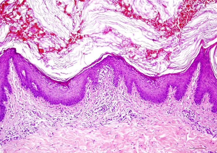
A characteristic feature of ichthyosis vulgaris is a markedly reduced, often completely
absent stratum granulosum. The stratum corneum exhibits mild compact orthohyperker-
atosis (Figure 2). Frequently, there is also hyperkeratosis of hair follicles and acrosyringia.
The epidermis may be slightly widened (acanthotic) but also atrophic. Isolated mild perivas-
cular lymphocytic infiltrates can be found in the dermis. Associated signs of spongiform
dermatitis may be encountered in the setting of atopy.
Immunohistochemically, a deficiency of filaggrin can be quantified, which correlates
with the number of mutations (one or two mutations in the filaggrin gene) and thus, the
severity of ichthyosis. Ultrastructurally, there is a defect in the keratohyalin granules,
which appear diminished and crumbly.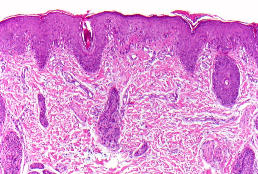
Dermatopathology 2021, 8 111
Dermatopathology 2021, 8, 5
Figure2.2.Ichthyosis
Figure Ichthyosisvulgaris.
vulgaris. Note
Note the
the absent
absentstratum
stratumgranulosum
granulosumand
andmild
mildcompact
compactorthohyperk-
orthohyper-
keratosis. Marked hyperkeratosis of the opening of the acrosyringium. Inflammatory infiltrates
eratosis. Marked hyperkeratosis of the opening of the acrosyringium. Inflammatory infiltrates are are
almost absent. HE stain, bar = 100 µm.
almost absent. HE stain, bar = 100 µm.
3.2. Differential
3.2. DifferentialDiagnoses
Diagnoses
AA thinned
thinned ororabsent
absentstratum
stratum granulosum
granulosum withwith mild
mild orthohyperkeratosis
orthohyperkeratosis is is also
also ob-
ob-
servedininpatients
served patientswith
with atopy
atopy andand other
other veryvery
rare rare ichthyoses,
ichthyoses, such
such as as Conradi–Hüner-
Conradi–Hünermann–
mann–Happle
Happle syndrome syndrome [6].
[6]. It can It can
also also be
be found in found in acquired
acquired ichthyosis-like
ichthyosis-like skin condi-
skin conditions (“ac-
tions (“acquired
quired ichthyoses”).
ichthyoses”). Causes are Causes are malignancies
malignancies (lymphomas), (lymphomas), Crohn0 s dis-
renal insufficiency,
renal insufficiency,
Crohn′s
ease, disease, autoimmune
autoimmune diseases (collagenoses),
diseases (collagenoses), GvHD, infections GvHD,
(HIV,infections (HIV, leprosy),
leprosy), endocrinopathies
(hypothyroidism), sarcoidosis, malnutrition
endocrinopathies (hypothyroidism), (vitamin
sarcoidosis, A) or drugs
malnutrition (lipid-lowering
(vitamin A) or drugsdrugs,
(lipid-
psychotropic
lowering drugs,drugs) [11].
psychotropic drugs) [11].
4.
4. Autosomal
Autosomal Recessive
Recessive Congenital
CongenitalIchthyosis
Ichthyosis
Autosomal
Autosomalrecessive
recessivecongenital
congenital ichthyosis
ichthyosis (ARCI)
(ARCI) represents
represents aa genetically
genetically heteroge-
heteroge-
neous
neous group of non-syndromic congenital ichthyoses with widely varying severity.The
group of non-syndromic congenital ichthyoses with widely varying severity. The
group
group comprises
comprises lamellar
lamellar ichthyosis,
ichthyosis, which
which isis most
most often
often due
due to
to tranglutaminase
tranglutaminase−1 −1defi-
de-
ficiency (Table 3), congenital ichthyosiform erythroderma, and the most severe
ciency (Table 3), congenital ichthyosiform erythroderma, and the most severe but rare but rare
subtype
subtypeof ofharlequin
harlequinichthyosis
ichthyosis[12].
[12].Newborns
Newbornscan canbe
beborn
bornwith
withaatight
tightand
andshiny
shinystratum
stratum
corneum,
corneum, which is associated with ectropion, eclabium, fluid loss, and thermal dysregu-
which is associated with ectropion, eclabium, fluid loss, and thermal dysregu-
lation,
lation, and
andresulting
resultingininpotentially
potentiallylife-threatening
life-threateningcomplications.
complications. However,
However,thetheclinical
clinical
presence of a collodion membrane is also encountered in other ichthyoses [5].
presence of a collodion membrane is also encountered in other ichthyoses [5].
Later, the collodion membrane is replaced by dark brown, adherent, plate-like scales
Later, the collodion membrane is replaced by dark brown, adherent, plate-like scales
(classic lamellar ichthyosis; Figure 3) or a whitish, poorly adherent, fine scale on reddened
(classic lamellar ichthyosis; Figure 3) or a whitish, poorly adherent, fine scale on reddened
skin (non-bullous congenital ichthyosiform erythroderma). To varying degrees, there are as-
skin (non-bullous congenital ichthyosiform erythroderma). To varying degrees, there are
sociated palmoplantar keratoderma, nail dystrophies, fibrosing alopecia, and hypohidrosis
associated palmoplantar keratoderma, nail dystrophies, fibrosing alopecia, and hypohi-
with heat intolerance [5].
drosis with heat intolerance [5].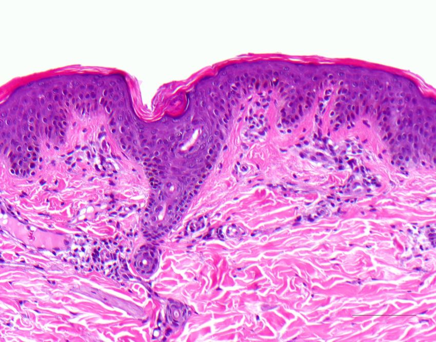
,   Dermatopathology 2021, 8                                                                                                   112   6

          Figure 3. Lamellar
              Figure 3. Lamellarichthyosis.   Dark
                                 ichthyosis. Dark    brownish
                                                  brownish      lamellar
                                                           lamellar         scaling
                                                                    scaling in        inwith
                                                                               a patient a patient  with transglutaminase−1
                                                                                             transglutaminase −1 deficiency.
          deficiency.
                                   The ARCI forms are caused by different mutations. In 30–40% of cases, a mutation is
                              present in the transglutaminase−1 gene, resulting in a disruption of protein cross-linking and
               The ARCI forms the are  caused of
                                  esterification  byceramides
                                                      different    mutations.Using
                                                                in corneocytes.   In 30–40%     of cases,
                                                                                       biotinylated          a mutation
                                                                                                    donor substrates,   suchis
                              as the amine   donor  monodansylcadaverine,     transglutaminase   activity
          present in the transglutaminase−1 gene, resulting in a disruption of protein cross-linking      can be  visualized
                              immediately in situ by fluorescence labeling based on the incorporation of monodansyl-
          and the esterification   of ceramides
                              cadaverine   on sectionsinofcorneocytes.       Using [13].
                                                           unfixed frozen biopsies   biotinylated
                                                                                         Mutations aredonor     substrates,
                                                                                                         also present  in the
          such as the amine donor     monodansylcadaverine,               transglutaminase       activity   can   be
                              ATB-binding cassette transporter (ABCA12) gene, which, unlike harlequin-ichthyosis, has visual-
          ized immediately in residual
                                 situ activity  in milder ARCIlabeling
                                       by fluorescence           cases. ABCA12  is required
                                                                            based   on theinincorporation
                                                                                              epidermal lipid transport   via
                                                                                                                   of mono-
                              the lamellar bodies. Other mutations involve the ichthyin, lipoxygenase, or cytochrome P450
          dansylcadaverine on     sections
                              oxidase         of unfixed
                                      genes FLJ39501.         frozencorrelation
                                                        A definitive    biopsiesof [13].  Mutations
                                                                                   this mutation  with a are  also
                                                                                                         specific    present
                                                                                                                  phenotype
          in the ATB-binding cassette   transporter
                              of ARCI has               (ABCA12)
                                            not been fully  establishedgene,
                                                                         [8]. which, unlike harlequin-ichthyosis,
          has residual activity   in milder ARCI cases. ABCA12 is required in epidermal lipid
                             4.1. Histology
          transport via the lamellar     bodies. Other mutations involve the ichthyin, lipoxygenase, or
                                   Histologically, there is compact orthohyperkeratosis, a slightly widened stratum
          cytochrome P450 oxidase   genesacanthosis,
                             granulosum,     FLJ39501.and  A papillomatosis
                                                              definitive correlation      of this
                                                                             of the epidermis.       mutation
                                                                                                 In the  papillarywith
                                                                                                                   body, a
          specific phenotype the  vessels appear
                              of ARCI     has not dilated
                                                     beenand  spiraling,
                                                            fully        and lymphocytic
                                                                  established    [8].     infiltrates are scarce or mild
                                   (Figure 4) [7].

          4.1. Histology           4.2. Differential Diagnoses
                                    The various forms of ARCI cannot be differentiated histologically, except for harlequin
               Histologically, there  is compact orthohyperkeratosis, a slightly widened stratum granu-
                               ichthyosis. Similarly, X-linked ichthyosis presents with almost identical pathology. Lichen
          losum, acanthosis, and    papillomatosis
                               simplex                  of thewith
                                        chronicus presents      epidermis.    In inflammation
                                                                    more severe  the papillaryand body,   theinvessels
                                                                                                     fibrosis            ap-
                                                                                                                 the papil-
          pear dilated and spiraling,   and lymphocytic infiltrates are scarce or mild (Figure 4) [7].
                               lary body.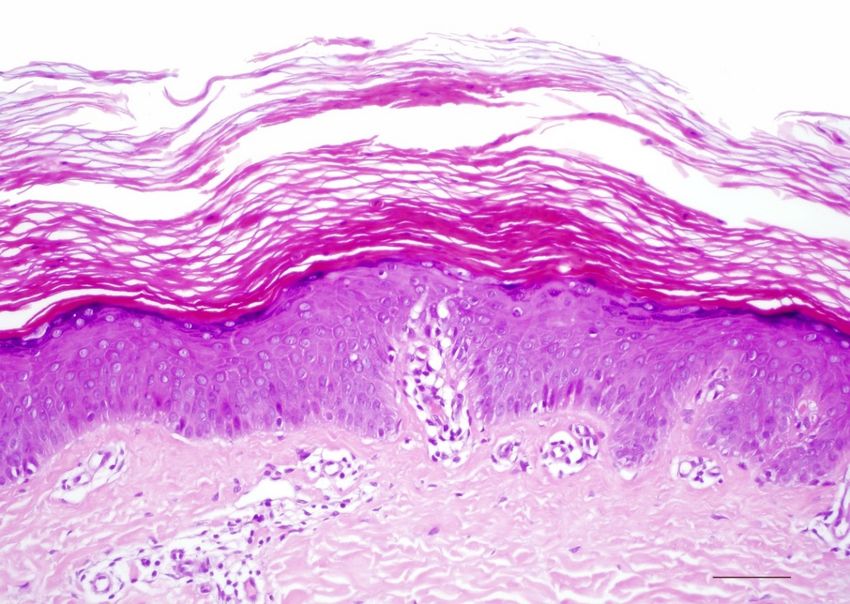
Dermatopathology 2021, 8                                                                                                              113
    Dermatopathology 2021, 8,                                                                                                  7

                                 Figure 4.
                                Figure    4.Autosomal
                                             Autosomal recessive lamellar
                                                           recessive      ichthyosis.
                                                                       lamellar       Acanthotic
                                                                                ichthyosis.      epidermis with
                                                                                              Acanthotic        well-developed
                                                                                                           epidermis           stra-
                                                                                                                       with well-developed
                                 tum granulosum and compact orthohyperkeratosis without further signs of inflammation. HE
                                stratum granulosum and compact orthohyperkeratosis without further signs of inflammation. HE
                                 stain, original magnification, bar = 100 µm.
                                stain, original magnification, bar = 100 µm.
                                4.2. Differential Diagnoses
                                5. Keratinopathic Ichthyosis
                                     The various forms of ARCI cannot be differentiated histologically, except for harle-
                                quin Epidermolytic    ichthyosis,
                                      ichthyosis. Similarly,        formerly
                                                             X-linked         also
                                                                      ichthyosis    called with
                                                                                 presents  bullous  congenital
                                                                                                almost identical ichthyotic
                                                                                                                  pathology. erythro-
                                derma                   to a  mutation   of keratin 1 or keratin 10
                                Lichen simplex chronicus presents with more severe inflammation and fibrosis in theclassified
                                        Brocq,  is due                                               and is therefore    pa-      as
                                keratinopathic
                                pillary body. ichthyosis (Table 3) [5]. Neonates present with erythroderma with blistering,
                                sometimes pronounced, and later develop (spiky) keratoses, preferentially on the extremi-
Dermatopathology 2021, 8,        5. Keratinopathic
                                ties                      with a keratin 1 mutation also have palmoplantar keratosis, which is8
                                                   Ichthyosis
                                     (Figure 5). Patients
                                absent  in patients with  a keratin
                                      Epidermolytic ichthyosis,     10 mutation
                                                                formerly         because
                                                                         also called      this
                                                                                     bullous   keratin isichthyotic
                                                                                             congenital    not expressed   there [14].
                                                                                                                    erythro-
                                derma Brocq, is due to a mutation of keratin 1 or keratin 10 and is therefore classified as
                                keratinopathic ichthyosis (Table 3) [5]. Neonates present with erythroderma with blister-
                                ing, sometimes pronounced, and later develop (spiky) keratoses, preferentially on the ex-
                                tremities (Figure 5). Patients with a keratin 1 mutation also have palmoplantar keratosis,
                                which is absent in patients with a keratin 10 mutation because this keratin is not expressed
                                there [14].

                                Figure5.
                                Figure 5. Epidermolytic
                                          Epidermolytic ichthyosis.
                                                         ichthyosis. Diffuse
                                                                     Diffuse palmoplantar
                                                                             palmoplantar keratoderma
                                                                                          keratoderma(keratin
                                                                                                      (keratin11mutation).
                                                                                                                mutation).

                                5.1. Histology
                                     There is massive orthohyperkeratosis and acanthosis of the epidermis. The supraba-
                                sal keratinocytes reveal vacuolization and distinct hypereosinophilic granules. In the stra-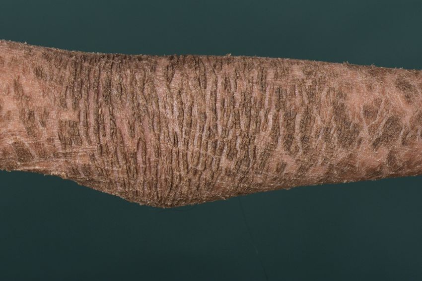
Dermatopathology 2021, 8                                                                                                     114

                            5.1. Histology
                                  There is massive orthohyperkeratosis and acanthosis of the epidermis. The suprabasal
                            keratinocytes reveal vacuolization and distinct hypereosinophilic granules. In the stratum
                            granulosum, the keratohyalin granules are coarse and irregular. The boundaries between
Dermatopathology 2021, 8,   keratinocytes are poorly demarcated, and clefts and blisters occur. Minor lymphocytic9
                            infiltrates may impose in the dermis (Figure 6) [8].

                            Figure 6. Epidermolytic
                            Figure     Epidermolyticichthyosis.   Acanthotic
                                                        ichthyosis.           epidermis
                                                                     Acanthotic          with with
                                                                                   epidermis  massive orthohyperkeratosis.
                                                                                                   massive                 Su-
                                                                                                           orthohyperkeratosis.
                            prabasal keratinocytes
                            Suprabasal              vacuolated
                                        keratinocytes  vacuolatedwith distinct
                                                                    with       hypereosinophilic
                                                                          distinct               granules
                                                                                   hypereosinophilic      and irregular
                                                                                                     granules           kerato-
                                                                                                              and irregular  ker-
                            hyalin granules. HE  stain, bar = 50 µm.
                            atohyalin granules. HE stain, bar = 50 µm.

                             5.2. Differential Diagnoses
                                  Electron microscopically,    the hypereosinophilic granules correspond to clumps of
                            the keratin   skeleton.  A collapse
                                  The histologic reaction patternof the
                                                                      ofmutant    keratinshyperkeratosis
                                                                         epidermolytic     causes the vacuolar
                                                                                                          is alsoaspect
                                                                                                                   foundofinthe
                                                                                                                             su-
                            cytoplasm    and results inichthyosis
                             perficial epidermolytic     mechanical   instability.
                                                                   with   a keratin 2 mutation (ichthyosis bullosa Siemens),
                             or epidermal nevi in the setting of mosaicism of keratinopathic ichthyoses [15,16]. Very
                            5.2. Differential
                             discrete          Diagnoses
                                       and circumscribed,   these changes are also found incidentally in normal skin (pref-
                                  The   histologic reaction  pattern oforepidermolytic
                             erentially in the vicinity of epithelial                    hyperkeratosis
                                                                          melanocytic tumors),    as well is
                                                                                                          as also found
                                                                                                              in cysts,   in su-
                                                                                                                        scars, or
                            perficial  epidermolytic   ichthyosis
                             various inflammatory dermatoses.      with  a keratin 2 mutation  (ichthyosis   bullosa Siemens),
                            or epidermal nevi in the setting of mosaicism of keratinopathic ichthyoses [15,16]. Very
                            discrete  and circumscribed, these changes are also found incidentally in normal skin (pref-
                             6. Erythrokeratoderma
                            erentially in the vicinity of epithelial or melanocytic tumors), as well as in cysts, scars, or
                                  Erythrokeratodermas are defined by localized erythematous keratoses on the body
                            various inflammatory dermatoses.
                             and are now classified in the ichthyosis group [5]. They are caused by mutations of con-
                             nexin
                            6.      30.3 or 31 (Table 3). These transmembrane protein gap junctions are essential for
                               Erythrokeratoderma
                             intercellular communication and, thus, for epidermal differentiation [17].
                                  Erythrokeratodermas are defined by localized erythematous keratoses on the body
                                  Autosomal dominantly inherited erythrokeratodermia variabilis (Mendes da Costa
                            and are now classified in the ichthyosis group [5]. They are caused by mutations of
                             syndrome) initially manifests with migratory figured erythema, and later persistent kera-
                            connexin 30.3 or 31 (Table 3). These transmembrane protein gap junctions are essential for
                             toses. The expression varies between intra- and interfamilial, and sometimes only circum-
                            intercellular communication and, thus, for epidermal differentiation [17].
                             scribed keratoses are found on pressure-exposed areas of the sole of the foot. Progressive
                                  Autosomal dominantly inherited erythrokeratodermia variabilis (Mendes da Costa
                             symmetric erythrokeratodermia (Gottron) is no longer distinguished as a separate entity
                            syndrome) initially manifests with migratory figured erythema, and later persistent ker-
                             from erythrokeratodermia variabilis [18].
                            atoses. The expression varies between intra- and interfamilial, and sometimes only circum-
                            Histology
                                The epidermis shows acanthosis and undulating surface with hyperkeratosis, focal
                            parakeratosis, dyskeratotic keratinocytes, and preserved stratum granulosum. Superficial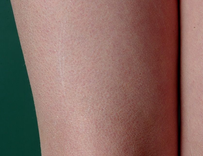
Dermatopathology 2021, 8 115
scribed keratoses are found on pressure-exposed areas of the sole of the foot. Progressive
symmetric erythrokeratodermia (Gottron) is no longer distinguished as a separate entity
from erythrokeratodermia variabilis [18].
Histology
The epidermis shows acanthosis and undulating surface with hyperkeratosis, focal
parakeratosis, dyskeratotic keratinocytes, and preserved stratum granulosum. Superficial
perivascular lymphocytic infiltrate may be present (Figure 7). Overall, the histologic
changes mentioned are highly variable and complicate diagnosis. The deficiency of the
Dermatopathology 2021, 8, affected connexin can be easily visualized by immunohistochemistry; at the same time, 10
compensatory connexin 43 expression is increased.
Figure7.7.Erythrokeratoderma.
Figure Erythrokeratoderma.Acanthotic
Acanthoticepidermis
epidermiswith
withorthohyperkeratosis,
orthohyperkeratosis,focal
focalparakeratosis,
parakerato-
sis, dyskeratotic
dyskeratotic keratinocytes,
keratinocytes, andand preserved
preserved stratum
stratum granulosum.
granulosum. Discrete
Discrete superficial
superficial perivascu-
perivascular
lar lymphocytic infiltrate. HE stain, bar = 100 µm.
lymphocytic infiltrate. HE stain, bar = 100 µm.
7. KID
7. KID Syndrome
Syndrome and
and HID
HID Syndrome
Syndrome
Keratitis–ichthyosis–deafness(KID)
Keratitis–ichthyosis–deafness (KID)syndrome
syndromeand andhystrix-like–ichthyosis–deafness
hystrix-like–ichthyosis–deafness
(HID)syndrome
(HID) syndromeareare different
different forms
forms of anofautosomal
an autosomal dominant
dominant inherited
inherited ichthyosis
ichthyosis caused
by a mutation
caused of connexin
by a mutation 26 (Table263)(Table
of connexin [19]. Because this connexin
3) [19]. Because performs
this connexin important
performs im-
functions in the inner
portant functions ear,
in the neurosensory
inner hearinghearing
ear, neurosensory loss also exists.
loss Patients
also exists. with KID
Patients withsyn-
KID
drome develop
syndrome sharply
develop circumscribed
sharply circumscribedwart-like hyperkeratotic
wart-like plaques
hyperkeratotic on the
plaques onface
the and
face
extremities;
and extremities; in HID syndrome, hystrix-like generalized ichthyosis predominates.the
in HID syndrome, hystrix-like generalized ichthyosis predominates. In In
setting of this
the setting ofsyndromic ichthyosis,
this syndromic keratitis,
ichthyosis, alopecia,
keratitis, nail dystrophy,
alopecia, dental abnormalities,
nail dystrophy, dental abnor-
or hypohidrosis,
malities, and an increased
or hypohidrosis, risk of infection
and an increased risk ofand carcinoma
infection occur.
and carcinoma occur.
7.1.
7.1. Histology
Histology
The
Theepidermis
epidermisisisacanthotic
acanthoticwith
withaapartially
partiallyverruciform
verruciformappearance.
appearance.TheThehyperkera-
hyperker-
totic stratum corneum contains parakeratoses with large round nuclear remnants,
atotic stratum corneum contains parakeratoses with large round nuclear remnants, and and
oc-
casionally shadow cells with vacuolated nuclei (Figure 8). Dyskeratotic keratinocytes
occasionally shadow cells with vacuolated nuclei (Figure 8). Dyskeratotic keratinocytes with
perinuclear halo (“bird’s eye”) appear as a dominant criterion. The stratum granulosum
with perinuclear halo (“bird’s eye”) appear as a dominant criterion. The stratum granu-
may be absent or strongly pronounced. Subepidermal dense lymphocytic infiltrates occur
losum may be absent or strongly pronounced. Subepidermal dense lymphocytic infiltrates
in some cases. The openings of the hair follicles and sweat glands are highly keratinized.
occur in some cases. The openings of the hair follicles and sweat glands are highly kerat-
inized. The sweat glands may be diminished and atrophic. Highly differentiated squa-
mous cell carcinoma may also occur at a young age [8].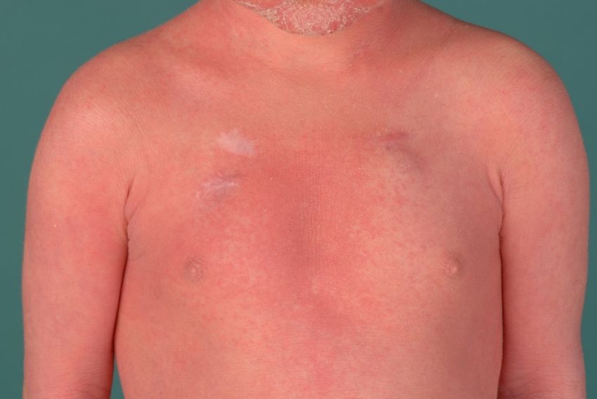
Dermatopathology 2021, 8 116
Dermatopathology 2021, 8, The sweat glands may be diminished and atrophic. Highly differentiated squamous cell11
carcinoma may also occur at a young age [8].
Figure8.8.KID
Figure KIDsyndrome
syndrome(keratitis–ichthyosis–deafness).
(keratitis–ichthyosis–deafness). Dyskeratotic
Dyskeratotic keratinocytes
keratinocytes with
with peri-
perinuclear
nuclear halo (“bird’s eye”). HE stain, bar = 100 µm.
halo (“bird’s eye”). HE stain, bar = 100 µm.
7.2.Differential
7.2. DifferentialDiagnoses
Diagnoses
Verrucaevulgares
Verrucae vulgaresalso
also show
show vacuolated
vacuolated cells,
cells, butbut in KID/HID
in KID/HID syndrome
syndrome these
these per-
persist
sist
in theinstratum
the stratum corneum.
corneum. Vacuolization
Vacuolization is absent
is absent in erythrokeratodermia.
in erythrokeratodermia.
8.8.Ichthyoses
Ichthyoseswith
withInflammatory
InflammatoryPsoriasiform
PsoriasiformPattern
Pattern
Some
Somehereditary
hereditaryichthyoses
ichthyoseshave
haveaahistologic
histologicpattern
patternthat
thatclosely
closelyresembles
resemblespsoriasis
psoriasis
vulgaris or chronic dermatitis in the setting of atopic eczema, which is why misdiagnosis
vulgaris or chronic dermatitis in the setting of atopic eczema, which is why misdiagnosis is
common (Table 2). Because of the significant and sometimes lethal complications associated
is common (Table 2). Because of the significant and sometimes lethal complications asso-
with
ciatedthiswith
group
thisofgroup
ichthyoses, prompt dermatohistologic
of ichthyoses, diagnosis diagnosis
prompt dermatohistologic is important [20].
is important
[20].
9. Netherton Syndrome
In autosomal
9. Netherton recessive Netherton syndrome, there is a mutation of the SPINK5 gene,
Syndrome
which encodes LEKTI (“lymphoepithelial Kazal-type related inhibitor”), a major serine
In autosomal recessive Netherton syndrome, there is a mutation of the SPINK5 gene,
protease inhibitor of the epidermis and thymus (Table 3) [21]. Patients are born with
which encodes LEKTI (“lymphoepithelial Kazal-type related inhibitor”), a major serine
marked erythroderma, which later often changes into anular eyrthema with a typical
protease inhibitor of the epidermis and thymus (Table 3) [21]. Patients are born with
double-edged scale (“ichthyosis linearis circumflexa”) (Figure 9). Later, brittle hairs are
marked erythroderma, which later often changes into anular eyrthema with a typical dou-
also noticeable (“bamboo hairs”, trichorrhexis invaginata). Type 1 allergies, elevated IgE
ble-edged scale (“ichthyosis linearis circumflexa”) (Figure 9). Later, brittle hairs are also
levels, and hypereosinophilia, as well as immunodeficiency and enteropathy, which can
noticeable
lead (“bamboo
to massive failurehairs”, trichorrhexis
to thrive, invaginata).
especially in Type
the first year 1 allergies,
of life, elevated
are associated IgEthis
with lev-
els, and hypereosinophilia,
cornification as well
disorder. Electrolyte as immunodeficiency
disturbances and sepsisand enteropathy,
are lethal risks forwhich can lead
infants.
to massive failure to thrive, especially in the first year of life, are associated with this corni-
fication disorder. Electrolyte disturbances and sepsis are lethal risks for infants.Dermatopathology 2021, 8, 12
Dermatopathology 2021, 8 117
Dermatopathology 2021, 8, 12
Figure 9. Netherton syndrome. Erythema and scaling of the trunk and face.
Figure9.
Figure 9. Netherton
Netherton syndrome.
syndrome. Erythema
Erythema and
and scaling
scaling of
of the
the trunk
trunk and
and face.
face.
9.1. Histology
9.1.
9.1. Histology
Histology
There is psoriasiform hyperplasia with a moderately widened stratum corneum
There
showing isis psoriasiform
Therefocal parakeratosishyperplasia
psoriasiform hyperplasia with
with aa moderately
and accumulations moderately
of neutrophils.widened
widened stratum
stratum
The stratum corneum
corneum
granulosum
showing
showing focal
is absent focal parakeratosis
parakeratosis
or severely and accumulations
and accumulations
diminished. of neutrophils.
of neutrophils.
The papillary dermis The stratum
The stratum
is papillomatously granulosum
granulosum
elongated and
is
is absent
absent or
or severely
severely diminished.
diminished. The
The papillary
papillary dermis
dermis isispapillomatously
papillomatously elongated
elongated and
and
contains dilated vessels and inflammatory infiltrates with lymphocytes, neutrophils, and
contains
contains dilated
dilated vessels
vessels and
and inflammatory
inflammatory infiltrates
infiltrates with
withlymphocytes,
lymphocytes, neutrophils,
neutrophils, and
and
eosinophilic granulocytes (Figure 10). Sometimes, however, there are histologic changes,
eosinophilic
eosinophilic granulocytes
granulocytes (Figure
(Figure 10). Sometimes, however,
10). Sometimes, however, there are
there histologic changes, as
as found in atopic dermatitis. Immunohistochemically, staining forare histologic
LEKTI changes,
is absent in the
found
as in atopic
found inand
atopicdermatitis. Immunohistochemically, staining for LEKTI is absent in the
epidermis hairdermatitis. Immunohistochemically,
follicles (Figure 11) [22]. staining for LEKTI is absent in the
epidermis
epidermis and
and hair
hairfollicles
follicles(Figure
(Figure11)11)[22].
[22].
Figure10.
Figure 10.Netherton
Netherton syndrome.
syndrome. Regular
Regular (psoriasiform)
(psoriasiform) hyperplasia
hyperplasiawith
withfocal
focalparakeratosis
parakeratosisand
and
Figure 10. Netherton syndrome. Regular (psoriasiform) hyperplasia with focal parakeratosis and
thinned stratum granulosum. Dilated vessels in the papillary dermis and inflammatory infiltrates.
thinned stratum granulosum. Dilated vessels in the papillary dermis and inflammatory infiltrates.
thinned
HE stain,stratum granulosum.
bar = 100 µm. Dilated vessels in the papillary dermis and inflammatory infiltrates.
HE stain, bar = 100 µm.
HE stain, bar = 100 µm.Dermatopathology 2021, 8 118
Dermatopathology 2021, 8, 13
(a) (b)
Figure
Figure 11.
11. Netherton
Nethertonsyndrome,
syndrome, immunohistochemistry,
immunohistochemistry, barbar
= 200 µmµm
= 200 (a,b). Immunohistochemistry
(a,b). shows
Immunohistochemistry a lackaoflack
shows stain-
of
ing for LEKTI in the epidermis and hair follicles (a); regular expression of LEKTI in the upper layers of the epidermis
staining for LEKTI in the epidermis and hair follicles (a); regular expression of LEKTI in the upper layers of the epidermis of
healthy skin,
of healthy barbar
skin, = 200 µmµm
= 200 (b).(b).
Immunoperoxidase
Immunoperoxidase staining.
staining.
9.2. Differential Diagnoses
9.2. Differential Diagnoses
Psoriasis vulgaris or atopic dermatitis cannot always be differentiated histologically.
Psoriasis vulgaris or atopic dermatitis cannot always be differentiated histologically.
PAS-positive granules in the stratum corneum cannot always be detected and are not spe-
PAS-positive granules in the stratum corneum cannot always be detected and are not
cific. The immunohistochemically detectable lack of LEKTI expression is important evi-
specific. The immunohistochemically detectable lack of LEKTI expression is important
dence
evidencefor for
Netherton syndrome.
Netherton syndrome.Other forms
Other of ichthyosis
forms withwith
of ichthyosis psoriasis-like histology
psoriasis-like are
histology
listed in Table 2.
are listed in Table 2.
10.
10. Peeling
Peeling Skin
Skin Disease
Disease
In
In peeling skin disease
peeling skin disease (peeling
(peeling skin
skin syndrome
syndrome B), B), generalized
generalized erythema
erythema withwith super-
super-
ficial skin detachment is evident
ficial skin detachment is evident from birth and persists throughout life with seasonal
variation (Figure 12). In In addition,
addition, episodic
episodic detachment
detachment of of the
the nail
nail plates
plates (onychomadesis)
(onychomadesis)
may
mayoccur.
occur.Hair status
Hair is inconspicuous
status except
is inconspicuous for a transient
except slight epilation
for a transient of fine hairs
slight epilation of [23].
fine
hairs [23].
There is a mutation of corneodesmosin, an important adhesion protein expressed in the
extracellular sections of desmosomes in the stratum corneum of the epidermis, as well as
at the inner hair root sheath of hair follicles (Table 3). Ultrastructurally, there is detachment
of intact corneocytes from the stratum granulosum (extracellular cleft formation) [20,23].
Autosomal dominant mutations in other domains of corneodesmosin cause hypotrichosis
simplex.
Concomitant barrier disruption leads to inflammation with massive pruritus, urticaria,
angioedema, food allergy, and asthma with elevated IgE levels and blood eosinophilia.021, 8, Dermatopathology 2021, 8 14119
Figure
Figure 12. Peeling
12. Peeling skin disease.
skin disease. Diffuse with
Diffuse erythema erythema withskin
superficial superficial skin
detachment detachment
is evident is evident
from birth from
and persists
birth and
throughout persists
life with throughout
seasonal variation. life with seasonal variation.
10.1. Histology
There is a mutation of corneodesmosin, an important adhesion protein expressed in
The epidermis
the extracellular sections of desmosomes is hyperplastic
in thewith prominent
stratum rete ridges.
corneum There
of the is mild hyperkeratosis
epidermis, as well
with focal parakeratosis and thinned stratum granulosum. Some biopsies show a focal
as at the inner hair root sheath of hair follicles (Table 3). Ultrastructurally, there is detach-
detachment of the stratum corneum, and in some cases, the stratum corneum is completely
ment of intact corneocytes fromthese
absent. However, thechanges
stratum granulosum
cannot (extracellular
always be detected cleftsection.
on a paraffin formation)
There are
[20,23]. Autosomal dominant
superficial and mutations
perivascular in other domains
lymphocytic ofwith
infiltrates corneodesmosin cause
single neutrophils, which hypo-
are also
trichosis simplex. found in the stratum corneum. The papillary body is elongated and edematous, vessels
are not dilated [7]. Immunohistochemically, staining for corneodesmosin is absent in the
Concomitant barrier disruption leads to inflammation with massive pruritus, urti-
stratum corneum [23].
caria, angioedema, food allergy, and asthma with elevated IgE levels and blood eosino-
philia. 10.2. Differential Diagnosis
Psoriasis vulgaris, Netherton syndrome, and CHILD syndrome cannot be differenti-
10.1. Histology ated without immunohistochemistry.
The epidermis11. is hyperplastic
CHILD Syndromewith prominent rete ridges. There is mild hyperkera-
tosis with focal parakeratosis and thinned with
Congenital Hemidysplasia stratum granulosum.
Ichthyosiform nevus andSomeLimb biopsies show syn-
Defect (CHILD) a
focal detachmentdrome
of theisstratum
a very rare X-linked dominant
corneum, and in somedisorder that the
cases, is usually
stratumlethal for male offspring.
corneum is com- It
pletely absent. However, these changes cannot always be detected on a paraffin section. in
is characterized by unilateral inflammatory, often waxy, yellow skin lesions, emphasized
the large flexures and perianogenital region [8,24]. Extracutaneous symptoms range from
There are superficial and
discrete perivascular
hypoplasia lymphocytic
of the limbs infiltratesMonosymptomatic
to severe deformities. with single neutrophils,
cases are often
which are also found in the stratum
misdiagnosed corneum.
as psoriasis or ILVEN The
[25].papillary body is elongated and edem-
atous, vessels are notThe
dilated [7].isImmunohistochemically,
disorder caused by nonsense or missense staining for corneodesmosin
mutations in the so-called NSDHLis
gene, which lead
absent in the stratum corneum [23]. to a disturbance of cholesterol biosynthesis (Table 3) [26,27].
11.1. Histology
10.2. Differential Diagnosis
Hyperplastic epidermis with elongated rete ridges and marked orthohyperkerato-
sis withNetherton
Psoriasis vulgaris, focal parakeratosis
syndrome,are found. The stratum
and CHILD granulosum
syndrome maybe
cannot bedifferenti-
prominent in
ated without immunohistochemistry.
11. CHILD SyndromeDermatopathology 2021, 8 120
some areas but can also be absent. A perivascular lymphocytic infiltrate and xanthoma-
tous macrophages that are markedly immunoreactive for adipophilin are apparent in the
papillary body [7].
11.2. Differential Diagnoses
Verruciform xanthomas also contain xanthomatous macrophages, which are absent
in the other major differential diagnoses as psoriasis inversa and epidermal nevus. The
presence of verruciform xanthomas or verruciform xanthoma-like changes in the setting of
CHILD syndrome is possible.
12. Severe Dermatitis, Multiple Allergies, Metabolic Wasting Syndrome
(SAM Syndrome)
SAM syndrome was identified as a severe life-threatening genodermatosis by Liat
Samuelov et al. in 2013 [28]. The acronym stands for severe dermatitis, multiple allergies,
metabolic wasting syndrome. It is caused by a mutation of desmoglein−1 (DSG1) (Table 3),
a major desmosomal adhesion molecule also involved in pemphigus disease. Later, a desmo-
plakin mutation was also identified [29]. The disease is inherited in an autosomal recessive
manner; heterozygous carriers of the DSG1 mutation develop only striate palmoplantar
keratoderma [30].
Clinically, there is ichthyosiform erythroderma in newborns similar to autosomal re-
cessive lamellar ichthyosis, Netherton syndrome, or peeling skin disease. Other symptoms
are pruritus, hypotrichosis, food allergies with elevated IgE, dysphagia, decreased growth,
and recurrent skin and respiratory infections. In varying degrees, pustular formation,
palmoplantar keratoses, onychodystrophy, dental anomalies, cardiac abnormalities, and
eosinophilic esophagitis are found. There is marked inter- and intrafamilial variability [30].
The accompanying inflammation can be explained by proinflammatory activity in ker-
atinocytes in the context of impaired barrier function and downregulated blockage of signal
transduction pathways [31]. Furthermore, the intracytoplasmic portion of DSG1 blocks the
RAS-RAF signaling pathway and, thus, affects epidermal differentiation [32,33]. Similar
to Netherton syndrome, anti-inflammatory therapies with biologics improve the clinical
picture (Oji V, unpublished).
12.1. Histology
Histologically, there is a superficial lymphocytic dermatitis with hyperplastic epider-
mis, parakeratosis, and neutrophil granulocytes that strongly resembles psoriasis (psoriasi-
form dermatitis). However, there are typically dilated intercellular spaces of the epidermis
without blistering (Figure 13). Intracellular edema and serum exudate (as in spongiotic
dermatitis), and rounding of keratinocytes and pyknosis of nuclei (as in pemphigus dis-
ease), hypereosinophilia of cytoplasm (as in M. Darier or Hailey–Hailey), ballooning, and
typical viropathic nuclear changes, as in herpes disease, are absent.Dermatopathology 2021, 8, 16
Dermatopathology 2021, 8 121
Figure 13. SAM syndrome. Psoriasiform dermatitis with dilated intercellular spaces of the epider-
Figure 13. SAM syndrome. Psoriasiform dermatitis with dilated intercellular spaces of the epidermis
mis without blistering (“desmosomal acantholysis”). HE stain, bar = 100 µm.
without blistering (“desmosomal acantholysis”). HE stain, bar = 100 µm.
12.2.Differential
12.2. DifferentialDiagnoses
Diagnoses
Allgenodermatoses
All genodermatoseswith withmutations
mutationsofofdesmosomal
desmosomalproteins
proteinsleading
leadingtotothe
thepattern
pattern
of “desmosomal acantholysis” (Metze, unpublished). These include
of “desmosomal acantholysis” (Metze, unpublished). These include keratosis palmoplan- keratosis
palmoplantaris
taris areata
areata et striata et striata
(striate (striate palmoplantar
palmoplantar keratodermakeratoderma
types 1 andtypes 1 and 2), Carva-
2), Carvajal-Huerta
jal-Huerta Naxos
syndrome, syndrome, Naxos ectodermal
syndrome, syndrome, ectodermal dysplasia
dysplasia skin skin
fragility fragility(McGrath
syndrome syndrome
(McGrath syndrome),
syndrome), or peeling
or peeling skin diseaseskin
[34].disease [34].
AuthorContributions:
Author Contributions:Conceptualization,
Conceptualization, D.M.
D.M. andand K.S.;
K.S.; methodology,
methodology, D.M.
D.M. and and
K.S.;K.S.; investiga-
investigation,
tion, K.S.
D.M., D.M.,andK.S. and
H.T.; H.T.; resources,
resources, D.M.; dataD.M.; dataD.M.
curation, curation, D.M.
and K.S.; and K.S.; writing—original
writing—original draft
draft preparation,
preparation,
D.M. and K.S.;D.M. and K.S.; writing—review
writing—review and editing,
and editing, D.M., K.S. andD.M.,
H.T.; K.S. and H.T.; D.M.;
visualization, visualization, D.M.;
supervision,
supervision,
H.T.; H.T.; project administration,
project administration, D.M.have
D.M. All authors All authors
read andhave readto
agreed and
theagreed to theversion
published published
of
version
the of the manuscript.
manuscript.
Funding:This
Funding: Thisresearch
researchreceived
receivedno
noexternal
externalfunding.
funding.
InstitutionalReview
Institutional ReviewBoard
BoardStatement:
Statement:Not
Notapplicable.
applicable.
InformedConsent
Informed ConsentStatement:
Statement:Not
Notapplicable.
applicable.
Conflictsof
Conflicts ofInterest:
Interest: The authors
authors declare
declareno
noconflict
conflictofofinterest.
interest.
Abbreviations
Abbreviations
Congenital hemidysplasia with ichthyosiform erythroderma and limb defects
CHILD syndrome Congenital
CHILD syndrome syndrome;hemidysplasia with ichthyosiform erythroderma
CRIE and limb defects
Congenital syndrome
reticular ichthyosiform erythroderma;
CRIE
EI Congenital reticular ichthyosiform erythroderma
Epidermolytic ichthyosis;
EIKID syndrome Epidermolytic ichthyosis
Keratitis ichthyosis deafness syndrome;
KID syndrome Keratitis ichthyosis deafness syndrome
Congenital erythroderma–hypotrichosis–recurrent infections–multiple food
SAM syndrome Congenital erythroderma–hypotrichosis–recurrent infections–multiple
SAM syndrome allergies syndrome;
food allergies syndrome
SEI Superficial epidermolytic ichthyosis.
SEI Superficial epidermolytic ichthyosisDermatopathology 2021, 8 122
References
1. Krug, M.; Oji, V.; Traupe, H.; Berneburg, M. Ichthyoses-Part 1: Differential diagnosis of vulgar ichthyoses and therapeutic options.
J. Dtsch. Dermatol. Ges. 2009, 7, 511–519. [CrossRef]
2. Krug, M.; Oji, V.; Traupe, H.; Berneburg, M. Ichthyoses-Part 2: Congenital ichthyoses. J. Dtsch. Dermatol. Ges. 2009, 7, 577–588.
[CrossRef] [PubMed]
3. Traupe, H. The Ichthyoses. In A Guide to Clinical Diagnosis, Genetic Counseling, and Therapy; Springer: Berlin, Germany, 1989;
pp. 103–138.
4. Oji, V.; Traupe, H. Ichthyoses: Differential diagnosis and molecular genetics. Eur. J. Dermatol. 2006, 16, 349–359. [PubMed]
5. Oji, V.; Tadini, G.; Akiyama, M.; Blanchet Bardon, C.; Bodemer, C.; Bourrat, E.; Coudiere, P.; DiGiovanna, J.J.; Elias, P.; Fischer, J.;
et al. Revised nomenclature and classification of inherited ichthyoses: Results of the First Ichthyosis Consensus Conference in
Sorèze 2009. J. Am. Acad. Dermatol. 2010, 63, 607–641. [CrossRef]
6. Metze, D.; Traupe, H. Hereditäre Verhornungsstörungen und epidermale Fehlbildungen. In Histopathologie der Haut, 2nd ed.;
Cerroni, L., Garbe, C., Metze, D., Kutzner, H., Kerl, H., Eds.; Springer: Berlin, Germany, 2016; Chapter 20; pp. 404–438.
7. Metze, D. Disorders of Keratinization. In McKee’s Pathology of the Skin, 5th ed.; 2-Volume-Set; Calonje, E., Brenn, T., Lazar, A.,
McKee, P.H., Eds.; Elsevier: Amsterdam, The Netherlands, 2019; Volume 1, Chapter 3, pp. 53–117.
8. Oji, V.; Metze, D.; Traupe, H. Inherited disorders of cornification. In Rook’s Textbook of Dermatology, 9th ed.; Burns, T., Breathnach,
S., Cox, N., Griffiths, C., Eds.; Wiley-Blackwell: Hoboken, NJ, USA, 2016; Volume 2, Part 6; Chapter 65, pp. 1–75.
9. Majmundar, V.D.; Baxi, K. Hereditary and Acquired Ichthyosis Vulgaris. In Treasure Island (FL); StatPearls Publishing: Treasure
Island, FL, USA, 2021.
10. Süßmuth, K.; Gruber, R.; Rodriguez, E.; Traupe, H.; Amler, S.; Sánchez-Guijo, A.; Valentin, F.; Tarinski, T.; Straub, N.;
Metze, D.; et al. Increased prevalence of filaggrin deficiency in 51 patients with recessive X-linked ichthyosis presenting for
dermatologic examination. J. Investig. Dermatol. 2018, 138, 709–711. [CrossRef] [PubMed]
11. Kütting, B.; Traupe, H. Der erworbene Ichthyosis-ähnliche Hautzustand. Hautarztand 1995, 46, 836–840. [CrossRef]
12. Hotz, A.; Kopp, J.; Bourrat, E.; Oji, V.; Komlosi, K.; Giehl, K.; Bouadjar, B.; Bygum, A.; Tantcheva-Poor, I.; Hellström Pigg, M.; et al.
Meta-Analysis of Mutations in ALOX12B or ALOXE3 Identified in a Large Cohort of 224 Patients. Genes 2021, 12, 80. [CrossRef]
13. Raghunath, M.; Hennies, H.C.; Velten, F.; Wiebe, V.; Steinert, P.M.; Reis, A.; Traupe, H. A novel in situ method for the detection of
deficient transglutaminase activity in the skin. Arch. Dermatol. Res. 1998, 290, 621–627. [CrossRef]
14. Rothnagel, J.A.; Dominey, A.M.; Dempsey, L.D.; Longley, M.A.; Greenhalgh, D.A.; Gagne, T.A.; Huber, M.; Frenk, E.; Hohl, D.;
Roop, D.R. Mutations in the rod domains of keratins 1 and 10 in epidermolytic hyperkeratosis. Science 1992, 257, 1128–
1130. [CrossRef]
15. Rothnagel, J.A.; Traupe, H.; Wojcik, S.; Huber, M.; Hohl, D.; Pittelkow, M.R.; Saeki, H.; Ishibashi, Y.; Roop, D.R. Mutations in the
rod domain of keratin 2e in patients with ichthyosis bullosa of Siemens. Nat. Genet. 1994, 7, 485–490. [CrossRef]
16. Traupe, H.; Kolde, G.; Hamm, H.; Happle, R. Ichthyosis bullosa of Siemens: A unique type of epidermolytic hyperkeratosis. J.
Am. Acad. Dermatol. 1986, 14, 1000–1005. [CrossRef]
17. Avshalumova, L.; Fabrikant, J.; Koriakos, A. Overview of skin diseases linked to connexin gene mutations. Int. J. Dermatol. 2014,
53, 192–205. [CrossRef] [PubMed]
18. van Steensel, M.A.M.; Oranje, A.P.; van der Schroeff, J.G.; Wagner, A.; van Geel, M. The missense mutation G12D in connexin30.3
can cause both erythrokeratodermia variabilis of Mendes da Costa and progressive symmetric erythrokeratodermia of Gottron.
Am. J. Med. Genet. A 2009, 149A, 657–661. [CrossRef]
19. van Geel, M.; van Steensel, M.A.; Küster, W.; Hennies, H.C.; Happle, R.; Steijlen, P.M.; König, A. HID and KID syndromes are
associated with the same connexin 26 mutation. Br. J. Dermatol. 2002, 146, 938–942. [CrossRef]
20. Süßmuth, K.; Traupe, H.; Metze, D.; Oji, V. Ichthyoses in everyday practice: Management of a rare group of diseases. J. Dtsch.
Dermatol. Ges. 2020, 18, 225–243. [CrossRef]
21. Chavanas, S.; Bodemer, C.; Rochat, A.; Hamel-Teillac, D.; Ali, M.; Irvine, A.D.; Bonafé, J.L.; Wilkinson, J.; Taïeb, A.;
Barrandon, Y.; et al. Mutations in SPINK5, encoding a serine protease inhibitor, cause Netherton syndrome. Nat. Genet. 2000, 25,
141–142. [CrossRef] [PubMed]
22. Leclerc-Mercier, S.; Bodemer, C.; Furio, L.; Hadj-Rabia, S.; de Peufeilhoux, L.; Weibel, L.; Bursztejn, A.C.; Bourrat, E.; Ortonne, N.;
Molina, T.J.; et al. Skin biopsy in Netherton Syndrome: A Histological review of a large series and new findings. Am. J.
Dermatopathol. 2016, 38, 83–91. [CrossRef] [PubMed]
23. Oji, V.; Eckl, K.M.; Aufenvenne, K.; Nätebus, M.; Tarinski, T.; Ackermann, K.; Seller, N.; Metze, D.; Nürnberg, G.; Fölster-Holst, R.;
et al. Loss of corneodesmosin leads to severe skin barrier defect, pruritus, and atopy: Unraveling the peeling skin disease. Am. J.
Hum. Genet. 2010, 87, 274–281. [CrossRef] [PubMed]
24. Ramphul, K.; Kota, V.; Mejias, S.G. Child Syndrome. In Treasure Island (FL); StatPearls Publishing: Treasure Island, FL, USA, 2021.
25. Happle, R.; Koch, H.; Lenz, W. The CHILD syndrome. Congenital hemidysplasia with ichthyosiform erythroderma and limb
defects. Eur. J. Pediatr. 1980, 134, 27–33. [CrossRef]
26. Bergqvist, C.; Abdallah, B.; Hasbani, D.J.; Abbas, O.; Kibbi, A.G.; Hamie, L.; Kurban, M.; Rubeiz, N. CHILD syndrome: A
modified pathogenesis-targeted therapeutic approach. Am. J. Med. Genet. A 2018, 176, 733–738. [CrossRef]
27. König, A.; Happle, R.; Bornholdt, D.; Engel, H.; Grzeschik, K.H. Mutations in the NSDHL gene, encoding a 3beta-hydroxysteroid
dehydrogenase, cause CHILD syndrome. Am. J. Med. Genet. 2000, 90, 339–346. [CrossRef]Dermatopathology 2021, 8 123
28. Samuelov, L.; Sarig, O.; Harmon, R.M.; Rapaport, D.; Ishida-Yamamoto, A.; Isakov, O.; Koetsier, J.L.; Gat, A.; Goldberg, I.;
Bergman, R.; et al. Desmoglein 1 deficiency results in severe dermatitis, multiple allergies and metabolic wasting. Nat. Genet.
2013, 45, 1244–1248. [CrossRef]
29. McAleer, M.A.; Pohler, E.; Smith, F.J.D.; Wilson, N.J.; Cole, C.; MacGowan, S.; Koetsier, J.L.; Godsel, L.M.; Harmon, R.M.;
Gruber, R.; et al. Severe dermatitis, multiple allergies, and metabolic wasting syndrome caused by a novel mutation in the
N-terminal plakin domain of desmoplakin. J. Allergy. Clin. Immunol. 2015, 136, 1268–1276. [CrossRef]
30. Taiber, S.; Samuelov, L.; Mohamad, J.; Barak, E.C.; Sarig, O.; Shalev, S.A.; Lestringant, G.; Sprecher, E. SAM syndrome is
characterized by extensive phenotypic heterogeneity. Exp. Dermatol. 2018, 27, 787–790. [CrossRef] [PubMed]
31. Polivka, L.; Hadj-Rabia, S.; Bal, E.; Leclerc-Mercier, S.; Madrange, M.; Hamel, Y.; Bonnet, D.; Mallet, S.; Lepidi, H.; Ovaert, C.; et al.
Epithelial barrier dysfunction in desmoglein-1 deficiency. J. Allergy Clin. Immunol. 2018, 142, 702–706.e7. [CrossRef]
32. Hammers, C.M.; Stanley, J.R. Desmoglein-1, differentiation, and disease. J. Clin. Investig. 2013, 123, 1419–1422. [CrossRef] [PubMed]
33. Ishida-Yamamoto, A.; Igawa, S. Genetic skin diseases related to desmosomes and corneodesmosomes. J. Dermatol. Sci. 2014, 74,
99–105. [CrossRef] [PubMed]
34. Metze, D.; Oji, V. Palmoplantar Keratodermas. In Dermatology, Series: Expert Consult, 4th ed.; Bolognia, J., Schaffer, J., Cerroni, L.,
Eds.; Elsevier: Philadelphia, PA, USA, 2018; Volume 1, Chapter 58, pp. 924–943.You can also read