Modulation of dopamine D1 receptors via histamine H3 receptors is a novel therapeutic target for Huntington's disease
←
→
Page content transcription
If your browser does not render page correctly, please read the page content below
RESEARCH ARTICLE

                                        Modulation of dopamine D1 receptors via
                                        histamine H3 receptors is a novel
                                        therapeutic target for Huntington’s
                                        disease
                                        David Moreno-Delgado1,2†‡, Mar Puigdellı́vol2,3,4,5†§, Estefanı́a Moreno1,2,
                                        Mar Rodrı́guez-Ruiz1,2, Joaquı́n Botta1,5, Paola Gasperini5, Anna Chiarlone2,6,
                                        Lesley A Howell7, Marco Scarselli8, Vicent Casadó1,2, Antoni Cortés1,2,
                                        Sergi Ferré9, Manuel Guzmán2,6, Carmen Lluı́s1,2, Jordi Alberch2,3,4,
                                        Enric I Canela1,2, Silvia Ginés2,3,4†*, Peter J McCormick1,2,5,10†*
                                        1
                                         Department of Biochemistry and Molecular Biomedicine, Faculty of Biology,
                                        Institute of Biomedicine of the University of Barcelona (IBUB), University of
                                        Barcelona, Barcelona, Spain; 2Centro de Investigación Biomédica en Red sobre
                                        Enfermedades Neurodegenerativas, Madrid, Spain; 3Department of Biomedical
                                        Science, Faculty of Medicine, University of Barcelona, Institut of Neuroscience,
                                        Barcelona, Spain; 4Institut d´Investigacions Biomèdiques August Pi i Sunyer
*For correspondence:                    (IDIBAPS), Barcelona, Spain; 5School of Pharmacy, University of East Anglia,
silviagines@ub.edu (SG);                Norwich Research Park, Norwich, United Kingdom; 6Department of Biochemistry
p.mccormick@qmul.ac.uk (PJMC)
                                        and Molecular Biology I, School of Biology, Instituto Universitario de Investigación
†
 These authors contributed              Neuroquı́mica, and Instituto Ramón y Cajal de Investigación Sanitaria, Complutense
equally to this work
                                        University of Madrid, Madrid, Spain; 7School of Biological and Chemical Sciences,
Present address: ‡UCB                   Queen Mary University of London, London, United Kingdom; 8Department of
BioPharma SPRL, Chemin de               Translational Research and New Technologies in Medicine and Surgery, University
Foriest, Braine-l’Alleud, Braine-
                                        of Pisa, Pisa, Italy; 9National Institute on Drug Abuse, Intramural Research Program,
l’Alleud, Belgium; §Department
of Biochemistry, University of
                                        National Institutes of Health, Department of Health and Human Services, Baltimore,
Cambridge, Cambridge, United            United States; 10William Harvey Research Institute, Barts and the London School of
Kingdom                                 Medicine, Queen Mary University of London, London, United Kingdom
Competing interests: The
authors declare that no
competing interests exist.
                                        Abstract Early Huntington’s disease (HD) include over-activation of dopamine D1 receptors
Funding: See page 24                    (D1R), producing an imbalance in dopaminergic neurotransmission and cell death. To reduce D1R
Received: 14 August 2019                over-activation, we present a strategy based on targeting complexes of D1R and histamine H3
Accepted: 26 May 2020                   receptors (H3R). Using an HD mouse striatal cell model and HD mouse organotypic brain slices we
Published: 09 June 2020                 found that D1R-induced cell death signaling and neuronal degeneration, are mitigated by an H3R
Reviewing editor: Volker
                                        antagonist. We demonstrate that the D1R-H3R heteromer is expressed in HD mice at early but not
Dötsch, Goethe University,             late stages of HD, correlating with HD progression. In accordance, we found this target expressed
Germany                                 in human control subjects and low-grade HD patients. Finally, treatment of HD mice with an H3R
                                        antagonist prevented cognitive and motor learning deficits and the loss of heteromer expression.
    Copyright Moreno-Delgado et
                                        Taken together, our results indicate that D1R - H3R heteromers play a pivotal role in dopamine
al. This article is distributed under
the terms of the Creative
                                        signaling and represent novel targets for treating HD.
Commons Attribution License,
which permits unrestricted use
and redistribution provided that
the original author and source are
credited.

Moreno-Delgado et al. eLife 2020;9:e51093. DOI: https://doi.org/10.7554/eLife.51093                                                 1 of 31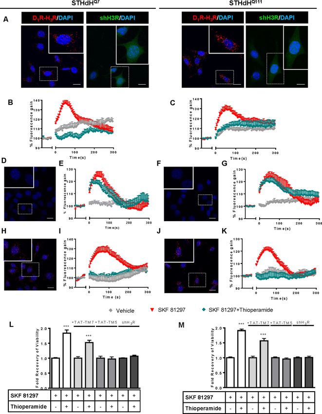
Research article                                                                                                   Neuroscience

                                   Introduction
                                   Huntington’s disease (HD) is a dominant inherited progressive neurodegenerative disorder caused
                                   by expansion of a CAG repeat, coding a polyglutamine repeat within the N-terminal region of hun-
                                   tingtin protein (Macdonald, 1993; Vonsattel and DiFiglia, 1998). Although dysfunction and death
                                   of striatal medium-sized spiny neurons (MSSNs) is a key neuropathological hallmark of HD
                                   (Ferrante et al., 1991; Vonsattel et al., 1985), cognitive deficits appear long before the onset of
                                   motor disturbances (Lawrence et al., 2000; Lemiere et al., 2004). It has been postulated that alter-
                                   ations in the dopaminergic system may contribute to HD neuropathology (Chen et al., 2013a;
                                   Jakel and Maragos, 2000), as dopamine (DA) plays a key role in the control of coordinated move-
                                   ments. Increased DA levels and DA signaling occur at early stages of the disease (Chen et al.,
                                   2013a; Garret et al., 1992; Jakel and Maragos, 2000), resulting in an imbalance in striatal neuro-
                                   transmission initiating signaling cascades that may contribute to striatal cell death (Paoletti et al.,
                                   2008; Ross and Tabrizi, 2011). Several studies demonstrated that DA receptor antagonists and
                                   agents that decrease DA content reduce chorea and motor symptoms while dopaminergic stimula-
                                   tion exacerbate such symptoms (Huntington Study Group, 2006; Mestre et al., 2009; Tang et al.,
                                   2007).
                                       Within the striatum, two different MSSNs populations can be distinguished: 1) MSSNs expressing
                                   enkephalin and dopamine D2 receptors (D2R), which give rise to the indirect striatal efferent path-
                                   way, and 2) MSSNs expressing substance P and dopamine D1 receptors (D1R), comprising the direct
                                   striatal efferent pathway. Recently, several studies with experimental models have changed the tradi-
                                   tional view that D2R-MSSNs are more vulnerable in HD (Cepeda et al., 2008; Kreitzer and Malenka,
                                   2007), proposing a new view in which D1R-MSSNs are more vulnerable to the HD mutation. In this
                                   view, it has been demonstrated that mutant huntingtin enhances striatal cell death through the acti-
                                   vation of D1R but not D2R (Paoletti et al., 2008). More recently, it has been described that, at early
                                   stages of the disease, HD mice show an increase in glutamate release onto D1R neurons but not D2R
                                   neurons while, later in the disease, glutamate release is selectively decreased to D1R cells
                                   (André et al., 2011a), indicating that several changes occur in D1R neurons at both early and late
                                   disease stages. Strategies that might reduce D1R signaling could prove successful towards prevent-
                                   ing HD (André et al., 2011a; André et al., 2011b; Ross and Tabrizi, 2011; Tang et al., 2007). How-
                                   ever, D1Rs are highly expressed in many tissues (Beaulieu and Gainetdinov, 2011) and broad use of
                                   D1R antagonists as a preventive treatment has important drawbacks including locomotor impair-
                                   ments (Giménez-Llort et al., 1997), or induce depression, parkinsonism and sedation in HD patients
                                   (Frank et al., 2008; Huntington Study Group, 2006).
                                       Histamine is an important neuromodulator with four known G protein-coupled receptors (GPCRs).
                                   H3Rs are expressed in brain regions involved in both motor function (striatum) and cognition, such
                                   as the cortex, thalamus, hypothalamus, hippocampus and amygdala (Panula and Nuutinen, 2013). It
                                   is known that in at least striatal GABAergic dynorphinergic neurons (Pillot et al., 2002; Ryu et al.,
                                   1994a; Ryu et al., 1994b), both D1R and H3R are co-expressed and we and others have found that
                                   they establish functional negative interactions by forming molecular complexes termed heteromers
                                   (Moreno et al., 2011; Sánchez-Lemus and Arias-Montaño, 2004). Hence, in this work, we hypothe-
                                   sized that targeting D1R through these receptor complexes of D1R and H3R might serve as a more
                                   efficient and targeted strategy to slow the progression of HD. Specifically, we demonstrate that
                                   D1R-H3R heteromers are expressed and functional in early HD stages but are lost in late stages. An
                                   H3R antagonist acting through D1R-H3R heteromers acts as a protective agent against dopaminergic
                                   imbalance in early HD stages improving learning and long-term memory deficits and rescuing the
                                   loss of D1R-H3R complexes at late stages of HD.

                                   Results
                                   Functional D1R-H3R heteromers are expressed in wild type STHdhQ7
                                   and HD STHdhQ111 striatal cell models
                                   To test whether D1R-H3R heteromers could indeed be targets for controlling D1R signaling in HD,
                                   we first analyzed the expression of both receptors in immortalized striatal cells expressing endoge-
                                   nous levels of full-length wild-type STHdhQ7 or mutant STHdhQ111 huntingtin (Ginés et al., 2010).
                                   Ligand binding determined that both STHdhQ7 and STHdhQ111 cells endogenously express similar

Moreno-Delgado et al. eLife 2020;9:e51093. DOI: https://doi.org/10.7554/eLife.51093                                                2 of 31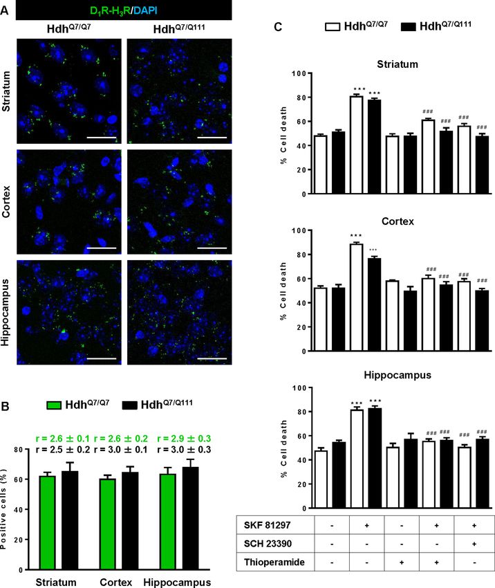
Research article Neuroscience
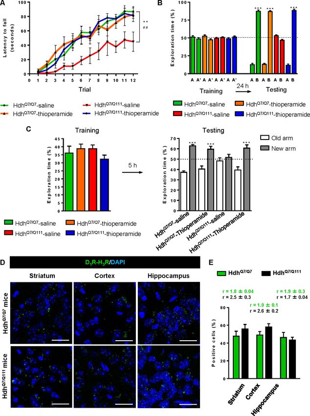
levels of D1R and H3R (Supplementary file 1). By proximity ligation assays (PLA), D1R-H3R hetero-
mers were detected as red spots surrounding the blue stained nuclei in both cell types (Figure 1A,
left panels of both cell types) and in cells treated with control lentivirus vector (Figure 1—figure sup-
plement 1A) but not in cells depleted of H3R (Figure 1A, right panels of both cell types) by shRNA,
as shown by RT-PCR and functionality (Figure 1—figure supplement 1B,C), or in negative controls
(Figure 1—figure supplement 1D). To ensure that D1R-H3R heteromers were functional in STHdh
cells, cell signaling experiments were performed. Using both STHdhQ7 and STHdhQ111 cells and con-
centrations of ligands previously shown to be optimal for receptor activation of the ERK1/2 pathway
(Ferrada et al., 2009; Moreno et al., 2014; Moreno et al., 2011), we observed that the D1R ago-
nist SKF 81297 was able to increase ERK1/2 phosphorylation whereas it was prevented by D1R
antagonist SCH 23390, and by the H3R antagonist thioperamide (Figure 1—figure supplement 2A,
B) via cross-antagonism. In addition, we tested a previously described alternative signaling pathway
activated downstream of D1R, Ca2+ mobilization (Chen et al., 2007; Jose et al., 1995). When cells
were treated with the D1R agonist SKF 81297 a robust and rapid increase in cytosolic Ca2+ was
detected in both STHdhQ7 and STHdhQ111 cells (Figure 1B,C). Importantly, this calcium release
could be dampened with the H3R antagonist thioperamide (cross-antagonism) (Figure 1B,C). The
above signaling data strongly support the presence of functional D1R-H3R heteromers in STHdh
cells.
To further demonstrate that an H3R antagonist is dampening D1R activation involving D1R-H3R
heteromers, we evaluated the effect of interfering peptides, which are synthetic peptides with the
amino acid sequence of domains of the receptors involved in the heteromeric interface. This
approach has been used by us and others to disrupt other heteromer complexes
(Bonaventura et al., 2015; Guitart et al., 2014; Hasbi et al., 2014; Lee et al., 2014; Viñals et al.,
2015). In a previous study we showed the efficacy of this approach in demonstrating heteromeriza-
tion of D1R with D3R, using a peptide with the sequence of D1R transmembrane domain 5 (TM5) but
not TM7 (Guitart et al., 2014). We therefore investigated whether synthetic peptides with the
sequence of TM5, and TM7 (as a negative control) of D1R, fused to HIV-TAT, were also able to dis-
rupt receptor D1R-H3R heteromers measured by PLA. In agreement with our hypothesis, there was a
near complete loss in PLA fluorescence signal when STHdhQ7 and STHdhQ111 cells were incubated
with TAT-TM five peptide (Figure 1D,F), but not for the negative control in which the TAT-TM seven
peptide was used (Figure 1H,J). We next evaluated whether TM5 or TM7 would interfere with the
observed cross-antagonism in calcium mobilization assays. Clearly, pretreatment of both STHdhQ7
and STHdhQ111 cells with the TAT-TM5 (Figure 1E,G) but not TAT-TM7 (Figure 1I,K) peptide dis-
rupts the ability of the H3R antagonist thioperamide to dampen D1R calcium signaling. These results
support that TM5 forms part of the interface of the D1R-H3R heteromer and demonstrate that the
H3R antagonist effect is driven through direct interaction between D1R and H3R.
H3R ligands prevent the D1R-induced cell death in STHdhQ7 and
STHdQ111 cells
It has been previously reported that upon activation of D1R, STHdh cell viability is reduced
(Paoletti et al., 2008). To explore whether H3R ligands could impair D1R activation through D1R-
H3R heteromers in a pathologically relevant readout, we used D1R-induced cell death as an output
of D1R activation in STHdh cells. As expected, STHdh cell viability decreased when treated with the
D1R agonist SKF 81297 in a concentration-dependent manner (Figure 1—figure supplement 2C).
Significant cell death did not occur until 30 mM SKF 81297 was used (Figure 1—figure supplement
2C), an effect prevented by the D1R antagonist SCH 23390 (Figure 1—figure supplement 2E). Pre-
treatment with the H3R antagonist thioperamide, which did not modify cell viability when adminis-
tered alone (Figure 1—figure supplement 2E), increased the number of surviving cells in the pres-
ence of the D1R agonist SKF 81297 in both cell types (Figure 1L,M and Figure 1—figure
supplement 2D). Importantly, the effect of the H3R antagonist thioperamide was specific since no
protection from D1R agonist-induced cell death was observed in cells depleted of H3R with shRNA
lentiviral infection (Figure 1L,M), but was observed in cells transfected with the control lentivirus
(Figure 1—figure supplement 2F). In addition, we also demonstrated that recovery of viability
induced by the H3R antagonist thioperamide was mediated by D1R-H3R heteromers since pre-incu-
bation with D1R TM5 peptide, but not D1R TM7 impaired the H3R antagonist protection from D1R
agonist-induced cell death (Figure 1L,M).
Moreno-Delgado et al. eLife 2020;9:e51093. DOI: https://doi.org/10.7554/eLife.51093 3 of 31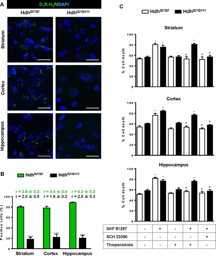
Research article                                                                                                               Neuroscience

Figure 1. Functional D1R-H3R heteromers are expressed in STHdhQ7 and STHdhQ111 cells. PLA were performed in STHdhQ7 and STHdhQ111 cells (A, D,
F, H and J) or in cells infected with shH3R to silence H3R, observed as green stained cells due to the GFP expression included in the plasmid (A). D1R-
H3R heteromers were visualized in STHdh cells as red spots around blue colored DAPI stained nucleus, but not in STHdh cells infected with shH3R
vector (A). Calcium increases were measured in STHdhQ7 (B, E and I) or STHdhQ111 (C, G and K). Cells were treated (20 min) or not with the H3R
Figure 1 continued on next page

Moreno-Delgado et al. eLife 2020;9:e51093. DOI: https://doi.org/10.7554/eLife.51093                                                              4 of 31
Research article                                                                                                              Neuroscience

Figure 1 continued
antagonist thioperamide (10 mM) before the addition of vehicle or SKF 81297 (1 mM). In (D, E, F, G, H, I, J and K), STHdHQ7 (D, E, H and I) or
STHdHQ111 (F, G, J and K) cells were also pre-treated for 60 min with 4 mM TM5 (D, E, F and G) or TM7 (H, I, J and K) peptides. Heteromers were
visualized as red spots around DAPI (blue) stained nucleus in cells pre-treated with TM7 peptide. Scale: 20 mm. For each calcium curve values are
expressed as a percentage increase with respect to untreated cells and are a mean ± SEM of 3 to 5 independent experiments. In (L and M), cell viability
was determined in STHdhQ7 (L) or STHdhQ111 cells (M) pre-treated for 60 min with vehicle (white columns), with 4 mM TAT-TM7 (pale grey columns) or
TAT-TM5 (grey columns) or infected with shH3R to silence H3R (dark grey columns) prior overstimulation with 30 mM SKF 81297. Values represent
mean ± SEM (n = 24 to 30) of cell viability recovery expressed as in-fold respect to SKF 81297 treated cells. Student’s t test showed a significant
(***p
Research article                                                                                                                   Neuroscience

Figure 2. Functional D1R-H3R heteromers are expressed in wild-type HdhQ7/Q7 and mutant HdhQ7/Q111 mice. Striatal, cortical or hippocampal slices
from 4-month-old HdhQ7/Q7 and HdhQ7/Q111 mice were used. In (A), by Proximity Ligation Assays (PLA) D1R-H3R heteromers were visualized in all slices
as green spots around blue colored DAPI stained nucleus. Scale bar: 20 mm. In (B), the number of cells containing one or more green spots is
expressed as the percentage of the total number of cells (blue nucleus). r values (number of green spots/cell containing spots) are shown above each
bar. Data (% of positive cells or r) are the mean ± SEM of counts in 600–800 cells from 4 to 8 different fields from three different animals. Student’s t test
showed no significant differences in heteromers expression in HdhQ7/Q7 and HdhQ7/Q111 mice. In (C), striatal, cortical or hippocampal organotypic slice
cultures from 4-month-old HdhQ7/Q7 and HdhQ7/Q111 mice were treated for 60 min with vehicle, the D1R antagonist SCH 23390 (10 mM) or H3R
antagonist thioperamide (10 mM) before the addition of SKF 81297 (50 mM). After 48 h cell death was determined. Values represent mean ± SEM (n = 3
to 19) of percentage of cell death. One-way ANOVA followed by Bonferroni post hoc tests showed a significant effect over non-treated organotypic
cultures (***p
Research article Neuroscience
(Figure 3—figure supplement 1 and Figure 3A,B), indicating that at more advanced disease stages
the D1R-H3R heteromer is lost. Although at 8 mo of age we detected a partial decrease in striatal
D1R expression in HdhQ7/Q111 compared with HdhQ7/Q7 mice using ligand binding experiments
(Supplementary file 2), the loss of heteromer expression is not due to a complete loss of receptor
expression since by radioligand binding (Supplementary file 2) and mRNA expression analysis
(Supplementary file 3) both receptors continue to be expressed.
To test the role of D1R-H3R heteromers, organotypic mouse striatal, cortical and hippocampal cul-
tures were obtained. Cell death was induced by the D1R agonist SKF 81297 (50 mM), and analysis of
DAPI and propidium iodide staining was performed. As expected, D1R agonist SKF 81297 treatment
increased the percentage of cell death in all three regions compared to vehicle-treated organotypic
cultures without significant differences between genotypes at 4 mo of age (Figure 2C). Importantly,
slices pre-treated with the H3R antagonist thioperamide, that does not modify cell death when
administered alone, protected cells from D1R elicited cell death in an equivalent manner to the D1R
antagonist SCH 23390 (Figure 2C), indicating that functional D1R-H3R heteromers are expressed in
different brain areas of HdhQ7/Q7 and HdhQ7/Q111 mice at early disease stages. The dramatic change
in heteromer expression in eight mo-old HdhQ7/Q111 mice was mirrored by the lack of protection of
the H3R antagonist thioperamide against SKF 81297-induced cell death in organotypic cultures
(Figure 3C), corroborating that the presence of D1R-H3R heteromers is needed for the H3R antago-
nist to prevent D1R-mediated cell death.
Treatment with thioperamide prevents cognitive and motor learning
deficits at early disease stages
To test whether the H3R antagonist thioperamide can exert beneficial effects in the initial stages of
the disease we evaluated the effect of chronic thioperamide treatment on motor learning and mem-
ory deficits in mutant HdhQ7/Q111 mice. Since cognitive decline is observed in these HD mice from 6
mo of age (Brito et al., 2014; Giralt et al., 2012; Puigdellı́vol et al., 2015) and the D1R-H3R hetero-
mers are expressed and functional until the age of 5 mo (Figure 4—figure supplement 1A,B), we
chose 5mo-old animals to start the thioperamide treatment (Figure 4—figure supplement 2). Corti-
costriatal function in saline and thioperamide-treated HdhQ7/Q7 and HdhQ7/Q111 mice was analyzed
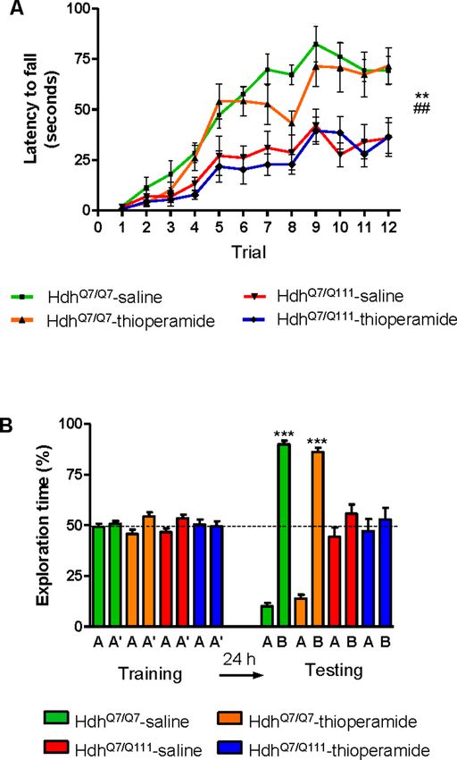
by using the accelerating rotarod task that evaluates the acquisition of new motor skills
(Puigdellı́vol et al., 2015). Saline-treated mutant HdhQ7/Q111 mice were unable to maintain their bal-
ance on the rotarod as wild-type HdhQ7/Q7 mice revealing impaired acquisition of new motor skills
(Figure 4A). Chronic treatment with thioperamide completely rescued motor learning deficits in
mutant HdhQ7/Q111 mice as evidenced by a similar latency to fall in the accelerating rotarod as wild-
type HdhQ7/Q7 mice. Next, recognition long-term memory (LTM) was analyzed by using the novel
object recognition test (NORT) (Figure 4B). After two days of habituation in the open field arena
(Figure 4—figure supplement 3), no significant differences were found between genotypes and/or
treatments, demonstrating no alterations in motivation, anxiety or spontaneous locomotor activity.
After habituation, animals were subjected to a training session in the open field arena in the pres-
ence of two similar objects (A and A’). Both saline and thioperamide-treated wild-type HdhQ7/Q7 and
mutant HdhQ7/Q111 mice similarly explored both objects indicating neither object nor place preferen-
ces (Figure 4B). After 24 hr, LTM was evaluated by changing one of the old objects (A’) for a novel
one (B). Whereas saline-treated HdhQ7/Q111 mice did not show any preference for the novel object
with respect to the familiar one, indicating recognition LTM deficits, thioperamide treatment
completely prevented this LTM deficit in mutant HdhQ7/Q111 mice (Figure 4B). Next, spatial LTM
was analyzed using the T-maze spontaneous alternation task (T-SAT) (Figure 4C). During the train-
ing, similar exploration time (Figure 4C, left panel) and similar number of arm entries (Figure 4—fig-
ure supplement 4, left panel) were found in all genotypes and treatments. After 5 hr, a testing
session showed that saline-treated HdhQ7/Q111 mice had no preferences between the novel arm and
the old arm, indicating spatial LTM deficits (Figure 4C, right panel). Interestingly, mutant HdhQ7/
Q111
mice treated with thioperamide spent more time in the novel versus the old arm, revealing pre-
served LTM (Figure 4C, right panel). Overall, these data demonstrate the effectiveness of thiopera-
mide treatment in restoring motor learning and preventing spatial and recognition LTM deficits in
mutant HdhQ7/Q111 mice.
We next tested if the reversion of the HD phenotype in mutant HdhQ7/Q111 mice induced by thio-
peramide treatment correlated with the preservation of D1R-H3R heteromer expression. By PLA we
Moreno-Delgado et al. eLife 2020;9:e51093. DOI: https://doi.org/10.7554/eLife.51093 7 of 31Research article Neuroscience Figure 3. Functional D1R-H3R heteromers are expressed in wild-type HdhQ7/Q7 but not in 8-month-old mutant HdhQ7/Q111 mice. Striatal, cortical or hippocampal slices from 8-month-old HdhQ7/Q7 and HdhQ7/Q111 mice were used. In (A), by Proximity Ligation Assays (PLA) D1R-H3R heteromers were visualized in HdhQ7/Q7 mice but not in HdhQ7/Q111 mice as green spots around blue colored DAPI stained nucleus. Scale bar: 20 mm. In (B), the number of cells containing one or more green spots is expressed as the percentage of the total number of cells (blue nucleus). r values (number of green spots/ cell containing spots) are shown above each bar. Data (% of positive cells or r) are the mean ± SEM of counts in 600–800 cells from 5 to 7 different fields from three different animals. Student’s t test showed a significant (***p
Research article                                                                                                              Neuroscience

Figure 4. Thioperamide chronic treatment prevents motor learning, long-term memory (LTM) deficits and the loss of receptor heteromerization in 6-
month-old HdhQ7/Q111 mice. In (A), curves illustrating the latency to fall in the accelerating rotarod of 6-month-old HdhQ7/Q7 and HdhQ7/Q111 mice
treated with saline or thioperamide from 5 months of age are shown. In (B), the exploration time for saline or thioperamide-treated HdhQ7/Q7 and
HdhQ7/Q111 mice during the training and the testing (24 hr delay, LTM) sessions in a novel-object recognition task showing that long-term recognition
Figure 4 continued on next page

Moreno-Delgado et al. eLife 2020;9:e51093. DOI: https://doi.org/10.7554/eLife.51093                                                             9 of 31
Research article                                                                                                                 Neuroscience

Figure 4 continued
memory deficits are rescued in the thioperamide-treated HdhQ7/Q111 mice. One-way ANOVA with Bonferroni post hoc showed significant differences
(***p
Research article                                                                                                             Neuroscience

Figure 5. Thioperamide treatment restored spinophilin-immunoreactive puncta reduction in the hippocampus and motor cortex of HdhQ7/Q111 mice
and exerts no effect on the clearance of mutant huntingtin accumulation. In (A) spinophilin-immunoreactive puncta were counted in the stratum oriens
and stratum radiatum of CA1 hippocampus and in (B) layers I, II/III and V of motor cortex area 1 (M1) of saline and thioperamide-treated HdhQ7/Q7 and
HdhQ7/Q111 mice. Quantitative analysis is shown as mean ± SEM (n = 9 images from three animals/group). Statistical analysis was performed using
Student’s two-tailed t test. *p
Research article Neuroscience
oligomeric forms (Figure 5C and Figure 5—figure supplement 1B). No significant differences
between groups were found when soluble monomeric mhtt levels were analyzed (Figure 5C and Fig-
ure 5—figure supplement 1B).
Thioperamide treatment does not rescue memory and motor learning
deficits in mutant HdhQ7/Q111 mice when D1R-H3R heteromers are lost
If the behavioral improvements observed after thioperamide treatment are mediated by the D1R-
H3R heteromer and not just by the blockade of the single H3R, then a treatment paradigm in the
absence of the heteromer should have no effect. To test this hypothesis, we used wild-type HdhQ7/
Q7
and mutant HdhQ7/Q111 mice at the age of 7 months, when we found the heteromer to be lost.
Animals were chronically treated with saline or thioperamide for 1 month and motor learning was
evaluated using the accelerating rotarod task. As expected, saline-HdhQ7/Q111 mice exhibited poor
performance in this task showing shorter latency to fall compared to wild-type HdhQ7/Q7 mice
(Figure 6A). Notably, thioperamide treatment had no effect on motor learning performance as both
saline- and thioperamide-treated mutant HdhQ7/Q111 mice were indistinguishable demonstrated by
similar latency to fall in the accelerating rotarod task (Figure 6A).
We next asked whether thioperamide treatment could improve cognitive function by rescuing
memory deficits in these same animals. Saline-treated 8-mo-old HdhQ7/Q111 mice exhibited long-
term memory deficits when recognition memory was analyzed using the novel object recognition
test (NORT) (Figure 6B). Similar to motor learning results, chronic treatment with thioperamide did
not rescue HdhQ7/Q111 mice from memory deficits (Figure 6B). Overall, these results demonstrate
that the effect of thioperamide in learning and memory in HdhQ7/Q111 mice requires the proper
expression and function of D1R-H3R heteromers.
D1R-H3R heteromer expression changes occur in other rodent HD
models and in HD patients
The fact that thioperamide treatment 1) prevents cognitive and motor learning deficits, 2) amelio-
rates striatal neuropathology, 3) ameliorates morphological alterations and 4) prevents the loss of
D1R-H3R heteromers at 6 mo and 8 mo of age in a mouse model of HD is suggestive that thiopera-
mide, or a future pharmacologically improved H3R antagonist specifically targeting D1R-H3R hetero-
mers, can be used to treat HD symptoms. To test this, we investigated D1R-H3R heteromer
expression in other transgenic HD mouse models and in human caudate-putamen slices using PLA.
The loss of heteromer expression compared with wild-type littermates was also observed in other
mouse models of HD, the R6/1 and R6/2 mice transgenic for the human huntingtin exon 1 (Fig-
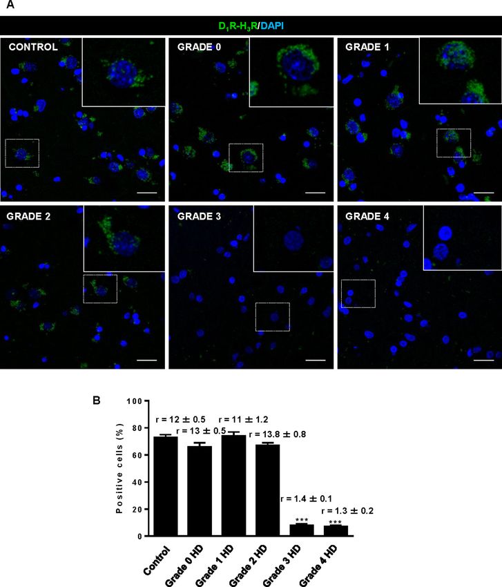
ure 7—figure supplement 1A,B, respectively). Importantly, D1R-H3R heteromers were detected as
green spots surrounding the blue stained nuclei in human caudate-putamen slices from control indi-
viduals and low-grade (grade 0, 1 and 2) HD patients (Figure 7A,B). In contrast, green spots were
almost absent in samples from high-grade (grade 3 or grade 4) HD patients (Figure 7A,B). These
results show that D1R-H3R heteromer formation changes during disease progression and, impor-
tantly, that humans express D1R-H3R heteromers at early disease stages.
Discussion
The imbalance of dopamine inputs throughout HD progression represents a potential ‘point of no
return’ for HD patients as this disequilibrium can eventually lead to substantial neuronal dysfunction
and cell death. In the present study, we demonstrate that 1) excess dopamine signaling via D1R
leads to cell death by activating the p38 pathway; 2) D1R-H3R complexes are found within the stria-
tum, cortex and hippocampus of WT mice and in HD mice at early but not late disease stages; 3) tar-
geting D1R via D1R-H3R complexes can slow progression of the disease in early but not late stages
when the complexes are lost; and 4) D1R-H3R complexes are expressed in the human brain and thus
represent potential therapeutic targets. This is the first demonstration of GPCR heteromers as
potential targets to treat HD. Together, these data support a novel role for D1R-H3R complexes in
neuroprotection and HD.
Several studies have revealed that dopamine neurotoxicity increases the sensitivity of MSSNs to
glutamate inputs and leads to striatal neurodegeneration, a role ascribed to aberrant D1R and not
D2R (Cepeda and Levine, 1998; Flores-Hernández et al., 2002; Paoletti et al., 2008; Tang et al.,
Moreno-Delgado et al. eLife 2020;9:e51093. DOI: https://doi.org/10.7554/eLife.51093 12 of 31Research article Neuroscience
Figure 6. Thioperamide chronic treatment does not prevent motor learning and long-term memory (LTM) deficits
in 8-month-old HdhQ7/Q111 mice when the D1R-H3R heteromer is not expressed. In (A), curves illustrating the
latency to fall in the accelerating rotarod of 8-month-old HdhQ7/Q7 and HdhQ7/Q111 mice treated with saline or
thioperamide from 7 months of age are shown. Two-way ANOVA with repeated measures showed significant
differences (**pResearch article Neuroscience
Figure 7. Striatal D1R-H3R heteromers are expressed in human control subjects and grade 2 HD patients but not
in grade 3–4 HD patients. In (A), by Proximity Ligation Assays (PLA), D1R-H3R heteromers were visualized as green
spots around blue colored DAPI stained nucleus in human striatal slices from age matched control subjects and 0–
2 grade HD patients but not in 3–4 grade HD patients. Scale bar: 20 mm. In (B), the number of cells containing one
or more green spots is expressed as the percentage of the total number of cells (blue nucleus). r values (number
of green spots/cell containing spots) are shown above each bar. Data are mean ± SEM of counts in 600–800 cells
from 10 different fields from subject described in Materials and Methods. Student’s t test showed a significant
(***pResearch article Neuroscience
2007). Thus, pharmacological treatments aimed to reduce D1R signaling may be beneficial to pre-
vent or slow striatal cell death. Although we cannot rule out the participation of D2R in striatal
degeneration, our results suggest that D1R is a major executor of the final signaling cascades that
lead to cell death in HD. This is further supported by the fact that D1R is in excess over D2R in the
striatum, so it is plausible that the former will be more significantly activated than the latter at
increased DA levels. We have demonstrated that a toxic but not sub-toxic concentration of
SKF81297 activates the p38 pro-apoptotic pathway, despite both concentrations triggering calcium
release, albeit at different levels. Accordingly, p38 inhibitors completely abrogated the cell death
induced by SKF81297 treatment, supporting the benefits of modulation of D1R signaling as potential
treatment in HD. However, direct manipulation of DA production and/or D1R signalling via a specific
antagonist has limited therapeutic ability due to associated deleterious side effects. An alternative
approach is to modify D1R signalling via the histamine neuromodulator. An interaction between H3R
and the dopaminergic system has been previously reported by us and others (Kononoff Vanhanen
et al., 2016; Rapanelli et al., 2016; Rapanelli et al., 2014). In this frame, we have demonstrated
that H3R ligands completely abrogate striatal cell death induced by D1R, likely by inhibition of D1R-
mediated calcium influx and p38 activation. Importantly, D1R-H3R complexes were found in the stria-
tum, cortex and hippocampus from wild-type HdhQ7/Q7 and mutant HdhQ7/Q111 mice, regions known
to be affected by mutant huntingtin toxicity (Reiner et al., 1988; Rosas et al., 2003; Vonsattel and
DiFiglia, 1998).
The mechanisms of action of D1R-H3R heteromers can be multiple including allosteric effects.
Indeed, the efficacy of the disrupting peptides supports protein-protein-driven effects. A second
and potentially additional mechanism is that heteromer formation may alter the trafficking of D1R,
which could have pleiotropic consequences on signaling. For example it is known that overstimula-
tion of D1R induces receptor internalization promoting rapid intracellular signaling (Kotowski et al.,
2011) and that receptor internalization can activate secondary signaling pathways (Lohse and Cale-
biro, 2013). We observe in vitro that thioperamide treatment maintains the PLA signal while in vivo
we see similar effects. The signaling effects we observe appears to be on a variety of concentrations
and timescales in agreement with previous studies showing that GPCR signaling occurs with varied
kinetics (Calebiro et al., 2010a; Calebiro et al., 2010b). Indeed, part of the concern of trying to tar-
get GPCR heteromers for therapeutic purposes is the uncertainty around their stability and thus indi-
rectly whether they can impact GPCR signaling at every timescale. For the case of D1R-H3R
heteromers, it appears that they are stable enough that they can affect both rapid receptor signaling
(e.g., Ca2+ mobilization) and longer cell signaling pathways like p38, two events that have previously
been involved in neuronal cell death in HD (Dau et al., 2014; Fan et al., 2012; Muller and Leavitt,
2014; Taylor et al., 2013; Wang et al., 2013). It is unclear what controls D1R-H3R heteromer forma-
tion or why it is lost during progression of HD. Whether it is a change in expression of an accessory
protein, a post-translational modification or a change in cell physiology/morphology remains to be
explored.
Our findings do not rule out that H3R ligands by targeting D2R-H3R heteromers (Ferrada et al.,
2008) could block D2R signaling and contribute to cell death protection. However, several findings
argue in favor of D1R-H3R heteromer as uniquely responsible for the effects of thioperamide on cell
death reduction. First, D1R over-activation induces cell death-related pathways and D1R-H3R disrup-
tion. In addition, pre-treatment with H3R ligands can block D1R-induced cell death and prevent D1R-
H3R loss. Finally, the effect of TAT-peptide analogues of D1R transmembrane domains in D1R-H3R
stability and function demonstrate that we are observing specific D1R-H3R, and not D2R-H3R, signal-
ing and function. Thioperamide has recently been suggested to act via the H4R receptor. However,
several pieces of our data suggest H4R is not responsible for the observed effects. First, we mea-
sured similar effects using VUF 5681, a different H3R antagonist. In addition we lose all effects of thi-
operamide in cells where H3R expression was silenced or when D1R-H3R heteromers are lost in the
mice yet H4R should still be expressed. Finally, H4R is thought to be mainly expressed peripherally,
while our data from brain slices and from mice which are predominatly cognitive in nature, strongly
implicate the CNS.
Besides striatal and cortical cell death, growing evidence points to neuronal dysfunction as
responsible for the earliest HD disturbances in cognitive and behavioral changes (Lemiere et al.,
2004; Puigdellı́vol et al., 2016). Despite these early changes, no effective treatments are currently
available to treat cognitive decline in HD. Moreover, the timing of intervention is also critical since
Moreno-Delgado et al. eLife 2020;9:e51093. DOI: https://doi.org/10.7554/eLife.51093 15 of 31Research article Neuroscience
atrophy and dysfunction progress with age and treatment may be different according to the stage
of illness. In this scenario, and given the well-known role of both dopamine and histamine in synaptic
plasticity and memory (Cahill et al., 2014; Ellender et al., 2011; Haas et al., 2008; Komater et al.,
2005; López de Maturana and Sánchez-Pernaute, 2010; Mohsen et al., 2014; Orsetti et al.,
2002; Pascoli et al., 2009; Wiescholleck and Manahan-Vaughan, 2014), it is possible that the ther-
apeutic potential of H3R ligands as modulators of D1R-H3R heteromers could also be extended to
improve learning impairments and cognitive decline in HD. This is supported by our data showing
that chronic treatment with the H3R antagonist thioperamide at 5 months of age prevented motor
learning deficits, as well as impaired spatial and recognition memories in mutant HdhQ7/Q111 mice.
Importantly, thioperamide treatment does not induce off-target effects (such as alterations in spon-
taneous locomotor activity or anxiety-like behaviors) neither in wild-type HdhQ7/Q7 nor in mutant
HdhQ7/Q111 mice. In addition, early chronic treatment with thioperamide prevented disruption of the
heteromer at 6 and 8 months of age and the subsequent cognitive decline. It seems unlikely that
there is a direct link between D1R-H3R heteromers and cognitive deficits, but the data do suggest
that whatever neuronal changes occur during progression of the disease they are blocked or at mini-
mum delayed. Importantly, we can say that D1R-H3R heteromers are required for this effect as thio-
peramide treatment at 7 months of age (when the heteromer is lost in HD mice) is not able to
prevent cognitive and motor learning deficits. This latter result might explain the results of the
effects that GSK189254, an H3R antagonist, have in a Q175 mouse model of HD (Whittaker et al.,
2017). The authors saw no change in motor performance and mild improvement in exploratory
behavior as measured in the Open Field test and in cognitive function as measured by a T-maze.
Our data suggest that D1R-H3R heteromer expression is crucial to the efficacy of H3R antagonists as
a therapeutic option in HD.
What disease-driven neuronal changes are prevented by H3R antagonism through the D1R-H3R
heteromer is not completely clear. However, we did find that chronic thioperamide treatment at
early stages completely rescue the reduction in the density of spinophilin-immunoreactive puncta in
HD mice in both hippocampal and cortical areas, suggesting that adequate dopaminergic signaling
is required for normal forms of synaptic structural plasticity and cognitive processes. Substantial data
support the importance of dopamine receptors for synaptic plasticity in the cortex and hippocampus
(Levy and Goldman-Rakic, 2000; Robbins, 2000; Sajikumar and Frey, 2004). In this view, any
dopamine imbalance with both suboptimal and supra-optimal dopamine activity has been reported
to modify cognitive performance (Mattay et al., 2003; Vijayraghavan et al., 2007). As the early
stages of HD may reflect a hyperdopaminergic stage (Chen et al., 2013a; Mochel et al., 2011),
treatments reducing dopamine signaling may have therapeutic benefits. In fact, dopamine-depleting
drugs such as tetrabenazine or dopamine-stabilizers as pridopidine showed neuroprotective effects
in HD mice (Wang et al., 2010), and improve motor coordination abnormalities in HD patients
(Huntington Study Group, 2006; de Yebenes et al., 2011), while specific D1R inhibition rescues
electrophysiological changes in excitatory and inhibitory synaptic transmission in full-length HD
mouse models (André et al., 2011b). However, none of these treatments have demonstrated cogni-
tive improvements. The suggestion that D1R-H3R heteromers may be legitimate targets for the treat-
ment of HD shines a spotlight on what continues to be an elusive drug target. Indeed, in the context
of this study, the loss of the heteromer in disease progression despite the fact that the receptors
themselves are still expressed and functional, points to the heteromers as optimal targets rather
than the single receptors. The concept of heteromers have been known for over a decade but physi-
ologic examples have only recently come to be appreciated (Bonaventura et al., 2015;
Viñals et al., 2015; Baba et al., 2013; Fribourg et al., 2011; González et al., 2012a
González et al., 2012b; Kern et al., 2012; Navarro et al., 2015). In sum, our study showing that
H3R antagonists can prevent learning and memory deficits by blocking D1R in D1R-H3R complexes,
along with the role of these heteromers on neuronal cell death, predict a critical role of the histamin-
ergic system as modulator of the dopamine imbalance in HD, and may help to overcome the delete-
rious effects of directly manipulating DA-production and/or signaling, thus opening new and
important alternatives for HD therapeutics.
Moreno-Delgado et al. eLife 2020;9:e51093. DOI: https://doi.org/10.7554/eLife.51093 16 of 31Research article Neuroscience
Materials and methods
Key resources table
Reagent type Additional
(species) or resource Designation Source or reference Identifiers information
Cell line HEK293 (Human American Type
(H. sapiens) embryonic kidney293 cells) Culture Collection
Cell line STHdhQ7; STHdhQ111 Dr M Macdonald
(M. musculus) (mouse striatal (Center for Genomic
neuronal progenitor cells) Medicine, Boston, USA)
Strain, strain HdhQ7/Q111; HdhQ7/Q7 Dr M Macdonald HdhQ111
background (Center for Genomic MGI:1861935
(Mus musculus) Medicine, Boston, USA)
Strain, strain R6/1; R6/2 The Jackson Laboratory R6/1:
background (Bar Harbor, ME, USA) MGI:2389466
(Mus musculus) For R6/2:
MGI:2386951
Strain, Post-mortem Tissue Bank at Hospital For details and
strain background human brain Universitario Fundación characteristics of
(H. sapiens) sections containing Alcorcón (Madrid, Spain) human samples see:
caudate-putamen Netherlands Brain Bank “Moreno E., et al.,
(Amsterdam, The Netherlands) Neuropsychopharmacology.
2018
PMID:28102227’
Antibody anti-D1R (guinea pig) Frontier Institute Cat. # D-1R-GP-Af500 Dilution: 1/200; 1/100
RRID:AB_2571595
Antibody anti-H3R Alpha diagnostic Cat. # H3R31-A Dilution: 1/200
(rabbit polyclonal) RRID:AB_1617140
Antibody goat Alexa Fluor Jackson Cat. #106-545-003 Dilution: 1/100
488 anti-guinea Immunoresearch RRID:AB_2337438
pig antibody Laboratories
Antibody anti-phospho-p38 Cell Signaling Cat. #9211S Dilution: 1/1,000
MAPK (Thr180/Tyr182) RRID:AB_331641
(rabbit polyclonal)
Antibody anti-b-tubulin Sigma Cat# SAB4200715 Dilution: 1/10,000
(mouse monoclonal) RRID:AB_2827403
Antibody IRDye 680 goat Li-cor Cat. #926–68071 Dilution: 1/10,000
anti-rabbit antibody RRID:AB_10956166
Antibody IRDye 800 goat Li-cor Cat. # 926–32210 Dilution: 1/10,000
anti-mouse antibody RRID:AB_621842
Antibody anti-spinophilin Millipore Cat# 06–852 Dilution: 1/250
(rabbit polyclonal) RRID:AB_310266
Antibody Cy3 anti-rabbit Jackson Immuno Cat# 111-165-003 Dilution: 1/200
secondary antibodies Research RRID:AB_2338000
Laboratories
Antibody Anti-1C2 Millipore Cat# MAB1574 Dilution: 1/1,000
(mouse monoclonal) RRID:AB_94263
Recombinant Clone V3LHS_638095 Thermo Scientific
DNA
reagent
Recombinant Clone V3LHS_638091 Thermo Scientific
DNA
reagent
Recombinant psPAX2 Addgene#12260
DNA reagent
Recombinant pMD2.G Addgene#12259
DNA reagent
Recombinant RHS4346 Thermo Scientific
DNA reagent
Continued on next page
Moreno-Delgado et al. eLife 2020;9:e51093. DOI: https://doi.org/10.7554/eLife.51093 17 of 31Research article Neuroscience
Continued
Reagent type Additional
(species) or resource Designation Source or reference Identifiers information
Recombinant H3R-shRNA and This study See Materials
DNA reagent control-shRNA and methods
Sequence- RT-qPCR primers This study See Materials
based reagent and methods
Peptide, TAT-TM peptides This study See Materials
recombinant protein and methods
Commercial Duolink II in situ PLA Sigma Cat. #DUO92008
assay or kit detection reagent red Kit
Commercial Duolink II PLA probe Sigma Cat. #DUO92010
assay or kit anti-guinea pig minus
Commercial Duolink II PLA probe Sigma Cat. #DUO92002
assay or kit anti-rabbit plus RRID:AB_2810940
Commercial High Capacity cDNA Applied Biosystems Cat. #4368814
assay or kit Reverse Transcription Kit
Commercial Amplified Luminiscent AlphaScreen SureFire Cat. # TGRESB
assay or kit Proximity Homogeneous p-ERK 1/2
Assay kit (Thr202/Tyr204)
Assay Kits
PerkinElmer
Commercial [3H] SCH 23390 PerkinElmer Cat. # NET930 0.02 nM to 10 nM
assay or kit
Commercial [3H] R-a-methyl histamine Perkinelmer Cat. # NET1027 0.1 nM to 20 nM
assay or kit
Commercial SB 203580 Tocris Cat. # 1402 1 mM; 10 mM
assay or kit (see Materials and methods)
Commercial SKF 81297 Tocris Cat. # 1447 100 nM; 1 mM; 30 mM; 50 mM
assay or kit (see Materials and methods)
Commercial SCH 23390 Tocris Cat. # 0925 one to 50 mM
assay or kit (see Materials and methods)
Commercial Thioperamide Sigma-Aldrich Cat. #T123 10 mM (cells)
assay or kit maleate salt 10 mg/kg
(mice)
Software, Grafit Erithacus
algorithm (http://www.erithacus.com/grafit/)
Software, ImageJ ImageJ RRID:SCR_003070
algorithm (https://imagej.nih.gov/ij/)
Software, SMART junior Panlab RRID:SCR_012154
algorithm (http://www.panlab.com/
panlabWeb/Software/
php/displaySoft.php?name
Soft=SMART JUNIOR)
Software, GraphPad Prism GraphPad Prism RRID:SCR_015807 Version 6
algorithm (https://www.graphpad.com/)
Human brain slices
Paraffin-embedded post-mortem 4 mm-thick brain sections containing caudate-putamen were
obtained and provided by the Tissue Bank at Hospital Universitario Fundación Alcorcón (Madrid,
Spain) and the Netherlands Brain Bank (Amsterdam, The Netherlands) according to the standardized
procedures of both institutions. The samples analyzed were from patients with HD (1 grade 0; 1
grade 1; 2 grade 2; 3 grade 3 and 3 grade four patients) and from age matched controls with no
neurological disease (three subjects). All protocols were approved by the institutional ethic
committees.
Moreno-Delgado et al. eLife 2020;9:e51093. DOI: https://doi.org/10.7554/eLife.51093 18 of 31Research article Neuroscience
Cell cultures
Mouse striatal wild-type STHdhQ7 and mutant STHdhQ111 cell lines were provided by Dr M. Macdon-
ald (Center for Genomic Medicine, Boston, USA) and confirmed by PCR. These conditionally immor-
talized wild-type STHdhQ7 and mutant STHdhQ111 striatal neuronal progenitor cell lines expressing
endogenous levels of normal and mutant huntingtin with 7 and 111 glutamines, respectively, have
been described previously (Trettel et al., 2000). These cells do not exhibit amino-terminal inclusions
allowing the study of changes involved in early HD pathogenesis (Trettel et al., 2000). Striatal cells
were grown at 33˚C in DMEM (Sigma-Aldrich), supplemented with 10% fetal bovine serum (FBS), 1%
streptomycinpenicillin, 2 mM L-glutamine, 1 mM sodium pyruvate, and 400 g/ml G418 (Geneticin;
Invitrogen).
HEK293 cells were purchased from ATCC and kept below passage 20. Cells were grown in Dul-
becco’s modified Eagle’s medium (DMEM) (Gibco, Paisley, Scotland, UK) supplemented with 2 mM
L-glutamine, 100 mg/ml sodium pyruvate, 100 U/ml penicillin/streptomycin, essential medium non-
essential amino acids solution (1/100) and 5% (v/v) heat inactivated fetal bovine serum (Invitrogen,
Paisley, Scotland, UK) and were maintained at 37˚C in an atmosphere with 5% CO2. Cells were tran-
siently transfected with the corresponding fusion protein cDNA using Lipofectamine 3000 (Invitro-
gen, Paisley, Scotland, UK). Both cell lines were routinely test for mycoplasma contamination
monthly by PCR.
Animal models of HD
Knock-in mice, with targeted insertion of 109 CAG repeats that extends the glutamine segment in
murine huntingtin to 111 residues, and the corresponding littermates having seven glutamine resi-
dues were maintained on a C57BL/6 genetic background (Lloret et al., 2006). HdhQ7/Q111 heterozy-
gous males and females were intercrossed to generate age-matched HdhQ7/Q111 heterozygous and
HdhQ7/Q7 wild-type littermates. Only males were used for all experiments. Hemizigous male mice
transgenic for exon 1 of the human huntingtin gene with a greatly expanded CAG repeat (~115
CAG repeats in R6/1 mice and ~160 CAG repeats in R6/2 mice) (Mangiarini et al., 1996) and wild-
type littermates were used when indicated in proximity ligation assays. Animals were housed under
a 12 hr light/dark cycle with food and water ad libitum.
Mouse brain slices preparation
For PLA experiments, 2-, 4-, 6- and 8-month-old HdhQ7/Q7 and HdhQ7/Q111 mice were deeply anes-
thetized and immediately perfused transcardially with saline (PBS) followed by 4% paraformaldehyde
(PFA)/phosphate buffer. Brains were removed and post-fixed overnight in the same solution, cryo-
protected by immersion in 10, 20, 30% gradient sucrose (24 hr for each sucrose gradient) at 4˚C and
then frozen in dry ice-cooled methylbutane. Serial coronal cryostat sections (30 mm) through the
whole brain were collected in PBS-0.025% azide as free-floating sections and stored at 4˚C until PLA
experiments were performed. For cell death determination, HdhQ7/Q111 and HdhQ7/Q7 mice were
killed by cervical dislocation at the age of 4, 5 and 8 months. Mouse brains were rapidly removed
and placed in ice-cold oxygenated (O2/CO2: 95%/5%) Krebs-HCO3bold- buffer (124 mM NaCl, 4 mM
KCl, 1.25 mM NaH2PO4, 1.5 mM MgSO4, 1.5 mM CaCl2, 10 mM glucose and 26 mM NaHCO3, pH
7.4). Cerebral hemisferes were split and sliced coronally using a McIlwain chopper (Ted Pella, Inc,
California) in sterile conditions. Striatum, cortex and hippocampal slices (300 mm thick) were kept at
4˚C in Krebs-HCO3bold- buffer during the dissection and transferred into a Millicell Insert (Millipore).
Cell death determination in striatal cells and in mouse organotypic slice
cultures
Striatal STHdhQ7 or STHdhQ111 cells were grown to reach 50% of confluence on 12-well plates con-
taining 3 cm2-glass coverslips. Medium was then replaced by a new supplemented medium contain-
ing 0.5% FBS. Vehicle, SCH 23390, thioperamide or SB 203580 were added at the indicated
concentrations to cells and incubated for 1 hr before the addition of D1R. When TAT-TM peptides
were applied to cell cultures, these were added 4 hr before the addition of D1R agonist. After ago-
nist addition, an additional incubation period of 24 hr was performed. Then cells were washed twice
in cold-PBS and fixed with 4% paraformaldehyde for 1 hr at 4˚C. Sample nuclei were stained with
Hoechst 1:1000. Stained cells were then washed with PBS and mounted under glass coverslips with
Moreno-Delgado et al. eLife 2020;9:e51093. DOI: https://doi.org/10.7554/eLife.51093 19 of 31Research article Neuroscience
Mowiol. A minimum of 10 fields were taken from each coverslip using a fluorescence microscope
and the plugin Image-based Tool for Counting Nuclei for ImageJ was used for the quantification of
the total nuclei. In mouse organotypic cultures, brain slices (300 mm thickness, see above) were cul-
tured for 24 hr into a Millicell Insert in Neurobasal medium supplemented with 20% horse serum,
0.5% B27, 2 mM L-glutamine, 100 mg/ml sodium pyruvate, non-essential amino acids solution (1/100)
and 100 units/ml penicillin/streptomycin (all supplements were from Invitrogen, Paisley, Scotland,
UK) before replacing with fresh medium. Vehicle, SCH 23390, thioperamide were added at the indi-
cated concentrations to organotypic cultures and incubated for 1 hr before the addition of D1R ago-
nist. TAT-TM peptides were applied to cell cultures 4 hr before the addition of D1R agonist. After
agonist addition, an additional incubation period of 48 hr was performed. Then, 10 mM propidium
iodide (PI) was added to organotypic cultures and maintained at 37˚C for 1 hr. Organotypic cultures
were washed twice in cold-PBS and fixed with 4% paraformaldehyde for 1 hr at 4˚C. Total nuclei
were stained with Hoechst 1:1000. The Hoechst stained and PI positive nuclei in organotypic cultures
were counted to evaluate cell death in the brain slices. Quantification was performed using Leica
SP2 confocal microscope (20x; UV, 561 lasers) and the quantification performed with the program
Image-based Tool for Counting Nuclei for ImageJ. Cell death is expressed as the percentage of PI
positive cells in the total Hoechst-stained nuclei.
Lentivirus production and cell transduction
Silencing lentiviral vectors were produced by co-transfecting HEK293 producing cellsT with lentiviral
silencing plasmids GIPZ Human histamine H3 receptor shRNA (Clone V3LHS_638095 or Clone
V3LHS_638091, Thermo Scientific) with packing plasmid psPAX2 and envelope coding plasmid
pMD2.G (Addgene#12260 and #12259, respectively) using the calcium phosphate method. For pro-
duction of control non silencing lentiviral particles the H3R silencing plasmid were substituted with
GIPZ Non-silencing Lentiviral shRNA Control (RHS4346, Thermoscientific). Infectious lentiviral par-
ticles were harvested at 48 hr post-transfection, centrifuged 10 min at 900 g to get rid of cell debris,
and then filtered through 0.45 mm cellulose acetate filters. The titer of recombinant lentivirus was
determined by serial dilution on HEK293T cells. For lentivirus transduction, striatal cells were subcul-
tured to 50% confluence, cells were transduced with H3R-shRNA-expressing lentivirus obtained with
plasmid (Clone V3LHS_638095) or control-shRNA-expressing lentivirus (LV control) at a multiplicity
of infection (MOI) of 10 in the presence of polybrene 5 mg/ml. Virus-containing supernatant was
removed after 3 hr. Puromycin was added to the culturing media at the final concentration of 1 mg/
ml 2 days after infection. 5 days after puromycin selection cells were transduced with the second
H3R-shRNA-expressing lentivirus obtained with plasmid Clone V3LHS_638091 to improve the level
of silencing achieved. LV control infected cells were re-infected with control-shRNA-expressing lenti-
virus. The second infection was carried out as the first one. Cells were tested 72 hr after the second
transduction was performed.
RNA and real-time PCR
RNA was extracted using TRIzol Reagent (Molecular Research Center). 10 mg of total RNA were
treated with RQ1 RNAse free DNAse (Promega) according to manufacturer instruction. DNAse
treated DNA was quantified again and cDNA was synthesized using 2 mg total RNA with a High
Capacity cDNA Reverse Transcription Kit; (Applied Biosystems). The mRNAs of actin, H3R and D1R
were amplified by real-time (RT)-PCR using 1 mL cDNA and power SYBER green PCR Master Mix
(Applied Biosystems) on a 7500 Real Time PCR system (Applied Biosystems). Primer sequences are
as follows: MsACT For: ATGAGCTGCCTGACGGCCAGGTCAT, MsACT Rev: TGGTACCACCAGA-
CAGCAC TGTGTT, H3R For: GCAACGCGCTGGTCATGCTC, H3R Rev: CCCCGGCCAAAGG
TCCAACG, D1R FOR: ACCTCTGTGTGATCAGCGTG, AND D1R REV: GCGTATGTCCTGCTCAACC
T. Thermal cycling conditions for amplification were set at 50˚C for 2 min and 95˚C for 10 min,
respectively. PCR denaturing was set at 95˚C for 15 s and annealing/extending at 60˚C for 60 s for
40 cycles. mRNA levels normalized for actin are expressed as fold change relative to control cells.
The results were quantified with the comparative Ct method (known as the 2 ddCt method).
Moreno-Delgado et al. eLife 2020;9:e51093. DOI: https://doi.org/10.7554/eLife.51093 20 of 31Research article Neuroscience
In Situ Proximity Ligation Assays (PLA)
Cells or mouse or human brain slices were mounted on glass slides and treated or not with the indi-
cated concentrations of receptor ligands or TAT-TM peptides for the indicated time. Then, cells or
slices were thawed at 4˚C, washed in 50 mM Tris-HCl, 0.9% NaCl pH 7.8 buffer (TBS), permeabilized
with TBS containing 0.01% Triton X-100 for 10 min and successively washed with TBS. Heteromers
were detected using the Duolink II in situ PLA detection Kit (OLink; Bioscience, Uppsala, Sweden)
following the instructions of the supplier. A mixture of equal amounts of the primary antibodies:
guinea pig anti-D1R antibody (1/200 Frontier Institute, Ishikari, Hokkaido, Japan) and rabbit anti-H3R
antibody (1:200, Alpha diagnostic, San Antonio, Texas, USA) were used to detect D1R-H3R hetero-
mers together with PLA probes detecting guinea pig or rabbit antibodies, Duolink II PLA probe anti-
guinea pig minus and Duolink II PLA probe anti-rabbit plus. Then samples were processed for liga-
tion and amplification with a Detection Reagent Red and were mounted using a DAPI-containing
mounting medium. Samples were observed in a Leica SP2 confocal microscope (Leica Microsystems,
Mannheim, Germany) equipped with an apochromatic 63X oil-immersion objective (N.A. 1.4), and a
405 nm and a 561 nm laser lines. For each field of view a stack of two channels (one per staining)
and 9 to 15 Z stacks with a step size of 1 mm were acquired. For PLA with brain slices, after image
processing, the red channel was depicted in green color to facilitate detection on the blue stained
nucleus and maintaining the color intensity constant for all images. A quantification of cells contain-
ing one or more spots versus total cells (blue nucleus) and, in cells containing spots, the ratio r (num-
ber of red spots/cell containing spots) were determined, using the Fiji package (http://pacific. mpi-
cbg.de/), considering a total of 600–800 cells from 4 to 10 different fields within each brain region
from three different mice per group or from three human control subjects, 3 human grade 3 or
grade 4 HD patients, 2 grade 0 or grade 1 HD patients or 1 grade 2 HD patient. Nuclei and spots
were counted on the maximum projections of each image stack. After getting the projection, each
channel was processed individually. The nuclei were segmented by filtering with a median filter, sub-
tracting the background, enhancing the contrast with the Contrast Limited Adaptive Histogram
Equalization (CLAHE) plug-in and finally applying a threshold to obtain the binary image and the
regions of interest (ROI) around each nucleus. Red spots images were also filtered and thresholded
to obtain the binary images. Red spots were counted in each of the ROIs obtained in the nuclei
images.
Membrane preparation and radioligand binding
Striatal cells or mouse striatal, cortical or hippocampal tissue were homogenized in 50 mM Tris-HCl
buffer, pH 7.4, containing a protease inhibitor mixture (1/1000, Sigma). The cellular debris was
removed by centrifugation at 13,000 g for 5 min at 4˚C, and membranes were obtained by centrifu-
gation at 105,000 g for 1 hr at 4˚C. Membranes were washed three more times at the same condi-
tions before use. Ligand binding was performed with membrane suspension (0.2 mg of protein/ml)
in 50 mM Tris–HCl buffer, pH 7.4 containing 10 mM MgCl2, at 25˚C. To obtain saturation curves,
membranes were incubated with increasing free concentrations of [3H] SCH 23390 (0.02 nM to 10
nM, PerkinElmer, Boston, MO, USA) or [3H] R-a-methyl histamine (0.1 nM to 20 nM, PerkinElmer,
Boston, MO, USA) providing enough time to achieve stable equilibrium for the lower ligand concen-
trations. Nonspecific binding was determined in the presence of 30 mM non-labeled ligand. Free and
membrane bound ligand were separated by rapid filtration of 500 ml aliquots in a cell harvester
(Brandel, Gaithersburg, MD, USA) through Whatman GF/C filters embedded in 0.3% polyethyleni-
mine that were subsequently washed for 5 s with 5 ml of ice-cold Tris–HCl buffer. The filters were
incubated overnight with 10 ml of Ecoscint H scintillation cocktail (National Diagnostics, Atlanta, GA,
USA) at room temperature and radioactivity counts were determined using a Tri-Carb 1600 scintilla-
tion counter (PerkinElmer, Boston, MO, USA) with an efficiency of 62%. Protein was quantified by
the bicinchoninic acid method (Pierce Chemical Co., Rockford, IL, USA) using bovine serum albumin
dilutions as standard. Monophasic saturation curves were analyzed by non-linear regression, using
the commercial Grafit software (Erithacus Software), by fitting the binding data to the equation pre-
viously deduced (equation (3) in Gracia et al., 2013.
Moreno-Delgado et al. eLife 2020;9:e51093. DOI: https://doi.org/10.7554/eLife.51093 21 of 31You can also read