Natural Presentation of Glycosaminoglycans in Synthetic Matrices for 3D Angiogenesis Models
←
→
Page content transcription
If your browser does not render page correctly, please read the page content below
ORIGINAL RESEARCH
                                                                                                                                             published: 04 October 2021
                                                                                                                                         doi: 10.3389/fcell.2021.729670

                                            Natural Presentation of
                                            Glycosaminoglycans in Synthetic
                                            Matrices for 3D Angiogenesis Models
                                            Cornelia Zapp 1,2 , Patricia Mundinger 1,2 and Heike Boehm 1,2*
                                            1
                                              Department of Cellular Biophysics, Max Planck Institute for Medical Research, Heidelberg, Germany, 2 Institute for Physical
                                            Chemistry, Heidelberg University, Heidelberg, Germany

                                            Glycosaminoglycans (GAGs) are long, linear polysaccharides that occur in the
                                            extracellular matrix of higher organisms and are either covalently attached to protein
                                            cores, as proteoglycans or in free form. Dependent on their chemical composition and
                                            structure, GAGs orchestrate a wide range of essential functions in tissue homeostasis.
                                            Accordingly, GAG-based biomaterials play a major role in tissue engineering. Current
                            Edited by:
                 Claudia Tanja Mierke,
                                            biomaterials exploit crosslinks between chemically modified GAG chains. Due to
           Leipzig University, Germany      modifications along the GAG chains, they are limited in their GAG-protein interactions
                         Reviewed by:       and accessibility to dissect the biochemical and biophysical properties that govern
                  Krishna Mohan Poluri,
                                            GAG functions. Herein, a natural presentation of GAGs is achieved by a terminal
         Indian Institute of Technology
                          Roorkee, India    immobilization of GAGs to a polyethylene glycol (PEG) hydrogel. A physicochemical
                       Tanaya Walimbe,      characterization showed that different end-thiolated GAGs can be incorporated within
         University of California, Davis,
                           United States
                                            physiological concentration ranges, while the mechanical properties of the hydrogel
           Yanuar Dwi Putra Limasale,       are exclusively tunable by the PEG polymer concentration. The functional utility of this
 Leibniz Institute of Polymer Research
                                            approach was illustrated in a 3D cell culture application. Immobilization of end-thiolated
                         (LG), Germany
                                            hyaluronan enhanced the formation of capillary-like sprouts originating from embedded
                  *Correspondence:
                        Heike Boehm         endothelial cell spheroids. Taken together, the presented PEG/GAG hydrogels create a
             heike.boehm@mr.mpg.de          native microenvironment with fine-tunable mechanobiochemical properties and are an
                                            effective tool for studying and employing the bioactivity of GAGs.
                    Specialty section:
         This article was submitted to      Keywords: glycosaminoglycans, terminal thiolation of glycosaminoglycans, hyaluronan, hydrogels, thiol-Michael
         Cell Adhesion and Migration,       addition, angiogenesis
                a section of the journal
  Frontiers in Cell and Developmental
                                Biology
                                            INTRODUCTION
           Received: 23 June 2021
      Accepted: 13 September 2021
                                            The mammalian extracellular matrix (ECM) is a complex and highly dynamic meshwork consisting
        Published: 04 October 2021
                                            of diverse bioactive macromolecules, such as fibrous structural proteins, glycosaminoglycans
                               Citation:    and adhesion mediating molecules, including glycoproteins and proteoglycans (Theocharis
            Zapp C, Mundinger P and
                                            et al., 2016). The ECM provides structural and functional support for the cells, although
Boehm H (2021) Natural Presentation
  of Glycosaminoglycans in Synthetic
                                            the exact composition differs tremendously among tissues within the body. Resident cells
Matrices for 3D Angiogenesis Models.        constantly synthesize, degrade and remodel the extracellular molecules, which in turn affect
       Front. Cell Dev. Biol. 9:729670.     cellular processes, like adhesion, migration, proliferation or development (Manou et al.,
      doi: 10.3389/fcell.2021.729670        2019). A balanced and dynamic bidirectional interaction among cells and their surrounding

Frontiers in Cell and Developmental Biology | www.frontiersin.org                  1                                           October 2021 | Volume 9 | Article 729670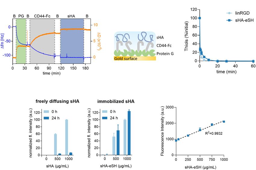
Zapp et al.                                                                                                                        PEG/GAG Hydrogel

  GRAPHICAL ABSTRACT

molecules is critical for tissue homeostasis, as well as during                 Its high hydrophilicity, biocompatibility, biodegradability
wound healing and development. As key components of the                     and possible extraction from animal independent sources with
ECM, glycosaminoglycans (GAGs) play a key role in tissue                    high purity, qualifies HA to be used as a natural polymer in
hydration, structural scaffolding, as well as cell signaling,               regenerative medicine (Xu et al., 2012). Particularly advantageous
hence modulating a wide range of cellular processes (Soares                 is that HA, as well as all GAGs, exhibits several functional groups
da Costa et al., 2017; Karamanos et al., 2018). Among the                   that can easily be used for chemical modifications, enabling
GAGs, hyaluronan (HA) is abundant in almost all tissues and                 conjugation of bioactive cues and crosslinking or extending
outstanding due to its non-sulfated composition and direct                  the lifetime by a reduced degradation rate (Köwitsch et al.,
cellular secretion into the extracellular space (Dicker et al., 2014;       2018; Serban and Skardal, 2019). Moreover, a combination of
Cowman et al., 2015). Hyaluronan is a linear, negatively charged            GAGs with a multitude of other materials, whether biologically
polysaccharide, build-up of repeating disaccharide units of β-1,4-          derived or synthetically manufactured, enables adaption to
D-glucuronic acid and β-1,3-N-acetyl-D-glucosamine, reaching                desired features of biomaterials. Hence, natural and synthetic
a molecular weight between 1 ×10⁵ Da and 1 × 10⁷ Da and a                   hydrogels can be applied to either generate ECM mimics or
polymer length of up to 25 nm (Toole, 2004). Under physiological            to deconstruct and evaluate how specific ECM parameters
conditions, HA chains form a hydrated random coil, occupying                (e.g., stiffness, ligand presentation, and dimensionality) provoke
a large volume dependent on the bonding between adjacent                    particular cellular behaviors.
saccharides and counterbalancing of its negative charge by                      Despite the increasing knowledge on HA and its bioactivity,
cations and water (Kutálková et al., 2020).                                 the importance of HA’s molecular weight and organization within
    During physiological or pathological remodeling, lower                  the ECM is not completely understood. Therefore, it is of
molecular weight HA fragments accumulate within the tissue                  particular importance to develop platforms that are suitable to
by depolymerization via reactive oxygen species and enzymatic               dissect the contribution of HA from additional ECM parameters.
cleavage by hyaluronidases (HYAL1-3, PH-20) (Lepperdinger                   Current successful hydrogel systems apply crosslinks between
et al., 2001; Wu et al., 2014). Dependent on its molecular weight           modified HA chains (Burdick and Prestwich, 2011; Broguiere
(MW) and temporal/spatial distribution in the tissue, HA is                 et al., 2016). However, these hydrogel types are unsuitable for
involved in numerous biological processes, by modulating both               studying GAG bioactivities as: (1) the side modifications impair
its physical properties as well as its direct biochemical signaling         the CD44-HA interactions, important for CD44-dependent cell
(Tavianatou et al., 2019). Hence, HA provides structural support,           signaling (Teriete et al., 2004; Banerji et al., 2007; Bencherif et al.,
maintains tissue hydration and serves as a lubricant in certain             2008; Bhattacharya et al., 2017) and (2) the GAG concentrations
tissues (Dicker et al., 2014). Furthermore, through interactions            govern the network, impeding an independent tuning of the
with specific HA surface receptors, such as CD44 or RHAMM,                  GAG concentrations.
HA contributes to the activation of complex cellular signaling                  In this context, we designed a hydrogel, which decouples the
pathways involved in cell adhesion, differentiation, proliferation          main hydrogel features from the presence of HA and further
and migration (Itano et al., 2002; Kim and Kumar, 2014; Simpson             GAGs in a user-friendly set-up, which allows analyzing the
et al., 2016; Wong et al., 2017). Dysfunction of HA within                  bioactive role of HA in a defined 3D environment. Careful design
the ECM is associated with various diseases, such as tumor                  of HA-based ECM models could benefit from HA-mediated
progression and metastasis, articular degeneration or chronic               cellular signaling, which goes beyond exploiting HA as an inert
inflammation (Liang et al., 2016). In the last decades, the role of         and non-adhesive scaffold. Within our highly controlled model
HA in disease and health was increasingly subject of investigation          of the ECM, it is possible to dissect and analyze the impact of
and led to the development of HA-based biomaterials applied as              cell adhesion cues, the abundance of HA and the varying stiffness
a dermal filler, drug delivery vehicle or ECM mimetic in tissue             of soft tissues on cell behavior. Especially because previous
engineering (Kim et al., 2017; Hong et al., 2020).                          research focused mainly on the bioactivity of free diffusing HA

Frontiers in Cell and Developmental Biology | www.frontiersin.org       2                                      October 2021 | Volume 9 | Article 729670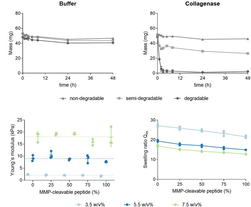
Zapp et al.                                                                                                                    PEG/GAG Hydrogel

in soluble form, the question arises, how a presentation of               Glycosaminoglycan Modifications
immobilized HA, as part of an artificial ECM, impacts cellular            sHA was functionalized at the reducing N-acetylglucosamine unit
signaling and behavior.                                                   according to the procedure of Lee et al. (2008). Briefly, sHA
    We applied polyethylene glycol (PEG) hydrogels as a solid             (100 mg), cysteamine hydrochloride (120 mg, 100 mM, Sigma-
base and coupled end-modified GAGs in a controlled manner                 Aldrich, Germany) and sodium chloride (467.5 mg, 400 mM,
to the hydrogels. We prepared the hydrogel backbone via thiol-            Merck, Germany) were dissolved in borate buffer (20 mL,
Michael addition reaction between an 8-arm, branched PEG with             100 mM, pH 8.5, Sigma-Aldrich, Germany) for 2 h at room
terminal vinyl sulfone groups and a linear dithiol linker, giving         temperature. After adding sodium cyanoborohydride (251 mg,
the advantage to work in mild, physiological conditions, suitable         200 mM, Sigma-Aldrich, Germany), the reaction mixture was
to directly embed cells. In general, 8-arm PEG-based hydrogels            stirred for 5 days at 40◦ C in an oil bath. Afterward, the reaction
have a higher shear modulus and swell significantly less compared         mixture was incubated with dithiothreitol (DTT, 150 mg, Sigma-
to those generated from 4-arm PEG (Rezakhani et al., 2020). The           Aldrich, Germany) for 2 h at 40◦ C. The reaction mixture
synthetic PEG polymer is considered biologically inert and hence          was dialyzed (MWCO: 3500 Da, Carl Roth, Germany) against
prevents unspecific protein adsorption and cell adhesion (Gibas           acidified deionized water with sodium chloride (pH 5) for 2 h
and Janik, 2010). The bioactivity of these hydrogels is modulated         and then against deionized water for 2 days. The thiolated sHA
by the incorporation of bioactive molecules, like bioactive               (sHA-eSH) was recovered by freeze-drying and stored at −80◦ C.
peptides or growth factors (Ravi et al., 2015). End-thiolated             For verification, the end-terminal thiol modification of sHA-
GAGs or cell-adhesive peptides can be conjugated covalently               eSH was quantified to 81.4 ± 3.0% by an adapted colorimetric
to the backbone, transforming the otherwise inert hydrogel                Ellman’s assay (Ellman, 1958). The same procedure was applied
into a highly controlled system, recapitulating features of the           for thiolation of the polysaccharides chondroitin sulfate A (CS),
ECM environment systematically. This conjugation enables us               dermatan sulfate (DS) and heparin (Hep), however, without
to investigate the effects of immobilized HA in comparison                sodium chloride in the reaction mix and dialysis with deionized
to freely diffusing HA. Within physiological concentration                water only. Terminal thiolation was quantified to 73.6% ± 0.4%
ranges, the fine-tunable mechanical properties are independent            (CS), 108.6% ± 9.3% (DS), 108.8% ± 2.1% (Hep) by the
of HA incorporation. Additionally, as the platform is not                 colorimetric Ellman’s assay.
limited to HA and adaptable for immobilization of other                       For the fluoresceinamine (FA, isomer 1, Sigma-Aldrich,
GAGs, it enables the co-presentation of carbohydrates and                 Germany) fluorophore conjugation, sHA (50 mg) and 1-ethyl-
proteins independently. Within this study, we comprehensively             3-[3-dimethylaminopropyl] carbodiimide (EDC, 10 mg, Sigma-
characterized the hydrogel properties and finally demonstrated            Aldrich, Germany) were dissolved in PBS (15 mL, 100 mM,
the versatility of this user-friendly platform for cell experiments       pH 5.8) for 30 min and FA (2.5 mg/mL in DMSO) was added
with an example: We demonstrate that PEG/GAG hydrogel                     dropwise (final conc. 0.1 mg/mL). The reaction mixture was
provides a degradable, 3D cell culture system for analyzing               incubated at room temperature for 5 h and dialyzed as before.
the influence of HA on the sprouting of endothelial cells                 To synthesize an end-thiolated and labeled sHA-eSH, the same
during angiogenesis.                                                      labeling protocol was performed before the thiolation reaction.
                                                                          To determine the success of this reaction, the degree of labeling
                                                                          (DOL) was determined. Therefore, the fluorescence intensity
MATERIALS AND METHODS                                                     of the products and a serial dilution of the fluorophore was
                                                                          measured at 488 nm/533 nm in the plate reader (Spark, Tecan,
Materials for Cell Culture and Hydrogel                                   Swiss). The degree of labeling is specified as molar percentage
Preparation                                                               of dye to HA monomer content (sHA: 14.9% ± 1.0%, sHA-eSH:
Hyaluronan (sHA, MW 10 kDa) was purchased from LifeCore                   0.07% ± 0.02%).
Biomedical (United States). Dermatan sulfate (DS, MW 30 kDa)
was purchased from Merck (Germany). Chondroitin sulfate A
(CS, MW 10–30 kDa) and heparin (Hep, MW 16.5 kDa) were                    Hydrogel Formation
obtained from Biosynth-Carbosynth (United Kingdom). PEG-                  Preparation of hydrogels was accomplished through thiol-
dithiol (MW 1 kDa), methylcellulose (4,000 cP) and OptiPrep               Michael addition reaction of an 8-arm PEG with terminal
(density gradient medium) were purchased from Sigma-Aldrich               vinyl sulfone groups (PEG-VS) and a PEG-dithiol crosslinker.
(Germany). The linRGD peptide (sequence: GCGWGRGDSPG)                     Hydrogels were formed with a series of PEG polymer
was purchased from PSL GmbH (Germany), the crosslinker                    concentrations (3.5 w/v%, 5.5 w/v%, and 7.5w/v%). For example
peptide (sequence: GCREGPQGIWGQERCG) from Pepscan                         to prepare 100 µL of 3.5 w/v% non-degradable hydrogels, 14.6
(Netherlands). 8-arm PEG with terminal vinyl sulfone groups               µL of a 20 w/v% (10 mM) PEG-VS stock was mixed with
(PEG-VS, MW 20 kDa, tripentaerythritol core) was obtained                 29.2 µL of a 2 w/v% (20 mM) crosslinker stock in 56.2 µL
from JenKem Technology (United States). Recombinant human                 PBS. For the preparation of functionalized hydrogels, PEG-VS
VEGF was purchased from R&D Systems (United States).                      was incubated together with the adhesive RGD motif (0.5 mM,
Human umbilical vein endothelial cells (HUVECs, pooled donor,             linRGD) and end-thiolated GAGs (0–1 mg/ml) for 20 min in
cryopreserved), endothelial cell growth medium (ECGM) and                 PBS. This functionalization step will result mainly in single
basal medium were purchased from Promocell (Germany).                     functionalized PEG-VS, since the added thiol containing species

Frontiers in Cell and Developmental Biology | www.frontiersin.org     3                                    October 2021 | Volume 9 | Article 729670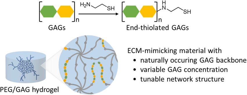
Zapp et al.                                                                                                                              PEG/GAG Hydrogel

are far less than equivalence [for highest functionalization                        settings. For comparison, fluorescence intensities of hydrogels
with 500 µM linRGD and 1 mg/mL sHA-eSH (76.9 µM)                                    before and after washing were plotted for both soluble sHA and
the molar VS:SH ratios are 1:0.049 (3.5 w/v%), 1:0.031 (5.5                         immobilized sHA-eSH.
w/v%) and 1: 0.023 (7.5 w/v%)]. In a polymerization step,                               Immobilized GAGs within hydrogels were also visualized
the preincubated PEG-VS solution was mixed with crosslinker                         by staining with the cationic carbocyanine dye Stains-All
and incubated for 1 h at 37 ◦ C. Thereby, the molar ratio                           (Sigma-Aldrich, Germany). 3.5 w/v% hydrogels (10 µL) were
of reactive thiol groups (total thiols from crosslinker and                         prepared with the PEG-dithiol linker and functionalized with
functionalization) and vinyl sulfone groups (from PEG-VS)                           1 mg/mL end-thiolated GAGs in triplicates. Hydrogels were
were stoichiometrically balanced (see detailed calculation in                       incubated for 24 h in 50 µL TRIS-borate-EDTA buffer (89 mM
Supplementary Material). Similar degradable hydrogels were                          tris(hydroxymethyl)aminomethane base (TRIS), 89 mM boric
crosslinked with a peptide, which is degradable by a range                          acid and 2 mM ethylenediaminetetraacetic acid (EDTA), pH
of matrix metalloproteinases (MMP-cleavable peptide). Semi-                         8.3, all Sigma-Aldrich, Germany) with two buffer exchanges.
degradable hydrogels were prepared with a 1:1 molar ratio of                        Hydrogels were stained with 50 µL of a Stains-All solution (0.005
the PEG-dithiol crosslinker and the MMP-cleavable peptide.                          w/v% in 70 v/v% ethanol) for 4 h and destained for 2 × 1 h with
The crosslinker concentrations for non-degradable hydrogels are                     50 µL 10 v/v% ethanol, before taking an image.
summarized in Table 1 (for other crosslinkers see Table 1 in                            Carbazole-based quantification of hyaluronic acid within
Supplementary Material). The stock solutions of sHA-eSH and                         hydrogels comprised hydrolyzation of sHA in its monomers
the MMP-cleavable peptide, dissolved in PBS, were adjusted to                       and a color reaction of glucuronic acid with carbazole (Plätzer
pH 7.4 with 1 M NaOH.                                                               et al., 1999; Frazier et al., 2008). A serial dilution of sHA in
                                                                                    water in triplicates was used as an external standard. Lyophilized
Time Resolved Conjugation of                                                        PEG hydrogels (90 µL) functionalized with 0.5 mM linRGD
                                                                                    and varying amounts of sHA-eSH as well as 50 µL of serial
End-Thiolated Hyaluronan and linRGD to
                                                                                    dilution samples were incubated with 200 µL 25 mM sodium
Polyethylene Glycol-Vinyl Sulfone                                                   tetraborate in sulfuric acid at 100 ◦ C for 10 min and cooled
The Measure-ITTM Thiol Assay Kit (Invitrogen, M30550,                               at room temperature for 15 min. Then 50 µL of a 0.125%
Thermo Fischer Scientific, Germany) was used to monitor the                         carbazole solution (Carl Roth, Germany) in absolute ethanol
conjugation of PEG-VS with either sHA-eSH or linRGD by                              was added. After incubation at 100◦ C for 10 min and at room
detecting a decrease in thiol groups over time. The kit was                         temperature for 15 min, the absorbance was measured at 550 nm
used according to the manufacturer’s instructions. sHA-eSH                          in a plate reader.
or linRGD peptide was reacted with PEG-VS for up to 1 h,
before reacting with 100 µL working solution of the assay.
The decrease in thiol groups is presented as a percentage
                                                                                    Crosslinking Efficiency and
of initial thiols over time. Concentrations are according to a                      Quantification of Thiols in Hydrogels
functionalization step of a 5.5 w/v% hydrogel with 0.5 mM                           To quantify the crosslinking efficiency, all types of hydrogels
linRGD and 1 mg/mL sHA-eSH.                                                         were prepared in triplicates, freeze-dried and weighed [m(non-
                                                                                    extracted)]. Then hydrogels were incubated in PBS for 2 days
Glycosaminoglycan Detection in                                                      at 37◦ C, freeze-dried and weighed again (m(extracted)). The
                                                                                    crosslinking efficiency was calculated by dividing m(extracted) by
Hydrogels                                                                           m(non-extracted).
The physical entrapment of sHA within a pure PEG hydrogel was                          The percentage of free thiols within hydrogels (40 µL)
assessed. Therefore, 3.5 w/v% hydrogels (10 µL) were prepared                       was determined using Ellman’s assay. Hydrogels and an
with different amounts of either a soluble fluorescently-labeled                    unpolymerized sample (only thiol linker in buffer, to determine
sHA or an end-thiolated fluorescently-labeled sHA (0–1 mg/mL).                      the initial thiol concentration) were prepared in triplicates. TRIS
The fluorescence intensity of the hydrogels after polymerization                    buffer (784 µL, 1 M, pH 8) and Ellman’s reagent (784 µL, 2 mM
was measured in the plate reader (488 nm/533 nm). After                             5,50 -Dithiobis(2-Nitrobenzoic-acid) and 50 mM sodium acetate
washing in 70 µL PBS at 37◦ C for 24 h in the dark (PBS was                         in water, both Sigma-Aldrich, Germany) was added per sample.
exchanged after 5 h and 20 h), the fluorescence intensity of the                    After incubation for 20 min at 350 rpm, the absorption of the
hydrogels was measured again in the plate reader using the same                     supernatant was measured at 412 nm. Values are in percentage of
                                                                                    free thiol groups measured in the unpolymerized sample.

TABLE 1 | Crosslinker concentrations (in mM) for non-degradable hydrogels.
                                                                                    Equilibrium Swelling and Mesh Size
Crosslinker         Functionalization       3.5 w/v%      5.5 w/v%   7.5 w/v%       Calculations
Non-degradable      PEG only                   11.7          18.3       25.0
                                                                                    After measuring the mass of hydrogels (90 µL) swollen in PBS
                    500 µM linRGD              11.3          17.9       24.6
                                                                                    or endothelial cell growth medium, hydrogels were freeze-dried
                    500 µM linRGD +            11.2          17.9       24.5
                                                                                    and weighed again. The swelling ratio (Qm ) was calculated as the
                    76.9 µM sHA-eSH                                                 ratio of the swollen hydrogel mass by its dry mass. Their mesh size
                                                                                    ξ was calculated based on the Flory-Rehner theory, as described

Frontiers in Cell and Developmental Biology | www.frontiersin.org               4                                    October 2021 | Volume 9 | Article 729670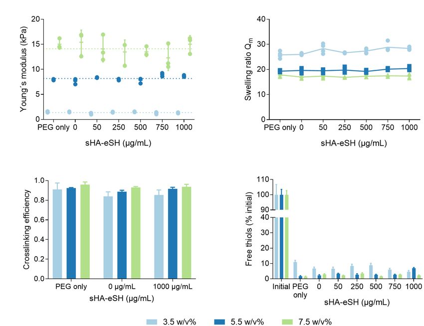
Zapp et al.                                                                                                                    PEG/GAG Hydrogel

previously (Peppas et al., 2000; Zustiak and Leach, 2010). First,          Sigma-Aldrich, Germany), 137 mM sodium chloride, 1 mM
the molecular weight between crosslinks (Mc ) was calculated by:           calcium dichloride (Carl Roth, Germany), 3 mM sodium azide
                       v
                                                      2
                                                                          (Riedel-de Haën, Germany), pH 7.4) overnight. Ca2+ ions are
          1     2     V 1 ln 1−v2,s + v2,s + X1 v2,s                       necessary for collagenase activity. Hydrogels were then incubated
             =     −
                                                                           with 300 µL buffer or 2 mg/mL collagenase (Type 1, from

        Mc     Mn                  v2,s 1/3    v
                            v2,r v2,r       − 2v2,s
                                                 2,r                       clostridium histolyticum, Sigma-Aldrich, Germany) at 37◦ C and
                                                                           their mass was weighed over 48 h. After each measurement,
   Where Mn is the number-average molecular weight of the                  solutions were renewed.
non-crosslinked polymer chains, V1 is the molar volume of the
solvent (18 cm3 /mol for water), v is the specific volume of the           Quartz Crystal Microbalance With
polymer (ρs /ρp with polymer density ρp = 1.12 g/cm3 and solvent
density ρs = 1.0 g/cm3 ), v2,s is the polymer volume fraction              Dissipation Monitoring
in swollen hydrogels, v2,r is the polymer volume fraction in               Experiments were performed with the manually handled Q-Sense
the relaxed state (immediately after polymerization), X1 is the            E4 instrument, equipped with a 4-channel system, open modules
polymer-solvent interaction parameter (0.426 for PEG in water)             and gold-coated quartz crystal electrodes (AT cut, 4.95 MHz,
(Lu and Anseth, 2000). Then, the mesh size was calculated by:              Biolin Scientific AB, Sweden). Electrodes were cleaned in a
                                    s                                      washing solution consisting of 5:1:1 (volume parts) deionized
                            1         2Cn Mc                               water, 25% ammonia (Merck, Germany) and 30% hydrogen
                     ξ= p         l                                        peroxide (Sigma-Aldrich, Germany) at 75◦ C for 10 min, followed
                          3
                            V 2,s      Mr
                                                                           by UV/ozone treatment (ProCleaner, Bioforce Nanosciences, IA,
   Where l is the average bond length across the backbone of               United States) for 10 min. The frequency and the dissipation of
the polymer (0.146 nm for PEG), Cn is the Flory characteristic             the 3rd, 5th, 7th, 9th, 11th, and 13th overtones were monitored.
ratio (4 for PEG polymers) and Mr is the molecular weight of the           After setting a baseline in PBS, 200 µL of the desired solution
repeating polymer units (44 g/mol for PEG) (Mellott et al., 2001).         was pipetted on the electrodes. The solutions remained on the
Thereby, contributions of the peptide crosslinker to the Flory             electrodes until a stable plateau in both the dissipation and
characteristic ratio and the molecular weight of the repeating             frequency was maintained for at least 5 min. Each adsorption
units were neglected according to the approach of Schwartz                 step was followed by washing the electrodes with PBS to remove
et al. (2009) and Rehmann et al. (2017), who studied similar               non-interacting molecules. Each sample was tested in duplicates.
hydrogel systems.                                                          For validating CD44-sHA interaction, protein G (BioVision, CA,
                                                                           United States), sHA/sHA-eSH, CD44-Fc-tag (Acro Biosystems,
Rheological Measurements of Swollen                                        DE, United States) and bovine serum albumin (Sigma-Aldrich,
Hydrogels                                                                  Germany) concentrations were 10 µg/mL, 0.5 mg/mL, 3
Material properties were assessed using a rotational rheometer             µg/mL, and 0.1 mg/mL in PBS, respectively. Data of the 7th
(Kinexus Pro+, Malvern, United Kingdom) equipped with an                   overtone are shown.
Active Hood Peltier plate cartridge and a parallel-plate geometry
(8 mm) with a solvent trap. For all hydrogel conditions, a strain          Cell Culture and Generation of
sweep between 0.1 and 100% at a constant frequency of 1 Hz and             Endothelial Cell Spheroids
a frequency sweep with frequencies between 0.01 and 100 Hz at              Human umbilical vein endothelial cells (HUVECs) were cultured
a constant shear strain was performed to determine the linear              in endothelial cell growth medium (ECGM) at 37◦ C with
viscoelastic regime. Swollen hydrogels were fixed between the              humidified 5% CO2 atmosphere up to passage 6. After
plates by a normal force set to 0.1 N (3.5 w/v%) or 0.2 N (5.5             reaching 90% confluency HUVECs were detached with 0.05%
w/v% and 7.5 w/v%). All stiffness measurements were performed              trypsin-EDTA (Thermo Fisher Scientific, Germany), collected,
at 25◦ C, for 2 min with an interval of 5 s with a frequency of 1 Hz       centrifuged at 230 × g for 3 min and reseeded at an appropriate
and a shear strain of 5%. The Young’s modulus E is related to the          density. Endothelial cell spheroids were generated using the
shear modulus G by:                                                        hanging-drop method. HUVECs were harvested and suspended
                                                                           in ECGM containing 0.25 w/v% methylcellulose. After deposition
                              E = G 2 (1 + v)
                                                                           of 25 µL drops on the lid of a humidified chamber, the hanging
   with a Poisson’s ratio v assumed as 0.5. The gelation time was          droplets were cultivated for 24 h. Under these conditions, the
determined for several hydrogel conditions. The evolution of the           suspended cells aggregate and form a single spheroid of defined
storage and loss modulus (G0 and G00 , respectively) was recorded          size and cell number (600 cells per spheroid).
at 37◦ C with an interval time of 2 min over a period of 1 h, at a
frequency of 1 Hz and a constant strain of 1%.                             In vitro Angiogenesis Assay
                                                                           For the in vitro angiogenesis assay, spheroids were harvested
Enzymatic Degradation of Hydrogels                                         and embedded into degradable 3.5 w/v% PEG hydrogels,
3.5 w/v% hydrogels (40 µL) were prepared as described                      functionalized with 0.5 mM linRGD and varying amounts of
and swollen in HEPES buffered saline (10 mM 2-[4-(2-                       sHA-eSH (0–1 mg/mL). Hydrogels were prepared as described
hydroxyethyl)piperazin-1-yl]ethanesulfonic acid (HEPES,                    previously with 15 v/v% OptiPrep to prevent sedimentation

Frontiers in Cell and Developmental Biology | www.frontiersin.org      5                                   October 2021 | Volume 9 | Article 729670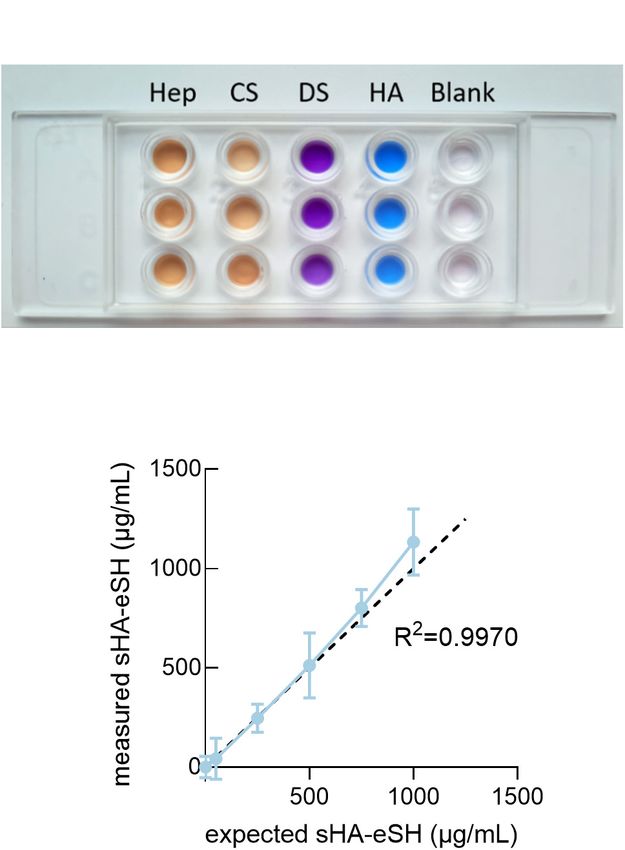
Zapp et al. PEG/GAG Hydrogel
of spheroids prior to polymerization of the hydrogel. For the impact on cellular behaviors. For this purpose, PEG hydrogels
polymerization step, the spheroid and crosslinker solutions were were formed as a solid base and end-thiolated GAGs were
added to the functionalized PEG-VS and 10 µL hydrogels conjugated in a controlled manner to the hydrogel. PEG
were formed in 96-well angiogenesis µ-plates (Ibidi GmbH, hydrogels were prepared via thiol-Michael addition reaction of
Germany). After polymerization, hydrogels were overlaid with an 8-arm branched PEG-vinyl sulfone (PEG-VS) and a linear
70 µL cell culture medium (basal medium with 15% FBS). dithiol linker, as described in previous studies (Lutolf et al.,
For stimulation of the HUVECs, the cell culture medium was 2003a). Synthesis of the PEG hydrogels is conducted in two steps
supplemented with 50 ng/mL VEGF165 . The gels were incubated (Figure 1). In a first functionalization step, bioactive ligands
at 37◦ C in a humidified 5% CO2 atmosphere and imaged after are conjugated to a branched 8-arm PEG monomacromer. In a
48 h. Phase-contrast images were acquired with a Zeiss Primovert subsequent gelation step, a dithiol linker molecule is added and
(Zeiss, Germany), equipped with an Axiocam 208 color and 20 the gel is polymerized.
× objective (Zeiss, Germany, Plan-Achromat, Ph1, 20 ×/0.30). Prior to PEG/HA hydrogel formation, the interaction of the
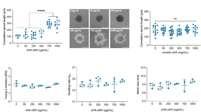
Sprouting was quantified by measuring the cumulative sprout small hyaluronan oligosaccharides (sHA), used within this study,
length, which had grown out of each spheroid, using the and its standard transmembrane receptor CD44 was proven in a
imaging software FIJI. label-free QCM-D experiment (Figures 2A,B). Dissipation and
frequency changes monitored over time indicate binding of sHA
Statistical Analysis to the receptor CD44, immobilized via a recombinant Fc-tag to
Data are presented as mean ± standard deviation. One-way a protein G adlayer. Corresponding dissipation and frequency
analysis of variance (ANOVA) followed by Tukey post-hoc shifts were observed with sHA-eSH (Supplementary Figure 1).
test was used to compare between conditions with GraphPad After verifying the sHA-CD44 interaction, the presentation of
Prism 8.0 software. sHA within a 3D scaffold was achieved with a straightforward
method: sHA was modified at the reducing end with a short thiol
linker (named sHA-eSH or end-thiolated sHA) and subsequently
RESULTS immobilized to PEG-VS, a building block for the hydrogel
formation. After successful end-thiolation of sHA, its conjugation
Presentation of Glycosaminoglycans to the branched 8-arm PEG-VS was tested. The decrease in
terminal free thiol groups of the sHA-eSH, due to its conjugation
Within a Highly Controllable and Tunable to PEG-VS, was detected in a time resolved assay (Figure 2C).
Set-Up An exponential decay of terminal thiols proved the conjugation
Conjugation of Glycosaminoglycans to Polyethylene of end-thiolated sHA to PEG-VS. A comparable reaction rate
Glycol Hydrogels was detected for the conjugation of the cell adhesive linear RGD
Within this study, we developed and characterized a PEG/GAG (linRGD) peptide motif (Figure 2C). A complete conversion of
hydrogel, which helps us to investigate the bioactive role of HA both biomolecules is achieved within 20 min. Consequently, the
and further GAGs as part of a well-defined 3D environment. functionalization was left to proceed for 20 min before addition
By altering independently the matrix stiffness as well as the of the crosslinker. In a subsequent step, these functionalized PEG
GAG and peptide presentation in a modular set-up, we can macromolecules are crosslinked into a network by addition of
better understand the interplay of these parameters and their a dithiol linker.
Figure 1 | Schematic illustration of PEG/GAG hydrogel formation. Branched 8-arm poly (ethylene glycol) PEG with terminal vinyl sulfone groups (VS) is functionalized
with end-thiolated GAGs and peptide binding motifs (e.g., RGD) via thiol-Michael addition reaction. Following this functionalization, PEG macromolecules are
crosslinked with a PEG-dithiol linker or matrix-metalloprotease (MMP)-cleavable di-cysteine peptide. The peptide linker sequence is derived from collagen with a
single amino acid exchange from A to W (blue) for a higher enzyme affinity and enhanced degradability (cleavage site underlined).
Frontiers in Cell and Developmental Biology | www.frontiersin.org 6 October 2021 | Volume 9 | Article 729670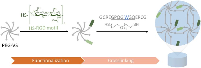
Zapp et al.                                                                                                                                           PEG/GAG Hydrogel

  Figure 2 | (A) QCM-D measurement confirms CD44-sHA interaction. The binding experiment shows the formation of a stable adlayer of protein G (PG) on a gold
  surface, followed by immobilization of Fc-tagged CD44 and its binding to sHA. Data from the 7th overtone are shown, frequency changes depicted in blue and
  dissipation changes in orange. (B) Schematic diagram of immobilization strategy on QCM-D sensor. (C) Kinetics of thiol-Michael addition reaction of sHA-eSH and
  linRGD. Progression of the conjugation to 8-arm PEG-VS macromonomers was monitored over time by detecting the decrease of free terminal thiol groups; mean
  and standard deviation of triplicates are presented. (D) Detection of varying amounts of fluorescently-labeled hyaluronan within hydrogels after incubation in PBS for
  24 h. 3.5 wt/vol% PEG hydrogels with varying amounts of fluorescent sHA (soluble sHA) or sHA-eSH (immobilized sHA) were prepared by thiol-Michael addition
  reaction. (E) Fluorescence intensity values of hydrogels after washing in PBS are plotted over the hyaluronan concentration per mL of hydrogel solution; mean and
  standard deviation of quintuplicates are presented.

   Due to sHA’s flexibility and small size, its physical entrapment                     GAGs. To demonstrate this, further common GAGs, more
within PEG hydrogels was assessed. Therefore, 3.5 w/v%                                  precisely chondroitin sulfate, dermatan sulfate and heparin, were
PEG hydrogels were loaded with varying amounts of soluble                               modified with an end-terminal thiol group and their presence
fluorescently-labeled sHA. The fluorescence intensities of these                        within hydrogels was detected by staining with the cationic
hydrogels were measured before and after washing in PBS. A drop                         carbocyanine dye Stains-All. Stains-All is a metachromatic
in the detected fluorescence intensities of these hydrogels was                         dye capable of staining different GAG types in distinct and
observable when hydrogels were incubated in PBS for 24 h                                characteristic colors (Andrade et al., 2018). Hydrogels were
(Figure 2C). This indicates that sHA freely diffuses out of the                         prepared with 1 mg/mL of end-thiolated GAGs, incubated for
hydrogel. Next, hydrogels were prepared with a fluorophore-                             24 h in borate buffer and stained with Stains-All. Stains-All
labeled and end-thiolated sHA. In this case, similar fluorescence                       incubation showed distinct colors for each of the GAG in
intensities of the hydrogels before and after incubation in PBS                         comparison to the almost transparent water control (Figure 3A).
were observed (Figure 2D). It can be concluded that thiol-                              It can be concluded, that the approach of immobilizing
mediated immobilization of sHA-eSH to the PEG backbone                                  sHA via a short thiol linker is extendable to further GAGs.
prevents the diffusion of sHA out of the hydrogel. These                                Next to these qualitative verifications, immobilized sHA-eSH
two experiments in combination demonstrate the necessity for                            within hydrogels was quantified in absolute numbers by a
conjugating sHA to the PEG hydrogel for a reliable presentation                         colorimetric assay. Measured sHA-eSH concentrations of each
of HA as part of a 3D microenvironment. Moreover, a very good                           sample are plotted against the expected theoretical values
correlation between an increasing amount of sHA-eSH within                              (Figure 3B). Overall, a good correlation occurs between these
the hydrogel and the detected fluorescence intensity values was                         values. In accordance with the previous diffusion experiments,
observable (Figure 2E).                                                                 a presentation of hyaluronan as part of a network is achievable.
   Additionally, this approach of immobilizing end-thiolated                            Finally, this immobilization strategy enables the independent co-
sHA is adaptable for immobilization of other end-thiolated                              presentation of GAGs and peptides. This facilitates the direct

Frontiers in Cell and Developmental Biology | www.frontiersin.org                   7                                           October 2021 | Volume 9 | Article 729670
Zapp et al.                                                                                                                                       PEG/GAG Hydrogel

sensing of biological signals by the cells within the otherwise                      PEG/HA hydrogels generally increase with an increase in the total
inert PEG hydrogels.                                                                 polymer fraction, as determined by rheological measurements
                                                                                     (Figure 4A). It was revealed that the incorporation of both
Physicochemical Characterization of Hydrogels With                                   types of bioactive molecules, linRGD motif (0.5 mM) and sHA-
Varying Amounts of End-Thiolated Hyaluronan                                          eSH (up to 1 mg/mL, equals 0.077 mM), barely influences
The physical and chemical properties of PEG/GAG hydrogels                            the Young’s modulus. Hydrogels have Young’s moduli of 1.4
were analyzed. Detailed characterization referred to small                           kPa ± 0.2 kPa (3.5 w/v%), 8.2 kPa ± 0.5 kPa (5.5 w/v%), and
hyaluronan oligosaccharides (sHA), the most studied GAG                              14.1 kPa ± 2 kPa (7.5 w/v%), and thus are within the range
type. Non-functionalized hydrogels were named “PEG only”,                            of physiological elasticity of soft tissues (Engler et al., 2006).
while hydrogels classified as “0–1,000 µg/mL sHA-eSH” are                            The process of polymerization was monitored over time in
co-functionalized hydrogels with a constant adhesive linRGD                          rheological measurements (Supplementary Figure 2, Supporting
motif concentration (0.5 mM) and varying amounts of                                  Information). The gel point, defined as the time of crossover of
hyaluronic acid.                                                                     storage modulus G0 and loss modulus G00 , was below 2 min under
   Mechanical properties of hydrogels were tuned by varying                          the present conditions (pH 7.4, 37◦ C). Both storage and loss
the PEG polymer concentration (3.5 w/v%, 5.5 w/v%, and 7.5                           modulus increase until reaching a plateau, indicating complete
w/v%), modulating the number of backbone polymers in a given                         gelation. The gelation time depends mainly on the total polymer
volume within the hydrogel network. The Young’s moduli of the                        fraction, and less on the applied degree of functionalization
                                                                                     [15 min (7.5 w/v%), 20 min (5.5 w/v%), and 25 min (3.5 w/v%)].
                                                                                     Due to an equal stoichiometric ratio of free VS to SH groups,
                                                                                     the PEG-VS and crosslinker concentrations increase with an
                                                                                     increase of the PEG polymer concentration from 3.5 w/v% to
                                                                                     7.5 w/v%. Additionally, with higher degree of functionalization
                                                                                     the crosslinker concentration is slightly reduced (Table 1).
                                                                                     Overall, applied gelation times of 60 min were sufficient for
                                                                                     exhaustive crosslinking.
                                                                                         As part of a physicochemical characterization, the swelling
                                                                                     behavior was investigated including the swelling ratio Qm in
                                                                                     PBS and cell culture medium. Swelling ratios for equilibrium-
                                                                                     swollen hydrogels in PBS were in general higher in comparison
                                                                                     to hydrogels swollen in cell culture medium (Table 2). In both
                                                                                     cases, the swelling ratio decreased with an increase in hydrogel
                                                                                     stiffness and was independent of a modification with linRGD
                                                                                     and sHA-eSH (Figure 4B). A further parameter to characterize
                                                                                     the network structure is the mesh size, defined as the linear
                                                                                     distance between neighboring crosslinks. Calculations of the
                                                                                     mesh size are based on swelling ratios and follow the trend
                                                                                     observed for the bulk elasticity as well as for swelling ratios.
                                                                                     With rising PEG polymer concentrations, the hydrogel stiffness
                                                                                     increases, while the mesh size decreases. Thereby, swelling ratios,
                                                                                     as well as mesh sizes for functionalized hydrogels are unaffected
                                                                                     by the varying amounts of sHA-eSH and the presence of the
                                                                                     adhesive linRGD motif (Supplementary Figure 2). Thus, the
                                                                                     mean and standard deviation are shown, independent of the
                                                                                     functionalization (Table 2). In each case, the mesh size is larger
                                                                                     than the bulk size of the immobilized hyaluronan with ∼4.4 nm
                                                                                     (Takahashi et al., 1999).

  Figure 3 | Detection of GAGs in PEG hydrogels. (A) PEG hydrogels were
  prepared with 1 mg/mL end-thiolated GAGs [heparin (Hep), chondroitin
  sulfate (CS), dermatan sulfate (DS) and hyaluronan (HA)] and stained with          TABLE 2 | Elasticity, swelling ratio and mesh size for PEG/HA hydrogels.
  Stains-All after incubation in borate buffer for 24 h. Stained gels reveal a
  detection of GAGs with distinct colors. Blank samples were prepared with           w/v%                   3.5                5.5                 7.5
  water and appeared colorless. (B) Quantification of sHA-eSH in a
                                                                                     Elasticity [kPa]        1.4 ± 0.2          8.2 ± 0.5          14.1 ± 2.0
  carbazole-based assay. PEG hydrogels were functionalized with 0.5 mM
  linRGD and varying amounts of sHA-eSH (0–1 mg/mL). Measured HA-eSH                 Qm (PBS)               27.3 ± 1.5         19.7 ± 0.7          17.3 ± 0.6
  concentrations are plotted against expected concentrations. Hydrogels were         Qm (medium)            22.0 ± 0.6         17.5 ± 1.0          15.8 ± 2.6
  swollen in PBS > 20 h and lyophilized, before performing the assay; mean           Mesh size [nm]         11.7 ± 0.6          9.1 ± 0.4           8.2 ± 0.3
  and standard deviation of triplicates/quadruplicates are presented.                Gelation [min]               25                 20                  15

Frontiers in Cell and Developmental Biology | www.frontiersin.org                8                                           October 2021 | Volume 9 | Article 729670
Zapp et al. PEG/GAG Hydrogel Figure 4 | Physicochemical characterization of PEG/HA hydrogels. Hydrogels were prepared in three different PEG polymer concentrations by thiol-Michael addition reaction with a PEG-dithiol crosslinker. Hydrogels were unmodified (PEG only) or functionalized with 0.5 mM linRGD and varying concentrations of sHA-eSH (0–1 mg/mL). (A) Bulk elasticity. The Young’s moduli of hydrogels were determined by shear rheological measurements and range from 1.4 to 14.1 kPa, dependent on the PEG polymer concentration; mean and standard deviation of triplicates are presented. (B) Swelling ratios. Hydrogels were equilibrium-swollen in PBS; mean and standard deviation of quadruplicates are presented. (C) Crosslinking efficiency based on the sol-gel fraction of PEG/HA hydrogels; mean and standard deviation of triplicates are presented. (D) Detection of free thiol groups within non-degradable hydrogels using Ellman’s reagent. Initial corresponds to an unpolymerized hydrogel (all thiol groups free); mean and standard deviation of triplicates are presented. Defects in the network structure occur either based on non- thiols, following the trend observed for the crosslinking efficiency reacted groups or the formation of disulfide bonds, loops and before. Again, the reaction conversion seems unaffected by the entanglements. To get more insight into the network structure, functionalization with linRGD and sHA-eSH. the sol fraction and crosslinking efficiency were derived by weight changes due to the extraction of non-reacted PEG. The Matrix-Metalloprotease-Cleavable Peptide Linker: crosslinking efficiency was found to correlate with the PEG Degradable Hydrogels for 3D Cell Culture polymer concentration of the hydrogel formulation (Figure 4C). To recapitulate the ECM degradability, essential for several This is in accordance with the trends for the bulk stiffness, biological processes, PEG-VS macromeres are often crosslinked swelling ratios and mesh size. Overall, hydrogels show high with an MMP-cleavable di-cysteine peptide. The MMP-sensitive reaction conversion rates. Crosslinking efficiency was lowest for peptide (16 amino acids) is derived from the cleavage site of 3.5 w/v% hydrogels with 0.87 ± 0.05 and highest for 7.5 w/v% natural collagen with a single amino acid mismatch for enhanced hydrogels with 0.94 ± 0.02. The crosslinking efficiency seems degradation (Lutolf et al., 2003b; Patterson and Hubbell, 2010). to be unaffected by the modifications with linRGD and sHA- The so far applied PEG-dithiol linker of 1 kDa with a contour eSH. Additionally, to get more insights into the network defects, length of ∼6.16 nm (Oesterhelt et al., 1999) was chosen according the number of free thiol groups was assessed immediately after to the contour length of the peptide (∼6.08 nm; Ainavarapu et al., polymerization. Free thiols were quantified in an Ellman’s assay, 2007). Similar contour lengths were preferred to achieve similar and correspond to non-reacted crosslinker molecules within network structures for both types of crosslinker. a hydrogel (Figure 4D). Hydrogels prepared with increasing Degradation characteristics of hydrogels, crosslinked with PEG polymer concentrations showed lower amounts of free three different ratios of the MMP-cleavable di-cysteine peptide Frontiers in Cell and Developmental Biology | www.frontiersin.org 9 October 2021 | Volume 9 | Article 729670
Zapp et al.                                                                                                                      PEG/GAG Hydrogel

and the PEG-dithiol linker, were evaluated (Figures 5A,B).                   PEG-dithiol crosslinker have been identified as inapplicable
Previously swollen hydrogels were enzymatically degraded with                for the spheroid-sprouting assay, as the hydrogel stiffness
collagenase, while their change in total weight was monitored                and degradability constrained sprouting of HUVECs (see
over time. As a control, hydrogels were incubated in buffer over             Supplementary Figure 3). A life/dead staining assay of evenly
the same period. Hydrogels incubated in buffer were almost                   distributed HUVECs shows > 80% cell viability at day 2
constant in weight, thus indicating the stability of the hydrogels           (Supplementary Figure 3).
over 48 h, regardless of the crosslinker composition. Hydrogels                 Evaluation of six peptide motifs, derived from ECM proteins,
formed with the MMP-cleavable peptide linker (degradable)                    revealed the highest sprouting capacity for the linRGD binding
show complete enzymatic degradation within 2 h. Hydrogels,                   motif in degradable 3.5 w/v% hydrogels (Supplementary
crosslinked with a linker mix of the PEG-dithiol and MMP-                    Figure 4). Thus, spheroids were embedded in degradable
cleavable peptide (semi-degradable, molar ratio 1:1), lose their             3.5 w/v% PEG/HA hydrogels functionalized with varying
mass to a certain degree upon incubation with collagenase. This              physiological concentrations of sHA-eSH (0–1 mg/mL), while
mass decrease might be explained by the partial degradation of               the linRGD concentration was maintained constant at 0.5 mM.
the network. PEG-dithiol crosslinked hydrogels maintain their                Cumulative sprout length analyses reveal an increased sprout
weight during incubation with collagenase, verifying an expected             formation when spheroids are enclosed by hydrogels containing
non-degradability. Altogether, the degradability of the hydrogels            more than 0.75 mg/mL sHA-eSH (Figure 6A). Overall, HUVECs,
can be controlled by the ratio of degradable peptide linker to non-          invading into PEG/HA hydrogels with increasing sHA-eSH
degradable PEG-dithiol linker. Next, the influence of a stepwise             concentrations, seem to form sprouts with more branched and
replacement of PEG-dithiol with the MMP-cleavable di-cysteine                bulkier structures (Figure 6B). As previously, an analysis of the
peptide on the physicochemical characteristics including the                 elasticity, swelling ratio and mesh size of degradable PEG/HA
mechanical properties and swelling behavior was investigated.                hydrogels revealed a stable network structure, unaffected by
   As seen previously, Young’s moduli in shear rheological                   the immobilization of up to 1 mg/mL sHA-eSH (Figures 6D–
measurements of hydrogels, prepared with varying ratios of the               F). Additionally, a presentation of immobilized HA, as part
PEG crosslinker and the MMP-cleavable di-cysteine peptide,                   of an artificial ECM was compared to freely diffusing HA
highly depend on the PEG polymer concentration. The impact                   in soluble form. This time, the cumulative sprout length
of the crosslinker ratio on the Young’s moduli seems limited                 remained unaffected by the increasing concentration of soluble
(Figure 5C). The observed swelling ratios for these hydrogels                sHA (Figure 6C). These findings suggest that hyaluronan may
decrease with an increasing percentage of the peptide linker                 enhance endothelial cell invasion in the presence of covalently
(Figure 5D). This trend is more pronounced for the softer                    immobilized sHA, while the presence of soluble sHA does
hydrogels (3.5 w/v%).                                                        not affect this behavior. These data demonstrate that the
   Overall, the developed PEG/GAG hydrogel system allows                     PEG/GAG hydrogels provide a chemically defined environment
a presentation of GAGs in physiological concentrations                       for the culture of endothelial cells and thus could overcome
independent from the presentation of peptides as cell adhesive               current existing systems by recapitulating the protein and GAG
binding motifs. Moreover, the hydrogels are fine-tunable in their            content of the ECM.
degradability via the MMP-sensitivity of the dithiol linker and
their stiffness via the PEG polymer concentration. Including
GAGs as part of the natural ECM in synthetic hydrogels enables               DISCUSSION
either the generation of ECM mimics or the deconstruction
and evaluation of how specific ECM parameters induce specific                As key components of the ECM, GAGs mediate tissue hydration,
cellular behaviors with a bottom-up approach.                                structural scaffolding, local presentation of soluble molecules
                                                                             and cell signaling. Despite the increasing knowledge of GAGs
                                                                             and their bioactivity, the importance of their molecular weight,
Application as Well-Defined Artificial 3D                                    chemical structure and organization within the ECM is not
Microenvironment                                                             completely understood. To systematically dissect the bioactive
Polyethylene Glycol/Hyaluronan Hydrogels Support                             role of GAGs, we established a versatile biomaterial for a natural
Endothelial Cell Sprouting                                                   presentation of GAGs in a well-defined 3D environment.
To illustrate the functional utility of this approach in a biological           For this purpose, previously end-thiolated GAGs were
setting, PEG/HA hydrogels were applied in a 3-dimensional                    conjugated to a PEG hydrogel, formed by thiol-Michael
in vitro angiogenesis model and the cell-instructive role of                 addition reaction between a vinyl sulfone 8-arm PEG and a
HA was analyzed.                                                             thiol containing crosslinker (Figure 1). We demonstrated the
   It was assessed whether PEG/HA hydrogels could support                    successful terminal modification of varying GAGs (heparin,
the formation of capillary-like sprouts in a routinely used                  chondroitin sulfate, dermatan sulfate, hyaluronic acid) and
spheroid sprouting assay. Therefore, spheroids from human                    their immobilization to a PEG hydrogel. Diffusion experiments
umbilical vein endothelial cells (HUVECs) were embedded                      with fluorescently-labeled hyaluronan proved the necessity of
in degradable 3.5 w/v% hydrogels and the cumulative sprout                   conjugation to the backbone when the mesh size exceeds the
length originating from each spheroid was quantified. Hydrogels              radius of the GAGs (Figure 2). This GAG-conjugation approach
with higher PEG polymer concentration and non-degradable                     provides a reproducible and defined presentation of desired

Frontiers in Cell and Developmental Biology | www.frontiersin.org       10                                   October 2021 | Volume 9 | Article 729670
Zapp et al. PEG/GAG Hydrogel Figure 5 | Effect of a stepwise replacement of the PEG-dithiol linker with the MMP-cleavable peptide on degradation, elasticity and swelling ratio. (A,B) Enzymatic and hydrolytic degradation of non-degradable, semi-degradable and degradable hydrogels. 3.5 w/v% hydrogels were prepared by thiol-Michael addition reaction using either the PEG-dithiol (non-degradable), a mixture of PEG-dithiol and the MMP-cleavable di-cysteine peptide (semi-degradable, molar ratio 1:1) or the peptide alone (degradable). Hydrogels were swollen overnight in buffer and mass changes due to enzymatic degradation were monitored over 48 h when incubated in buffer (A) or with collagenase (B). (C,D) Hydrogels were formed with three different PEG polymer concentrations by thiol-Michael addition reaction with a stepwise replacement of the PEG-dithiol crosslinker by the MMP-cleavable di-cysteine peptide. (C) The Young’s moduli of hydrogels were determined by shear rheological measurements. (D) Swelling ratios of hydrogels with increasing degradability. Swelling ratios were calculated by dividing the mass of the equilibrium-swollen hydrogel by the dry mass; mean and standard deviation of triplicates are presented. GAG types in physiological relevant concentrations. The sulfated allow the incorporation of new features, but also critically GAG content from diverse biological samples and decellularized compromise the chemical structure and change the specificity ECMs is in the lower range of µg/mg tissue with an often of protein-GAG interactions (Kjellén and Lindahl, 2018; Shi tissue- and cell type-specific GAG expression (Barbosa et al., et al., 2021). To address this shortcoming for studying the 2003; Wolf et al., 2012; Ventura et al., 2019). The successful GAG bioactivity, a terminal GAG immobilization strategy was quantification of sHA in swollen hydrogels (Figure 3) revealed established herein, which enables a natural presentation of efficient incorporation within the physiological range of the GAGs within a 3D model while their backbone structure is human body (e.g., 200 µg/g for large intestine and heart, conserved. Dynamic modifications (e.g., acetylation, sulfation, 500 µg/g in the dermis) (Stern and Maibach, 2008; Cowman epimerization) of the GAG backbone produce a tremendous et al., 2015). Importantly, the PEG/GAG hydrogels introduced heterogeneity and guide interactions with soluble GAG-binding here require only a slight change at the reducing-end of signaling molecules (Muthana et al., 2012). Sulfated GAGs the GAG polymer chain, while most developed GAG-based modulate the trafficking, signaling activity and stability of growth biomaterials yet exploit a crosslinking between GAG chains, factors and cytokines due to mainly electrostatic interactions which requires side-modifications of the repeating structure with the highly anionic GAGs (Proudfoot et al., 2017; Köwitsch (Burdick and Prestwich, 2011; Broguiere et al., 2016; Ornell et al., 2018; Hachim et al., 2019). Using a hydrogel system in et al., 2019; Limasale et al., 2020). In general, modifications which heparin (derivatives) are crosslinked with 4-arm PEG, Frontiers in Cell and Developmental Biology | www.frontiersin.org 11 October 2021 | Volume 9 | Article 729670
Zapp et al.                                                                                                                                    PEG/GAG Hydrogel

  Figure 6 | Influence of immobilized sHA on angiogenic sprouting. (A) Analysis of the cumulative sprout length originating from HUVEC spheroids, embedded in 3.5
  w/v% PEG hydrogel functionalized with 0.5 mM linRGD and varying concentrations of sHA-eSH. Spheroids were stimulated with 50 ng/mL VEGF for 48 h. Statistical
  significance was calculated using one-way ANOVA followed by Tukey’s test (∗∗∗∗ p < 0.0001, n ≥ 3). (B) Representative phase-contrast images of VEGF-induced
  sprouting of spheroids embedded in PEG/HA hydrogels. Scale bar 50 µm. (C) Analysis of the cumulative sprout length originating from HUVEC spheroids,
  embedded in 3.5 w/v% PEG hydrogel functionalized with 0.5 mM linRGD and incubated with varying concentrations of freely diffusing sHA. (D) Bulk elasticity
  determined by shear rheological measurements, (E) swelling ratio of hydrogels in PBS and (F) mesh size of degradable 3.5 w/v% hydrogels prepared by
  thiol-Michael addition reaction with a MMP-cleavable di-cysteine peptide and functionalized with 0.5 mM linRGD and varying concentrations of sHA-eSH; mean and
  standard deviation of triplicates are presented.

the group around Uwe Freudenberg was able to show that                               that increasing consumption of PEG-VS arms might alter the
the heparin concentration and the sulfation pattern at heparin                       network structure. Thereby, changes in storage modulus, gelation
derivatives control the spatiotemporal availability of soluble                       and swelling ratio upon high degrees of modifications are less
growth factors (Limasale et al., 2020). This type of hydrogel                        profound for the 8-arm PEG-VS than for 4-arm PEG systems, as
is very successful for studying the controlled administration                        demonstrated by Kim et al. (2016).
of soluble signaling molecules and their biological activity                             Detailed analysis of the PEG/HA network structure revealed
(Chwalek et al., 2014; Freudenberg et al., 2019). The herein                         that elasticity, swelling behavior and network defects are
presented terminal GAG immobilization strategy mimics the                            controlled by the PEG polymer concentration, but not by
natural occurring conjugation of GAGs to proteins, and enables                       the total HA content (up to 1 mg/mL sHA-eSH, Figure 4).
a comparison between a GAG-containing network and its                                In line with comparable PEG hydrogels (Kim et al., 2016;
GAG-free counterpart. Research focusing on growth factor-                            Rezakhani et al., 2020), designed for decoupling of the
independent effects of GAGs (e.g.,CD44-HA interactions) and                          mechanical and biochemical properties, the PEG polymer
their biological implications, might benefit from an interaction                     concentration dictates the swelling ratio and stiffness. Tuning
with an unmodified GAG chain. Terminally modified GAGs                               the stiffness in this manner mimics variances in stiffness
are often applied to study specific GAG-protein interactions                         of the ECM and are within the range of physiological
in QCM-D studies (Wolny et al., 2010; Dyer et al., 2017;                             elasticity of soft tissues (Engler et al., 2006; Chaudhuri et al.,
Bano et al., 2018). In a previous study a reduced HA-                                2020). Moreover, constant thiol concentrations measured in an
protein interaction was detected for side-modified HA in                             Ellman’s assay suggest an unaltered network formation upon
comparison to terminally conjugated HA (Minsky et al., 2016).                        functionalization with bioactive ligands. Increased crosslinking
Furthermore, circumventing the use of side-modified GAGs for                         efficiencies were observed for a higher PEG concentration.
the formation of PEG/GAG hydrogels offers an uncoupling                              Structural defects in end-linked networks mainly appear at
of the network structure from the presence of GAGs, which                            lower PEG polymer concentrations, due to entanglements and
could not be achieved in exiting systems yet. Herein, HA                             intramolecular crosslinking and contribute to the determination
concentrations in PEG/HA hydrogels were varied between 0 and                         of macroscopic properties (Lutolf and Hubbell, 2003). Recently,
1 mg/mL at constant soft mechanical properties. Even higher                          Rezakhani et al. (2020) accomplished networks with minimal
HA concentrations are possible, although it must be considered                       structural defects at low solid content by rearranging the

Frontiers in Cell and Developmental Biology | www.frontiersin.org               12                                        October 2021 | Volume 9 | Article 729670
You can also read