Novel regulation mechanism of adrenal cortisol and DHEA biosynthesis via the endogen ERAD inhibitor small VCP interacting protein
←
→
Page content transcription
If your browser does not render page correctly, please read the page content below
www.nature.com/scientificreports

                OPEN             Novel regulation mechanism
                                 of adrenal cortisol and DHEA
                                 biosynthesis via the endogen ERAD
                                 inhibitor small VCP‑interacting
                                 protein
                                 Recep Ilhan1, Göklem Üner2, Sinem Yilmaz3,4, Esra Atalay Sahar3, Sevil Cayli5,
                                 Yalcin Erzurumlu1,7, Oguz Gozen6 & Petek Ballar Kirmizibayrak1,3*

                                 Endoplasmic reticulum-associated degradation (ERAD) is a well-characterized mechanism of protein
                                 quality control by removal of misfolded or unfolded proteins. The tight regulation of ERAD is critical
                                 for protein homeostasis as well as lipid metabolism. Although the mechanism is complex, all ERAD
                                 branches converge on p97/VCP, a key protein in the retrotranslocation step. The multifunctionality of
                                 p97/VCP relies on its multiple binding partners, one of which is the endogenous ERAD inhibitor, SVIP
                                 (small VCP-interacting protein). As SVIP is a promising target for the regulation of ERAD, we aimed to
                                 assess its novel physiological roles. We revealed that SVIP is highly expressed in the rat adrenal gland,
                                 especially in the cortex region, at a consistently high level during postnatal development, unlike the
                                 gradual increase in expression seen in developing nerves. Steroidogenic stimulators caused a decrease
                                 in SVIP mRNA expression and increase in SVIP protein degradation in human adrenocortical H295R
                                 cells. Interestingly, silencing of SVIP diminished cortisol secretion along with downregulation of
                                 steroidogenic enzymes and proteins involved in cholesterol uptake and cholesterol biosynthesis. A
                                 certain degree of SVIP overexpression mainly increased the biosynthesis of cortisol as well as DHEA
                                 by enhancing the expression of key steroidogenic proteins, whereas exaggerated overexpression
                                 led to apoptosis, phosphorylation of eIF2α, and diminished adrenal steroid hormone biosynthesis.
                                 In conclusion, SVIP is a novel regulator of adrenal cortisol and DHEA biosynthesis, suggesting that
                                 alterations in SVIP expression levels may be involved in the deregulation of steroidogenic stimulator
                                 signaling and abnormal adrenal hormone secretion.

                                 Abbreviations
                                 3β-HSD	3β-Hydroxysteroid dehydrogenase
                                 AIF	Apoptosis-inducing factor
                                 Ang II	Angiotensin II
                                 ACTH	Adrenocorticotropin
                                 CYPs	P450 heme-containing monooxygenases
                                 EEA1	Early endosome antigen 1
                                 ERAD	Endoplasmic reticulum-associated degradation
                                 ER	Endoplasmic reticulum
                                 HMGR	3-Hydroxy-3-methylglutary coenzyme A reductase
                                 IB	Immunoblotting

                                 1
                                  Department of Biochemistry, Faculty of Pharmacy, Ege University, 35100 Bornova, Izmir, Turkey. 2Department of
                                 Bioengineering, Izmir Institute of Technology, 35430 Urla, Izmir, Turkey. 3Department of Biotechnology, Graduate
                                 School of Natural and Applied Sciences, Ege University, Izmir, Turkey. 4Department of Bioengineering, Faculty of
                                 Engineering, University of Alanya Aladdin Keykubat, Antalya, Turkey. 5Department of Histology and Embryology,
                                 Medical Faculty, Ankara Yıldırım Beyazıt University, Ankara, Turkey. 6Department of Physiology, School of
                                 Medicine, Ege University, Izmir, Turkey. 7Present address: Suleyman Demirel University, Faculty of Pharmacy,
                                 Isparta, Turkey. *email: petek.ballar@ege.edu.tr

Scientific Reports |   (2022) 12:869                | https://doi.org/10.1038/s41598-022-04821-y                                               1

                                                                                                                                          Vol.:(0123456789)
www.nature.com/scientificreports/
IHC Immunohistochemistry
KCl Potassium chloride
LAMP1 Lysosome associated membrane protein-1
LDRL Low-density lipoprotein receptor
MBP Myelin basic protein
OMM Outer mitochondrial membrane
PDI Protein disulfide isomerase
RCAS Receptor binding cancer antigen expressed on SiSo cells
S.D Standard deviation
StAR Steroidogenic acute regulatory protein
SVIP Small VCP-interacting protein
UPR Unfolded protein response
ZF/R Zona fasciculata/reticularis
p97/VCP (CDC48p in yeast) is a highly abundant molecular chaperone that functions in diverse cellular processes
such as ubiquitin-dependent proteolysis, retrograde transport of protein from the endoplasmic reticulum (ER),
cell cycle progression, DNA replication, chromosome condensation, and autophagy1–3. The functional versatility
of p97/VCP is mainly due to the presence of its multiple interacting proteins. For instance, while the p47-p97/
VCP complex functions in Golgi assembly during mitosis, the Ufd1-Npl4-p97/VCP complex participates in
endoplasmic reticulum-associated degradation (ERAD), a process that not only functions as a quality control
mechanism by removing unfolded proteins from the ER, but also regulates the abundance of properly folded
proteins4. The interaction of p97/VCP with the ubiquitin ligase gp78 and the putative retrotranslocation chan-
nel Derlin1 is essential for the degradation of ERAD s ubstrates2,5. SVIP (small p97/VCP-interacting protein)
is a membrane-anchored 76-amino acid protein that binds to p97/VCP in a mutually exclusive manner of p47,
Ufd1-Npl4, and gp784,6. Both SVIP and gp78 directly interact with p97/VCP via their shared p97/VCP-interacting
motif. SVIP was identified as the first endogenous ERAD inhibitor through its regulatory role in the formation of
the ERAD machinery that includes p97/VCP, gp78, and D erlin16. Overexpression of SVIP inhibits gp78-mediated
ERAD by uncoupling gp78 from its functional partners, namely p97/VCP and Derlin-1, therefore suppressing the
degradation of gp78’s ERAD substrates CD3δ, the Z variant of α-1-antitrypsin, and CFTRΔF5086,7. Interestingly,
prolonged ER stress significantly upregulates SVIP, which inhibits ERAD and may promote autophagy8. Indeed,
it has been shown that SVIP regulates the autophagy process by modulating LC3 processing, p62 expression,
and sequestration of polyubiquitinated proteins into autophagosomes8.
Mounting evidence indicates that similar to p97/VCP, SVIP is also a multifunctional protein. SVIP regulates
the size of lipid droplets in fibrotic rat liver by modulating the levels of Rab7, a protein that plays critical role in
the fusion of lysosomes with autophagosomes as well as lipid d roplets9. Concomitantly, SVIP was reported to
be present in the proteome of VLDL transport vesicles, where it colocalizes and interacts with apolipoprotein-
B10010. Furthermore, silencing of SVIP in hepatocytes caused a significant decrease in VLDL transport vesicle
formation and VLDL secretion11. Besides its regulatory role in the lipid metabolism of hepatocytes, SVIP was
shown to be highly expressed in the cerebrum and cerebellum of the brain in a tissue distribution assay using
tissue extracts made from different mouse o rgans8. Intriguingly, SVIP expression was highly correlated with
levels of myelin basic protein in the developing nerves, while it was barely detectable in the early postnatal stage
and strongly increased at later stages12. Moreover, while SVIP and p97/VCP were co-localized in neuronal cell
bodies, they are not co-localized in the peripheral nerve myelin, suggesting that SVIP localizes and functions in
compact myelin in a manner independent of its interactions with p97/VCP12.
There are several additional studies by our group and others highlighting the possible role of SVIP in tumo-
rigenesis as well. Firstly, the ERAD pathway is reported to be regulated by androgen in androgen-responsive
prostate cancer cells13. Regulation of the levels of ERAD components leads to enhanced ERAD proteolytic activ-
ity, which was found to be positively related with prostate t umorigenesis13. Moreover, this androgen-mediated
downregulation of SVIP was also reported to be present in the glioma cells and involved in the cell proliferation
regulation of glioma cells with wild-type p5314. Lastly, proteomics and metabolomics analysis revealed that
epigenetic loss of SVIP induces metabolic programming of cancer cells via depletion of mitochondrial enzymes
and oxidative respiration a ctivity15.
Herein, we report for the first time that SVIP is highly expressed in the rat adrenal gland tissue and is involved
in the regulation of human adrenal cortisol and dehydroepiandrosterone (DHEA) biosynthesis. We investigated
the postnatal developmental levels and zonal expression pattern of SVIP using rat adrenal gland tissues, while
the functional role of SVIP in adrenal gland was studied in the human adrenal corticocarcinoma H295R cell
line, which is generally used to study the adrenal cortex biology in vitro. Our findings indicate a novel role for
SVIP in cortisol and DHEA biosynthesis and the homeostasis of adrenal cortex cells.
Material and methods
Materials. The H295R cell line was grown in DMEM-F12 medium (GIBCO) with Nu serum and ITS-
Premix (Corning). Antibodies against SVIP (Sigma-Aldrich, HPA039807), CYP17A1 (Santa Cruz, sc-374244),
CYP11A1(CST, 14217), CYP11B1 (ABCAM, Ab-229884), HSD3β2 (ABCAM, Ab-75710), HMGR (ABCAM,
Ab-174830), StAR (ABCAM, Ab-58013), LDLR (ABCAM, Ab-52818), caspase 3 (CST, 9665), cleaved-caspase
3 (CST, 9664), PARP-1 (CST, 9542), eIF2α (CST, 9722), p-eIF2α (CST, 9721), anti-LC3 (CST, 12741), anti-p62
(CST, 5114), actin (Sigma-Aldrich, A5316), and GAPDH (CST, 5174) were used for protein quantity determina-
tion by immunoblotting. The Organelle Localization IF Antibody Sampler Kit (CST, 8653) was used in immuno-
Scientific Reports | (2022) 12:869 | https://doi.org/10.1038/s41598-022-04821-y 2
Vol:.(1234567890)
www.nature.com/scientificreports/
fluorescence co-localization experiments. HRP-conjugated anti-mouse or anti-rabbit IgG was purchased from
Pierce.
Cycloheximide (66-81-9) was purchased from Calbiochem and 8Br-cAMP (Ab-141448), forskolin (Ab-
120058), and angiotensin II (Ab-120183) were purchased from ABCAM.
Immunohistochemistry. Laboratory animals were obtained from the Gaziosmanpasa University Experi-
mental Animal Research Laboratory and the experimental procedures were reviewed and approved by the Gazi-
osmanpasa University ethics committee (No: 2014 HADYEK-004). The study was conducted in accordance with
the National Research Council’s Guide for the Care and Use of Laboratory Animals and the ARRIVE guide-
lines 2.0. Same-age animals were euthanized by administering an overdose of sodium pentobarbital (150 mg/kg,
intraperitoneally [ip]) before organ removal. One adrenal gland from each animal was fixed in 4% formalin for
12 h immediately upon collection, then dehydrated and embedded in paraffin for immunohistochemistry. The
contralateral adrenal gland from each animal was frozen for protein analysis.
Immunohistochemistry was performed according to a previously described procedure16. Briefly, the serial
sections, 5 µm thick, were collected on poly-l-lysine-coated slides (Sigma-Aldrich) and incubated overnight at
56 °C. The tissue sections were then deparaffined with xylene and then rehydrated with ethanol. The sections
were treated with 10 mmol/L citrate buffer (pH 6.0) twice for 5 min in the microwave oven and allowed to cool
for 20 min. Endogenous peroxidase activity was blocked using 3% hydrogen peroxide for 20 min. The tissue
section slides were incubated with polyclonal anti-SVIP antibody (1:100) at room temperature for 1 h. Negative
control sections were treated with an isotype of mouse IgG and rabbit IgG antibodies. Next, the samples were
incubated with HRP-conjugated secondary antibody (BA-1000; 1:400; Vector Laboratories) and DAB chromogen
(Sigma, Laboratories, Utah) added sequentially. The sections were stained simultaneously with Mayer hema-
toxylin (ScyTek Laboratories) and microscope images were obtained with a Leica microscope (Leica DM2500,
Nussloch, Germany).
Cell culture and treatments. Human adrenocortical carcinoma cell line H295R was obtained from Amer-
ican Type Culture Collection (ATCC, USA). Routine growth and experimental procedures with the H295R cell
line were carried out in accordance with ATCC and OECD standard operating procedures. Briefly, the cells were
cultured in 100-mm cell culture dishes in Dulbecco’s modified Eagle medium/HamF12 cell culture medium sup-
plemented with 1% ITS Premix and 2.5% Nu Serum. The medium was changed once every 2–3 days. The cells
were passaged when the cell density reached 80% and used in experiments after 5 to 15 passages.
All chemicals used, including steroidogenic stimulating agents, were prepared at 1000× concentration so that
the DMSO ratio did not exceed 0.1%.
The Lipofectamine 3000 kit (Invitrogen) was used following the manufacturer’s instructions, in order to
manipulate protein expression levels either by overexpression or silencing. Silencer® Negative Control siRNA
(Ambion, 4611) and SVIP siRNA (AM16104, sense sequence: GACAAAAAGAGGCUGCAUC) were ordered
from Ambion. The pCIneo-SVIP-His plasmid has been previously r eported6.
For the cycloheximide experiment, cells transfected with SVIP-encoding plasmid or treated with forskolin
were incubated with 100 µg/ml cycloheximide (Calbiochem) for the indicated time periods. At the end of the
experiment, the cells were harvested and the proteins of interest were examined by immunoblotting.
Double immunofluorescence. H295R cells or cryostat sections of rat adrenal cortex were fixed with 4%
paraformaldehyde in 1× PBS at 4 °C for 30 min, and then subjected to immunofluorescent staining as previously
reported2.
Preparation of protein samples and immunoblotting. Cell lysates were prepared using RIPA buffer
(1X PBS, 1% nonidet P-40, 0.5% sodium deoxycholate, and 0.1% SDS, pH 8.0). The total protein content was
determined using bicinchoninic acid (BCA) protein assay (Thermo Fisher Scientific, USA). Samples (typically
40 µg) were loaded onto gels after 1-h treatment with 4× Laemmli buffer at 37 °C. Following SDS-PAGE, gels
were transferred to the PVDF membrane, which was then, PVDF membrane was treated with primary antibod-
ies and secondary antibodies to detect the protein of interest. Proteins were visualized with a Vilber Loumart
FX-7 (Vilber Lourmat, Thermo Fisher Scientific, US) using the chemiluminescence method for protein quanti-
fication and analyzed with ImageJ software (http://imagej.nih.gov/ij/).
Total RNA isolation and RT‑PCR experiments. Biorad Aurum Total RNA Mini Kit (Bio-Rad, USA)
was used for RNA extraction as per the manufacturer’s instructions. Prior to cDNA synthesis, RNA concentra-
tions were determined on a Beckman Coulter Du730 instrument capable of measuring at 260/280 nm wave-
length. The iScript cDNA Synthesis Kit (Bio-Rad, USA) was used to obtain cDNA from 1 µg of RNA according
to the instructions. Quantitative RT-PCR was performed using SYBR Green I (Bio-Rad, USA) and LightCy-
cler480 thermocycler (Roche). Gene expression analysis was conducted using specific primers for CYP11A1 (F:
GCTTTGCCTTTGAGTCCATCA, R: CTCGGGGTTCACTACTTCCTC), CYP11B1 (F: GGACCCACCTCT
TGTTTCATAG, R: GAATGGAACTGGCGTCCTTAT), CYP17A1 (F: TCACAATGAGAAGGAGTGGCAC, R:
TACTGACGGTGAGATGAGCTGG), HSD3β2 (F: CAGAGATGTGCATGTGGGTAT, R: GTTGGGCATTGT
GTGAAAGAG), LDLR (F: GCCTCTGAAATGCCTCTTCT, R: CCCAGAAGCCACTCATACATAC), HMGR
(F: TGATTGACCTTTCCAGAGCAAG, R: CTAAAATTGCCATTCCACGAGC), CYP21A2 (F: CAAGCTGGT
GTCTAGGAACTACC, R: TCTCATGCGCTCACAGAACTC), and SVIP (F: CAAAAAGAGGCTGCATCT
CGG, R: AACTGTCCACCTAAGTCCACC) in 10-μL reactions with 300 nM of primer pairs. Fold change for
the transcripts was normalized against the housekeeping genes; TBP and 36B4. The Ct values (threshold cycle
Scientific Reports | (2022) 12:869 | https://doi.org/10.1038/s41598-022-04821-y 3
Vol.:(0123456789)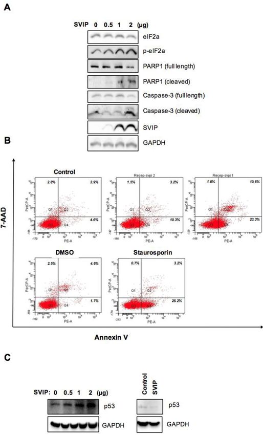
www.nature.com/scientificreports/
value) determined for each sample were analyzed with the QCt relative quantification method using the Qiagen
REST 2009 program. At least two independent biological replicates with three technical replicates per experi-
ment were used for each PCR.
Flow cytometry. Cell flow cytometry experiments were conducted using the PE Annexin V Apoptosis
Detection Kit I (BD Biosciences, 559763) according to the manufacturer’s instructions. Briefly, transfected cells
were suspended in 100 μl of Annexin V binding buffer and then 5 μl of FITC-conjugated Annexin V and 7-AAD
added to samples. After the incubation at room temperature for 15 min in the dark, the stained cells were exam-
ined using a FacsCanto Flow Cytometry (BD Bioscience, USA).
Hormone analysis. As stated in the OECD document and various literature, hormone analyses were per-
formed using H295R cells with 4 to 10 passages. Cells were grown on 6-well plates and transfected as indicated.
The media was replenished 6 or 24 h post-transfection of plasmid encoding SVIP or SVIP siRNA, respectively.
The growth media and cell lysates were collected 48 h later. Cortisol and DHEA levels were determined using
ELISA kits (Enzo Life Sciences, ADI-900-071 and ADI-900-093), and hormone concentrations were normalized
to the total protein concentration and calculated as a fold change17,18.
Statistical analysis. Data are presented as means ± standard deviation (SD). One-way ANOVA with post
hoc test or Student’s t-test was performed for statistical analyses by using GraphPad Prism software. The signifi-
cance threshold was accepted as p < 0.05.
Results
SVIP is highly expressed in adrenal gland. We first examined the tissue distribution of SVIP using 14
different mouse tissue extracts. Our results revealed that SVIP is highly expressed in the medulla spinalis and
adrenal gland in addition to the previously reported cerebrum, cerebellum, and sciatic n erve8 (Fig. 1A). The
expression of SVIP was particularly high in adult tissues but very low at postnatal day 2, 4, and 7 in the medulla
spinalis as well as the cerebrum, cerebellum, and sciatic nerve as previously reported, suggesting that SVIP may
function in one or many of the changes that occur during postnatal maturation of the central nervous system.
Interestingly, SVIP was expressed at a low level in a subset of other non-neuronal adult tissues other than the
adrenal gland. Unlike in the central nervous system, SVIP was highly expressed in the adrenal gland in all
postnatal development stages (Fig. 1B). A similar developmental expression pattern was observed in rat adrenal
gland, with a slight increase at day 60 (Fig. 1C).
Next, we determined the localization of SVIP in rat adrenal gland by immunohistochemistry (IHC). At post-
natal day 0, SVIP immunoreactivity was strongly identified in the inner zones but not in the outer layer of the
adrenal cortex, i.e. the zona glomerulosa (Fig. 2A). Weak SVIP immunoreactivity was observed in the adrenal
medulla. The strong expression pattern of SVIP in the cortex persisted at postnatal day 5 and 15 (Fig. 2B–F). At
60 days of postnatal age, SVIP expression was dominant in the cortex of the adrenal gland but weak expression
in the medulla of the adrenal gland (Fig. 2G–I). SVIP expression was found to be specific to the neuroendocrine
chromaffin cells of the adrenal medulla by day 60 (Fig. 2G,H). In line with IHC data, immunofluorescence labe-
ling of SVIP also showed that SVIP was mainly expressed in the inner zones of the rat adrenal cortex but not in
the zona glomerulosa (Fig. 2J,K). Together, these data indicate that SVIP is preferentially expressed in the zona
fasciculata/reticularis (ZF/R) of the rat adrenal gland.
After the initial SVIP expression screening in rodent organs and the detection of adrenal cortex as the only
tissue that expresses SVIP at all developmental stages, we aimed to analyze the role of SVIP in adrenal function.
However, there are significant differences between rodent and human adrenal physiology. Both the zonation and
the repertoire of steroidogenic enzymes vary between rodents and h uman19,20. Since the adrenal cortex of adult
mice and rats lacks CYP17A1, corticosterone is the principal glucocorticoid secreted, and adrenal androgens
are not biosynthesized in rodents. Thus, to further study the role and regulation of SVIP in adrenal cortex cells,
H295R human pluripotent adrenocortical cells were used as an in vitro model because they express all the ster-
oidogenic enzymes and have steroid hormone secretion and regulation pattern mimicking primary cultures of
adrenal cortex cells21. Immunofluorescence analysis revealed that SVIP was localized in numerous punctuated
structures on P60 rat adrenal cortex cells (Fig. 3A), and SVIP was also associated with various small and large
punctate structures in H295R cells (Fig. 3B,C). Furthermore, SVIP did not co-localize with CYP17A1 in H295R
cells (Supplementary Fig. 1).
Next, we analyzed the co-localization of SVIP with well-defined organelle markers. Analysis of merged
pictures obtained from double-labeling immunofluorescence showed no positive co-localizations of ectopic
SVIP with the ER marker PDI (protein disulfide isomerase) (Fig. 4A), the mitochondria marker AIF (apoptosis-
inducing factor) (Fig. 4B), the Golgi marker RCAS (receptor-binding cancer antigen expressed on SiSo cells)
(Fig. 4C), and the early endosome marker EEA1 (early endosome antigen 1) (Fig. 4D). On the other hand,
SVIP was found to be highly co-localized with the lysosome marker LAMP1 (lysosome-associated membrane
protein-1) (Fig. 4E). Furthermore, LAMP1 is normally localized in vesicles throughout the cells, while in cells
with high SVIP overexpression we observed lysosomal clustering in the juxtanuclear regions both in basal con-
ditions (Fig. 4E) and when steroidogenesis was stimulated with forskolin and angiotensin II (Ang II) (Fig. 4F).
Additionally, overexpression of SVIP caused cellular relocalization of p97/VCP in adrenal cortex cells (Fig. 4G).
SVIP is downregulated by stimulators of steroidogenesis in H295R cells. Forskolin or 8Br-cAMP
treatment causes differentiation of H295R cells into “zona fasciculata-like cells”, which in turn increases the
biosynthesis of cortisol and androgens via cAMP and the protein kinase A pathway22. Therefore, we incubated
Scientific Reports | (2022) 12:869 | https://doi.org/10.1038/s41598-022-04821-y 4
Vol:.(1234567890)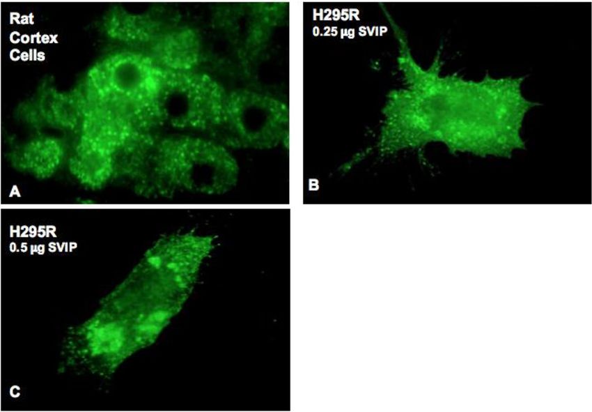
www.nature.com/scientificreports/
Figure 1. SVIP is highly expressed in adrenal gland. (A) Equal amounts of total protein from each organ
extract were analyzed by immunoblotting (IB) with antibodies against SVIP and p97/VCP. (B) Mouse tissues
were obtained at postnatal day 2, 4, 7, and 28 and processed for IB. (C) SVIP expression was investigated in
the adrenal glands of rats at the indicated ages. The left shows representative Western blot images and the right
shows the results of quantitative densitometric analysis of the changes in SVIP protein expression. Error bars
indicate standard deviation (*p < 0.05).
H295R human adrenocortical cells were incubated with 10 µM forskolin or 0.5 mM 8-Br-cAMP for increasing
time periods up to 24 h and evaluated SVIP expression by immunoblotting. Both treatments decreased the lev-
els of SVIP protein while upregulating StAR (steroidogenic acute regulatory protein) (Fig. 5A,B), a protein that
shuttles cholesterol from the outer to the inner mitochondrial membranes in a rate-limiting s tep23. H295R cells
can also be differentiated into more “glomerulosa-like cells” and synthesize more aldosterone via pre-treatment
with angiotensin II (Ang II) or potassium chloride (KCl)24. Our results showed that both KCl and Ang II also
decreased SVIP protein levels similar to forskolin or 8Br-cAMP (Fig. 5C,D).
In order to substantiate our findings and determine the mechanism underlying the downregulation of
SVIP protein levels, we also evaluated the transcriptional regulation of SVIP by steroidogenic stimulators. Our
data revealed that all tested stimulators diminished the SVIP mRNA compared to vehicle-treated control cells
Scientific Reports | (2022) 12:869 | https://doi.org/10.1038/s41598-022-04821-y 5
Vol.:(0123456789)www.nature.com/scientificreports/
Figure 2. Distribution of SVIP in the rat adrenal gland during the postnatal development. SVIP
immunoreactivity was detected in the rat adrenal gland at postnatal day 0 (A), 5 (B,C), 15 (D–F), and 60 (G–I)
by IHC. Strong expression of SVIP is observed in the cortex (C) compared to medulla (M) in postnatal rat
adrenal glands. SVIP immunopositivity is seen in the secretory vesicles (arrows) of adrenal cortex cells (arrows,
F and I). SVIP localization was also determined by immunofluorescence labeling in the rat adrenal gland at
postnatal day 15 (J), 30 (K), and 60 (L). Scale bars: 200 µm (A,B,D,G,J,K); 100 µm (C,H); 20 µm (E,F,I,L).
(Fig. 5E). Next, we carried out cycloheximide chase assay to determine whether SVIP protein stability is regulated
by forskolin. Indeed, in addition to the downregulation of SVIP mRNA levels, forskolin treatment enhanced SVIP
protein degradation (Fig. 5F). These findings indicate that steroidogenic stimulators reduce SVIP transcription
as well as accelerate SVIP protein degradation.
Next, we sought to determine whether stimulating steroidogenesis could also alter the expression levels of
ubiquitin ligase gp78 and retrotranslocation protein p97/VCP, two proteins reported to physically and func-
tionally interact with SVIP. We found that forskolin treatment up to 12 h augmented expression of gp78 and
p97/VCP, suggesting that steroidogenic stimulator treatment physiologically regulates the levels of the ERAD
components (Fig. 5G).
SVIP regulates cortisol and DHEA secretion via modulation of steroidogenesis‑related protein
levels. As SVIP levels were regulated by steroidogenic stimulators, we next investigated the effect of SVIP on
steroid hormone secretion. Surprisingly, the cortisol secretion was significantly augmented in a dose-dependent
manner up to certain SVIP overexpression level, but the highest tested SVIP level lost its ability to increase the
cortisol levels (Fig. 6A). Concomitantly, when SVIP expression was decreased by RNAi, basal cortisol secretion
was significantly diminished (Fig. 6B). We also investigated the effect of SVIP on other major steroid hormones
Scientific Reports | (2022) 12:869 | https://doi.org/10.1038/s41598-022-04821-y 6
Vol:.(1234567890)www.nature.com/scientificreports/
Figure 3. SVIP is localized at punctate structures as juxtanuclear vacuoles in rat adrenal cortex cells and H295R
cell line. (A) Location of endogenous SVIP was determined in postnatal day 60 rat adrenal cortex cells by
immunostaining using confocal microscopy. (B,C) H295R cells were transfected with (B) 0.25 μg or (C) 0.5 μg
pCIneo-SVIP-His plasmid, then cells were stained with anti-His antibody.
such as aldosterone and DHEA. While aldosterone levels were not changed by SVIP overexpression (Fig. 6D),
our data revealed that SVIP overexpression moderately enhanced DHEA biosynthesis (Fig. 6C). Furthermore,
when the cumulative effect was investigated, SVIP was found to further enhance forskolin-stimulated cortisol
biosynthesis (Supplementary Fig. 2). Together, these data suggest that SVIP regulates adrenal cortisol and DHEA
pathways but not the aldosterone biosynthesis pathway.
Steroid hormones are essential signaling molecules that regulate multiple physiological processes. In adrenal
steroidogenesis, the biosynthesis of cortisol and DHEA occurs from cholesterol via the concerted action of P450
heme-containing monooxygenases (CYPs) and 3β-hydroxysteroid dehydrogenase (3β-HSD) enzymes in adrenal
cortex25–27. Furthermore, 3-hydroxy-3-methylglutary coenzyme A reductase (HMGR) and low-density lipopro-
tein receptor (LDLR) are equally important in assuring sufficient amounts of cholesterol for steroid hormone
production28,29. Since the transcriptional regulation of these genes is highly studied, we first analyzed the effect of
SVIP overexpression in mRNA expression levels of some key genes involved in cholesterol uptake, biosynthesis
or mobilization and steroidogenesis. Overexpression of SVIP did not significantly change the mRNA levels of
CYP11A1, CYP11B1, CYP17A1, or HMGR levels. LDLR and CYP21A2 mRNA levels were slightly increased,
while HSD3β2 mRNA levels showed almost two-fold upregulation with SVIP overexpression (Fig. 7A).
SVIP has been reported to be involved in cellular protein degradation by regulating ERAD and autophagy6,8,9.
Therefore, we also determined levels of the key proteins involved in steroid hormone biosynthesis. While a certain
degree of SVIP overexpression consistently enhanced StAR, CYP11A1, CYP11B1, CYP17A1, HSD3β2, HMGR,
and LDLR protein levels, its high overexpression resulted in a significant downregulation of all of these proteins
except HSD3β2 (Fig. 7B), consistent with the increase in HSD3β2 mRNA levels observed previously (Fig. 7A).
HSD3β2 protein level was found to be augmented even at higher SVIP overexpression level. Moreover, SVIP
overexpression further enhanced forskolin-stimulated CYP17A1 and StAR levels even at higher concentrations,
suggesting the high level of induction obtained with forskolin masks the inhibitory effect of high levels of SVIP
(Supplementary Fig. 3). On the other hand, the protein levels of StAR, CYP11A1, CYP11B1, CYP17A1, HSD3β2,
HMGR, and LDLR were significantly decreased by silencing SVIP in H295R cells (Fig. 7C), which is well cor-
related with cortisol secretion data (Fig. 6).
Since increased SVIP expression enhanced the levels of tested proteins but did not affect their transcription
levels, except for HSD3β2, LDLR, and CYP21A2, we next investigated the effect of SVIP mainly on the deg-
radation rate of CYP17A1, an enzyme that catalyzes the key branching point in the adrenal steroid hormone
synthesis pathway towards cortisol and DHEA biosynthesis. Indeed, CYP17A1 degradation was found to be
slowed even at high SVIP overexpression levels in the cycloheximide chase assay (Fig. 7D). Thus, our data showed
that consistent with its role as ERAD inhibitor, SVIP overexpression decreased the turnover rate of CYP17A1.
Scientific Reports | (2022) 12:869 | https://doi.org/10.1038/s41598-022-04821-y 7
Vol.:(0123456789)www.nature.com/scientificreports/
Figure 4. SVIP is co-localized with lysosomes and p97/VCP. H295R cells were transfected with 0.5 μg pCIneo-
SVIP-His plasmid, then cells were co-stained with anti-His antibody and specific antibodies against organelle
markers: (A) PDI (ER marker), (B) AIF (mitochondria marker), (C) RCAS (Golgi marker), (D) EEA1 (early
endosomes marker), and (E) LAMP1 (lysosomal marker). (F) SVIP overexpressed cells were treated with 0.1 μM
angiotensin II or 10 μM forskolin for 24 h and double immunostaining of SVIP and LAMP1 was performed. (G)
SVIP relocalized p97/VCP. Images were taken after double immunostaining with anti-His antibody for SVIP
and anti-VCP antibody for endogenous p97/VCP.
Scientific Reports | (2022) 12:869 | https://doi.org/10.1038/s41598-022-04821-y 8
Vol:.(1234567890)www.nature.com/scientificreports/
Figure 4. (continued)
Scientific Reports | (2022) 12:869 | https://doi.org/10.1038/s41598-022-04821-y 9
Vol.:(0123456789)www.nature.com/scientificreports/
Figure 5. Stimulation of steroidogenesis downregulates SVIP. H295R cells were treated with (A) 10 μM
forskolin, (B) 0.5 mM 8Br-cAMP, (C) 0.1 μM angiotensin II or (D) 14 mM KCl for the indicated times and SVIP
protein levels were determined via immunoblotting (IB). Actin antibody was hybridized to the same membranes
to verify equal protein loading. (E) SVIP mRNA was quantified by RT-qPCR. The experiment was repeated
twice with at least three replicates. Error bars represent standard error (*p < 0.05). (F) The degradation of SVIP
was analyzed by cycloheximide (CHX) chase on forskolin or vehicle-treated H295R cells. The SVIP and StAR
levels were determined via IB (l.e.: longer exposure). Densitometric analysis of SVIP levels by ImageQuant is
shown in the right panel (mean ± S.D., n = 3) (*p < 0.05 and **p < 0.01) (G) Following treatment with forskolin
for the indicated times, expression levels of gp78, p97/VCP, SVIP, and StAR were detected by IB using antibodies
against them. Actin was used as a loading control.
Scientific Reports | (2022) 12:869 | https://doi.org/10.1038/s41598-022-04821-y 10
Vol:.(1234567890)www.nature.com/scientificreports/
Figure 5. (continued)
These results further suggest that the downregulation of proteins at high SVIP levels is not due to the enhanced
protein degradation.
One possible explanation for the decreased protein levels in cells that express SVIP above a certain level
might be global translational attenuation. Therefore, we next sought to investigate the phosphorylation of eIF2α,
which is the best characterized mechanism for the regulation of translational initiation30. We observed that SVIP
overexpression increased p-eIF2α levels in a dose-dependent manner (Fig. 8A). Furthermore, SVIP enhanced the
levels of cleaved caspase-3 and PARP-1 levels implying that high SVIP expression promoted apoptosis in H295R
cells (Fig. 8A). Apoptosis of cells transfected with SVIP expressing plasmid was significantly higher than that
in vector-transfected control cells (Fig. 8B). Additionally, apoptotic morphology was observed in SVIP overex-
pressing cells (Supplementary Fig. 4). Because it was previously reported that SVIP as an ERAD inhibitor may
delay the degradation of tumor suppressor p 5314, which regulates the cell cycle and acts as guardian of genome
stability, we next evaluated the effect of SVIP on the p53 levels in H295R cells and found that p53 levels were
increased with SVIP overexpression and decreased with SVIP silencing (Fig. 8C). In summary, our data suggest
that exaggerated SVIP expression levels triggers caspase-dependent apoptosis along with an increase in eIF2α
phosphorylation of and p53 abolishment in H295R cells.
Discussion
ERAD is a well-characterized protein quality control mechanism functioning in the degradation of misfolded ER
proteins. It also plays a role in the destruction of some properly folded proteins, the best-known example being
HMGR, which is the key enzyme of the sterol biosynthesis p athway31–33. The degradation of HMGR by ERAD
is a vital feedback inhibition system for sterol homeostasis both in yeast and mammalian cells. Several ERAD
ubiquitin ligases such as gp78, TRC8, Hrd1, March6, and RNF145 have been demonstrated to play a role in the
turnover of HMGR34,35. Besides HMGR, other proteins involved in sterol biosynthesis, such as Insig proteins
and squalene monooxygenase are reported to be ERAD substrates32,36. It has also indicated that gp78 medi-
ates the degradation of apolipoprotein B100, one of the main LDL-VLDL lipoproteins involved in cholesterol
transport37. Further evidence that ERAD has a conserved role in sterol regulation is the biosynthesis of sterols
Scientific Reports | (2022) 12:869 | https://doi.org/10.1038/s41598-022-04821-y 11
Vol.:(0123456789)www.nature.com/scientificreports/
Figure 6. SVIP biphasically regulates cortisol and DHEA secretion in H295R cell line. (A) H295R cells
were transfected with 0.25, 0.5, 0.75, and 1.5 μg SVIP. Six hours later, the cell culture media was replenished
and both cells and cell culture supernatant were harvested after 48 h. The amount of cortisol in the growth
media was quantified by ELISA. Cortisol amounts were normalized to the cellular protein concentration and
presented as fold change compared to the cells that did not overexpress SVIP. The data graphed represent the
mean ± SD. SVIP expression levels were determined by IB. GAPDH was used as loading control. (B) H295R
cells were transfected with control or SVIP siRNA. At 24 h after transfection, fresh growth media was added.
Both cells and cell culture supernatant were harvested 48 h later. Cortisol measurements and evaluation of
SVIP expression levels were done as described for (A). (C,D) H295R cells were transfected with 0.5, 0.75, and
1.0 μg SVIP and processed as described for (A). The amount of DHEA (C) or aldosterone (D) in growth media
was quantified by ELISA. Hormone levels were normalized to the cellular protein concentration and presented
as fold change compared to cells that did not overexpress SVIP. The data graphed represent the mean ± SD
(*p < 0.05, **p < 0.001, n = 3).
Scientific Reports | (2022) 12:869 | https://doi.org/10.1038/s41598-022-04821-y 12
Vol:.(1234567890)www.nature.com/scientificreports/
and sterol-derived metabolites in plants38. Therefore, in addition to protein quality control, ERAD has emerged
as a key mechanism that controls the flux of metabolic pathways by altering the abundance of sterol biosynthetic
enzymes and their regulators. Considering its role in lipid and protein homeostasis, tight regulation of ERAD
is critical for cells. Although there are several diverse ERAD branches, they all converge on the cytosolic p97/
VCP ATPase complex, which extracts ubiquitinated substrates from the ER membrane and delivers them to the
proteasome for degradation39. Therefore, SVIP is notable as an ERAD component for being the first identified
endogenous inhibitor of ERAD through its interaction with p97/VCP.
As the adaptor protein for multifunctional p97/VCP, SVIP has also been shown to be involved in multiple
cellular processes such as the regulation of vacuole formation, autophagy, and ERAD inhibition4,6,8. In an SVIP
expression screening assay, we showed that unlike its gradually increasing expression in the developing nervous
tissues such as cerebrum, cerebellum, sciatic nerve, and medulla spinalis, SVIP expression in the adrenal gland
remained at a consistently high level during postnatal development, suggesting that SVIP may function through-
out the developmental process of the adrenal gland. Our immunohistochemistry and double immunofluorescence
data further demonstrated that SVIP expression is not only organ specific but also differs between the regions of
the rat adrenal gland, where SVIP is exclusively expressed in the adrenal cortex (Fig. 2). Since the adrenal cortex
was the only tissue found to express SVIP at all developmental stages, we aimed to analyze the role of SVIP in
adrenal function. Importantly, there are significant differences between rodent and human adrenal physiology,
mainly in terms of the zonation and the repertoire of steroidogenic e nzymes19,20. Thus, to further study the role
and regulation of SVIP in adrenal cortex cells, we utilized the H295R pluripotent adrenocarcinoma cell line, one
of the best characterized cellular models for studying adrenal cortex cell biology. This cell line expresses genes that
encode all the key enzymes for steroidogenesis, having the physiological characteristics of zonally undifferentiated
human adrenal cells and producing all of the steroid hormones found in the adult adrenal cortex24. We observed
that SVIP localized as punctuated structures both in rat adrenal cortex cells and H295R cells (Fig. 3). Further-
more, we demonstrated that the expression of ectopic SVIP displays co-localization with lysosomal membrane
protein LAMP1 and causes lysosomal clustering (Fig. 4). As shown previously in HeLa c ells8, overexpression of
SVIP relocalizes p97/VCP from the cytosol to the juxtanuclear vacuoles in H295R cells as well. Very recently,
Drosophila SVIP was also shown to recruit p97/VCP to lysosomes in muscle c ells40.
Adrenal steroidogenesis is a tightly regulated dynamic process, as adrenal steroid hormones are key regulators
of a wide variety of physiological functions including blood pressure, inflammation, glucose metabolism, and
stress41–43. Pre-synthesized hormones are not stored for immediate release and de novo biosynthesis of adrenal
steroid is controlled by multiple regulatory mechanisms (e.g., transcriptional, post-translational and substrate
transportation)44. Downregulation of SVIP both at the transcriptional and post-translational levels by steroido-
genic stimulating agents in H295R cells suggests that SVIP may have a role in the steroid hormone biosynthesis
pathway. However, when the effect of SVIP on steroid hormone levels was determined, SVIP overexpression up
to certain levels caused an increase in cortisol and DHEA secretion while SVIP silencing diminished cortisol
levels. This unprecedented result may suggest that SVIP plays a critical role in the termination of intracellular
signaling events triggered by steroidogenic stimulators.
Besides steroidogenic enzymes, proteins involved in cholesterol biosynthesis and uptake are also regulatory
factors of steroid hormone production45,46. Our data clearly demonstrate that the change in cortisol secretion
capability of H295R cells via SVIP expression levels is highly correlated with the modulation of the protein levels
such as StAR, HMGR, and CYP17A1. The downregulation of SVIP by steroidogenic stimulators and diminished
levels of key proteins accompanied by a decrease in cortisol secretion by silencing of SVIP expression provide
further evidence of the involvement of SVIP in the termination of steroid hormone biosynthesis signals in
adrenal cells to prevent exaggerated steroidogenesis. As a glucocorticoid, cortisol biosynthesis is regulated by
adrenocorticotropin (ACTH) in addition to the general regulatory mechanism. As cortisol is an overall catabolic
hormone, excess cortisol reduces lean body mass, and muscle mass, induces insulin resistance, and may increase
energy expenditure. Moreover, Cushing’s syndrome is one of the well-known physiological disorders associated
with excessive cortisol biosynthesis47. Thus, negative regulation of cortisol biosynthesis is important, and several
negative feedback mechanisms have been reported, such as hypothalamic–pituitary–adrenal axis activity and
decreased ACTH secretion via cortisol48,49. Nevertheless, our findings strongly suggest that modulation of SVIP
expression may function as a novel regulatory mechanism of adrenal steroid biosynthesis.
Notably, we also found that the regulation of SVIP protein levels is extremely critical in adrenal cortex, where
at immense overexpression of SVIP led to reduced levels of steroidogenic proteins along with diminished cortisol
production (Fig. 7A). Our findings that SVIP at this high concentration still decreased the degradation rate of
CYP17A1, which catalyzes the key branching point in adrenal steroid hormone synthesis pathway towards corti-
sol and DHEA biosynthesis (Fig. 7D), proposed that the opposite steroidogenic response at high SVIP levels was
not directly linked to regulation of the degradation rate of tested proteins. Therefore, we hypothesized that over-
expression of SVIP at high levels might impose stress on the ER via ERAD inhibition, causing the accumulation
of unfolded or misfolded proteins in the ER lumen. To restore the normal ER functions, cells launch the Unfolded
Protein Response (UPR), which primarily involves attenuating general protein synthesis, increasing the lumenal
folding capacity, and increasing the degradation of misfolded proteins through ERAD or autophagy to resolve
the ER o verload50,51. In response to ER stress, cells attenuate global translation through PERK-mediated eIF2α
phosphorylation52. Our results also demonstrated that exaggerated expression of SVIP significantly increased
levels of p-eIF2α, which functions in translational attenuation during ER stress. Therefore, we speculate that the
downregulation of the tested steroidogenic proteins at high SVIP levels may be due to inhibition of translation,
but further experiments such as metabolic pulse labeling of newly synthesized proteins are required to confirm
the relation between SVIP and translational attenuation in adrenal cortex cells.
When ER stress is not resolved, the prolonged UPR induces apoptosis to remove the stressed cells from the
organism53. Interestingly, prolonged ER stress associated with the accumulation of misfolded proteins in the ER
Scientific Reports | (2022) 12:869 | https://doi.org/10.1038/s41598-022-04821-y 13
Vol.:(0123456789)www.nature.com/scientificreports/
Figure 7. SVIP modulates the expression levels of several proteins related to steroidogenesis. (A) After ▸
H295R cells were transfected with 0.75 μg SVIP, mRNA levels of CYP17A1, CYP11A1, CYP11B1, HSD3β2,
LDLR, HMGR, CYP21A2, and SVIP were examined by RT-qPCR. The experiment was repeated at least three
times with three replicates. Error bars represent standard error (*p < 0.05 and ***p < 0.001) (B and C) Protein
expression levels of CYP17A1, CYP11A1, CYP11B1, HSD3β2, LDLR, StAR, HMGR, and SVIP were determined
by IB of cells transfected with (B) 0.5 and 1.5 μg plasmid encoding SVIP or (C) SVIP siRNA. (D) Cells were
transfected with 1.5 μg SVIP and then treated with cycloheximide for the indicated times. CYP17A1 and SVIP
levels were determined via IB. The degradation rates of substrates were calculated using three independent
experiments (*p < 0.05). GAPDH was used as loading control.
was shown to significantly upregulate SVIP6. It has also been shown that overexpression of SVIP increased the
levels of tumor suppressor p53, which regulates the cell cycle and leads to apoptosis14. Our data also indicated
that high SVIP levels promotes apoptosis in H295R cells. Considering a previous report indicating that mito-
chondrial dysfunction contributes to the decline of steroidogenesis in human granulosa c ells54, it is plausible to
speculate that SVIP-mediated induction of apoptosis may also decrease cortisol synthesis via apoptosis-related
mitochondrial dysfunction.
Considering the role of ubiquitin ligase gp78 in ERAD and cholesterol biosynthesis together with its func-
tional inhibition by SVIP, it is highly possible that gp78 may have a role in steroid hormone biosynthesis. There-
fore, it is essential to further investigate the regulation of adrenal steroidogenesis by ERAD and its interplay with
the activities of other organelles (such as mitochondria), since the biosynthesis of cortisol requires the activities of
enzymes between the ER and mitochondria. Interestingly, p97/VCP functions in the extraction of ubiquitinated
proteins from the outer mitochondrial membrane and presents ubiquitinated proteins to the proteasome for
degradation55. Therefore, by removing p97/VCP from its functional complex, SVIP may also affect mitochondrial
protein degradation. Proteomics and metabolomics analyses utilizing SILAC assay have consistently pointed out
that epigenetic loss of SVIP is associated with depletion of some mitochondrial enzymes and oxidative respiration
activity which reverts upon SVIP restoration15.
In conclusion, our results suggest that modulation of SVIP expression alters not only the expression levels
of steroidogenic genes and hormone output, but also the expression of genes required for de novo cholesterol
biosynthesis, uptake and trafficking. Furthermore, SVIP constitutes a double-edged sword in adrenal steroido-
genesis: on one hand, SVIP positively regulates steroid hormone biosynthesis, while on the other, excessive SVIP
expression induces apoptosis. Our study may suggest a key link between ERAD and sterol biology by providing
evidence of the role of SVIP in adrenal hormone biosynthesis.
Scientific Reports | (2022) 12:869 | https://doi.org/10.1038/s41598-022-04821-y 14
Vol:.(1234567890)www.nature.com/scientificreports/
Scientific Reports | (2022) 12:869 | https://doi.org/10.1038/s41598-022-04821-y 15
Vol.:(0123456789)www.nature.com/scientificreports/
Figure 7. (continued)
Scientific Reports | (2022) 12:869 | https://doi.org/10.1038/s41598-022-04821-y 16
Vol:.(1234567890)www.nature.com/scientificreports/
Figure 8. High expression of SVIP induces apoptosis in the H295R cell line. (A) H295R cells were transfected as indicated
and harvested 24 h post-transfection. The expression levels of the proteins of interest were investigated using IB. (B) H295R
cells were stained by Annexin V/7-AAD and analyzed via flow cytometry 24 h after transfection. Additionally, 1 μM
staurosporine (Sta) was used to induce apoptosis and DMSO treatment was used as its control. (C) H295R cells transfected
with plasmid encoding SVIP (left) or SVIP siRNA (right). The p53 protein levels were investigated in H295R cells by IB.
GAPDH was used as loading control.
Scientific Reports | (2022) 12:869 | https://doi.org/10.1038/s41598-022-04821-y 17
Vol.:(0123456789)www.nature.com/scientificreports/
Data availability
The data that support the findings of this study are available from the corresponding author (PBK) upon reason-
able request.
Received: 12 July 2021; Accepted: 31 December 2021
References
1. Meyer, H. & Weihl, C. C. The VCP/p97 system at a glance: Connecting cellular function to disease pathogenesis. J. Cell Sci. 127,
3877–3883 (2014).
2. Zhong, X. et al. AAA ATPase p97/valosin-containing protein interacts with gp78, a ubiquitin ligase for endoplasmic reticulum-
associated degradation. J. Biol. Chem. 279, 45676–45684 (2004).
3. Yamanaka, K., Sasagawa, Y. & Ogura, T. Recent advances in p97/VCP/Cdc48 cellular functions. Biochim. Biophys. Acta Mol. Cell
Res. 1823, 130–137 (2012).
4. Nagahama, M. et al. SVIP is a novel VCP/p97-interacting protein whose expression causes cell vacuolation. Mol. Biol. Cell 14,
262–273 (2003).
5. Ballar, P., Shen, Y., Yang, H. & Fang, S. The role of a novel p97/valosin-containing protein-interacting motif of gp78 in endoplasmic
reticulum-associated degradation. J. Biol. Chem. 281, 35359–35368 (2006).
6. Ballar, P. et al. Identification of SVIP as an endogenous inhibitor of endoplasmic reticulum-associated degradation. J. Biol. Chem.
282, 33908–33914 (2007).
7. Ballar, P., Ors, A. U., Yang, H. & Fang, S. Differential regulation of CFTRΔF508 degradation by ubiquitin ligases gp78 and Hrd1.
Int. J. Biochem. Cell Biol. 42, 167–173 (2010).
8. Wang, Y. et al. SVIP induces localization of p97/VCP to the plasma and lysosomal membranes and regulates autophagy. PLoS ONE
6, e24478 (2011).
9. Jia, D. et al. SVIP alleviates CCl 4-induced liver fibrosis via activating autophagy and protecting hepatocytes. Cell Death Dis. 10,
71 (2019).
10. Rahim, A. et al. Proteomic analysis of the very low density lipoprotein (VLDL) transport vesicles. J. Proteomics 75, 2225–2235
(2012).
11. Tiwari, S., Siddiqi, S., Zhelyabovska, O. & Siddiqi, S. A. Silencing of small valosin-containing protein-interacting protein (SVIP)
reduces very low density lipoprotein (VLDL) secretion from rat hepatocytes by disrupting its endoplasmic reticulum (ER)-to-Golgi
trafficking. J. Biol. Chem. 291, 12514–12526 (2016).
12. Wu, J., Peng, D., Voehler, M., Sanders, C. R. & Li, J. Structure and expression of a novel compact myelin protein—small VCP-
interacting protein (SVIP). Biochem. Biophys. Res. Commun. 440, 173–178 (2013).
13. Erzurumlu, Y. & Ballar, P. Androgen mediated regulation of endoplasmic reticulum-associated degradation and its effects on
prostate cancer. Sci. Rep. 7, 40719 (2017).
14. Bao, D. et al. Regulation of p53wt glioma cell proliferation by androgen receptor-mediated inhibition of small VCP/p97-interacting
protein expression. Oncotarget 8, 23142–23154 (2017).
15. Llinàs-Arias, P. et al. Epigenetic loss of the endoplasmic reticulum-associated degradation inhibitor SVIP induces cancer cell
metabolic reprogramming. JCI Insight 4(8), e125888 (2019).
16. Cayli, S. et al. Developmental expression of p97/VCP (Valosin-containing protein) and Jab1/CSN5 in the rat testis and epididymis.
Reprod. Biol. Endocrinol. 9, 117 (2011).
17. Felizola, S. J. A. et al. PCP4: A regulator of aldosterone synthesis in human adrenocortical tissues. J. Mol. Endocrinol. 52, 159–167
(2014).
18. Lu, J. Y. & Sewer, M. B. p54 nrb /NONO regulates cyclic AMP-dependent glucocorticoid production by modulating phosphodi-
esterase mRNA splicing and degradation. Mol. Cell. Biol. 35, 1223–1237 (2015).
19. Yates, R. et al. Adrenocortical development, maintenance, and disease. Curr. Top. Dev. Biol. 106, 239–312 (2013).
20. Pihlajoki, M., Dörner, J., Cochran, R. S., Heikinheimo, M. & Wilson, D. B. Adrenocortical zonation, renewal, and remodeling.
Front. Endocrinol. 6, 27 (2015).
21. Romero, D. G. et al. Adrenal transcription regulatory genes modulated by angiotensin II and their role in steroidogenesis. Physiol.
Genomics 30, 26–34 (2007).
22. Oskarsson, A., Ullerås, E., Plant, K. E., Hinson, J. P. & Goldfarb, P. S. Steroidogenic gene expression in H295R cells and the human
adrenal gland: Adrenotoxic effects of lindane in vitro. J. Appl. Toxicol. 26, 484–492 (2006).
23. Christenson, L. K. & Strauss, J. F. Steroidogenic acute regulatory protein: An update on its regulation and mechanism of action.
Arch. Med. Res. 32, 576–586 (2001).
24. Rainey, W. E., Saner, K. & Schimmer, B. P. Adrenocortical cell lines. Mol. Cell. Endocrinol. 228, 23–38 (2004).
25. Miller, W. Steroidogenic enzymes. Endocr. Dev. 13, 1–18 (2008).
26. Payne, A. H. & Hales, D. B. Overview of steroidogenic enzymes in the pathway from cholesterol to active steroid hormones. Endocr.
Rev. 25, 947–970 (2004).
27. Sewer, M. B., Dammer, E. B. & Jagarlapudi, S. Transcriptional regulation of adrenocortical steroidogenic gene expression. Drug
Metab. Rev. 39, 371–388 (2007).
28. Brown, M. S. & Goldstein, J. L. How LDL receptors influence cholesterol and atherosclerosis. Sci. Am. 251, 58–69 (1984).
29. Friesen, J. A. & Rodwell, V. W. The 3-hydroxy-3-methylglutaryl coenzyme-A (HMG-CoA) reductases. Genome Biol. 5, 248 (2004).
30. De Haro, C., Méndez, R. & Santoyo, J. The eIF-2α kinases and the control of protein synthesis1. FASEB J. 10, 1378–1387 (1996).
31. Hampton, R. Y., Gardner, R. G. & Rine, J. Role of 26S proteasome and HRD genes in the degradation of 3-hydroxy-3-methylglu-
taryl-CoA reductase, an integral endoplasmic reticulum membrane protein. Mol. Biol. Cell 7, 2029–2044 (1996).
32. Song, B. L., Sever, N. & DeBose-Boyd, R. A. Gp78, a membrane-anchored ubiquitin ligase, associates with Insig-1 and couples
sterol-regulated ubiquitination to degradation of HMG CoA reductase. Mol. Cell 19, 829–840 (2005).
33. Cao, J. et al. Ufd1 is a cofactor of gp78 and plays a key role in cholesterol metabolism by regulating the stability of HMG-CoA
reductase. Cell Metab. 6, 115–128 (2007).
34. Menzies, S. A. et al. The sterol-responsive RNF145 E3 ubiquitin ligase mediates the degradation of HMG-CoA reductase together
with gp78 and hrd1. Elife 7, 1–30 (2018).
35. Zelcer, N. et al. The E3 ubiquitin ligase MARCH6 degrades squalene monooxygenase and affects 3-hydroxy-3-methyl-glutaryl
coenzyme A reductase and the cholesterol synthesis pathway. Mol. Cell. Biol. 34, 1262–1270 (2014).
36. Foresti, O., Ruggiano, A., Hannibal-Bach, H. K., Ejsing, C. S. & Carvalho, P. Sterol homeostasis requires regulated degradation of
squalene monooxygenase by the ubiquitin ligase Doa10/Teb4. Elife 2, e00953 (2013).
37. Liang, J. S. et al. Overexpression of the tumor autocrine motility factor receptor Gp78, a ubiquitin protein ligase, results in increased
ubiquitinylation and decreased secretion of apolipoprotein B100 in HepG2 cells. J. Biol. Chem. 278, 23984–23988 (2003).
38. Doblas, V. G. et al. The SUD1 gene encodes a putative E3 ubiquitin ligase and is a positive regulator of 3-hydroxy-3-methylglutaryl
coenzyme a reductase activity in Arabidopsis. Plant Cell 25, 728–743 (2013).
Scientific Reports | (2022) 12:869 | https://doi.org/10.1038/s41598-022-04821-y 18
Vol:.(1234567890)www.nature.com/scientificreports/
39. Wu, X. & Rapoport, T. A. Mechanistic insights into ER-associated protein degradation. Curr. Opin. Cell Biol. 53, 22–28 (2018).
40. Johnson, A. E. et al. SVIP is a molecular determinant of lysosomal dynamic stability, neurodegeneration and lifespan. Nat. Com-
mun. 12, 513 (2021).
41. Timmermans, S., Souffriau, J. & Libert, C. A general introduction to glucocorticoid biology. Front. Immunol. 10, 1545 (2019).
42. Bollag, W. B. Regulation of aldosterone synthesis and secretion. Compr. Physiol. 3, 1017–1055 (2014).
43. Kuo, T., McQueen, A., Chen, T. C. & Wang, J. C. Regulation of glucose homeostasis by glucocorticoids. Adv. Exp. Med. Biol. 872,
99–126 (2015).
44. Turcu, A. F. & Auchus, R. J. Adrenal steroidogenesis and congenital adrenal hyperplasia. Endocrinol. Metab. Clin. North Am. 44,
275–296 (2015).
45. Miller, W. L. & Bose, H. S. Early steps in steroidogenesis: Intracellular cholesterol trafficking. J. Lipid Res. 52, 2111–2135 (2011).
46. van der Sluis, R. J., Van Eck, M. & Hoekstra, M. Adrenocortical LDL receptor function negatively influences glucocorticoid output.
J. Endocrinol. 226, 145–154 (2015).
47. Christiansen, J. J. et al. Effects of cortisol on carbohydrate, lipid, and protein metabolism: Studies of acute cortisol withdrawal in
adrenocortical failure. J. Clin. Endocrinol. Metab. 92, 3553–3559 (2007).
48. Faghih, R. T., Savla, K., Dahleh, M. A. & Brown, E. N. A feedback control model for cortisol secretion. In Annu. Int. Conf. IEEE
Eng. Med. Biol. Soc. IEEE Eng. Med. Biol. Soc. Annu. Int. Conf. Vol. 2011, 716–719 (2011).
49. Ramamoorthy, S. & Cidlowski, J. A. Corticosteroids: Mechanisms of action in health and disease. Rheum. Dis. Clin. North Am.
42, 15–31 (2016).
50. Mohammed Thangameeran, S. I. et al. A role for endoplasmic reticulum stress in intracerebral hemorrhage. Cells 9, 750 (2020).
51. Tsai, Y. C. & Weissman, A. M. The unfolded protein response, degradation from the endoplasmic reticulum, and cancer. Genes
Cancer 1, 764–778 (2010).
52. Back, S. H. et al. Translation attenuation through eIF2α phosphorylation prevents oxidative stress and maintains the differentiated
state in β cells. Cell Metab. 10, 13–26 (2009).
53. Fribley, A., Zhang, K. & Kaufman, R. J. Regulation of apoptosis by the unfolded protein response BT—apoptosis: methods and
protocols, second edition. In (eds Erhardt, P. & Toth, A.) 191–204 (Humana Press, 2009). https://doi.org/10.1007/978-1-60327-
017-5_14.
54. Sreerangaraja Urs, D. B. et al. Mitochondrial function in modulating human granulosa cell steroidogenesis and female fertility.
Int. J. Mol. Sci. 21, 3592 (2020).
55. Taylor, E. B. & Rutter, J. Mitochondrial quality control by the ubiquitin–proteasome system. Biochem. Soc. Trans. 39, 1509–1513
(2011).
Acknowledgements
This research was funded by the Scientific and Technological Research Council of Turkey (TUBITAK, SBAG-
116S444) and by Ege University internal funds (16/ECZ/006). We thank the Pharmaceutical Sciences Research
Centre (FABAL, Ege University, Faculty of Pharmacy) and Biotechnology and Bioengineering Application and
Research Centre (BIYOMER, İzmir Institute of Technology) for equipment support, Burcu ERBAYKENT TEPE-
DELEN, Selin GÜNAL, and Aysegül KAYMAK for their technical assistance and scientific support. The authors
would also like to thank Jacqueline Gutenkunst at Gözen Translation & Editing (http://www.gozen.net) for her
assistance in editing the manuscript.
Author contributions
R.I. carried out most of the experiments and analyzed data. G.U. designed experiments related to apoptosis and
the dose-dependent effect of SVIP on steroidogenesis and co-wrote the manuscript. S.Y., G.U., and R.I. contrib-
uted in steroidogenic protein level detection. E.A.S. and R.I. contributed to cortisol secretion experiments. S.C.
carried out immunohistochemical analysis. Y.E. and O.G. contributed to mouse organ screening and optimization
of ELISA and RT-PCR experiments. P.B.K. conceived the study, took the lead in writing the manuscript, and was
in charge of overall direction and planning.
Competing interests
The authors declare no competing interests.
Additional information
Supplementary Information The online version contains supplementary material available at https://doi.org/
10.1038/s41598-022-04821-y.
Correspondence and requests for materials should be addressed to P.B.K.
Reprints and permissions information is available at www.nature.com/reprints.
Publisher’s note Springer Nature remains neutral with regard to jurisdictional claims in published maps and
institutional affiliations.
Open Access This article is licensed under a Creative Commons Attribution 4.0 International
License, which permits use, sharing, adaptation, distribution and reproduction in any medium or
format, as long as you give appropriate credit to the original author(s) and the source, provide a link to the
Creative Commons licence, and indicate if changes were made. The images or other third party material in this
article are included in the article’s Creative Commons licence, unless indicated otherwise in a credit line to the
material. If material is not included in the article’s Creative Commons licence and your intended use is not
permitted by statutory regulation or exceeds the permitted use, you will need to obtain permission directly from
the copyright holder. To view a copy of this licence, visit http://creativecommons.org/licenses/by/4.0/.
© The Author(s) 2022
Scientific Reports | (2022) 12:869 | https://doi.org/10.1038/s41598-022-04821-y 19
Vol.:(0123456789)You can also read