Differential modes of crosslinking establish spatially distinct regions of peptidoglycan in Caulobacter crescentus
←
→
Page content transcription
If your browser does not render page correctly, please read the page content below

Please do not remove this page

Differential modes of crosslinking establish
spatially distinct regions of peptidoglycan in
Caulobacter crescentus
Stankeviciute, Gabriele; Miguel, Amanda V.; Radkov, Atanas; et.al.
https://scholarship.libraries.rutgers.edu/discovery/delivery/01RUT_INST:ResearchRepository/12643446180004646?l#13643534310004646

Stankeviciute, G., Miguel, A. V., Radkov, A., Chou, S., Huang, K. C., & Klein, E. A. (2019). Differential modes
of crosslinking establish spatially distinct regions of peptidoglycan in Caulobacter crescentus. In Molecular
Microbiology (Vol. 111, Issue 4, pp. 995–1008). Rutgers University. https://doi.org/10.7282/t3-geqz-g349

This work is protected by copyright. You are free to use this resource, with proper attribution, for
research and educational purposes. Other uses, such as reproduction or publication, may require the
permission of the copyright holder.
Downloaded On 2021/10/23 15:35:11 -0400

Page 1 of 45                                         Molecular Microbiology

         1     Differential modes of crosslinking establish spatially distinct regions of peptidoglycan in

         2     Caulobacter crescentus

         3

         4     Running title: Distinct domains of peptidoglycan in C. crescentus

         5

         6     Gabriele Stankeviciute1, Amanda V. Miguel2, Atanas Radkov3, Seemay Chou3,5, Kerwyn Casey

         7     Huang2,4,5, and Eric A. Klein1,6,#

         8
                                       Fo

         9     1Center   for Computational and Integrative Biology, Rutgers University-Camden, Camden, NJ
                                              rP

        10     08102

        11     2Department   of Bioengineering, Stanford University, Stanford, CA 94305
                                                    ee

        12     3Department   of Biochemistry and Biophysics, University of California San Francisco, San
                                                         rR

        13     Francisco, CA 94158
                                                                ev

        14     4Department   of Microbiology and Immunology, Stanford University School of Medicine,

        15     Stanford, CA 94305
                                                                      iew

        16     5Chan   Zuckerberg Biohub, San Francisco, CA 94158

        17     6Biology   Department, Rutgers University-Camden, Camden, NJ 08102

        18

        19     # Correspondence:

        20     Eric A. Klein

        21     eric.a.klein@rutgers.edu

        22     (856) 225-6335

        23

                                                                                                             1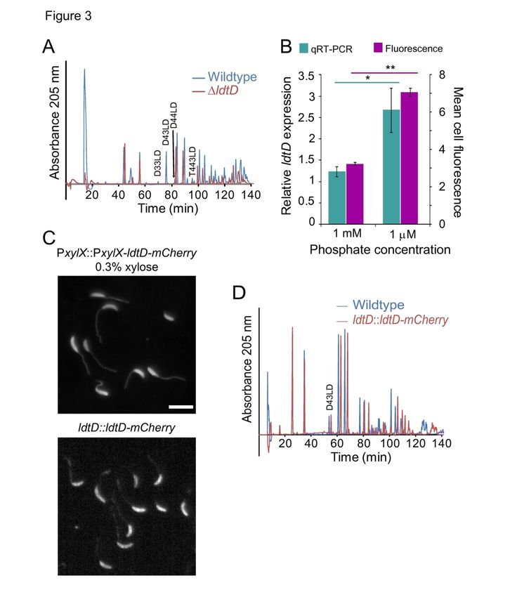
Molecular Microbiology                                           Page 2 of 45

24   Keywords: transpeptidase, stalk biosynthesis, LD crosslinks, LD transpeptidases, lysozyme, cell

25   shape

26

                           Fo
                                  rP
                                        ee
                                              rR
                                                     ev
                                                           iew

                                                                                                       2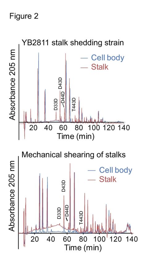
Page 3 of 45                                          Molecular Microbiology

        27     Summary

        28

        29     The diversity of cell shapes across the bacterial kingdom reflects evolutionary pressures that

        30     have produced physiologically important morphologies. While efforts have been made to

        31     understand the regulation of some prototypical cell morphologies such as that of rod-shaped

        32     Escherichia coli, little is known about most cell shapes. For Caulobacter crescentus, polar stalk

        33     synthesis is tied to its dimorphic life cycle, and stalk elongation is regulated by phosphate

        34     availability. Based on the previous observation that C. crescentus stalks are lysozyme-resistant,
                                      Fo

        35     we compared the composition of the peptidoglycan cell wall of stalks and cell bodies and
                                             rP

        36     identified key differences in peptidoglycan crosslinking. Cell-body peptidoglycan contained

        37     primarily DD-crosslinks between meso-diaminopimelic acid and D-alanine residues, whereas
                                                    ee

        38     stalk peptidoglycan had more LD-transpeptidation (meso-diaminopimelic acid-meso-
                                                          rR

        39     diaminopimelic acid), mediated by LdtD. We determined that ldtD is dispensable for stalk
                                                                 ev

        40     elongation; rather, stalk LD-transpeptidation reflects an aging process associated with low

        41     peptidoglycan turnover in the stalk. We also found that lysozyme resistance is a structural
                                                                        iew

        42     consequence of LD-crosslinking. Despite no obvious selection pressure for LD-crosslinking or

        43     lysozyme resistance in C. crescentus, the correlation between these two properties was

        44     maintained in other organisms, suggesting that DAP-DAP crosslinking may be a general

        45     mechanism for regulating bacterial sensitivity to lysozyme.

                                                                                                                   3
Molecular Microbiology                                           Page 4 of 45

46   Introduction

47           The bacterial kingdom contains species with a diverse array of morphologies, virtually all

48   of which are determined by the peptidoglycan cell wall. Among the best-studied unusual

49   morphologies are the stalks of the Alphaproteobacteria of the Caulobacteraceae family. The stalk

50   is a unipolar extension of the cell envelope formed in a cell cycle-dependent manner in the model

51   organism Caulobacter crescentus. The dimorphic life cycle of C. crescentus produces one motile

52   (swarmer) cell and one adherent (stalked) cell upon each cell division (Stove Poindexter and

53   Cohen-Bazire, 1964). The swarmer cell has a polar flagellum and pili, and is non-replicative. To
                             Fo

54   enable cell division, the swarmer initiates a differentiation program in which it sheds its
                                   rP

55   flagellum and, at the same pole, produces an adhesive holdfast (Tsang et al., 2006). After the

56   holdfast is secreted, the stalk forms and elongates from the holdfast pole, thereby causing the
                                          ee

57   holdfast to be pushed away from the cell body and localized to the tip of the stalk. During cell
                                                 rR

58   division, a new flagellum is synthesized at the pole opposite the stalk. As a result, following
                                                        ev

59   cytokinesis, the stalked cell maintains its stalk and immediately reenters the cell cycle, while the

60   swarmer daughter cell is flagellated and enters a quiescent state from which it needs to emerge
                                                              iew

61   before synthesizing a new stalk and beginning a proliferative cycle. In addition to its regulation

62   by the cell cycle, stalk elongation is dramatically induced during phosphate limitation (Gonin et

63   al., 2000).

64           Since the stalk elongates upon phosphate limitation, one hypothesis is that the stalk acts

65   as a “nutrient antenna” (Ireland et al., 2002). Under diffusive conditions, nutrient uptake is

66   proportional to cell length; therefore, having a long, thin appendage would be optimal for

67   maximizing cell length while minimizing surface area (Wagner et al., 2006). An alternative

68   hypothesis is that by adhering to surfaces via the holdfast at the stalk tip, stalk elongation can

                                                                                                          4
Page 5 of 45                                           Molecular Microbiology

        69     elevate C. crescentus cells off the surface to gain access to convective fluid flow, thereby

        70     increasing nutrient availability (Klein et al., 2013).

        71            Despite these hypotheses for the physiological role of stalk elongation, the molecular

        72     mechanisms of stalk synthesis remain unknown. This knowledge gap is due, in part, to

        73     uncertainty regarding the chemical composition of the stalk. Initial studies comparing the

        74     peptidoglycan of C. crescentus stalks and cell bodies were contradictory; their findings differed

        75     regarding the relative molar ratios of the amino acids alanine, diaminopimelic acid (DAP), and

        76     glutamic acid as well as between the glycan sugars N-acetylglucosamine (NAG) and N-
                                       Fo

        77     acetylmuramic acid (NAM) (Fujiki et al., 1976, Goodwin and Shedlarski, 1975, Poindexter and
                                              rP

        78     Hagenzieker, 1982). Despite these contradictory findings, physiological evidence supports the

        79     hypothesis that stalk peptidoglycan is somehow distinct from cell-body peptidoglycan.
                                                     ee

        80     Treatment of C. crescentus cells with lysozyme to induce spheroplast formation leads to a
                                                           rR

        81     peculiar “lollipop” morphology in which the cell body swells while the stalk retains its shape
                                                                   ev

        82     (Schmidt and Stanier, 1966), suggesting that stalk peptidoglycan may be more resistant to

        83     lysozyme-mediated degradation than cell-body peptidoglycan due to modifications in
                                                                        iew

        84     peptidoglycan composition.

        85            The identification of enzymatic machinery directly responsible for stalk elongation has

        86     also been challenging. Peptidoglycan synthesis is mediated by a family of mono- and bi-

        87     functional penicillin-binding proteins (PBPs) that have glycosyltransferase and/or transpeptidase

        88     activities. Given the presence of peptidoglycan within the stalk (Schlimpert et al., 2012), one or

        89     more of the PBPs is likely required for stalk elongation. Two recent studies reported that

        90     systematic deletion of the C. crescentus bi-functional PBPs and the single monofunctional

        91     glycosyltransferase, either individually or in combination, did not prevent stalk formation in low-

                                                                                                                    5
Molecular Microbiology Page 6 of 45
92 phosphate conditions (Strobel et al., 2014, Yakhnina and Gitai, 2013); any single PBP (except
93 PbpZ) was sufficient for growth and stalk biogenesis. These data suggest that either the
94 redundancy of this activity allows any PBP to synthesize stalk peptidoglycan, or, consistent with
95 its potentially unique composition, there are yet unidentified enzymes involved in stalk
96 peptidoglycan insertion.
97 To distinguish among these possibilities, in this study we determined that stalk
98 peptidoglycan is resistant to digestion by several lytic, cell wall-degrading hydrolases, including
99 lysozyme and an interbacterial peptidoglycan amidase toxin. Muropeptide analysis via high-
Fo
100 performance liquid chromatography (HPLC) revealed that this resistance in the stalk is due to
rP
101 increased cell wall LD-crosslinks between meso-DAP residues on adjacent peptidoglycan
102 peptide stems. Further, we identified LdtD as the LD-transpeptidase required for stalk LD-
ee
103 crosslinking.
rR
104
ev
105 Results
iew
106 C. crescentus stalks and cell bodies have different sensitivities to wall-degrading enzymes
107 The previous finding that the creation of C. crescentus spheroplasts with lysozyme leads
108 to swollen cell bodies, while the stalk remains relatively unaffected (Schmidt and Stanier, 1966)
109 (Fig. 1A), is surprising because high-resolution cryo-electron micrographs appear to show that
110 the peptidoglycan cell wall is continuous and uniform from the cell body through the stalk
111 (Schlimpert et al., 2012). We hypothesized that this phenomenon could be explained by two
112 mechanisms: 1) although lysozyme efficiently degrades stalk peptidoglycan, the stalk does not
113 swell due to lower turgor pressure and/or reduced mechanical stress in the wall (compared to the
114 cell body) because the stalk has a narrower radius than the cell body, or 2) stalk peptidoglycan is
6Page 7 of 45 Molecular Microbiology
115 insensitive to lysozyme degradation. To directly test these hypotheses, we uniformly labeled the
116 peptidoglycan of C. crescentus cells expressing periplasmic RFP by growing cells in Hutner-
117 Imidazole-Glucose-Glutamate (HIGG) minimal medium containing 1 µM phosphate (HIGG + 1
118 µM phosphate) for 48 h in the presence of the fluorescent D-amino acid hydroxy coumarin-
119 carbonyl-amino-D-alanine (HADA) (Kuru et al., 2012). Cells were permeabilized with
120 chloroform-saturated Tris buffer to ensure that the peptidoglycan would be accessible for
121 lysozyme degradation. We confirmed outer-membrane permeability by visualizing the release of
122 periplasmic RFP (Fig. 1B). Exposed peptidoglycan was treated with 25 µg mL-1 lysozyme for 15
Fo
123 min at 37 C and the digested cells were imaged with fluorescence microscopy. While cell
rP
124 bodies were entirely degraded, stalks retained HADA fluorescence (Fig. 1B), demonstrating that
stalks are more resistant to lysozyme than cell bodies.
ee
125
126 To gain insight into the underlying properties of the stalk that provide lysozyme
rR
127 resistance, we asked whether lytic peptidoglycan-degrading enzymes that target other bonds
ev
128 within the cell wall degrade stalk peptidoglycan. Type VI secretion (T6S) systems are used by
129 some bacteria to attack and kill neighboring cells; multiple effectors of type VI secretion systems
iew
130 function by degrading peptidoglycan components, leading to cell lysis (Russell et al., 2011).
131 Salmonella enterica serovar Typhi T6S amidase effector 3 (Tae3) specifically hydrolyzes DD-
132 crosslinks between meso-DAP and D-alanine on adjacent peptide stems (Russell et al., 2012). As
133 with lysozyme, treatment of permeabilized C. crescentus cells with Tae3 led to complete
134 digestion of the cell body while leaving stalks intact (Fig. 1C), indicating a difference in Tae3
135 activity between the two compartments.
136
7Molecular Microbiology Page 8 of 45
137 Stalk and cell-body cell walls have distinct compositions and affinities for peptidoglycan-binding
138 proteins
139 One potential explanation for the difference in enzymatic sensitivity between the two
140 regions of the cell is a difference in peptidoglycan composition. We hypothesized that such a
141 difference alters the affinity of proteins that bind peptidoglycan. The gp144 endolysin gene from
142 Pseudomonas aeruginosa phage ϕKZ contains an N-terminal peptidoglycan binding domain
143 (Briers et al., 2007). We purified a KZ144-YFP fusion protein and labeled permeabilized C.
144 crescentus cells with it. Strikingly, KZ144-YFP exclusively labeled cell-body peptidoglycan and
Fo
145 not stalk peptidoglycan (Fig. 1D), suggesting that stalk peptidoglycan is distinct from that of the
rP
146 cell body.
147 To identify compositional differences between these two compartments, we compared the
ee
148 muropeptide content of C. crescentus cells grown in HIGG + 1 mM or 1 µM phosphate,
rR
149 conditions under which cells have short or long stalks, respectively (Schlimpert et al., 2012).
ev
150 Sacculi were purified and digested with mutanolysin prior to muropeptide analysis via HPLC
151 (Experimental Methods). Overall, the muropeptide content was qualitatively similar under both
iew
152 growth conditions, with the exception of one major peak that was uniquely present under low
153 phosphate (elongated stalk) conditions (Fig. 1E, Table 1). Given the specificity of Tae3 for DD-
154 crosslink digestion (Fig. 1F), the inability of Tae3 to degrade C. crescentus stalks suggests that
155 the stalk compartment is enriched in LD-crosslinks between meso-DAP residues on neighboring
156 peptide stems. Electrospray ionization mass spectrometry (ESI-MS) analysis of the peak
157 observed during phosphate limitation confirmed that it is an LD-crosslinked muropeptide dimer
158 (D43LD) (Fig. S1; the muropeptide naming convention is described in the legend for Table 1).
159
8Page 9 of 45 Molecular Microbiology
160 Stalk-specific LD-transpeptidation increases during stalk elongation
161 To determine whether LD-crosslinks are uniquely present in the stalk, we separated stalks
162 from cell bodies and collected sacculi for muropeptide analysis. For validation, stalks were
163 purified via two complementary methods. First, we harvested stalks from strain YB2811 (a
164 derivative of NY111d1 with a pstS::miniTn5 insertion), which spontaneously sheds stalks into
165 the medium (Ireland et al., 2002, Poindexter, 1978). Second, stalks were collected from wild-
166 type cells by mechanically detaching the stalks (Experimental Methods) (Wagner et al., 2006). In
167 each case, peaks corresponding to LD-transpeptidation only appeared in stalk fractions (Fig. 2,
Fo
168 Table 2); the identified LD-peaks correspond to dimers D33LD and D43LD and trimers T443LD
rP
169 and T443NLD as determined by confirmation via mass spectrometry (Fig. S2) and by
170 comparison to published chromatograms (Glauner, 1988). While the two stalk-isolation methods
ee
171 yielded similar results with respect to LD-crosslinks, we consistently observed an increase in the
rR
172 pentapeptide M5G in mechanically sheared stalk samples; the reason for this enrichment is
ev
173 unclear.
174 To identify candidate LD-transpeptidases in C. crescentus, we used RPS-Blast
iew
175 (Marchler-Bauer et al., 2017) to search for proteins containing conserved LD-transpeptidase
176 domains (NCBI CDD database: COG2989, COG3034, COG3786, Pfam03734, PRK10594) and
177 identified two candidates. CCNA_01579 has homology to E. coli LdtD (26% identical, 42%
178 similar; henceforth referred to as LdtD) and CCNA_03860 is homologous to Streptomyces
179 purpurogeneiscleroticus YkuD (42% identical, 53% similar; henceforth referred to as YkuD).
180 Both proteins contain the conserved active site histidine and cysteine residues (Fig. S3A)
181 (Magnet et al., 2007). Analysis of stalk muropeptides in the ∆ldtD strain confirmed that this
182 enzyme is responsible for 91% of the LD-crosslinking (Fig. 3A and Table 2). By contrast,
9Molecular Microbiology Page 10 of 45
183 deletion of ykuD either alone or in combination with ldtD did not reduce LD-crosslinking (Table
184 2). Complementation of the ldtD deletion by expressing xylose-inducible ldtD confirmed the role
185 of LdtD in the formation of LD-crosslinks (Fig. S3B-D). We note that in the ΔldtD and
186 ΔldtDΔykuD strains we detected low levels of LD-crosslinks, particularly D33D and T443DN
187 (Table 2), suggesting C. crescentus may express an additional, as yet unidentified, LD-
188 transpeptidase.
189 The expression of ldtD, as determined by qRT-PCR and fluorescence intensity of an
190 LdtD-mCherry fusion at its native promoter, was significantly higher during growth in low (1
Fo
191 µM) phosphate compared with high (1 mM) phosphate, consistent with the hypothesized role of
rP
192 LdtD in stalk peptidoglycan crosslinking (Fig. 3B). Despite the restriction of LD-crosslinks to
193 the stalk compartment, LdtD-mCherry was broadly localized throughout the entire cell envelope
ee
194 when expressed either from the inducible xylX locus or from the native chromosomal locus under
rR
195 its endogenous promoter (Fig. 3C). The fluorescence intensity was approximately constant
ev
196 throughout the cell body and stalk (Fig. S4A); we observed ~15% higher fluorescence in the cell
197 body, although that increase may be an optical artifact due to the larger volume of that
iew
198 compartment. The muropeptide composition of the ldtD::ldtD-mCherry strain in HIGG + 1 µM
199 phosphate was comparable to that in wild-type cells (Fig. 3D), demonstrating that the fluorescent
200 protein fusion is functional. While LD-transpeptidation regulates stalk peptidoglycan
201 muropeptide composition, deletion of ldtD or ykuD either alone or in combination had no effect
202 on stalk elongation (Fig. S4B). None of the LD-transpeptidase mutants had a growth defect in
203 either high or low phosphate (Fig. S4C). Taken together, these data establish LdtD as the major
204 LD-transpeptidase during stalk synthesis.
205
10Page 11 of 45 Molecular Microbiology
206 LD-transpeptidation mediates lysozyme sensitivity and KZ144 binding
207 To confirm the relationship between stalk LD-crosslinking and lysozyme resistance, we
208 labeled wild-type and ΔldtD peptidoglycan with HADA, permeabilized the outer membrane, and
209 treated cells with lysozyme. Abrogation of LD-transpeptidation increased the sensitivity of stalk
210 peptidoglycan to lysozyme-mediated degradation (Fig. 4A). Similarly, deletion of ldtD enabled
211 stalk labeling by the phage peptidoglycan-binding protein KZ144-YFP (Fig. 4B). We note that
212 KZ144-YFP binding was not entirely uniform within the cell body, with decreased labeling at
213 the non-stalked pole in both wild-type and ldtD cells (Fig. S5). These data suggest that there
Fo
214 may be local variations in PG composition along the cell body.
rP
215 Since the stalk is ~20% the width of the cell body (Wagner and Brun, 2007), we also
considered whether geometrical constraints could contribute to the subcellular specificity of
ee
216
217 KZ144-YFP binding. If so, we expected that binding of KZ144-YFP would not be affected in
rR
218 wider rod-shaped cells, and would not be generally correlated with LD-transpeptidation levels.
ev
219 To address these possibilities, we labeled the peptidoglycan of Agrobacterium tumefaciens and
220 Hyphomonas neptunium. Although the average cell size of A. tumefaciens is similar to C.
iew
221 crescentus, the growing pole (as determined by the pole with narrower width (Cameron et al.,
222 2014)) in log-phase A. tumefaciens cells bound more KZ144-YFP than did the opposite pole in
223 the same cells (Fig. 4C). Interestingly, at least one LD-transpeptidase in A. tumefaciens localizes
224 to the growing cell pole (Cameron et al., 2014); although the precise distribution of LD-
225 crosslinks has not been determined, it is possible that they are asymmetrically localized along the
226 cell length, which could contribute to the gradient of KZ144-YFP binding. H. neptunium forms a
227 stalk that is morphologically similar to the C. crescentus stalk, but this organism does not form
228 DAP-DAP crosslinks (Cserti et al., 2017). KZ144-YFP efficiently labeled DD-crosslinked stalks
11Molecular Microbiology Page 12 of 45
229 in H. neptunium (Fig. 4D), leading us to conclude that binding affinity is regulated by the mode
230 of crosslinking rather than by peptidoglycan geometry.
231
232 LD-transpeptidation correlates with lysozyme sensitivity in Gram-negative bacteria
233 Since LD-crosslinks in the C. crescentus stalk provided protection from degradation by
234 lysozyme, we hypothesized that the 3-3 mode of transpeptidation may regulate lysozyme
235 sensitivity more generally. During log-phase growth, LD-crosslinks account for 3.5% of
236 muropeptides in E. coli (Pisabarro et al., 1985). The fraction of these crosslinks increases to
Fo
237 8.6% in deep stationary phase (Pisabarro et al., 1985). A. tumefaciens peptidoglycan is enriched
rP
238 with LD-crosslinks, containing ~23% DAP-DAP (Brown et al., 2012). To assess the relationship
239 between the percentage of LD-crosslinks and lysozyme sensitivity, we permeabilized log-phase
ee
240 and stationary-phase E. coli as well as log-phase A. tumefaciens with chloroform-saturated Tris
rR
241 buffer and digested them with lysozyme. The turbidity of the treated cells was measured over
ev
242 time by measuring OD600. LD-crosslink-deficient log-phase E. coli cells were the most
243 susceptible to lysozyme degradation (Fig. 5A, S6). By contrast, DAP-DAP-rich A. tumefaciens
iew
244 cells were highly resistant (Fig. 5A, S6). Additionally, we detected a strong correlation between
245 the initial rate of lysozyme degradation (0-60 s) and LD-crosslink content (R2=0.975, Fig. 5B),
246 suggesting that lysozyme sensitivity may be generally linked to LD-crosslinking.
12Page 13 of 45 Molecular Microbiology
247 Discussion
248 Based on the observation that C. crescentus lysozyme-induced spheroplasts display an
249 unusual “lollipop” morphology (Schmidt and Stanier, 1966), we investigated the chemical
250 composition of stalk peptidoglycan to determine the underlying mechanism of lysozyme
251 resistance in the stalk. The recent development of fluorescent D-amino acids that label
252 peptidoglycan in vivo (Kuru et al., 2012) enabled us to directly visualize the degradation of cell-
253 body and stalk peptidoglycan by lysozyme (Fig. 1B) and the DD-amidase Tae3 (Fig. 1C),
254 leading us to conclude that stalk peptidoglycan is more resistant to enzymatic digestion than cell-
Fo
255 body peptidoglycan. Muropeptide analysis revealed that low-phosphate conditions promoting
rP
256 stalk elongation led to the accumulation of cell wall LD-crosslinks due to the activity of the LD-
257 transpeptidase CCNA_01579 (LdtD) (Fig. 1E, 3A). LdtD specifically catalyzes transpeptidation
ee
258 in the stalk compartment (Fig. 2, 3A), and this particular mode of crosslinking confers resistance
rR
259 to lysozyme-mediated degradation within the stalk and in species with high levels of LD-
ev
260 crosslinks (Fig. 4A, 5A, B).
261
iew
262 Regulation of LdtD activity in the stalk
263 Despite the enrichment of LD-crosslinks within the stalk, LdtD protein localization
264 appears to be approximately uniform throughout the cell (Fig. 3C). Therefore, it is unclear how
265 LdtD activity becomes restricted to the stalk compartment. One possibility is the availability of
266 suitable substrates. Tetrapeptide DAP residues are the donor substrates for LD-crosslinking, and
267 these tetrapeptide substrates generally are generated via the activity of DD-carboxypeptidases,
268 which cleave between the fourth and fifth muropeptide residues. Comparing the muropeptide
269 content of isolated stalks and cell bodies showed that stalks have elevated levels of tripeptides
13Molecular Microbiology Page 14 of 45
270 and pentapeptides and reduced levels of tetrapeptides (Table 2), suggesting that
271 carboxypeptidase activity is not responsible for generating additional substrate tetrapeptides.
272 These data are consistent with the previously reported lack of carboxypeptidase activity in C.
273 crescentus (Markiewicz et al., 1983). Alternatively, DD-endopeptidases can cleave DD-
274 crosslinks to create free tetrapeptides. While C. crescentus does not appear to have direct
275 homologues to the known DD-endopeptidases PbpG, DacB, MepA, or EnvC, there are a large
276 number of more distantly related peptidases, including an annotated AmpH homologue
277 (CC_3489) that may serve this enzymatic function (Gonzalez-Leiza et al., 2011). Finally, the
Fo
278 high ratio of tetra-tetra dimers to tetra-tri dimers in the cell body fraction suggests that PBP
rP
279 enzymes prefer tetrapeptide substrates. In contrast, LdtD appears to make a significant number of
280 tetra-tri crosslinks; therefore, the increased abundance of tripeptides in the stalk may serve as a
ee
281 preferred acceptor substrate for LdtD, although the underlying mechanism for accumulating
rR
282 tripeptides is unknown.
ev
283
284 Function of LD-transpeptidation in the stalk
iew
285 For the majority of bacterial species that have been investigated to date, the dominant
286 mode of peptidoglycan crosslinking is DD-transpeptidation mediated by PBPs. In species that
287 use a polar growth mechanism, such as those of the orders Actinomycetales (Lavollay et al.,
288 2011) and Rhizobiales (Brown et al., 2012), LD-crosslinks can constitute ~40-80% of the total
289 crosslinks. In A. tumefaciens, the Ldt Atu0845 localizes to the growing pole, consistent with its
290 hypothesized role in polar elongation (Cameron et al., 2014). Given that the Alphaproteobacteria
291 C. crescentus and A. tumefaciens are in the same phylogenetic class, as well as the specific
292 accumulation of LD-crosslinks in the stalk (Fig. 2), it was tempting to hypothesize that stalk
14Page 15 of 45 Molecular Microbiology
293 elongation represents a form of Ldt-driven polar growth. However, deletion of ldtD did not result
294 in any stalk-elongation defect (Fig. S4B). Moreover, unlike Atu0845 (Cameron et al., 2014),
295 LdtD-mCherry was uniformly distributed throughout the cell body and stalk envelope (Fig. 3C).
296 An alternative model for stalk Ldt activity arises from the pattern of LD-crosslinking in
297 E. coli. DAP-DAP crosslinking increases during stationary phase (Pisabarro et al., 1985), when
298 incorporation of new peptidoglycan material is dramatically reduced (Blasco et al., 1988).
299 Similarly, since the C. crescentus stalk is built by inserting new peptidoglycan material at its
300 base at the cell pole (Hughes et al., 2013), there appears to be a very low rate of new
Fo
301 peptidoglycan insertion in the stalk relative to the cell body. Although the role of LD-
rP
302 transpeptidation in E. coli during stationary phase is unknown, C. crescentus LdtD may play a
303 similar role in peptidoglycan maintenance in the stalk.
ee
304
rR
305 LD-transpeptidation increases lysozyme resistance
ev
306 Typically, lysozyme resistance is attributed to one of several modifications of the glycan
307 strands, including MurNac O-acetylation, GlcNac N-deacetylation, and MurNac O-glycolylation
iew
308 (reviewed in (Davis and Weiser, 2011)). While the C. crescentus genome does not contain any
309 genes with obvious O-acetylation or O-glycolylation activity, it does encode two orthologs of
310 Streptococcus pneumoniae GlcNac N-deacetylase pgdA; one of these genes, hfsH, regulates the
311 adhesiveness of holdfast (Wan et al., 2013). The role of the second ortholog, CCNA_02236, is
312 unknown, leaving open the possibility that GlcNac N-deacetylation may play a role in regulating
313 lysozyme resistance in C. crescentus.
314 Lysozyme digestion of C. crescentus stalks (Fig. 4A) as well as whole-cell peptidoglycan
315 from E. coli and A. tumefaciens (Fig. 5A,B) suggests a relationship between the degree of LD-
15Molecular Microbiology Page 16 of 45
316 transpeptidation and lysozyme sensitivity. This relationship is somewhat surprising: lysozyme
317 acts by hydrolyzing the NAM-NAG glycosidic bond on the glycan backbone, whereas LD-
318 crosslinks occur on the peptide stem. Computational models of peptidoglycan structure propose
319 several conformational differences between LD- and DD-crosslinks that may explain this
320 phenomenon (de Pedro and Cava, 2015). First, lack of the D-alanine residue linking the meso-
321 DAP molecules in the LD-crosslink may make the stem peptides more rigid, leading to a more
322 extended conformation and a greater distance between glycan strands. Second, DD-crosslinks
323 allow the formation of additional hydrogen bonds that can affect the favored orientation of the
Fo
324 NAG-NAM disaccharides in the glycan strand. The overall effect of LD-transpeptidation on
rP
325 peptidoglycan conformation may explain the decreased activity and affinity of lysozyme as well
326 as those of the KZ144 peptidoglycan-binding domain.
ee
327 Our analysis of E. coli and A. tumefaciens peptidoglycan lysozyme susceptibility
rR
328 suggests that the relationship between LD-crosslinking and lysozyme insensitivity may be a
ev
329 more general cellular property. In the context of bacterial pathogenesis, lysozyme is a significant
330 player in host defense. For example, in a mouse model of lung infection by Klebsiella
iew
331 pneumoniae, lysozyme M knockout mice did not survive beyond 72 h post infection, whereas
332 25% of wild-type mice survived up to 120 h (Markart et al., 2004). Transcriptomic analysis of K.
333 pneumoniae exposed to pulmonary surfactant showed that relative to controls, interaction with
334 lung secretions increased ybiS LD-transpeptidase expression (Willsey et al., 2018). Upregulation
335 of ybiS expression may be part of a general envelope stress response, similar to the increase of E.
336 coli ldtD transcription during the Cpx envelope stress response (Bernal-Cabas et al., 2015);
337 alternatively, it may be a specific program for modulating peptidoglycan composition to avoid
338 killing by the host lysozyme defense. Increasing the proportion of LD-crosslinks also increases
16Page 17 of 45 Molecular Microbiology
339 bacterial pathogenicity by enhancing resistance against beta-lactam antibiotics (Cremniter et al.,
340 2006, Gupta et al., 2010, Hugonnet et al., 2016). In E. coli, inhibition of PBPs by beta-lactams
341 blocks DD-transpeptidation, leading to peptidoglycan instability and cell death; this phenotype is
342 suppressed by overexpression of LdtD and production of (p)ppGpp by the stringent response
343 (Hugonnet et al., 2016).
344
345 Robust maintenance of cell shape may benefit from redundancy in peptidoglycan-modifying
346 enzymes
Fo
347 For most rod-shaped bacteria, including C. crescentus, DD-transpeptidation is the
rP
348 dominant form of peptidoglycan crosslinking. Since peptidoglycan organization and stability are
349 critical for cellular integrity, it is perhaps not surprising that many of these organisms encode a
ee
350 highly redundant set of high-molecular weight PBPs with DD-transpeptidase activity. For
rR
351 example, E. coli, C. crescentus, and Bacillus subtilis encode five, seven, and 10 high-molecular
ev
352 weight PBPs, respectively (Blattner et al., 1997, Kunst et al., 1997, Nierman et al., 2001).
353 Redundancy among the bifunctional class A PBPs allows many of these genes to be deleted
iew
354 singly or in combination without losing viability; however, deletion of all members of this
355 enzyme class is lethal (Denome et al., 1999, Strobel et al., 2014, Yakhnina and Gitai, 2013). In
356 contrast, these species have relatively low abundances of LD-crosslinks, and LD-transpeptidase
357 activity is dispensable. E. coli and C. crescentus encode six and two Ldt enzymes, respectively;
358 deletion of these genes, even in combination, does not yield any observable phenotype under
359 normal growth conditions (Sanders and Pavelka, 2013) (Fig. S4B-D). The function of LD-
360 transpeptidases in these organisms remains largely unknown; however, they may have a role in
361 optimizing peptidoglycan architecture or responding to particular environmental stresses. Two of
17Molecular Microbiology Page 18 of 45
362 the E. coli Ldt enzymes are responsible for DAP-DAP crosslinking (Magnet et al., 2008), while
363 several others link the peptidoglycan to Braun’s lipoprotein (Magnet et al., 2007). While C.
364 crescentus does not contain Braun’s lipoprotein, our observation that deletion of ykuD has no
365 effect on PG composition suggests that there may be an alternative substrate for this enzyme as
366 well.
367 In contrast, LD-transpeptidases of Rhizobiales and Actinomycetales species that elongate
368 via polar growth may take on a more significant role. Mycobacterium tuberculosis has five Ldt
369 proteins, and the loss of just two isoforms (LdtMt1 and LdtMt2) causes defects in cell shape,
Fo
370 growth, and virulence (Schoonmaker et al., 2014). Most species of Rhizobiales, including A.
rP
371 tumefaciens, have 12-16 Ldt genes, leading to extensive redundancy (Cameron et al., 2015).
372 Thus, it appears that over a wide spectrum of organisms with varying growth modes, there is a
ee
373 selective pressure to maintain redundancy in the elongation machinery. This insight may be
rR
374 useful for predicting the elongation modes of species using metagenomic data based on their
ev
375 complement of PBP and Ldt genes, thereby providing a framework for understanding the link
376 between cell growth and environmental, ecological, and pathogenicity factors.
iew
377
18Page 19 of 45 Molecular Microbiology
378 Experimental Methods
379 Bacterial strains, plasmids, and growth conditions
380 The strains, plasmids, and primers used in this study are described in Tables S1, S2, and
381 S3, respectively. C. crescentus wild-type strain NA1000 and its derivatives were routinely
382 cultured at 30 C in peptone yeast extract medium (Poindexter, 1964). In experiments where
383 phosphate levels were varied, C. crescentus strains were grown in Hutner-Imidazole-Glucose-
384 Glutamate (HIGG) minimal medium supplemented with 1 or 1000 µM phosphate (Poindexter,
385 1978). E. coli strains were grown at 37 C in liquid lysogeny broth (LB). A. tumefaciens strain
Fo
386 C58 (kind gift from Zemer Gitai, Princeton University) was grown at 30 C in peptone yeast
rP
387 extract medium. H. neptunium strain 14-15 (ATCC) was grown at room temperature in Difco
ee
388 Marine Broth 2216. When necessary, antibiotics were added at the following concentrations:
389 kanamycin, 30 µg mL-1 in broth and 50 µg mL-1 in agar for E. coli and 5 µg mL-1 in broth and 25
rR
390 µg mL-1 in agar for C. crescentus; tetracycline, 12 µg mL-1 in broth and 12 µg mL-1 in agar for E.
ev
391 coli and 1 µg mL-1 in broth and 2 µg mL-1 in agar for C. crescentus; chloramphenicol, 1 µg mL-1
392 in broth and 1 µg mL-1 in agar for C. crescentus; gentamicin, 15 µg mL-1 in broth and 20 µg mL-1
iew
393 in agar for E. coli and 5 µg mL-1 in broth and 0.5 µg mL-1 in agar for C. crescentus. Where
394 noted, gene expression was induced in C. crescentus with 0.3% (w/v) xylose. Peptidoglycan was
395 fluorescently labeled by adding 0.25 mM HADA (purchased under a Material Transfer
396 Agreement from Indiana University) to the growth medium (Kuru et al., 2012).
397
398 Protein purification
399 KZ144-YFP was cloned into pET28a (Novagen) and expressed in BL21(DE3) cells (New
400 England Biolabs) to produce strain EK313. EK313 was grown overnight in LB, diluted 1:60 into
19Molecular Microbiology Page 20 of 45
401 20 mL LB, and grown to OD600 0.6. Expression was induced for 4 h with 1 mM IPTG. Five
402 milliliters of cells were collected via centrifugation (10 min, 3,800 x g, 4 C), and protein was
403 purified using the Qiagen Ni-NTA Spin Kit in accordance with the manufacturer’s protocol.
404 Tae3 was cloned into the pET29b+ vector (Novagen) and expressed in Shuffle Express T7 lysY
405 cells (New England Biolabs). Proteins were purified to homogeneity using previously reported
406 methods (Russell et al., 2012), except that in all steps no reducing agents or lysozyme were used.
407
408 Fluorescence microscopy and image analysis
Fo
409 Cells were spotted onto 1% agarose pads. Fluorescence microscopy was performed on a
rP
410 Nikon Ti-E inverted microscope equipped with a Prior Lumen 220PRO illumination system, CFI
Plan Apochromat 100X oil immersion objective (NA 1.45, WD 0.13 mm), Zyla sCMOS 5.5-
ee
411
412 megapixel camera (Andor), and NIS Elements v, 4.20.01 for image acquisition. Stalk lengths and
rR
413 envelope fluorescence profiles were measured using ImageJ v. 1.48q (NIH). Cell body length,
ev
414 total cell fluorescence, and centerline fluorescence were quantified using Morphometrics (Ursell
415 et al., 2017) and Matlab v. R2015b.
iew
416
417 Transmission electron microscopy
418 Droplets of cell suspensions were placed on a strip of Parafilm. Formvar-coated copper
419 grids (Electron Microscopy Sciences) were floated on top of the cell suspensions for 1 min. The
420 grids were washed twice with water and then negatively stained with 1% uranyl acetate (Electron
421 Microscopy Sciences). Following an additional wash with water, the grids were imaged on a
422 Zeiss EM902 transmission electron microscope.
423
20Page 21 of 45 Molecular Microbiology
424 Cell permeabilization and labeling
425 For experiments requiring enzymatic treatment of peptidoglycan for microscopy and
426 analysis, the outer membrane was removed via chloroform permeabilization. Chloroform-
427 saturated Tris buffer was prepared by mixing 50 mM Tris [pH 7.4] with chloroform (70:30) and
428 shaking the mixture at room temperature for 30 min. Cells to be permeabilized were collected via
429 centrifugation (2 min at 6,000 x g, 4 C) and resuspended in an equal volume of the aqueous
430 phase of the chloroform-saturated Tris buffer. Resuspended cells were rocked for 45 min at room
431 temperature and then washed twice in 50 mM Tris [pH 7.4] (via centrifugation for 10 min at
Fo
432 5,000 x g) to remove residual chloroform. Cells were labeled with purified KZ144-YFP (0.1-0.2
rP
433 mg mL-1; purification described above) for 5 min at room temperature and washed twice with 50
mM Tris [pH 7.4].
ee
434
435
rR
436 Muropeptide purification
ev
437 For whole C. crescentus cells, we started with 500-ml cultures grown in either high-
438 phosphate (1 mM) or low-phosphate (1 µM) HIGG growth media. To mechanically detach stalks
iew
439 from cell bodies, cells from 3 L cultures were sheared in a standard kitchen blender for 1 min at
440 maximum speed. For mechanically sheared cells and the stalk-shedding YB2811 strain, cell
441 bodies were purified via low-speed centrifugation (10 min at 8,000 x g, 4 C); excess cell bodies
442 were removed via 2-3 spins for 10 min at 10,000 x g. The removal of cell bodies was assessed by
443 phase microscopy. Subsequently, pure stalks were harvested from the washed supernatant (1 h
444 centrifugation at 47,056 x g). Peptidoglycan muropeptides were purified from C. crescentus as
445 previously described (Desmarais et al., 2014) and separated on a reversed-phase C18 column
446 (Thermo Scientific; 250 x 4.6-mm column, 3-µm particle size) held at 55 C. The LC solvent
21Molecular Microbiology Page 22 of 45
447 system consisted of 50 mM sodium phosphate [pH 4.35] with 0.4% sodium azide (solvent A)
448 and 75 mM sodium phosphate, pH 4.95 + 15% (v/v) methanol (solvent B). The solvent flow rate
449 was 0.5 mL min-1 and a linear gradient to 100% solvent B was performed over 135 min.
450 Muropeptide elution was monitored at 205 nm and sample fractions were collected at time points
451 as indicated. Collected fractions were dried via vacuum centrifugation and analyzed by ESI-MS
452 at the Rutgers Biological Mass Spectrometry Facility. Major peaks were labeled based on
453 previous analysis of C. crescentus muropeptides (Takacs et al., 2010). LD-crosslinks were
454 labeled based on previous analysis of E. coli muropeptides (Glauner, 1988) and confirmed by
Fo
455 LC/MS. Muropeptide abundances were measured using Chromanalysis v. 1.0 (Desmarais et al.,
rP
456 2015).
457
ee
458 Quantitative RT-PCR (qRT-PCR)
rR
459 Cells (0.5-5 mL) were collected via centrifugation (10 min, 3,800 x g, 4 C) and
ev
460 resuspended in Bacterial RNA Protect (Qiagen) in accordance with the manufacturer’s protocol.
461 RNA was purified using the RNeasy Mini Kit (Qiagen) and contaminating DNA was removed
iew
462 via on-column digestion (Qiagen). Purified RNA (5 ng µL-1) was reverse-transcribed using the
463 ABI High Capacity cDNA Reverse Transcription kit. Control samples did not contain reverse
464 transcriptase to assess the level of DNA contamination. cDNA was amplified using Power SYBR
465 Green Master Mix (Thermo Scientific) and analyzed on a Quantstudio 6 instrument (Thermo
466 Scientific). Quantification of qRT-PCR data was performed using the ∆∆Ct method (Livak and
467 Schmittgen, 2001) with rpoD as the endogenous control.
468
469
22Page 23 of 45 Molecular Microbiology
470 Cell growth assay
471 Cells were grown overnight in the media to be used for growth analysis. The optical
472 density at 660 nm (OD660) was normalized to 0.03 for each strain. Cell growth was assessed by
473 measuring OD660 in 96-well format using a CLARIOstar plate reader (BMG Labtech) incubated
474 at 30 C with shaking.
475
476 In vitro lysozyme sensitivity assay
477 Cells were chloroform-permeabilized as described above. The OD600 for each sample was
Fo
478 normalized to 0.08 by measuring the absorbance of a 15-µL sample in a 384-well plate using a
rP
479 CLARIOstar plate reader. To measure the rate of digestion, lysozyme was added to 15-µL cell
suspensions at a final concentration of 1 µg mL-1 and OD600 was measured at 30-s intervals over
ee
480
481 15 min without shaking. A linear regression to the first 60 s of data was used to determine the
rR
482 initial degradation rate.
ev
iew
23Molecular Microbiology Page 24 of 45
483 Acknowledgments
484 We thank Dr. Haiyan Zheng and the Rutgers Biological Mass Spectrometry Facility for their
485 assistance in acquiring mass spectrometry data. Funding was provided by NSF CAREER Award
486 MCB-1149328, the Stanford Center for Systems Biology under Grant P50-GM107615, and the
487 Allen Discovery Center at Stanford on Systems Modeling of Infection (to K.C.H); the UCSF
488 Program for Breakthrough Biomedical Research (to S.C.); and NSF CAREER Award MCB-
489 1553004 (to E.A.K.). S.C. and K.C.H. are Chan Zuckerberg Investigators.
490
Fo
491 Author Contributions
rP
492 GS, SC, KCH, and EAK contributed to the conception and design of the project. GS, AM, AR,
493 SC, KCH, and EAK contributed to the acquisition, analysis, and/or interpretation of the data.
ee
494 EAK wrote and GS, SC, and KCH edited the manuscript. All authors reviewed the manuscript
rR
495 prior to submission.
ev
496
iew
24Page 25 of 45 Molecular Microbiology
497 References
498 Bernal-Cabas, M., Ayala, J. A. and Raivio, T. L. (2015) The Cpx envelope stress response
499 modifies peptidoglycan cross-linking via the L,D-transpeptidase LdtD and the novel
500 protein YgaU. J Bacteriol 197: 603-614.
501 Blasco, B., Pisabarro, A. G. and de Pedro, M. A. (1988) Peptidoglycan biosynthesis in
502 stationary-phase cells of Escherichia coli. J Bacteriol 170: 5224-5228.
503 Blattner, F. R., Plunkett, G., 3rd, Bloch, C. A., Perna, N. T., Burland, V., Riley, M., Collado-
504 Vides, J., Glasner, J. D., Rode, C. K., Mayhew, G. F., Gregor, J., Davis, N. W.,
Fo
505 Kirkpatrick, H. A., Goeden, M. A., Rose, D. J., Mau, B. and Shao, Y. (1997) The
rP
506 complete genome sequence of Escherichia coli K-12. Science 277: 1453-1462.
507 Briers, Y., Volckaert, G., Cornelissen, A., Lagaert, S., Michiels, C. W., Hertveldt, K. and
ee
508 Lavigne, R. (2007) Muralytic activity and modular structure of the endolysins of
rR
509 Pseudomonas aeruginosa bacteriophages phiKZ and EL. Mol Microbiol 65: 1334-1344.
ev
510 Brown, P. J., de Pedro, M. A., Kysela, D. T., Van der Henst, C., Kim, J., De Bolle, X., Fuqua, C.
511 and Brun, Y. V. (2012) Polar growth in the Alphaproteobacterial order Rhizobiales. Proc
iew
512 Natl Acad Sci U S A 109: 1697-1701.
513 Cameron, T. A., Anderson-Furgeson, J., Zupan, J. R., Zik, J. J. and Zambryski, P. C. (2014)
514 Peptidoglycan synthesis machinery in Agrobacterium tumefaciens during unipolar growth
515 and cell division. MBio 5: e01219-01214.
516 Cameron, T. A., Zupan, J. R. and Zambryski, P. C. (2015) The essential features and modes of
517 bacterial polar growth. Trends Microbiol 23: 347-353.
518 Cremniter, J., Mainardi, J. L., Josseaume, N., Quincampoix, J. C., Dubost, L., Hugonnet, J. E.,
519 Marie, A., Gutmann, L., Rice, L. B. and Arthur, M. (2006) Novel mechanism of
25Molecular Microbiology Page 26 of 45
520 resistance to glycopeptide antibiotics in Enterococcus faecium. J Biol Chem 281: 32254-
521 32262.
522 Cserti, E., Rosskopf, S., Chang, Y. W., Eisheuer, S., Selter, L., Shi, J., Regh, C., Koert, U.,
523 Jensen, G. J. and Thanbichler, M. (2017) Dynamics of the peptidoglycan biosynthetic
524 machinery in the stalked budding bacterium Hyphomonas neptunium. Mol Microbiol 103:
525 875-895.
526 Davis, K. M. and Weiser, J. N. (2011) Modifications to the peptidoglycan backbone help bacteria
527 to establish infection. Infect Immun 79: 562-570.
Fo
528 de Pedro, M. A. and Cava, F. (2015) Structural constraints and dynamics of bacterial cell wall
rP
529 architecture. Front Microbiol 6: 449.
530 Denome, S. A., Elf, P. K., Henderson, T. A., Nelson, D. E. and Young, K. D. (1999) Escherichia
ee
531 coli mutants lacking all possible combinations of eight penicillin binding proteins:
rR
532 viability, characteristics, and implications for peptidoglycan synthesis. J Bacteriol 181:
ev
533 3981-3993.
534 Desmarais, S. M., Cava, F., de Pedro, M. A. and Huang, K. C. (2014) Isolation and preparation
iew
535 of bacterial cell walls for compositional analysis by ultra performance liquid
536 chromatography. J Vis Exp: e51183.
537 Desmarais, S. M., Tropini, C., Miguel, A., Cava, F., Monds, R. D., de Pedro, M. A. and Huang,
538 K. C. (2015) High-throughput, highly sensitive analyses of bacterial morphogenesis using
539 ultra performance liquid chromatography. J Biol Chem 290: 31090-31100.
540 Fujiki, K., Fukuda, A. and Okada, Y. (1976) Amino acid composition of peptidoglycan in
541 Caulobacter crescentus. J Biochem 80: 1453-1455.
26Page 27 of 45 Molecular Microbiology
542 Glauner, B. (1988) Separation and quantification of muropeptides with high-performance liquid
543 chromatography. Anal Biochem 172: 451-464.
544 Gonin, M., Quardokus, E. M., O'Donnol, D., Maddock, J. and Brun, Y. V. (2000) Regulation of
545 stalk elongation by phosphate in Caulobacter crescentus. J. Bacteriol. 182: 337-347.
546 Gonzalez-Leiza, S. M., de Pedro, M. A. and Ayala, J. A. (2011) AmpH, a bifunctional DD-
547 endopeptidase and DD-carboxypeptidase of Escherichia coli. J Bacteriol 193: 6887-
548 6894.
549 Goodwin, S. D. and Shedlarski, J. G., Jr. (1975) Purification of cell wall peptidoglycan of the
Fo
550 dimorphic bacterium Caulobacter crescentus. Arch Biochem Biophys 170: 23-36.
rP
551 Gupta, R., Lavollay, M., Mainardi, J. L., Arthur, M., Bishai, W. R. and Lamichhane, G. (2010)
552 The Mycobacterium tuberculosis protein LdtMt2 is a nonclassical transpeptidase required
ee
553 for virulence and resistance to amoxicillin. Nat Med 16: 466-469.
rR
554 Hughes, H. V., Lisher, J. P., Hardy, G. G., Kysela, D. T., Arnold, R. J., Giedroc, D. P. and Brun,
ev
555 Y. V. (2013) Co-ordinate synthesis and protein localization in a bacterial organelle by the
556 action of a penicillin-binding-protein. Mol Microbiol 90: 1162-1177.
iew
557 Hugonnet, J. E., Mengin-Lecreulx, D., Monton, A., den Blaauwen, T., Carbonnelle, E., Veckerle,
558 C., Brun, Y. V., van Nieuwenhze, M., Bouchier, C., Tu, K., Rice, L. B. and Arthur, M.
559 (2016) Factors essential for L,D-transpeptidase-mediated peptidoglycan cross-linking and
560 beta-lactam resistance in Escherichia coli. Elife 5.
561 Ireland, M. M., Karty, J. A., Quardokus, E. M., Reilly, J. P. and Brun, Y. V. (2002) Proteomic
562 analysis of the Caulobacter crescentus stalk indicates competence for nutrient uptake.
563 Mol Microbiol 45: 1029-1041.
27Molecular Microbiology Page 28 of 45
564 Klein, E. A., Schlimpert, S., Hughes, V., Brun, Y. V., Thanbichler, M. and Gitai, Z. (2013)
565 Physiological role of stalk lengthening in Caulobacter crescentus. Commun Integr Biol 6:
566 e24561.
567 Kunst, F., Ogasawara, N., Moszer, I., Albertini, A. M., Alloni, G., Azevedo, V., Bertero, M. G.,
568 Bessieres, P., Bolotin, A., Borchert, S., Borriss, R., Boursier, L., Brans, A., Braun, M.,
569 Brignell, S. C., Bron, S., Brouillet, S., Bruschi, C. V., Caldwell, B., Capuano, V., Carter,
570 N. M., Choi, S. K., Cordani, J. J., Connerton, I. F., Cummings, N. J., Daniel, R. A.,
571 Denziot, F., Devine, K. M., Dusterhoft, A., Ehrlich, S. D., Emmerson, P. T., Entian, K.
Fo
572 D., Errington, J., Fabret, C., Ferrari, E., Foulger, D., Fritz, C., Fujita, M., Fujita, Y.,
rP
573 Fuma, S., Galizzi, A., Galleron, N., Ghim, S. Y., Glaser, P., Goffeau, A., Golightly, E. J.,
574 Grandi, G., Guiseppi, G., Guy, B. J., Haga, K., Haiech, J., Harwood, C. R., Henaut, A.,
ee
575 Hilbert, H., Holsappel, S., Hosono, S., Hullo, M. F., Itaya, M., Jones, L., Joris, B.,
rR
576 Karamata, D., Kasahara, Y., Klaerr-Blanchard, M., Klein, C., Kobayashi, Y., Koetter, P.,
ev
577 Koningstein, G., Krogh, S., Kumano, M., Kurita, K., Lapidus, A., Lardinois, S., Lauber,
578 J., Lazarevic, V., Lee, S. M., Levine, A., Liu, H., Masuda, S., Mauel, C., Medigue, C.,
iew
579 Medina, N., Mellado, R. P., Mizuno, M., Moestl, D., Nakai, S., Noback, M., Noone, D.,
580 O'Reilly, M., Ogawa, K., Ogiwara, A., Oudega, B., Park, S. H., Parro, V., Pohl, T. M.,
581 Portelle, D., Porwollik, S., Prescott, A. M., Presecan, E., Pujic, P., Purnelle, B., et al.
582 (1997) The complete genome sequence of the gram-positive bacterium Bacillus subtilis.
583 Nature 390: 249-256.
584 Kuru, E., Hughes, H. V., Brown, P. J., Hall, E., Tekkam, S., Cava, F., de Pedro, M. A., Brun, Y.
585 V. and VanNieuwenhze, M. S. (2012) In situ probing of newly synthesized peptidoglycan
28Page 29 of 45 Molecular Microbiology
586 in live bacteria with fluorescent D-amino acids. Angew Chem Int Ed Engl 51: 12519-
587 12523.
588 Lavollay, M., Fourgeaud, M., Herrmann, J. L., Dubost, L., Marie, A., Gutmann, L., Arthur, M.
589 and Mainardi, J. L. (2011) The peptidoglycan of Mycobacterium abscessus is
590 predominantly cross-linked by L,D-transpeptidases. J Bacteriol 193: 778-782.
591 Livak, K. J. and Schmittgen, T. D. (2001) Analysis of relative gene expression data using real-
592 time quantitative PCR and the 2(-Delta Delta C(T)) Method. Methods 25: 402-408.
593 Magnet, S., Bellais, S., Dubost, L., Fourgeaud, M., Mainardi, J. L., Petit-Frere, S., Marie, A.,
Fo
594 Mengin-Lecreulx, D., Arthur, M. and Gutmann, L. (2007) Identification of the L,D-
rP
595 transpeptidases responsible for attachment of the Braun lipoprotein to Escherichia coli
596 peptidoglycan. J Bacteriol 189: 3927-3931.
ee
597 Magnet, S., Dubost, L., Marie, A., Arthur, M. and Gutmann, L. (2008) Identification of the L,D-
rR
598 transpeptidases for peptidoglycan cross-linking in Escherichia coli. J Bacteriol 190:
ev
599 4782-4785.
600 Marchler-Bauer, A., Bo, Y., Han, L., He, J., Lanczycki, C. J., Lu, S., Chitsaz, F., Derbyshire, M.
iew
601 K., Geer, R. C., Gonzales, N. R., Gwadz, M., Hurwitz, D. I., Lu, F., Marchler, G. H.,
602 Song, J. S., Thanki, N., Wang, Z., Yamashita, R. A., Zhang, D., Zheng, C., Geer, L. Y.
603 and Bryant, S. H. (2017) CDD/SPARCLE: functional classification of proteins via
604 subfamily domain architectures. Nucleic Acids Res 45: D200-D203.
605 Markart, P., Korfhagen, T. R., Weaver, T. E. and Akinbi, H. T. (2004) Mouse lysozyme M is
606 important in pulmonary host defense against Klebsiella pneumoniae infection. Am J
607 Respir Crit Care Med 169: 454-458.
29Molecular Microbiology Page 30 of 45
608 Markiewicz, Z., Glauner, B. and Schwarz, U. (1983) Murein structure and lack of DD- and LD-
609 carboxypeptidase activities in Caulobacter crescentus. J Bacteriol 156: 649-655.
610 Nierman, W. C., Feldblyum, T. V., Laub, M. T., Paulsen, I. T., Nelson, K. E., Eisen, J. A.,
611 Heidelberg, J. F., Alley, M. R., Ohta, N., Maddock, J. R., Potocka, I., Nelson, W. C.,
612 Newton, A., Stephens, C., Phadke, N. D., Ely, B., DeBoy, R. T., Dodson, R. J., Durkin,
613 A. S., Gwinn, M. L., Haft, D. H., Kolonay, J. F., Smit, J., Craven, M. B., Khouri, H.,
614 Shetty, J., Berry, K., Utterback, T., Tran, K., Wolf, A., Vamathevan, J., Ermolaeva, M.,
615 White, O., Salzberg, S. L., Venter, J. C., Shapiro, L. and Fraser, C. M. (2001) Complete
Fo
616 genome sequence of Caulobacter crescentus. Proc Natl Acad Sci U S A 98: 4136-4141.
rP
617 Pisabarro, A. G., de Pedro, M. A. and Vazquez, D. (1985) Structural modifications in the
618 peptidoglycan of Escherichia coli associated with changes in the state of growth of the
ee
619 culture. J Bacteriol 161: 238-242.
rR
620 Poindexter, J. S. (1964) Biological properties and classification of the Caulobacter group.
ev
621 Bacteriol Rev 28: 231-295.
622 Poindexter, J. S. (1978) Selection for nonbuoyant morphological mutants of Caulobacter
iew
623 crescentus. J Bacteriol 135: 1141-1145.
624 Poindexter, J. S. and Hagenzieker, J. G. (1982) Novel peptidoglycans in Caulobacter and
625 Asticcacaulis spp. J Bacteriol 150: 332-347.
626 Russell, A. B., Hood, R. D., Bui, N. K., LeRoux, M., Vollmer, W. and Mougous, J. D. (2011)
627 Type VI secretion delivers bacteriolytic effectors to target cells. Nature 475: 343-347.
628 Russell, A. B., Singh, P., Brittnacher, M., Bui, N. K., Hood, R. D., Carl, M. A., Agnello, D. M.,
629 Schwarz, S., Goodlett, D. R., Vollmer, W. and Mougous, J. D. (2012) A widespread
30Page 31 of 45 Molecular Microbiology
630 bacterial type VI secretion effector superfamily identified using a heuristic approach. Cell
631 Host Microbe 11: 538-549.
632 Sanders, A. N. and Pavelka, M. S. (2013) Phenotypic analysis of Eschericia coli mutants lacking
633 L,D-transpeptidases. Microbiology 159: 1842-1852.
634 Schlimpert, S., Klein, E. A., Briegel, A., Hughes, V., Kahnt, J., Bolte, K., Maier, U. G., Brun, Y.
635 V., Jensen, G. J., Gitai, Z. and Thanbichler, M. (2012) General protein diffusion barriers
636 create compartments within bacterial cells. Cell 151: 1270-1282.
637 Schmidt, J. M. and Stanier, R. Y. (1966) The development of cellular stalks in bacteria. The
Fo
638 Journal of cell biology 28: 423.
rP
639 Schoonmaker, M. K., Bishai, W. R. and Lamichhane, G. (2014) Nonclassical transpeptidases of
640 Mycobacterium tuberculosis alter cell size, morphology, the cytosolic matrix, protein
ee
641 localization, virulence, and resistance to beta-lactams. J Bacteriol 196: 1394-1402.
rR
642 Stove Poindexter, J. L. and Cohen-Bazire, G. (1964) The fine structure of stalked bacteria
ev
643 belonging to the family Caulobacteraceae. Journal of Cell Biology 23: 587-607.
644 Strobel, W., Moll, A., Kiekebusch, D., Klein, K. E. and Thanbichler, M. (2014) Function and
iew
645 localization dynamics of bifunctional penicillin-binding proteins in Caulobacter
646 crescentus. J Bacteriol 196: 1627-1639.
647 Takacs, C. N., Poggio, S., Charbon, G., Pucheault, M., Vollmer, W. and Jacobs-Wagner, C.
648 (2010) MreB drives de novo rod morphogenesis in Caulobacter crescentus via
649 remodeling of the cell wall. J Bacteriol 192: 1671-1684.
650 Tsang, P. H., Li, G., Brun, Y. V., Freund, L. B. and Tang, J. X. (2006) Adhesion of single
651 bacterial cells in the micronewton range. PNAS 103: 5764-5768.
31Molecular Microbiology Page 32 of 45
652 Ursell, T., Lee, T. K., Shiomi, D., Shi, H., Tropini, C., Monds, R. D., Colavin, A., Billings, G.,
653 Bhaya-Grossman, I., Broxton, M., Huang, B. E., Niki, H. and Huang, K. C. (2017) Rapid,
654 precise quantification of bacterial cellular dimensions across a genomic-scale knockout
655 library. BMC Biol 15: 17.
656 Wagner, J. K. and Brun, Y. V. (2007) Out on a limb: how the Caulobacter stalk can boost the
657 study of bacterial cell shape. Mol Microbiol 64: 28-33.
658 Wagner, J. K., Setayeshgar, S., Sharon, L. A., Reilly, J. P. and Brun, Y. V. (2006) A nutrient
659 uptake role for bacterial cell envelope extensions. Proc Natl Acad Sci U S A 103: 11772-
Fo
660 11777.
rP
661 Wan, Z., Brown, P. J., Elliott, E. N. and Brun, Y. V. (2013) The adhesive and cohesive
662 properties of a bacterial polysaccharide adhesin are modulated by a deacetylase. Mol
ee
663 Microbiol 88: 486-500.
rR
664 Willsey, G. G., Ventrone, S., Schutz, K. C., Wallace, A. M., Ribis, J. W., Suratt, B. T. and
ev
665 Wargo, M. J. (2018) Pulmonary surfactant promotes virulence gene expression and
666 biofilm formation in Klebsiella pneumoniae. Infect Immun 86.
iew
667 Yakhnina, A. A. and Gitai, Z. (2013) Diverse functions for six glycosyltransferases in
668 Caulobacter crescentus cell wall assembly. J Bacteriol 195: 4527-4535.
669
670
671
32You can also read