Activation of Sympathetic Signaling in Macrophages Blocks Systemic Inflammation and Protects against Renal Ischemia-Reperfusion Injury
←
→
Page content transcription
If your browser does not render page correctly, please read the page content below
BASIC RESEARCH            www.jasn.org

Activation of Sympathetic Signaling in Macrophages
Blocks Systemic Inflammation and Protects against
Renal Ischemia-Reperfusion Injury
Sho Hasegawa ,1,2 Tsuyoshi Inoue,2,3 Yasuna Nakamura,1,3 Daichi Fukaya,2,4 Rie Uni,1,2
Chia-Hsien Wu ,1,3 Rie Fujii,1,2 Wachirasek Peerapanyasut,2,5 Akashi Taguchi,6
Takahide Kohro,7 Shintaro Yamada,8,9 Mikako Katagiri,8 Toshiyuki Ko,8,9 Seitaro Nomura,8,9
Atsuko Nakanishi Ozeki,6 Etsuo A. Susaki,10,11 Hiroki R. Ueda,10,11 Nobuyoshi Akimitsu,6
Youichiro Wada,6 Issei Komuro,8 Masaomi Nangaku,1 and Reiko Inagi2
Due to the number of contributing authors, the affiliations are listed at the end of this article.

ABSTRACT
Background The sympathetic nervous system regulates immune cell dynamics. However, the detailed role
of sympathetic signaling in inflammatory diseases is still unclear because it varies according to the disease
situation and responsible cell types. This study focused on identifying the functions of sympathetic sig-
naling in macrophages in LPS-induced sepsis and renal ischemia-reperfusion injury (IRI).
Methods We performed RNA sequencing of mouse macrophage cell lines to identify the critical gene that
mediates the anti-inflammatory effect of b2-adrenergic receptor (Adrb2) signaling. We also examined the
effects of salbutamol (a selective Adrb2 agonist) in LPS-induced systemic inflammation and renal IRI.
Macrophage-specific Adrb2 conditional knockout (cKO) mice and the adoptive transfer of salbutamol-
treated macrophages were used to assess the involvement of macrophage Adrb2 signaling.
Results In vitro, activation of Adrb2 signaling in macrophages induced the expression of T cell Ig and mucin
domain 3 (Tim3), which contributes to anti-inflammatory phenotypic alterations. In vivo, salbutamol ad-
ministration blocked LPS-induced systemic inflammation and protected against renal IRI; this protection
was mitigated in macrophage-specific Adrb2 cKO mice. The adoptive transfer of salbutamol-treated mac-
rophages also protected against renal IRI. Single-cell RNA sequencing revealed that this protection was
associated with the accumulation of Tim3-expressing macrophages in the renal tissue.
Conclusions The activation of Adrb2 signaling in macrophages induces anti-inflammatory phenotypic
alterations partially via the induction of Tim3 expression, which blocks LPS-induced systemic inflammation
and protects against renal IRI.

JASN 32: ccc–ccc, 2021. doi: https://doi.org/10.1681/ASN.2020121723

The sympathetic nervous system (SNS) plays im-
portant roles in the maintenance of homeostasis.
The SNS is known to innervate lymphoid organs,                  Received December 9, 2020. Accepted February 15, 2021.
including the spleen and lymph nodes.1,2 This an-
atomic finding suggests that sympathetic neuro-                  Published online ahead of print. Publication date available at
                                                                www.jasn.org.
transmitters directly affect immune cell dynamics.
Although many previous reports have clarified that               Correspondence: Dr. Tsuyoshi Inoue, Department of Physiology
                                                                of Visceral Function and Body Fluid, Nagasaki University Grad-
SNS activity certainly affects immune systems,3,4               uate School of Biomedical Sciences, 3F Basic Medical Science
the direction of its immunologic regulation is not              Building, 1-12-4, Sakamoto, Nagasaki 852-8523, Japan. Email:
straightforward and varies according to the disease             ts-inoue@nagasaki-u.ac.jp

situation and responsible cell types. For example,              Copyright © 2021 by the American Society of Nephrology

JASN 32: ccc–ccc, 2021                                                                                             ISSN : 1046-6673/3207-ccc   1
BASIC RESEARCH   www.jasn.org

activation of sympathetic signaling enhances retention-
                                                                   Significance Statement
promoting signals and consequently inhibits lymphocyte
egress from lymph nodes (anti-inflammatory direction).5 In          The detailed role of neural activity in inflammatory diseases is still
contrast, SNS activation induces the accumulation of patho-        unclear because it varies according to the disease situation and
                                                                   responsible cell types. This study shows that activation of b2-
genic CD4-positive T cells in the fifth lumbar cord, which is
                                                                   adrenergic receptor (Adrb2) signaling in macrophages induces
involved in the pathogenesis of experimental autoimmune            the expression of T cell Ig and mucin domain 3 (Tim3), which con-
encephalomyelitis (proinflammatory direction).6 Moreover,           tributes to anti-inflammatory phenotypic alterations. Experiments
adrenergic receptors, which receive sympathetic signaling,         using conditional knockout mice reveal that macrophage Adrb2
are ubiquitously expressed in various cell types in the body,      signaling directly mitigates LPS-induced systemic inflammation and
                                                                   renal ischemia-reperfusion injury (IRI). The adoptive transfer of
which makes it difficult to precisely understand the sympa-
                                                                   Adrb2 signal–activated macrophages also protects against renal
thetic regulation of inflammatory diseases. Thus, the effect of     IRI, in association with the accumulation of Tim3-expressing mac-
sympathetic signal activation in vivo is heterogeneous, and        rophages in the renal tissue. These results indicate that macrophage
“the critical receiver of sympathetic signaling (immune cells,     Adrb2 signaling plays critical roles in the severity of AKI.
epithelial cells, or other cell types)” may differ depending on
the situation.
                                                                  molecules were isolated from the total RNA and then conver-
   Therefore, it is important to clarify the detailed role of
                                                                  ted to cDNA with poly A primers using a TruSeq RNA Sample
sympathetic signaling in specific cell types in specific inflam-
                                                                  Preparation kit v2 (Illumina). High-throughput sequenc-
matory situations. Hence, in this study, we focused on sym-
                                                                  ing was performed using a Hiseq2500 (Illumina) system.
pathetic signaling in macrophages and sought to determine its
                                                                  Sequenced paired-end reads were mapped onto the mouse
detailed roles in systemic inflammation (LPS-induced sepsis)
                                                                  genome build mm10 using hisat2 with the parameter “-q–
and local inflammation (renal ischemia-reperfusion injury).
                                                                  dta-cufflinks.” The SAM file was converted into BAM format,
                                                                  and the fragments per kilobase of transcript per million
METHODS                                                           mapped fragments were subsequently calculated using cuffdiff
                                                                  and cummerBund. Genes commonly induced by b2-adrenergic
Cell Culture and Reagents                                         receptor (Adrb2) signaling were extracted using the log fold
RAW 264.7 cells (mouse macrophage cell line) were main-           change, which was calculated from log(fragments per kilobase
tained in DMEM–high-glucose media (D5796; Sigma-                  of transcript per million mapped fragments 10.001) counts.
Aldrich, St. Louis, MO) containing 10% FBS (F7524, lot            The data were deposited in the Genomic Expression Archive
#BCBT 3830; Sigma-Aldrich). U937 cells (human monocyte            (GEA) under accession number E-GEAD-404.
cell line) were maintained in RPMI-1640 media (R8758;
Sigma-Aldrich) with 10% FBS. U937 cells were differentiated       Quantitative Real-Time PCR
into macrophages by 48-hour stimulation with 100 nM phor-         Total RNA was isolated using the RNeasy Mini Kit (74106) and
bol 12-myristate 13-acetate (P1585; Sigma-Aldrich). All cells     reverse transcribed using PrimeScript RT master mix (Takara,
were cultured in a humidified 5% CO2-enriched atmosphere           Shiga, Japan). The cDNA was then subjected to quantitative
at 37°C. TNF-a was induced by 100 ng/ml LPS (L4391; Sigma-        real-time PCR (qPCR) using the THUNDERBIRD qPCR Mix
Aldrich). L-NE (25304–31; Nacalai tesque, Kyoto, Japan), sal-     (Toyobo, Tokyo, Japan) and a CFX96 Real Time System (Bio-
butamol (S8260; Sigma-Aldrich), butoxamine hydrochloride          Rad). b-actin was used to standardize the mRNA expression
(B1385; Sigma-Aldrich), and the protein kinase A (PKA) in-        levels. The primer sequences are listed in Supplemental
hibitor 14–22 amide (476485; Sigma-Aldrich) were used in          Table 1.
this study.
                                                                  Small Interfering RNA Transfection
Measurement of TNF-a and ILs                                      Small interfering RNA (siRNA) transfection was conducted
The TNF-a concentration was measured using TNF-a Mouse            using Opti-MEM I Reduced Serum Medium (31985070;
Uncoated ELISA Kit with Plates (88–7324–22; Thermo Fisher         Thermo Fisher Scientific) and Lipofectamine RNAiMAX
Scientific, Waltham, MA) or TNF-a Human Uncoated ELISA             Transfection Reagent (13778150; Thermo Fisher Scientific).
Kit with Plates (88–7346–86; Thermo Fisher Scientific) ac-         T cell Ig and mucin domain 3 (Tim3) knockdown was con-
cording to the manufacture’s protocol. The concentrations         ducted by Silencer Select Pre-Designed siRNA for mouse Tim3
of IL-6 and IL-10 were measured using Mouse IL-6 ELISA            (s101150 [#1] and s101148 [#2]; Thermo Fisher Scientific).
Kit (KE10007; Proteintech, Rosemont, IL) and Mouse IL-10          Silencer Select Negative Control No. 1 siRNA (4390843;
ELISA Kit (KE10008; Proteintech).                                 Thermo Fisher Scientific) was used as the negative control.

RNA Sequencing                                                    Gene Overexpression
Total RNA was isolated using the RNeasy Mini Kit (74106;          Tim3 (Havcr2) plasmid (green fluorescent protein tagged;
Qiagen, Hilden, Germany). Poly (A)–containing mRNA                MG227499; Origene Technologies, Inc., Rockville, MD) or

2        JASN                                                                                                     JASN 32: ccc–ccc, 2021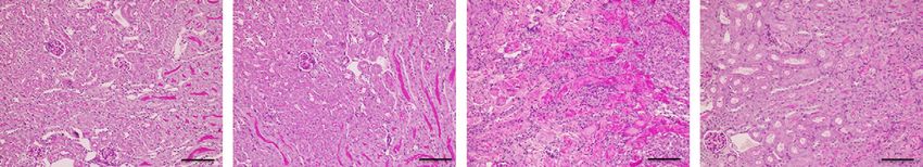
www.jasn.org      BASIC RESEARCH

empty plasmid was introduced using a Neon Transfection Sys-        the histologic examination of tubular necrosis. Semiquantita-
tem (Thermo Fisher Scientific) according to the manufac-            tive tubular injury scores were graded on the basis of the pro-
turer’s protocol. The overexpression of Tim3 was validated         portion of injured tubules as follows: (0) none, (1) ,25%, (2)
by immunocytochemistry.                                            25%–50%, (3) 50%–75%, and (4) .75%. The average score of
                                                                   four fields in the outer medulla was calculated for each sample.
Animal Experiments                                                     For Tim3 immunostaining, anti-Tim3 rabbit mAb (1:200,
Male mice (8–12 weeks of age, 20–25 g) were used for all           83882S; Cell Signaling, Danvers, MA) was used as the primary
experiments. Wild-type (WT) C57BL/6 mice were purchased            antibody. The sections were stained with Histofine Simple
from Nippon Bio-Supp. Center (Tokyo, Japan). Macrophage-           Stain Mouse MAX PO (R) (414341; Nichirei, Tokyo, Japan)
specific Adrb2 conditional knockout (cKO) mice were gener-          and ImmPACT DAB substrate (SK-4105; Vector, Burlingame,
ated by crossbreeding lysozyme M (LysM)-Cre and Adrb2 flox          CA), which were then counterstained with Mayer Hematoxy-
mice. Genotyping was confirmed by tail PCR using published          lin (Wako).
primers. LysM-Cre mice were obtained from the Jackson Lab-
oratory (Bar Harbor, ME). Adrb2 flox mice7 were provided by         Three-Dimensional Visualization of the SNS in the
Wataru Ogawa (Kobe University, Kobe, Japan) and Gerard             Spleen
Karsenty (Columbia University, New York, NY). All experi-          Three-dimensional (3D) visualization of the SNS in the spleen
ments were approved by the University of Tokyo Institutional       was performed using Clear, Unobstructed Brain/Body Imag-
Review Board (approval nos. P18–051 and H19–164). All an-          ing Cocktails and Computational (CUBIC)8–10 analysis as de-
imal procedures were performed according to the National           scribed in our previous papers.11,12 In brief, the fixed mouse
Institutes of Health guidelines (Guide for the Care and Use        spleen was immersed in CUBIC-L for delipidation and then
of the Laboratory Animals). BUN and plasma creatinine (Cre)        subjected to immunofluorescence staining. Finally, the refrac-
levels were measured by SRL Inc. (Osaka, Japan).                   tive index was matched by the placement of the sample in
                                                                   CUBIC-R1. The primary antibody used for staining was anti-
Preparation of Peritoneal Macrophages                              tyrosine hydroxylase antibody (sheep polyclonal, 1:100,
After the mouse was euthanized, 8 ml of sterile PBS was in-        ab113; Abcam, Cambridge, United Kingdom). The secondary
jected into the peritoneal cavity. The injected fluid was col-      antibody was Alexa Fluor 555–conjugated donkey anti-sheep
lected after a gentle massage of the peritoneum. The fluid was      IgG (1:100, A-21436; Invitrogen, Carlsbad, CA). The raw
centrifuged at 5003g for 5 minutes, and the supernatant was        image data were acquired with a custom-built light sheet
discarded. The cell pellets were resuspended in RPMI-1640          fluorescence microscopy (MVX10-LS; developed by Olympus,
media (R8758; Sigma-Aldrich) with 10% FBS and 1%                   Tokyo, Japan). The 3D-rendered image was visualized with
penicillin-streptomycin, and they were plated in culture           Imaris (Bitplane).
dishes. The cells were washed with PBS 1 hour after plating,
and the culture medium was refreshed. The next day, the cells      Adoptive Transfer of Splenic Macrophages
were used for experiments.                                         Spleens were harvested from donor mice, and single-cell sus-
                                                                   pensions were made through a 40-mm cell strainer with sterile
Renal Bilateral Ischemia-Reperfusion Injury                        PBS. The single-cell suspensions were labeled with anti-F4/80
Mice were anesthetized by the intraperitoneal administration       MicroBeads (130–110–443; Miltenyi Biotec, Bergisch Glad-
of medetomidine 0.3 mg/kg, butorphanol 5 mg/kg, and mid-           bach, Germany), and F4/80-positive splenocytes were selected
azolam 4 mg/kg. Renal bilateral ischemia-reperfusion injury        by the magnetic cell separation method. The cells were incu-
(bIRI) was performed by clamping the renal pedicles for            bated with vehicle or 100 mM salbutamol for 1 hour, and they
26 minutes. The clamps were removed, and the wound was             were washed twice with PBS. The macrophages were intrave-
sutured after restoration of blood flow was visually observed.      nously administered to recipient mice.
Sham-operated mice underwent the same procedure, without
clamping of the renal pedicles.                                    Single-Cell RNA Sequencing
                                                                   Digestion buffer was prepared as a mixture of DMEM–high-
Splenectomy                                                        glucose media (D5796) with 10% FBS (F7524, lot #BCBT
Mice were anesthetized by the intraperitoneal administration       3830), RQ1 RNase-Free DNase (20 U/ml, M6101; Promega,
of medetomidine 0.3 mg/kg, butorphanol 5 mg/kg, and mid-           Madison, WI), collagenase type 1 (2 mg/ml, CLS1; Worthing-
azolam 4 mg/kg. The splenic vasculature was ligated, and the       ton, Columbus, OH), collagenase type 2 (2 mg/ml, CLS2,
spleen was removed via a small incision.                           Worthington), and Dispase II (1 mg/ml, 04942078001; Roche,
                                                                   Mannheim, Germany). Kidneys were harvested, minced into
Immunohistochemistry                                               1-mm3 cubes, and digested using the digestion buffer with
Kidney samples were fixed in Mildform 10 N (133–10311;              shaking at 37°C. The supernatant buffer was collected after
Wako, Osaka, Japan) before being embedded in paraffin. Tis-         shaking for 10 minutes, and fresh digestion buffer was added
sue sections were subjected to periodic acid–Schiff staining for   to the remaining cell aggregations. After this process was

JASN 32: ccc–ccc, 2021                                                                      Macrophage Adrb2 Signaling in AKI     3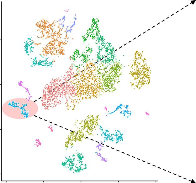
BASIC RESEARCH    www.jasn.org

                A                                                                                                                          ****
                          Plate RAW 264.7 cells
                                                                             4000                                     ****
                                                                                                                                                   ****
                                                                             3000

                                                            TNF-D (pg/mL)
                24h

                          LPS stimulation with
                                                                             2000
                             Vehicle
                             Norepinephrine (NE) 1 PM
                             NE 10 PM                                        1000
                 4h

                          Collect culture supernatant                                         0
                                                                                                      Vehicle 1 PM 10 PM            Vehicle 1 PM 10 PM
                                                                                                                  NE                                NE

                                                                                                            LPS (–)                         LPS (+)

                B
                          Plate RAW 264.7 cells                                                                  ****
                                                                                                                                         ***
                                                                                              5000
                                                                                                                                    **
                                                                                                                             n.s.
                24h
                                                                                              4000

                          LPS stimulation with
                                                                              TNF-D (pg/mL)

                             Vehicle                                                          3000
                             NE 1 PM
                             NE 1 PM + butoxamine 1-10 PM
                                                                                              2000

                 4h
                                                                                              1000

                          Collect culture supernatant
                                                                                                  0
                                                                                                            Vehicle     NE          1 PM       3 PM      10 PM
                                                                                                                                           Butoxamine
                                                                                                                                         NE 1 PM

                                                                                                                                LPS (+)

                C
                         Plate RAW 264.7 cells
                                                                             4000                                 ****                     ****
                                                                                                                                                   ****
                24h
                                                                             3000
                                                             TNF-D (pg/mL)

                         LPS stimulation with
                            Vehicle
                                                                             2000
                            Salbutamol 10 nM
                            Salbutamol 100 nM
                 4h
                                                                             1000

                         Collect culture supernatant
                                                                                              0
                                                                                                      Vehicle 10 nM 100 nM           Vehicle 10 nM 100 nM

                                                                                                              Salbutamol                        Salbutamol

                                                                                                            LPS (–)                            LPS (+)

Figure 1. Activation of sympathetic signaling suppresses the inflammatory response of macrophages via Adrb2. (A) TNF-a induction
by LPS was suppressed by NE treatment in RAW 264.7 cells (n56). (B) Butoxamine mitigated the anti-inflammatory effect of NE in RAW
264.7 cells (n58). (C) Salbutamol also suppressed TNF-a induction by LPS in RAW 264.7 cells (n56). All data are presented as means 6
SEM. Statistical comparisons were analyzed by (B) one-way ANOVA or (A and C) two-way ANOVA with a post hoc Tukey multiple
comparisons test. n.s., not significant. **P,0.01; ***P,0.001; ****P,0.0001.

4        JASN                                                                                                                                                JASN 32: ccc–ccc, 2021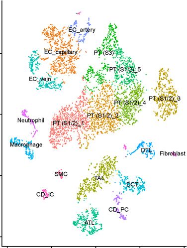
www.jasn.org BASIC RESEARCH
A B RAW 264.7 cells
(Tim3 expression)
**
relative expression/E-actin
707 genes induced by NE and
991 genes induced by salbutamol 1.5
suppressed by butoxamine
in Raw 264.7 cells
in Raw 264.7 cells
1.0
0.5
0.0
Vehicle Salbutamol
Pmacs
(Tim3 expression)
**
relative expression/E-actin
37 candidate genes 1.5
(including Tim3)
1,189 genes induced by 1.0
salbutamol in Pmacs
0.5
0.0
Vehicle Salbutamol
C
Plate RAW 264.7 cells
Tim3 expression
****
1.5
***
relative expression/E-actin
24h
Vehicle 1.0
Salbutamol 100 nM
Salbutamol 100 nM+ PKI 5 PM
0.5
4h
RNA extraction 0.0
Vehicle Salbutamol Salbutamol
+ PKI
D
Plate RAW 264.7 cells
(Two-way ANOVA interaction P value = 0.02)
24h ****
siNC (negative control) 4000 ****
or
–14%
siTim3 ****
24h 3000
–22%
TNF-D (pg/mL)
Cell passage
2000
24h
LPS stimulation with
1000
Vehicle
Salbutamol 10 nM
4h
0
Vehicle Salbutamol Vehicle Salbutamol
Collect culture supernatant
siNC siTim3
Figure 2. Tim3 is downstream of Adrb2 signaling and partially mediates its anti-inflammatory effects. (A) RNA sequencing was con-
ducted for three different in vitro macrophage models. The detailed experiment protocols are shown in Supplemental Figure 3: (1) 707
genes were extracted with log fold change (log FC) more than one (NE per vehicle) and log FC less than zero (NE 1 butoxamine/NE),
(2) 991 genes were extracted with log FC more than one (salbutamol per vehicle), and (3) 1189 genes were extracted with log FC more
JASN 32: ccc–ccc, 2021 Macrophage Adrb2 Signaling in AKI 5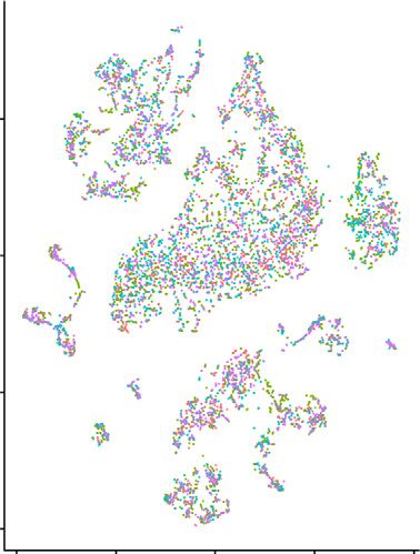
BASIC RESEARCH    www.jasn.org

repeated four times, the solution was passed through 70- and        The data were deposited in the GEA under accession number
40-mm cell strainers successively. The solution was then            E-GEAD-405.
centrifuged at 3003g for 5 minutes, and the resulting cell
pellet was diluted with the digestion buffer again. The solution    Statistical Analyses
was passed through 40-mm cell strainers three times, and a          All data are presented as means 6 SEM. An unpaired two-
single-cell suspension was obtained.                                tailed t test was used to analyze the data for only two groups.
   The single-cell suspension was loaded onto a well on the         For multiplex comparisons, a one-way or two-way ANOVA
103 Chromium Single Cell instrument (103 Genomics,                  followed by a post hoc Tukey multiple comparisons test, if
Pleasanton, CA). Bar coding and cDNA synthesis were per-            appropriate, was applied. P,0.05 was considered statistically
formed using Chromium Single Cell 3’ Reagent Kits v3.1              significant. All statistical analyses were performed with
(103 Genomics) according to the manufacturer’s instruc-             GraphPad Prism 8 software (GraphPad Software, San
tions. High-throughput sequencing was performed using a             Diego, CA).
NovaSeq6000 (Illumina) system at Takara Bio Inc. (Shiga,
Japan).
                                                                    RESULTS
Data Processing and Analysis of Single-Cell
RNA-Sequencing Data                                                 Activation of Sympathetic Signaling Suppresses the
The raw data were processed using Cell Ranger v3.1 (103             Inflammatory Response of Macrophages via Adrb2
Genomics) to obtain the filtered feature bar code matrices.          We first examined the effects of sympathetic signaling on the
Seurat v3 was used for the detailed analysis. We analyzed           inflammatory response of macrophages using RAW 264.7 cells
each sample separately and excluded cells with ,200 or              (mouse macrophage cell line). Norepinephrine (NE), a sym-
.5000 genes detected or with ,20,000 unique molecular               pathetic neurotransmitter, suppressed TNF-a induction by
identifiers detected. We also excluded cells with a relatively       LPS in a dose-dependent manner (Figure 1A). This anti-
high percentage of genes mapped to mitochondrial genes              inflammatory effect was mitigated by butoxamine, a selective
($50%). Subsequently, we log normalized the data and ob-            Adrb2 antagonist (Figure 1B). The dose-dependent anti-
tained 2000 highly variable genes for principal component           inflammatory effect was also induced by salbutamol, a selec-
analysis (PCA) from each dataset with “FindVariableFea-             tive Adrb2 agonist (Figure 1C, Supplemental Figure 1). Thus,
tures.” We then merged the list with the standard workflow           activation of sympathetic signaling suppresses the inflamma-
using “FindIntegrationAnchors” and “IntegrateData.” We              tory response of macrophages via Adrb2. Furthermore, we
subsequently performed PCA for the integrated data using            conducted the same experiment using mouse peritoneal mac-
the variable genes and determined significant principal com-         rophages and differentiated U937 cells (human macrophages)
ponents on the basis of the jackstraw. Clustering was per-          and confirmed that the anti-inflammatory effect of Adrb2 sig-
formed using “FindNeighbors” and subsequently, “FindClus-           naling was common in macrophages of various origins
ters” with a resolution of 0.6. We visualized the data on           (Supplemental Figure 2).
t-distributed stochastic neighbor embedding (tSNE) using
“RunTSNE.” Marker genes in each cluster were identified us-          Tim3 Is a Mediator of Anti-Inflammatory Effect Induced
ing “FindAllMarkers” with min.pct50.25 and logfc.thres-             by Adrb2 Signaling
hold50.25. The marker genes sorted by the average log fold          We attempted to identify the critical gene that mediates the
change are presented in Supplemental Table 2. Macrophage            anti-inflammatory effect of Adrb2 signaling. RNA sequencing
subclustering was performed as follows. First, we extracted the     was conducted for three different in vitro macrophage models.
macrophage cluster and changed the default assay from “in-          Adrb2 signaling–induced genes were respectively selected in
tegrated” to “RNA.” Then, we log normalized the data and            each model: (1) 707 genes, (2) 991 genes, and (3) 1189 genes
obtained 2000 highly variable genes for PCA with “FindVar-          (Figure 2A, Supplemental Figure 3). Among 37 genes, which
iableFeatures.” Clustering was performed using “FindNeigh-          were commonly selected in the three models (Supplemental
bors” and subsequently, “FindClusters” with a resolution            Table 3), we focused on Tim3, given its reported anti-
of 0.8. We visualized the data on tSNE using “RunTSNE.”             inflammatory role in immune cells. qPCR also confirmed

than one (salbutamol per vehicle). A total of 37 genes, including Tim3, were commonly induced by the activation of Adrb2 signaling in
the three different models. (B) qPCR confirmed that Tim3 expression was upregulated by the salbutamol treatment in RAW 264.7 cells
and peritoneal macrophages (Pmacs; n53). (C) Tim3 induction by salbutamol was counteracted by the inhibition of PKA in RAW 264.7
cells (n56). (D) Tim3 knockdown by siRNA partly inhibited the anti-inflammatory effect of salbutamol in RAW 264.7 cells (n56). All data
are presented as means 6 SEM. (B) An unpaired two-tailed t test was used to analyze the data for only two groups. For multiplex
comparisons, (C) a one-way ANOVA or two-way (D) ANOVA with a post hoc Tukey multiple comparisons test was applied. PKI, protein
kinase A inhibitor; siNC, negative control siRNA; siTim3, Tim3 knockdown by siRNA. **P,0.01; ***P,0.001; ****P,0.0001.

6        JASN                                                                                                    JASN 32: ccc–ccc, 2021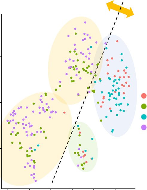
www.jasn.org BASIC RESEARCH
A
600 ****
LPS administration with
Vehicle
****
Salbutamol (15 mg/kg)
400
TNF-D (pg/mL)
4h
200 n.s.
Euthanize
C57BL/6 (WT)
0
Vehicle Salbutamol Vehicle Salbutamol
LPS (–) LPS (+)
*
15000
**** 5000 ***
4000
10000
IL-10 (pg/mL)
IL-6 (pg/mL)
3000
n.s.
2000
5000 n.s.
n.s.
1000
0 0
Vehicle Salbutamol Vehicle Salbutamol Vehicle Salbutamol Vehicle Salbutamol
LPS (–) LPS (+) LPS (–) LPS (+)
B (Two-way ANOVA interaction P value = 0.01)
LPS administration with n.s.
Vehicle 500
Salbutamol (15 mg/kg)
***
400 ****
4h
TNF-D (pg/mL)
300 –53%
Euthanize
–82%
200
100
Adrb2 flox/flox LysM-Cre: Adrb2 flox/flox
(Littermate WT) (Adrb2 cKO) 0
Vehicle Salbutamol Vehicle Salbutamol
Littermate WT Adrb2 cKO
Figure 3. Adrb2 signaling in macrophages plays a critical role in the in vivo systemic inflammatory response. (A) LPS was in-
traperitoneally administered to WT mice immediately after the intraperitoneal administration of vehicle or salbutamol. Plasma TNF-a
and IL-6 levels were reduced, whereas plasma IL-10 level was increased by the salbutamol treatment (n53 or n55). (B) Macrophage-
specific Adrb2 cKO mice were generated by crossbreeding LysM-Cre and Adrb2 flox mice. LPS was intraperitoneally administered to
Adrb2 cKO mice and their littermate WT mice immediately after the intraperitoneal administration of vehicle or salbutamol. Deletion of
Adrb2 on macrophages partially abolished the salbutamol-induced suppression of the systemic inflammatory response (n510). All data
are presented as means 6 SEM. Statistical comparisons were analyzed by a two-way ANOVA with a post hoc Tukey multiple com-
parisons test. n.s., not significant. *P,0.05; ***P,0.001; ****P,0.0001.
JASN 32: ccc–ccc, 2021 Macrophage Adrb2 Signaling in AKI 7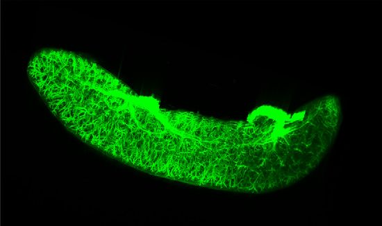
BASIC RESEARCH www.jasn.org
A 24h 24h
Vehicle • Sham Euthanize
• Renal blRI
Salbutamol (15 mg/kg)
C57BL/6 (WT)
Clamp
B **** ****
250 * 2.5 **
200 2.0
BUN (mg/dL)
Cre (mg/dL)
150 1.5
100 1.0
n.s. n.s.
50 0.5
0 0.0
Vehicle Salbutamol Vehicle Salbutamol Vehicle Salbutamol Vehicle Salbutamol
Sham bIRI Sham bIRI
C Vehicle Salbutamol Vehicle Salbutamol
(sham) (sham) (bIRI) (bIRI)
D **** E
Sympathetic nerves in the spleen
4
*
Tubular injury score
3
2
1 n.s.
0
2 mm
Vehicle Salbutamol Vehicle Salbutamol
Sham bIRI
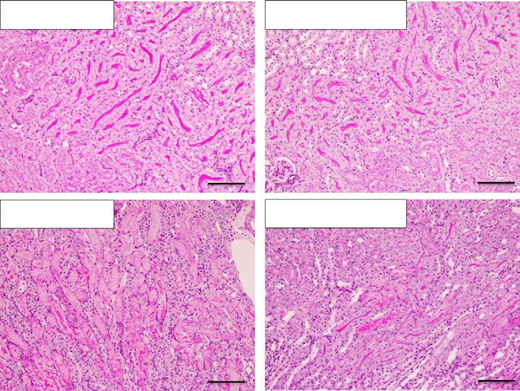
Figure 4. Pretreatment with salbutamol protects against renal ischemia-reperfusion injury. (A) The study protocol is shown. Vehicle or
salbutamol (15 mg/kg) was intraperitoneally administered to WT mice 24 hours before renal bIRI. Blood and kidney samples were
obtained 24 hours after bIRI. (B) BUN and plasma Cre levels are shown (n53 or n56). (C) Representative periodic acid–Schiff staining of
the renal outer medulla is shown. Scale bars: 100 mm. (D) Histologic tubular injury scores are shown (n53 or n56). (E) The distribution of
SNS in the spleen is visualized by tissue clearing and 3D immunofluorescence staining of tyrosine hydroxylase. All data are presented
as means 6 SEM. Statistical comparisons were analyzed by a two-way ANOVA with a post hoc Tukey multiple comparisons test. n.s.,
not significant. *P,0.05; **P,0.01; ****P,0.0001.
8 JASN JASN 32: ccc–ccc, 2021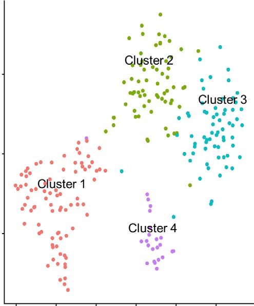
www.jasn.org        BASIC RESEARCH

                 A                                                               24h                                                             24h

                                                                                                    Renal bIRI                                                    Euthanize
                     A: Vehicle (to littermate WT mice)
                     B: Salbutamol (to littermate WT mice)
                     C: Salbutamol (to Adrb2 cKO mice)

                                                                                                          Clamp

                  Adrb2 flox/flox              LysM-Cre: Adrb2 flox/flox
                 (Littermate WT)                    (Adrb2 cKO)

   B                                                                                                                  D
                 400                                                            2.5        **                                                5             ****
                                  *                                                                                                                                    ****
                                                                                2.0
                                                                                                     **
                                           (P = 0.055)                                                                                       4

                                                                                                                      Tubular injury score
                 300
   BUN (mg/dL)

                                                                  Cre (mg/dL)

                                                                                1.5                                                          3
                 200
                                                                                1.0                                                          2

                 100
                                                                                0.5                                                          1

                     0                                                          0.0                                                          0
                             A         B                 C                             A        B            C                                         A          B           C

   C
                         A                                                        B                                               C

Figure 5. Macrophage Adrb2 signaling is critical for the protective effect of salbutamol pretreatment against renal ischemia-
reperfusion injury. (A) The study protocol is shown. Macrophage-specific Adrb2 cKO and littermate WT mice were used in this ex-
periment. Vehicle or salbutamol (15 mg/kg) was intraperitoneally administered 24 hours before renal bIRI. Blood and kidney samples
were obtained 24 hours after bIRI. (B) BUN and plasma Cre levels are shown (n515–17). (C) Representative periodic acid–Schiff staining
of the renal outer medulla is shown. Scale bars: 100 mm. (D) Histologic tubular injury scores are shown (n512–14). All data are pre-
sented as means 6 SEM. Statistical comparisons were analyzed by a one-way ANOVA with a post hoc Tukey multiple comparisons test.
*P,0.05; **P,0.01; ****P,0.0001.

that Tim3 expression was upregulated by the salbutamol treat-                                   downstream of Adrb2 signaling and partially mediates the
ment in RAW 264.7 cells and peritoneal macrophages                                              anti-inflammatory phenotypic alteration, which cannot be
(Figure 2B). Moreover, the inhibition of PKA, the canonical                                     simply explained by the conventional macrophage M1/M2
Adrb2 signaling downstream pathway component, counter-                                          axis (Supplemental Figure 6).
acted the salbutamol-induced upregulation of Tim3 expres-
sion in macrophages, confirming that Tim3 is downstream of                                       Adrb2 Signaling in Macrophages Plays a Critical Role in
Adrb2 signaling (Figure 2C).                                                                    the In Vivo Systemic Inflammatory Response
   Next, we examined the anti-inflammatory role of Tim3                                          Next, we attempted to clarify the role of macrophage Adrb2
expression in the macrophage inflammatory response. Tim3                                         signaling in the systemic inflammatory response using the
knockdown by siRNA partially inhibited the anti-                                                LPS-induced mouse septic model. First, LPS (5 mg/kg) was
inflammatory effect of salbutamol in RAW 264.7 cells                                             intraperitoneally administered to WT mice, immediately after
(Figure 2D, Supplemental Figure 4). In contrast, Tim3 over-                                     intraperitoneal administration of vehicle or salbutamol
expression suppressed the inflammatory response of                                               (15 mg/kg). We measured plasma cytokine levels as the pa-
RAW 264.7 cells (Supplemental Figure 5). Thus, Tim3 is                                          rameter of the systemic inflammatory response 4 hours after

JASN 32: ccc–ccc, 2021                                                                                                                       Macrophage Adrb2 Signaling in AKI         9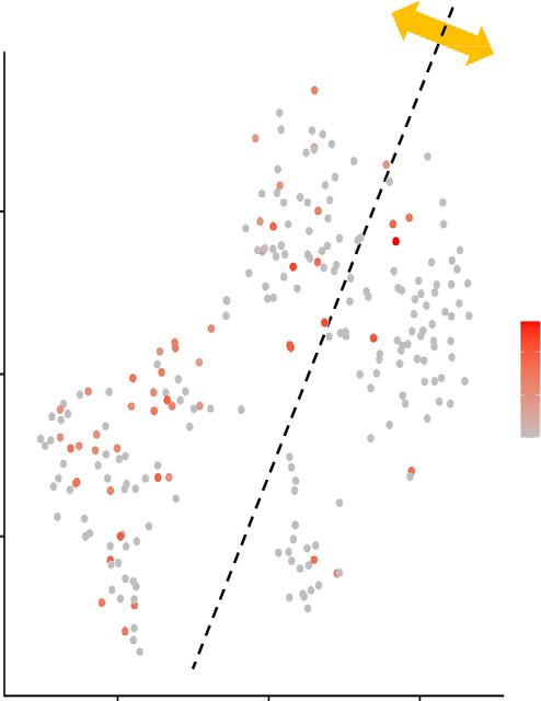
BASIC RESEARCH                    www.jasn.org

     A                                   Treatment (1h)
                                            Vehicle
                                                                                             18h                                               24h
                                            Salbutamol

          Isolate MM from                                     Adoptive transfer                          • Sham                                          Euthanize
          donors’ spleens                                      (1.6 × 10⁵, i.v.)                         • Renal blRI

                                             MM

                                          Treatment

         Donor                                                        Recipient                                         Clamp
         (WT)                                                          (WT)

     B                                       ***                                                                                          **
                          250
                                                                **                               2.5                                                       **

                          200                                                                    2.0
                                                                                   Cre (mg/dL)
            BUN (mg/dL)

                          150                                                                    1.5

                          100                                                                    1.0
                                      n.s.                                                                    n.s.
                          50                                                                     0.5

                           0                                                                      0
                                Vehicle Salbutamol        Vehicle Salbutamol                           Vehicle Salbutamol                            Vehicle Salbutamol
                                    Sham                       bIRI                                         Sham                                           bIRI

     C
         Vehicle (sham)                                    Salbutamol (sham)

                                                                                                                                      4
                                                                                                                                                          ***
                                                                                                                                                                               *
                                                                                                               Tubular injury score

                                                                                                                                      3

                                                                                                                                      2

         Vehicle (bIRI)                                    Salbutamol (bIRI)
                                                                                                                                      1
                                                                                                                                                 n.s.

                                                                                                                                      0
                                                                                                                                           Vehicle Salbutamol        Vehicle Salbutamol
                                                                                                                                                Sham                          bIRI

Figure 6. Adoptive transfer of salbutamol-treated macrophages protects against renal ischemia-reperfusion injury. (A) The study
protocol is shown. Salbutamol-treated macrophages from donor mice were adoptively transferred to recipient mice 18 hours before
bIRI. Blood and kidney samples were obtained 24 hours after bIRI. (B) BUN and plasma Cre levels are shown (n53 or n57). (C)
Representative periodic acid–Schiff staining of the renal outer medulla and histologic tubular injury scores are shown (n53 or n57). All
data are presented as means 6 SEM. Statistical comparisons were analyzed by a two-way ANOVA with a post hoc Tukey multiple
comparisons test. Mw, macrophage; n.s., not significant; i.v., intravenous. Scale bars: 100 mm. *P,0.05; **P,0.01; ***P,0.001.

10          JASN                                                                                                                                                          JASN 32: ccc–ccc, 2021
www.jasn.org BASIC RESEARCH
A Treatment (1 h)
• Vehicle
18h 24h
• Salbutamol
Isolate MM from Adoptive transfer • Sham Euthanize
donors' spleens (1.6 × 10⁵, i.v.) • Renal blRI pp
scRNA-seq
(renal tissue)
MM
Ctl_sham (BUN 23.2 mg/dL, Cre 0.05 mg/dL)
Treatment
Ctl_blRI (BUN 158.5 mg/dL, Cre 0.70 mg/dL)
Donor Recipient Sal_sham (BUN 24.2 mg/dL, Cre 0.09 mg/dL)
(WT) (WT)
Sal_blRI (BUN 121.9 mg/dL, Cre 0.46 mg/dL)
B
PT (S1/2)_1
EC_capillary
25 25 PT (S1/2)_2
PT (S1/2)_3
TAL
PT (S1/2)_4
PT(S3)
Ctl_sham PT (S1/2)_5
tSNE_2
tSNE_2
0 Ctl_bIRI 0 ATL
EC_vein
Sal_sham DCT
Sal_bIRI Macrophage
DTL
EC_artery
–25 –25 CD_PC
Neutrophil
Fibroblast
CD_IC
SMC
–50 –50
–50 –25 0 –25 50 –50 –25 0 –25 50
tSNE_1 tSNE_1
C Kim1 expressions in PT (S3) D Tim3 expressions in MM
4
2
3
Expression Level
Expression Level
2
1
1
0 0
Ctl_sham Ctl_bIRI Sal_sham Sal_bIRI Ctl_sham Ctl_bIRI Sal_sham Sal_bIRI
Figure 7. Tim3-expressing macrophages are accumulated in the injured kidney after salbutamol-treated macrophage transfer. (A) The
study protocol is shown. Four renal tissues from the adoptive transfer experiment were analyzed by scRNA-seq. The BUN and plasma
Cre levels of each mouse are shown. (B) tSNE plots of the single-cell data are visualized with sample information (left panel) and cell-
type information (right panel). The total numbers of cells in each sample are as follows: 1667 cells (sham operation after vehicle-treated
macrophage transfer [Ctl_sham]), 2128 cells (bilateral ischemia-reperfusion injury after vehicle-treated macrophage transfer [Ctl_bIRI]),
1653 cells (sham operation after salbutamol-treated macrophage transfer [Sal_sham]), and 1932 cells (bilateral ischemia-reperfusion
injury after salbutamol-treated macrophage transfer [Sal_bIRI]). (C) The violin plot of kidney injury molecule 1 (Kim1) expressions in the
PT (S3) cluster is drawn. The total numbers of cells in this cluster are as follows: 128 cells (Ctl_sham), 139 cells (Ctl_bIRI), 93 cells
(Sal_sham), and 136 cells (Sal_bIRI). (D) The violin plot of Tim3 expressions in the macrophage cluster is drawn. The total numbers of
JASN 32: ccc–ccc, 2021 Macrophage Adrb2 Signaling in AKI 11BASIC RESEARCH www.jasn.org
LPS administration (Figure 3A). As a result, plasma TNF-a clearing–based 3D immunofluorescence staining of sympa-
and IL-6 levels were significantly lower in the salbutamol- thetic nerves (Figure 4E). We conducted a splenectomy
treated group than in the vehicle-treated group, whereas the 10 days before the bIRI experiment. As a result, the protec-
level of IL-10, an anti-inflammatory cytokine, was higher in tive effects of salbutamol against renal bIRI were not ob-
the salbutamol-treated group. Although renal histologic in- served in splenectomized mice (Supplemental Figure 8),
jury was not apparently observed at this stage, the expression suggesting the importance of splenic immune cells in the
of neutrophil gelatinase–associated lipocalin, one of the AKI renoprotective effects of salbutamol.
biomarkers, was significantly reduced in the salbutamol-
treated group, suggesting the protective effects of Adrb2 sig- Macrophage Adrb2 Signaling Is Critical for the
naling against kidney injury (Supplemental Figure 7). Protective Effect of Salbutamol against Renal bIRI
However, we were unable to determine whether the sys- In order to determine whether macrophages are involved in
temic anti-inflammatory effect of salbutamol was associated the salbutamol-induced protection against renal bIRI, we con-
with macrophages because sympathetic signaling plays vari- ducted renal bIRI experiments using macrophage-specific
ous physiologic roles in many cell types in the body. Thus, we Adrb2 cKO and littermate WT mice (Figure 5A). As a result,
generated macrophage-specific Adrb2 cKO mice by cross- deletion of Adrb2 on macrophages abolished the salbutamol-
breeding LysM-Cre and Adrb2 flox mice and conducted the induced protection against renal bIRI, as shown by the reversal
same experiment (Figure 3B). Deletion of Adrb2 on macro- of plasma Cre levels (Figure 5B) and histologic tubular injury
phages then partially abolished the salbutamol-induced sup- scores (Figure 5, C and D). Thus, the protective effects of
pression of the systemic inflammatory response, showing the salbutamol against renal bIRI are primarily due to the activa-
importance of macrophages in this context. tion of Adrb2 signaling in macrophages.
Salbutamol Pretreatment Protects the Kidney from Adoptive Transfer of Salbutamol-Treated
bIRI Macrophages Protects the Kidney from bIRI
Thus far, we clarified that Adrb2 signaling on macrophages Given that Adrb2 signaling on macrophages is critical to pro-
plays a critical role in the systemic inflammatory response. tection against kidney injury, we pondered whether Adrb2
Next, we aimed to determine the role of macrophage Adrb2 signal–activated macrophages themselves provide protec-
signaling in local acute inflammation. For this purpose, we tion against renal bIRI and conducted the adoptive transfer
opted to utilize the renal bIRI model, which is generated by experiment (Figure 6A). Adoptive transfer of 1.63 10 5
26 minutes of renal ischemia followed by 24 hours of salbutamol-treated (Adrb2 signal–activated) macrophages
reperfusion. from donor mice protected the kidneys from bIRI in recip-
Vehicle or salbutamol (15 mg/kg) was intraperitoneally ient mice, as shown by the lower BUN, lower plasma Cre
administered 24 hours before renal bIRI. Blood and kidney (Figure 6B), and lesser degree of histologic tubular injury
samples were obtained 24 hours after renal bIRI (Figure 4A). (Figure 6C).
Pretreatment with salbutamol provided strong protection Next, we performed single-cell RNA sequencing (scRNA-
from kidney injury as shown by the lower BUN, lower plasma seq) of renal tissues to analyze the renoprotective role of
Cre (Figure 4B), and lesser degree of histologic tubular injury salbutamol-treated (Adrb2 signal–activated) macrophages in
(Figure 4, C and D). Thus, systemic pretreatment with salbutamol detail (Figure 7A). We visualized the single-cell datasets using
protects the kidney from bIRI. tSNE and identified 19 cell-type clusters (Figure 7B,
Supplemental Table 2). For example, the expression of kidney
Splenic Immune Cells May Play Important Roles in the androgen–regulated protein clearly identified proximal tubu-
Protective Effect of Salbutamol against Renal bIRI lar cells in the S3 segment, the most vulnerable sections to
Next, we tested whether immune systems were involved in the ischemic injury (Figure 7B, Supplemental Figure 9A). The
salbutamol-induced protection against renal bIRI. The spleen violin plot of kidney injury molecule 1 (Kim1) expression
is central to the immune system, and splenic immune cell in this cluster showed that the degree of tubular injury was
dynamics are thought to be influenced by the degree of sym- lower in the bIRI after salbutamol-treated macrophage trans-
pathetic signaling because the sympathetic nerves are fer (Sal_bIRI) condition than in the bIRI after vehicle-treated
densely distributed in the spleen, as visualized by our tissue macrophage transfer (Ctl_bIRI) condition (Figure 7C).
cells in this cluster are as follows: 30 cells (Ctl_sham), 71 cells (Ctl_bIRI), 45 cells (Sal_sham), and 91 cells (Sal_bIRI). Tim3-expressing
macrophages were accumulated in the injured kidney after salbutamol-treated macrophage transfer (17% [Ctl_sham], 21% [Ctl_bIRI],
4% [Sal_sham], and 33% [Sal_bIRI] of total macrophages in the renal tissue). ATL, ascending thin limb; CD_IC, intercalated cell of the
collecting duct; CD_PC, principal cell of the collecting duct; DCT, distal convoluted tubular cell; DTL, descending thin limb; EC,
endothelial cell; Mw, macrophage; PT (S1/2), proximal tubular cell in the S1/2 segments; PT (S3), proximal tubular cell in the S3
segment; SMC, smooth muscle cell; TAL, thick ascending limb; i.v., intravenous.
12 JASN JASN 32: ccc–ccc, 2021www.jasn.org BASIC RESEARCH
A Sub-clustering of macrophages
25
5
tSNE_2
tSNE_2
0
0
Macrophage
–25
–5
–50
–50 –25 0 –25 50 –15 –10 –5 0 5 10
tSNE_1
tSNE_1
B C Tim3
Circulating MM Circulating MM
Resident MM Resident MM
Cluster 2
(bIRI)
25 Cluster 3 5
(sham)
Cluster 1
tSNE_2
(bIRI) 2
tSNE_2
Ctl_sham
0 0
Ctl_bIRI 1
Sal_sham 0
Sal_bIRI
–5 –5
Cluster 4
(sham & bIRI)
–15 –10 –5 0 5 10 –10 0 10
tSNE_1 tSNE_1
Figure 8. The accumulating Tim3-expressing macrophages after bIRI are mainly composed of circulating macrophages. (A) Unbiased
subclustering of macrophages is visualized as the tSNE plots. (B) In the tSNE plots with sample information, clusters 1 and 2 are mostly
composed of macrophages from bIRI groups (circulating macrophages), whereas cluster 3 is composed of macrophages from sham
groups, and cluster 4 contains macrophages from both groups (tissue-resident macrophages). (C) The expression levels of Tim3 are
visualized on the tSNE plots. Tim3-expressing macrophages were preferentially distributed on the left side (circulating macrophages).
Ctl_bIRI, bilateral ischemia-reperfusion injury after vehicle-treated macrophage transfer; Ctl_sham, sham operation after vehicle-treated
macrophage transfer; Mw, macrophage; Sal_bIRI, bilateral ischemia-reperfusion injury after salbutamol-treated macrophage transfer;
Sal_sham, sham operation after salbutamol-treated macrophage transfer.
JASN 32: ccc–ccc, 2021 Macrophage Adrb2 Signaling in AKI 13BASIC RESEARCH www.jasn.org
We especially focused on Tim3 expression in the macro- restore kidney function after renal IRI via mitochondrial bio-
phage cluster because high Tim3 expression is a marker of genesis in the proximal tubules.20,21 In contrast, we observed
Adrb2 signal–activated macrophages according to our the anti-inflammatory effect of salbutamol pretreatment in
in vitro data (Figure 2). The macrophage cluster was clearly the acute phase of renal IRI, which was mainly derived from
identified in the tSNE plot of single-cell data (Figure 7B) by the macrophage Adrb2 signaling (Figures 4 and 5, Supplemental
expression of two representative markers, Cd68 and LysM Figure 8). The adoptive transfer of Adrb2 signal–activated
(Supplemental Figure 9B). The violin plot of Tim3 expression macrophages also protected the kidney from IRI (Figures
in this cluster illustrated that Tim3-expressing macrophages 6–8). Taken together, the critical receiver of Adrb2 signaling
(Adrb2 signal–activated macrophages) were particularly accu- in renal IRI may differ depending on the timing: Macrophages
mulated in the renal tissue of the bIRI after salbutamol-treated are important in the acute injury phase, and proximal tubules
macrophage transfer condition (Figure 7D), which was also are important in the recovery phase. Future studies are needed
confirmed by immunohistochemistry (Supplemental to clarify the interaction between these receivers of Adrb2
Figure 10). Furthermore, we investigated the origin of these signaling and the renal SNS. Prior denervation of renal SNS
Tim3-expressing macrophages from scRNA-seq data. Unbi- is known to alleviate the severity of renal IRI in animal stud-
ased subclustering of macrophages yielded four subclusters ies.22 In contrast, we have previously shown that the renal SNS
(Figure 8A). Clusters 1 and 2 were mostly composed of mac- is denervated after IRI, which results in NE depletion inside
rophages from bIRI groups (circulating macrophages), the kidney.11 Thus, determination of the influence of renal
whereas cluster 3 was composed of macrophages from sham SNS activity or damage on the dynamics of macrophages
groups, and cluster 4 contained macrophages from both and the proximal tubules may aid elucidation of the SNS’s
groups (tissue-resident macrophages) (Figure 8B). As Tim3 role in kidney disease progression.
was preferentially expressed on the left side of the tSNE plots In this study, we also found that Tim3 expression was in-
(Figure 8C), the accumulating Tim3-expressing macrophages duced by the activation of Adrb2 signaling in macrophages
after bIRI might be circulating macrophages. Therefore, (Figure 2). Tim3 was included in the 37 genes, which were
Adrb2 signal–activated macrophages with high Tim3 expres- commonly selected as Adrb2 signal–induced genes from our
sion may come from outside the renal tissues and play critical RNA sequencing data of three in vitro macrophage models
roles in the protection against renal bIRI. (Supplemental Table 3). Tim3 is expressed on many immune
cells, including T cells,23,24 natural killer cells,25 and dendritic
cells,26 and suppresses the immunologic activity as one of the
DISCUSSION immune checkpoints. Previous studies have shown that
Tim3 is also expressed on macrophages and plays immu-
In this study, we demonstrated that activation of Adrb2 sig- nosuppressive roles. 27–30 In our data, Tim3 knockdown
naling in macrophages blocks LPS-induced systemic inflam- promoted the inflammatory response of macrophages and
mation and protects the kidney from IRI. Furthermore, Adrb2 mitigated the anti-inflammatory effect of salbutamol (Figure 2D,
signaling induces Tim3 expression, which contributes to anti- Supplemental Figure 4). In contrast, Tim3 overexpression
inflammatory phenotypic alterations in macrophages. attenuated the inflammatory response of macrophages
Adrb2 agonists are known to exert anti-inflammatory ef- (Supplemental Figure 5). Taken together, these findings in-
fects in several inflammatory diseases, such as LPS-induced dicate that Tim3 is downstream of Adrb2 signaling and con-
sepsis3,13 and acute lung injury.14 However, these in vivo ob- tributes to its anti-inflammatory effect, which cannot be
servations could not precisely identify the cell type responsible simply explained by the conventional macrophage M1/M2
for the anti-inflammatory effect. In our data, the anti- axis (Supplemental Figure 6). In our scRNA-seq dataset,
inflammatory effect of salbutamol was mitigated in Tim3-expressing macrophages were accumulated in the re-
monocyte-derived macrophage-specific Adrb2 knockout nal tissue of IRI after the adoptive transfer of Adrb2 signal–
mice (Figure 3), demonstrating the importance of macro- activated macrophages (Figure 7D, Supplemental Figure 10),
phage Adrb2 signaling in this process. Although sympathetic in association with neutrophil’s phenotypic change and re-
signaling is known to affect other immune cells, including duced renal tubular injury (Figure 7C, Supplemental
CD4-positive T cells4 and tissue-resident macrophages,15,16 Figure 11–13). Thus, Tim3-expressing macrophages in-
our data clearly demonstrated the importance of monocyte- duced by Adrb2 signaling may elicit direct protective effects
derived macrophages as the critical receiver of sympathetic against renal IRI.
signaling in LPS-induced systemic inflammation and Although the mechanism of Tim3 induction by Adrb2 sig-
renal IRI. naling was not clarified in detail, we at least confirmed that
Adrb2 signaling has various effects on local inflammation Tim3 expression induced by Adrb2 activation was dependent
in the kidney (diabetic kidney disease,17 nephrotoxic injury,18 on PKA signaling (canonical downstream component of
and septic kidney injury19) because adrenergic receptors are Adrb2 signaling) in macrophages (Figure 2C). As cAMP/
ubiquitously expressed in the body. Of note, the Adrb2 agonist PKA signaling is reported to induce promoter/enhancer activ-
formoterol (postinjury administration) was reported to ity of Tim3 in Jurkat T cells,31 the induction of Tim3 in
14 JASN JASN 32: ccc–ccc, 2021www.jasn.org BASIC RESEARCH
macrophages may also be explained by promoter/enhancer providing the potential mechanism for the protection against
activity. renal IRI induced by the adoptive transfer of salbutamol-
The spleen is widely innervated by sympathetic nerves, treated macrophages. Second, our study did not examine
which is also illustrated in our immunofluorescence staining how the Adrb2 signal–activated macrophages affect other im-
(Figure 4E). Thus, splenic immune cells may be susceptible to mune cell dynamics, such as T cells, B cells, natural killer cells,
sympathetic signaling. In our experiments, prior splenectomy and dendritic cells. The renoprotective effect of the adoptive
abolished the protective effects of salbutamol against renal IRI transfer of salbutamol-treated macrophages might be partially
(Supplemental Figure 8). In addition, the adoptive transfer of induced by the interaction with other immune cells. The anal-
Adrb2 signal–activated splenic macrophages provided strong ysis of immune cell dynamics in the lymphoid tissues after the
protection against renal IRI (Figures 6–8). Thus, splenic mac- adoptive transfer of Adrb2 signal–activated macrophages
rophages may play important roles in the anti-inflammatory would provide additional insights concerning the role of
effects of systemic salbutamol administration. The critical role macrophages in the whole immune dynamics.
of the spleen in neuroimmune interactions was also reported In conclusion, the activation of Adrb2 signaling in macro-
in the cholinergic anti-inflammatory pathway.32–35 The cho- phages induces anti-inflammatory phenotypic alterations
linergic anti-inflammatory pathway, which is activated by partially via the induction of Tim3 expression, which blocks
electrical stimulation of the vagus nerve, is mediated by the LPS-induced systemic inflammation and protects against re-
spleen.36–38 Vagus nerve induces the activation of splenic sym- nal IRI. Our data provide important insights concerning
pathetic nerves, leading to the NE-induced activation of CD4- neuroimmune interactions in the pathophysiology of inflam-
positive T cells in the spleen.39–41 Various types of splenic matory diseases.
immune cells, including macrophages and T cells, may inter-
act with one another and alter the immunologic dynamics in
responding to sympathetic signaling. Further studies using DISCLOSURES
single-cell analysis are needed to elucidate the immunologic
dynamics in the spleen in response to splenic SNS activation. The Division of CKD Pathophysiology, University of Tokyo is financially
Finally, it is important to determine whether the anti- supported by Kyowa Kirin Co. Ltd. R. Inagi reports research funding from
inflammatory role of ADRB2 signaling can be applied in clin- Kyowa Kirin Co. Ltd. and Nipro; honoraria from Alexion, Bayer, Chugai,
Kyowa Kirin Co. Ltd., Mitsubishi Tanabe, MSD, Research and Development
ical treatment for inflammatory diseases. In our study, ADRB2
Support Center, Kowa, and Sumitomo Dainippon; scientific advisor or
activation induced an anti-inflammatory response in differ- membership as associate editor of Clinical and Experimental Pharmacology
entiated U937 cells (human macrophages) as well as mouse Physiology and K360 and as editorial board member of American Journal of
macrophages (Figure 1, Supplemental Figure 2), suggesting Physiology Renal Physiology, Kidney International, and Kidney Research and
that our concept can be applied to human inflammatory dis- Clinical Practice; and other interests/relationships as councilor and secretary
of the Japanese Society of Nephrology (international liaison committee) and
eases. Indeed, genetic variation of ADRB2 was associated with
the International Maillard Reaction Society (IMARS; chief editor of IMARS
increased mortality and more organ dysfunction in septic Highlights). I. Komuro reports research funding from AstraZeneca, Japan,
shock in a clinical study.42 However, a multicenter, random- Astellas Pharma Inc., Bayer Yakuhin, Ltd., Daiichi Sankyo Company, Limited,
ized controlled trial (BALTI-2) failed to show the benefits of Kowa Pharmaceutical Co. Ltd., Mitsubishi Tanabe Pharma Corporation, ONO
intravenous salbutamol treatment in the course of acute re- PHARMACEUTICAL CO., LTD., Otsuka Pharmaceutical Co., Ltd., Sanofi,
K.K., Sumitomo Dainippon Pharma Co., Ltd., Takeda Pharmaceutical Com-
spiratory distress syndrome.43 In this previous study, salbuta-
pany Limited, and Teijin Pharma Limited and honoraria from AstraZeneca,
mol treatment was poorly tolerated due to the problem of Japan, Daiichi Sankyo Company, Limited, Mitsubishi Tanabe Pharma Corpo-
hemodynamics, including tachycardia and arrhythmia.43 ration, ONO PHARMACEUTICAL CO., LTD., and Takeda Pharmaceutical
Thus, cell type–specific activation of ADRB2 signaling may Company Limited. M. Nangaku reports honoraria from AstraZeneca and
be needed to prevent such cardiac side effects. In our data, Boehringer Ingelheim and scientific advisor or membership with Akebia, As-
tellas, Bayer, Boehringer-Ingelheim, Daiichi-Sankyo, GlaxoSmithKline, Japan
macrophages were the critical receiver of Adrb2 signaling in
Tobacco Inc., Kyowa Kirin Co. Ltd., and Mitsubishi-Tanabe.E. A. Susaki and
the protection against renal IRI. As demonstrated in our ex- H. R. Ueda are coinventors of patents owned by RIKEN.E. A. Susaki reports
periment (Figures 6–8), the adoptive transfer of ADRB2 patents and inventions with Tokyo Chemical Industry, CO, LTD; speakers bu-
signal–activated immune cells is a promising candidate for reau with Tokyo Chemical Industry, CO, LTD; and other interests/relationships
clinical application, although infection risks during immune as a senior researcher at CUBICStars Co. H. R. Ueda reports ownership interest
in CUBICStars Inc.; research funding from Olympus Corporation; and patents
cell harvest and treatment cannot be ignored in the clinical and inventions with CUBICStars Inc. (inventions on CUBIC-HV reagents) and
setting. RIKEN (inventions on CUBIC reagents). All remaining authors have nothing
Our study has several limitations. First, the scRNA-seq data to disclose.
in our study lacked sufficient statistical power with respect to
the dispersion of Kim1 expression in proximal tubules and
Tim3 expression in macrophages in the renal tissues (Figure 7). FUNDING
Although we need to increase kidney samples to make a con-
clusion by the scRNA-seq data alone, the results were compat- This work was supported by Japan Society for the Promotion of Science
ible with in vitro and in vivo data in the rest of our study, (JSPS) Grant-in-Aid for Japan Society for JSPS Research Fellow JSPS
JASN 32: ccc–ccc, 2021 Macrophage Adrb2 Signaling in AKI 15BASIC RESEARCH www.jasn.org
KAKENHI grant 19J11928 (to S. Hasegawa); MSD Life Science Foundation (S. Supplemental Figure 9. Cell-type marker gene expression levels for unbi-
Hasegawa and T. Inoue); Japan Society for the Promotion of Science Grant-in- ased clustering of single-cell RNA sequencing data.
Aid for Scientific Research (B) JSPS KAKENHI grants 18H02727 (to R. Inagi) Supplemental Figure 10. Immunostaining of T cell Ig and mucin domain 3
and 18H02824 (to M. Nangaku); Kyowa Kirin Co. Ltd. (R. Inagi); Japan on renal tissues of the adoptive transfer experiment.
Agency for Medical Research and Development grants JP20gm6210013 (to Supplemental Figure 11. Assessment of infiltrating neutrophils in the sin-
T. Inoue) and JP20gm6210010 (to S. Nomura); Japan Society for the gle-cell RNA sequencing data.
Promotion of Science Grant-in-Aid for Research Activity Start-Up and for Supplemental Figure 12. M1/M2 marker gene expression levels on the mac-
Young Scientists JSPS KAKENHI grants 18H06192 (to T. Inoue) and rophage subclustering data in the single-cell RNA sequencing.
20K17242 (to T. Inoue); Kidney Foundation grant JKFB18-3 (to T. Inoue); Supplemental Figure 13. The adoptive transfer of salbutamol-treated mac-
Salt Science Research Foundation grant 1919 (to T. Inoue); Smoking Research rophages 2 days before renal ischemia-reperfusion injury does not provide
Foundation (to T. Inoue); Yukiko Ishibashi Foundation (to T. Inoue); protection.
Mochida Memorial Foundation (to T. Inoue); Takeda Science Foundation Supplemental Table 1. Primer sequences for the quantitative
(to T. Inoue); Astellas Foundation for Research on Metabolic Disorders (to real-time PCRs.
T. Inoue); and Suzuken Memorial Foundation (to T. Inoue). This work was Supplemental Table 2. A list of the marker genes of each cluster in the
partially carried out under the support of Isotope Science Center, University of single-cell RNA sequencing.
Tokyo. Part of this study was performed in collaboration with Olympus Cor- Supplemental Table 3. A list of the 37 genes commonly selected in the RNA
poration and with kind software support by Bitplane. sequencing from the three in vitro models.
ACKNOWLEDGMENTS REFERENCES
The authors are grateful to Dr. Naoki Kuramoto, Dr. Yu Hirata, Prof. Wataru
1. Felten DL, Felten SY, Carlson SL, Olschowka JA, Livnat S: Noradren-
Ogawa (Kobe University), and Prof. Gerard Karsenty (Columbia University)
ergic and peptidergic innervation of lymphoid tissue. J Immunol 135
for providing the Adrb2 flox mice. The authors also thank Ms. Ikumi
[Suppl]: 755s–765s, 1985
Okuaki, Ms. Rieko Matsuda, Ms. Kahoru Amitani, Ms. Sayaka Hayashi, and
2. Nance DM, Sanders VM: Autonomic innervation and regulation of the
Ms. Nanako Shida for their technical support.
immune system (1987-2007). Brain Behav Immun 21: 736–745, 2007
The illustrations of mice, kidneys, and petri dishes were obtained from
3. Agaç D, Estrada LD, Maples R, Hooper LV, Farrar JD: The b2-adrenergic
TogoTV (2016 DBCLS TogoTV).
receptor controls inflammation by driving rapid IL-10 secretion. Brain
Sho Hasegawa performed the main experiments, analyzed the data, and
Behav Immun 74: 176–185, 2018
wrote the original draft; Nobuyoshi Akimitsu, Rie Fujii, Daichi Fukaya,
4. Araujo LP, Maricato JT, Guereschi MG, Takenaka MC, Nascimento VM,
Tsuyoshi Inoue, Yasuna Nakamura, Atsuko Nakanishi Ozeki, Wachirasek
de Melo FM, et al.: The sympathetic nervous system mitigates CNS
Peerapanyasut, Rie Uni, and Chia-Hsien Wu performed or contributed to the
autoimmunity via b2-adrenergic receptor signaling in immune cells.
animal experiments; Takahide Kohro, Akashi Taguchi, and Youichiro Wada
Cell Rep 28: 3120–3130.e5, 2019
performed the Hiseq2500 run and data processing in the RNA sequencing
5. Nakai A, Hayano Y, Furuta F, Noda M, Suzuki K: Control of lymphocyte
analysis; Mikako Katagiri, Toshiyuki Ko, Issei Komuro, Seitaro Nomura, and
egress from lymph nodes through b2-adrenergic receptors. J Exp Med
Shintaro Yamada aided our scRNA-seq analysis; Etsuo A. Susaki and Hiroki R.
211: 2583–2598, 2014
Ueda aided 3D visualization using tissue clearing; Reiko Inagi, Tsuyoshi Inoue,
6. Arima Y, Harada M, Kamimura D, Park JH, Kawano F, Yull FE, et al.:
and Masaomi Nangaku supervised this study and revised the manuscript; and
Regional neural activation defines a gateway for autoreactive T cells to
all authors approved the final version of the manuscript.
cross the blood-brain barrier. Cell 148: 447–457, 2012
7. Hinoi E, Gao N, Jung DY, Yadav V, Yoshizawa T, Myers MG Jr, et al.: The
sympathetic tone mediates leptin’s inhibition of insulin secretion by
modulating osteocalcin bioactivity. J Cell Biol 183: 1235–1242, 2008
SUPPLEMENTAL MATERIAL 8. Susaki EA, Tainaka K, Perrin D, Kishino F, Tawara T, Watanabe TM,
et al.: Whole-brain imaging with single-cell resolution using chemical
This article contains the following supplemental material online at http:// cocktails and computational analysis. Cell 157: 726–739, 2014
jasn.asnjournals.org/lookup/suppl/doi:10.1681/ASN.2020121723/-/ 9. Kubota SI, Takahashi K, Nishida J, Morishita Y, Ehata S, Tainaka K, et al.:
DCSupplemental. Whole-body profiling of cancer metastasis with single-cell resolution.
Supplemental Figure 1. The effect of b2-adrenergic receptor signaling on Cell Rep 20: 236–250, 2017
various inflammatory cytokines. 10. Tainaka K, Murakami TC, Susaki EA, Shimizu C, Saito R, Takahashi K,
Supplemental Figure 2. The anti-inflammatory effect of b2-adrenergic re- et al.: Chemical landscape for tissue clearing based on hydrophilic re-
ceptor signaling is common in macrophages of various origins. agents. Cell Rep 24: 2196–2210.e9, 2018
Supplemental Figure 3. The detailed protocols of RNA sequencing 11. Hasegawa S, Susaki EA, Tanaka T, Komaba H, Wada T, Fukagawa M,
experiments. et al.: Comprehensive three-dimensional analysis (CUBIC-kidney) vi-
Supplemental Figure 4. The effect of T cell Ig and mucin domain 3 knock- sualizes abnormal renal sympathetic nerves after ischemia/reperfusion
down using another small interfering RNA on the inflammatory response of injury. Kidney Int 96: 129–138, 2019
macrophages. 12. Hasegawa S, Tanaka T, Saito T, Fukui K, Wakashima T, Susaki EA, et al.:
Supplemental Figure 5. The effect of T cell Ig and mucin domain 3 over- The oral hypoxia-inducible factor prolyl hydroxylase inhibitor enar-
expression on the inflammatory response of macrophages. odustat counteracts alterations in renal energy metabolism in the early
Supplemental Figure 6. The effect of b2-adrenergic receptor signaling on stages of diabetic kidney disease. Kidney Int 97: 934–950, 2020
macrophage M1/M2 markers. 13. Nakamura A, Imaizumi A, Yanagawa Y, Kohsaka T, Johns EJ: beta(2)-
Supplemental Figure 7. Assessment of renal damage in the LPS-induced Adrenoceptor activation attenuates endotoxin-induced acute renal
septic model. failure. J Am Soc Nephrol 15: 316–325, 2004
Supplemental Figure 8. Prior splenectomy abolishes the protective effect of 14. Grailer JJ, Haggadone MD, Sarma JV, Zetoune FS, Ward PA: Induction
salbutamol pretreatment against renal ischemia-reperfusion injury. of M2 regulatory macrophages through the b2-adrenergic receptor
16 JASN JASN 32: ccc–ccc, 2021You can also read