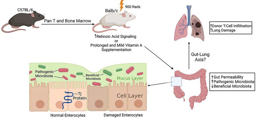
Retinoic Acid Signaling Modulates Recipient Gut Barrier Integrity and Microbiota After Allogeneic Hematopoietic Stem Cell Transplantation in Mice
←
→
Page content transcription
If your browser does not render page correctly, please read the page content below
ORIGINAL RESEARCH
                                                                                                                                        published: 25 October 2021
                                                                                                                                  doi: 10.3389/fimmu.2021.749002

                                          Retinoic Acid Signaling Modulates
                                          Recipient Gut Barrier Integrity
                                          and Microbiota After Allogeneic
                                          Hematopoietic Stem Cell
                                          Transplantation in Mice
                                          Pan Pan 1,2, Samantha N. Atkinson 3,4, Brian Taylor 1,2, Haojie Zhu 1,2, Dian Zhou 1,2,
                                          Philip Flejsierowicz 1,2, Li-Shu Wang 1,2, Matthew Morse 1,2, Chen Liu 5, Ian L. Gunsolus 6
                          Edited by:
                                          and Xiao Chen 1,2*
                       Xue-Zhong Yu,
 Medical University of South Carolina,    1 Division of Hematology & Oncology, Medical College of Wisconsin, Milwaukee, WI, United States, 2 Department of
                        United States
                                          Medicine, Medical College of Wisconsin, Milwaukee, WI, United States, 3 Center for Microbiome Research, Medical College
                      Reviewed by:        of Wisconsin, Milwaukee, WI, United States, 4 Department of Microbiology and Immunology, Medical College of Wisconsin,
                        Xuefang Cao,      Milwaukee, WI, United States, 5 Department of Pathology, Yale University School of Medicine, New Haven, CT, United
University of Maryland, United States     States, 6 Department of Pathology, Medical College of Wisconsin, Milwaukee, WI, United States
                            Meng Lv,
 Peking University People’s Hospital,
                               China      Graft-versus-host disease (GVHD) remains a major complication after allogeneic
                  *Correspondence:        hematopoietic stem cell transplantation (HSCT). An impaired intestinal epithelial barrier
                          Xiao Chen       is an important component of GVHD pathogenesis. However, contributing host factors
                    xchen@mcw.edu
                                          that modulate mucosal barrier integrity during GVHD are poorly defined. We hypothesized
                   Specialty section:     that vitamin A and retinoic acid (RA) exert positive impacts on maintaining intestinal barrier
         This article was submitted to    function after HSCT, thus preventing or dampening GVHD severity. Unexpectedly, we
    Alloimmunity and Transplantation,
               a section of the journal
                                          found that exogenous RA increased intestinal permeability of recipient mice after
              Frontiers in Immunology     allogeneic HSCT. Serum bacterial endotoxin levels were significantly higher in GVHD
           Received: 28 July 2021         mice fed a vitamin A-high (VAH) diet compared to those fed a vitamin A-normal (VAN) diet,
     Accepted: 30 September 2021          indicating a more compromised intestinal barrier function. Furthermore, VAH mice
       Published: 25 October 2021
                                          showed more severe lung GVHD with increased donor T cell infiltration in this tissue
                            Citation:
       Pan P, Atkinson SN, Taylor B,      and died significantly faster than VAN recipients. 16S rRNA sequencing of fecal samples
     Zhu H, Zhou D, Flejsierowicz P,      revealed significant differences in the diversity and composition of gut microbiota between
         Wang L-S, Morse M, Liu C,
     Gunsolus IL and Chen X (2021)
                                          VAN and VAH transplant recipients. Collectively, we show that retinoic acid signaling may
   Retinoic Acid Signaling Modulates      negatively impact intestinal barrier function during GVHD. Mild vitamin A supplementation
   Recipient Gut Barrier Integrity and    is associated with increased lung GVHD and more profound gut dysbiosis. Micronutrients
          Microbiota After Allogeneic
            Hematopoietic Stem Cell       such as vitamin A could modulate complications of allogeneic HSCT, which may be
             Transplantation in Mice.     mediated by shaping gut microbiota.
         Front. Immunol. 12:749002.
   doi: 10.3389/fimmu.2021.749002         Keywords: retinoic acid, intestinal barrier, gut microbiota, vitamin A, graft-versus-host disease

Frontiers in Immunology | www.frontiersin.org                                   1                                      October 2021 | Volume 12 | Article 749002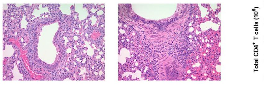
Pan et al.                                                                                                                         Vitamin A Regulates GVHD Risk

INTRODUCTION                                                                              intestinal barrier function after allogeneic HSCT remains
                                                                                          unclear. This is a clinically relevant question since recipient
Graft-versus-host disease (GVHD) remains a major complication                             vitamin A levels could potentially be modified to strengthen
after allogeneic hematopoietic stem cell transplantation (HSCT)                           the intestinal barrier and mitigate GVHD.
(1, 2). The gastrointestinal (GI) tract is one of the major target                            We hypothesized that vitamin A and RA exert positive
organs of GVHD and its involvement is often associated with a                             impacts on maintaining intestinal epithelial integrity during
poor prognosis (3–5). Since the GI tract is home to an enormous                           GVHD. In this study, we tested this hypothesis with dietary
amount of microbiota, its damage from the pretransplant                                   modification of vitamin A levels in recipient mice and examined
conditioning regimen and subsequent alloimmunity can lead to                              how a clinically relevant dose of vitamin A supplementation
leakage of immunostimulatory gram-negative bacterial fragments,                           affects GVHD risk. Contrary to our expectation, exogenous RA
such as lipopolysaccharide (LPS), into the circulation that can                           and mild vitamin A supplementation increased intestinal
intensify the inflammatory responses characteristic of GVHD (6).                           permeability after allogeneic HSCT. Vitamin A
In this regard, it has been shown that a breached mucosal                                 supplementation was associated with more severe lung damage
epithelial barrier is an important component of GVHD                                      and increased mortality of recipient mice after transplantation.
pathogenesis (7, 8). Accumulating evidence also shows that                                Fecal bacterial analysis revealed significant changes in diversity
protecting or restoring intestinal epithelial barrier function may                        and composition of microbiota in these animals. These results
be an effective approach in preventing or mitigating GVHD (9–11).                         demonstrate that RA signaling has the potential of modifying
Such a strategy is appealing because it deviates from conventional                        recipient intestinal barrier integrity and gut microbiota after
GVHD management by targeting recipient nonimmune cells                                    allogeneic HSCT. Furthermore, a diet with higher than normal
instead of donor immune cells, such as T cells and antigen                                vitamin A levels was associated with worse clinical outcomes in
presenting cells (12–14). Since this strategy does not interfere                          our GVHD model. These results have important implications for
with donor T cell activation and function, one can expect                                 the potential use of high-dose vitamin A in the clinical setting
maximal graft-versus-leukemia (GVL) response without overt                                of GVHD.
GVHD. However, host factors that are involved in maintaining
and modulating intestinal barrier function after allogeneic HSCT
are not well defined.
    Vitamin A is an essential nutrient that participates in a variety                     MATERIALS AND METHODS
of biological processes (15). In particular, it is well established
                                                                                          Mice
that vitamin A is an important factor that helps maintain
                                                                                          C57BL/6 (B6; H-2Kb) and Balb/c (H-2Kd) mice were purchased
mucosal epithelial barrier function. An epithelial monolayer of
                                                                                          from The Jackson Laboratory (Bar Harbor, ME) or bred in the
cells and tight junctions are two major components that form the
                                                                                          Biomedical Resource Center at the Medical College of Wisconsin
intestinal mucosal barrier. Tight junctions are important
                                                                                          (MCW). All experiments and procedures were carried out under
proteins that seal adjacent epithelial cells thus preventing the
                                                                                          protocols approved by the MCW Institutional Animal Care and
leakage of solutes and water through paracellular pathways.
                                                                                          Use Committee.
Vitamin A has been shown to upregulate ZO-1, Occludin, and
Claudin tight junction mRNA and protein expression (16).
Vitamin A deficiency increases the risk of GI tract infection                              Animal Experiments
and vitamin A supplementation in children can significantly                                Bone marrow transplantation (BMT) was performed as
reduce mortality associated with diarrhea (17). In the context of                         described previously (23). Briefly, bone marrow (BM) was
HSCT, we and others have shown that retinoic acid (RA), the                               flushed from femurs and tibias of B6 donor mice with
active metabolite of vitamin A, actively participates in regulating                       Dulbecco′s Modified Eagle′s Medium (DMEM) and passed
GVHD severity (18, 19). In these preclinical studies, the RA                              through sterile mesh filters to obtain single-cell suspensions.
signaling in donor T cells modulates intestinal GVHD risk by                              Splenic Pan-T cells were purified from donor mice using
influencing donor T cell polarization and migration. Exogenous                             EasySep™ Mouse T Cell Isolation Kit (StemCell Technologies,
RA intensifies GVHD and leads to increased mortality in mice                               Cambridge, MA) according to the manufacturer’s instructions.
undergoing allogeneic HSCT (18–20). In addition, it has been                              Balb/c recipients were conditioned with total body irradiation
shown that host dendritic cell RA signaling regulates GVHD                                administered as a single exposure of 900 Rads using a Shepherd
severity (21, 22). However, whether RA signaling affects                                  Mark I Cesium Irradiator (J. L. Shepherd and Associates, San
                                                                                          Fernando, CA). Irradiated recipients received a single
Abbreviations: ALT, alanine aminotransferase; AST, aspartate aminotransferase;
                                                                                          intravenous injection of bone marrow cells (6–10 ×106) plus
BM, bone marrow; BMT, bone marrow transplantation; FMT, fecal microbiota                  purified splenic Pan-T cells (0.3–0.5 ×10⁶) in the lateral tail vein.
transplantation; GVHD, graft-versus-host disease; GVL, graft-versus-leukemia;             Exogenous RA was given to recipient mice via intraperitoneal
HSCT, hematopoietic stem cell transplantation; LDA, Linear discriminant                   injection after BMT. For experimental purpose of vitamin A
analysis; LEfSe, Linear discriminant analysis effect size; LPS, lipopolysaccharide;       dietary modification, 3-week old male Balb/c mice were
MLNs, mesenteric lymph nodes; PCoA, Principal Coordinate Analysis; RA,
retinoic acid; RDA, Recommended Dietary Allowance; Tregs, regulatory T cells;
                                                                                          randomized into two groups and were fed a AIN-93G-based
UL, Tolerable Upper Intake Level; VAH, vitamin A-high; VAN, vitamin                       diet containing either normal (4 IU/g, VAN) or higher (10 IU/g,
A-normal.                                                                                 VAH) amount of vitamin A (Envigo, Indianapolis, IN) for at

Frontiers in Immunology | www.frontiersin.org                                         2                                 October 2021 | Volume 12 | Article 749002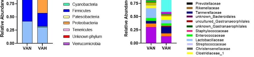
Pan et al.                                                                                                     Vitamin A Regulates GVHD Risk

least additional 8 weeks. BMT was performed on these recipient         a BD LSRII flow cytometer. Data were analyzed using FlowJo
mice as described above.                                               software (TreeStar, Ashland, OR).

Isolation of Colonic Epithelium                                        Cytometric Bead Array
Colon tissues were harvested upon termination of studies.              Serum cytokine levels were determined using the BD Cytometric
Colonic epithelium was isolated according to a modified                 Bead Array system (BD Biosciences) according to the
protocol (24). Briefly, the dissected colons were cut                   manufacturer’s protocol.
longitudinally, washed in cold PBS, and incubated in ice-cold
BD Cell Recovery Solution (BD Biosciences) for 10 minutes.             Serum FITC-Dextran Analysis
Colon tissues were then transferred to 2 ml of cold PBS and            After food and water were withheld for 3-4 hours, mice were
gently peeled/scraped by fine needles or scalpels. Detached             orally administered with 0.75 mg/g body weight of 4kDa FITC-
epithelial layers were collected by centrifugation at 1600 rpm         dextran (Sigma-Aldrich). Serum samples were collected 3-4
for 5 minutes.                                                         hours after oral administration. Fluorescence was measured
                                                                       spectrophotometrically in 96-well opaque plates (excitation:
                                                                       485 nm, emission: 528 nm). Serum FITC-dextran levels were
Caco-2 Cultures and Experiments                                        determined based on generated standard curves.
Caco-2 cells (HTB-37™, Lot# 70013347) were purchased from
the American Type Culture Collection (ATCC, Manassas, VA).
                                                                       Serum LPS Analysis
Caco-2 cells were grown in DMEM supplemented with 10%
                                                                       Serum LPS levels were measured using Pierce ™ LAL
heat-inactivated FBS, 2 mM L-glutamine, 1% Non-Essential
                                                                       Chromogenic Endotoxin Quantitation Kit (Thermo Fisher
Amino Acids (NEAA), and 1% penicillin/streptomycin in a
                                                                       Scientific) according to the manufacturer’s instructions.
humidified 37°C, 5% CO2 incubator. The culture medium was
changed every two to three days. Caco-2 cells were grown in 100-       Pathological Analysis
mm culture dishes (Thermo Fisher Scientific, Waltham, MA)               Representative samples of liver, colon, small intestine, and lung
and sub-cultured by digestion with 0.25% trypsin-EDTA. For             were fixed in formalin and paraffin embedded. The tissue
experimental purpose, Caco-2 cells were cultured and                   sections were then stained with hematoxylin and eosin.
maintained in 24-well plates for 14–21 days to differentiate           Histological analysis was performed by an experienced
before treatment. Cells were treated with TNF-a (20 ng/mL)             pathologist (C.L.) in a blinded fashion. A semiquantitative
and IL-6 (20 ng/mL) for 72 hours in the presence or absence of         scoring system was used to account for histological changes in
5 µM RA. Untreated cells were used as controls. Cultures were          the liver, colon, small intestine, and lung as previously
collected for RNA isolation, reverse transcription, and                described (25).
quantitative real-time PCR analysis.
                                                                       Gut Microbiome Analysis
Quantitative Real-Time PCR Analysis                                    Fecal samples were collected immediately before termination of
The TRIZOL® reagent (Sigma-Aldrich) was used to homogenize             studies and genomic DNA was purified using DNeasy
tissues/cells and isolate total RNA according to the                   PowerLyzer PowerSoil Kit (Qiagen) according to the
manufacturer’s instructions. The concentration of total RNA            manufacturer’s instructions. Purified genomic DNA was sent
was determined by absorbance at 260/280 nm using NanoDrop.             to the University of Wisconsin-Madison Biotechnology Center
Reverse transcription was performed using QuantiTect® Reverse          and the V3/V4 variable region of the 16S rDNA gene was
Transcription Kit (Qiagen, Hilden, Germany). Relative gene             sequenced with 2x300bp paired end technology. The site-
expression was measured using QuantiTect® SYBR Green PCR               specific primers used were: 341F:5’-CCTACGGGNGG
Kit (Qiagen). b-2-microglobulin (B2M) and glyceraldehyde-3-            CWGCAG-3’ and 806R:5’-GACTACHVGGGTATCTAATCC-
phosphate dehydrogenase (GAPDH) were used as the                       3’. Bioinformatic analyses of microbiome diversity, abundance,
housekeeping genes for mouse studies and Caco-2                        and functional pathways were performed by a senior
studies, respectively.                                                 Bioinformatics Analyst (S.N.A.) in a blinded fashion at the
                                                                       Center for Microbiome Research at the MCW. QIIME2
Flow Cytometric Analysis                                               (v. 2020.2) was used to analyze the paired-end 16S rRNA
Immune cells were labeled with monoclonal antibodies                   sequencing reads (26). Sequences were imported, trimmed
conjugated to fluorescein isothiocyanate (FITC), phycoerythrin          using CutAdapt, and summarized to check quality.
(PE), PE-Cy5, PE-Cy7, or allophycocyanin (APC). PE-anti-CD4            Representative sequences were chosen using DADA2, which
(clone GK1.5), FITC-anti-CD8a (clone 53-6.7), PE-anti-TCRb             also removes chimeric sequences. The representative sequences
(clone H57-597), FITC-anti-H-2Kb (clone AF6-88.5), APC-anti-           were then aligned, masked for hypervariable regions, and
CD8a (clone 53-6.7), and PE-Cy5-anti-CD8a (clone 53-6.7) were          phylogenetic trees were produced. A classifier was generated to
purchased from BD Biosciences. FITC-anti-CCR9 (clone CW-               assign taxonomy to the reads using the 99% similarity files of the
1.2), PE-anti-a4b7 (clone DATK32), PE-Cy7-anti-CD4 (clone              SILVA 132 release and the 341-806 region of the 16S gene.
GK1.5), and APC-anti-H-2Kb (clone AF6-88.5) were obtained              Taxonomy was assigned to the feature table to make taxonomy
from eBioscience (San Diego, CA). Samples were analyzed using          bar plots and to generate relative abundance tables. Diversity

Frontiers in Immunology | www.frontiersin.org                      3                                October 2021 | Volume 12 | Article 749002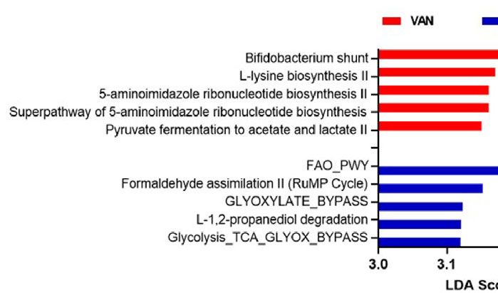
Pan et al.                                                                                                                            Vitamin A Regulates GVHD Risk

metrics were run using the core-metrics-phylogenetic command                          RESULTS
of QIIME2. Alpha and beta diversity were analyzed using their
respective commands, alpha/beta-group-significance (27). Alpha                         Retinoic Acid Increases Gut Permeability
and beta diversity boxplots were generated using R. Principal                         of Recipient Mice After Allogeneic BMT
Coordinate Analysis (PCoA) plots were examined using                                  We first examined the effects of RA, the active metabolite of
Emperor (28) and finalized figures were made using the                                  vitamin A, on intestinal barrier function in a culture system. To
qiime2R package in R. LEfSe, Linear Discriminant Analysis                             that end, we used Caco-2 cells, a widely used human intestinal
(LDA) effect size, was run to determine enriched organisms                            epithelial cell line. TNF-a and IL-6, two cytokines that are
from each group (29). PICRUSt2 was used to predict functional                         elevated in many gastrointestinal inflammatory disorders
capacity of the 16S reads (30, 31). Output from PICRUSt2 was                          including GVHD, have been shown to increase the
then put through LEfSe to determine differentially abundant                           permeability of Caco-2 cells (32, 33). Consistent with these
functional predictions.                                                               findings, treating Caco-2 cells with TNF-a and IL-6 led to a
                                                                                      significant increase in gene expression of Claudin-2, a tight
Hepatic Function Assay                                                                junction molecule that is associated with increased intestinal
Serum samples were collected upon termination of studies and                          permeability (34). It has been well documented that Claudin-2 is
sent to Wisconsin Diagnostic Laboratories for hepatic function                        a pore-forming protein that contributes to inducing a “leaky gut”
assay. Levels of alanine aminotransferase (ALT) and aspartate                         in several intestinal inflammatory disorders (35). Interestingly,
aminotransferase (AST) were determined using Roche cobas                              the expression of Claudin-2 was further increased in the presence
c702 analyzers.                                                                       of RA (Figure 1A). We also measured gene expression of other
                                                                                      tight junction molecules including Claudin-1 and ZO-1. There
Statistical Analysis                                                                  was a significant reduction in gene expression of Claudin-1 in
Statistical analysis was performed using GraphPad Prism (La                           Caco-2 cells after RA exposure, whereas ZO-1 expression levels
Jolla, CA). Survival comparisons were performed using the log-                        were not affected (Figures 1B, C). These results suggest that RA
rank test. Other differences between experimental groups were                         may further impair gut barrier integrity in an inflammatory
analyzed using an unpaired two-tailed Student’s t-test or one-                        environment by increasing Claudin-2 expression.
way ANOVA. Mann-Whitney U-test was used for group                                         We then investigated the effect of RA on intestinal barrier
comparison in microbiota experiments. A p value less than                             function in recipient mice undergoing allogeneic BMT. We
0.05 was considered as statistical significance in all experiments.                    treated recipient mice with RA or DMSO after transplantation

                          A                                B                                      C

                          D                                E                                  F

 FIGURE 1 | Retinoic acid increases gut permeability of recipient mice after allogeneic BMT. Caco-2 cells were maintained for 14–21 days and allowed to
 differentiate. The cultures were then treated with either DMSO or 5 µM RA for 4 days. They were exposed to TNF-a (20 ng/ml) and IL-6 (20 ng/ml) during the last 24
 hours of incubation. Culture wells that did not receive any treatments were used as controls. Gene expression of Claudin-2 (A), Claudin-1 (B), and ZO-1 (C) from
 different treatment groups was analyzed by real-time q-PCR. Data are normalized for b2‐microglobulin RNA and presented as fold increase over gene expression in
 no treatment (NT) group. Data are shown as Mean ± SEM and are cumulative results from two independent experiments. (D–F) Lethally irradiated Balb/c mice were
 transplanted with 7 x10⁶ BM plus 0.4 x 10⁶ purified T cells from B6 mice. Recipient mice then received intraperitoneal injections of either DMSO or 450 µg of RA
 every other day for 4 doses. On Day 7 post-BMT, FITC-dextran analysis was performed, and colonic epithelium was collected for real-time q-PCR analysis.
 (D) Serum levels of FITC-dextran in recipient mice (n = 5–6 per group). (E, F) Relative gene expression of Claudin-2 and apoptotic markers (n = 11 per group). Data
 are shown as Mean ± SEM and are cumulative results from two to three independent experiments. Statistics: *P ≤.05, **P ≤.01, ****P ≤.0001.

Frontiers in Immunology | www.frontiersin.org                                     4                                      October 2021 | Volume 12 | Article 749002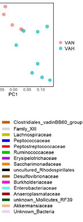
Pan et al.                                                                                                                            Vitamin A Regulates GVHD Risk

and measured intestinal permeability by fluorescein                                   integrity of intestinal epithelial barrier. On day 7 after
isothiocyanate (FITC)-dextran assay. In this assay, the                              transplantation, serum LPS levels were significantly higher in
translocation of orally applied FITC-dextran into circulation is                     VAH recipients compared to those of VAN mice (Figure 2A),
measured to reflect intestinal barrier integrity. On day 7 after                      indicating a more compromised intestinal barrier. Gene
transplantation, serum FITC-dextran levels were significantly                         expression of tight junction molecule Claudin-1 was also
higher in RA-treated mice compared to DMSO-treated mice                              significantly higher in VAH mice compared to that of VAN
(Figure 1D), demonstrating impaired gut barrier integrity. We                        mice (Figure 2B). However, there was no significant difference
further isolated the colonic epithelium and measured gene                            in Claudin-2 expression in intestinal epithelial cells on day 7 after
expression levels of tight junction molecules. Consistent with                       BMT (Figure 2C). Interestingly, mRNA levels of genes
in vitro studies, RA treatment significantly increased the                            associated with RA biosynthesis and signaling, such as aldh1a1
expression of Claudin-2 in purified colon epithelial cells                            and RAR-b, were significantly higher in colon epithelial cells of
(Figure 1E). In addition, gene expression of pro-apoptotic                           VAH mice (Figure 2D). Examination of inflammatory cytokines
marker Bax and caspase family members (Caspase-3 and                                 and apoptosis-associated markers also did not reveal differences
Caspase-9) was significantly increased in RA-treated mice                             between the two groups (Figures 2E, F). Thus, vitamin A
(Figure 1F). These results indicate that exogenous RA can                            supplementation is associatedwith decreased intestinal barrier
negatively impact intestinal barrier function of allogeneic BMT                      integrity without affectingClaudin-2 expression.
recipients by increasing Claudin-2 expression and promoting the
apoptosis of intestinal epithelial cells.                                            Dietary Vitamin A Supplementation Does
                                                                                     Not Affect Donor T-Cell Compartment
Recipient Mice Fed a Diet High in Vitamin                                            During GVHD
A Show Increased Gut Permeability After                                              To determine how vitamin A supplementation influences the in
Allogenic BMT                                                                        vivo expansion of donor T cells, we harvested the spleen and
To examine how endogenous RA signaling modulates intestinal                          mesenteric lymph nodes (MLNs) of VAN and VAH mice on day
barrier function after allogeneic BMT, we generated vitamin A                        7 after BMT. There were no significant differences in the
normal (VAN) and vitamin A high (VAH) Balb/c recipient mice                          percentage and absolute number of donor CD4 and CD8 T
through dietary modifications. In these studies, VAH mouse                            cells in the spleen and MLNs between VAN and VAH mice
chow contained 2.5-fold higher vitamin A levels compared to                          (Figures 3A, B, and data not shown). There were also no
VAN mouse chow. Pretransplant conditioning and subsequent                            significant differences in serum inflammatory cytokine levels
alloimmunity can cause significant damage to host intestinal                          between the two groups (Figure 3C). Retinoic acid is known to
epithelial barrier, leading to translocation of LPS into the                         imprint gut-homing specificity on T cells by increasing the
systemic circulation. Thus, serum LPS levels reflect the                              expression of integrin a4b7 and chemokine receptor CCR9.

                         A                      B                        C                      D

                                 E                                                    F

 FIGURE 2 | Recipient mice fed a diet high in vitamin A show increased gut permeability after allogenic BMT. Lethally irradiated VAN and VAH mice were
 transplanted with BM (6–10 x 106) plus 0.3–0.4 x 10⁶ purified T cells from B6 mice. On Day 7 post-BMT, serum and colonic epithelium were collected. (A) Serum
 LPS levels in recipient mice (n = 4–5 per group). Data are shown as Mean ± SEM and are derived from one of two representative experiments. (B–F) Relative gene
 expression of Claudin-1, Claudin-2, aldh1a1, RAR-b, inflammatory cytokines, and apoptotic markers of colonic epithelial cells (n = 8–9 per group). Data are shown as
 Mean ± SEM and are cumulative results from three independent experiments. Statistics: *P ≤.05, **P ≤.01.

Frontiers in Immunology | www.frontiersin.org                                    5                                       October 2021 | Volume 12 | Article 749002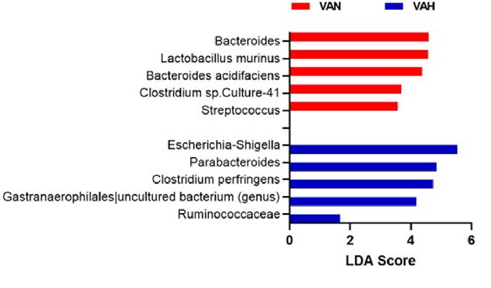
Pan et al.                                                                                                                      Vitamin A Regulates GVHD Risk

                        A                                   B                                     C

                        D                                   E                                         F

 FIGURE 3 | Dietary vitamin A supplementation does not affect donor T-cell compartment after allogeneic BMT. Lethally irradiated VAN and VAH mice were
 transplanted with BM (6–10 x 106) plus 0.3–0.4 x 10⁶ purified T cells from B6 mice. On Day 7 post-BMT, the percentage and absolute number of donor H‐2b+CD4+
 T cells in the spleen (A) and MLNs (B) were examined. (C) Serum proinflammatory cytokine levels were determined by cytometric bead array. Data are shown as
 Mean ± SEM and are cumulative results from three independent experiments. (D) The percentage of donor H‐2b+CD4+ T cells expressing a4b7 and CCR9 from
 MLNs of recipient mice was examined. Data are shown as Mean ± SEM and are cumulative results from two experiments. (E, F) BMT was performed as described
 above except Foxp3+EGFP+ mice were used as donors. On Day 7 post-BMT, the percentage and absolute number of donor H‐2b+CD4+Foxp3+ Tregs in the spleen
 (E) and MLNs (F) were examined (n = 7 per group). Data are shown as Mean ± SEM and are cumulative results from two independent experiments.

However, the percentage and absolute number of donor CD4                          mice is not attributable to increased hepatic GVHD. Pathological
and CD8 T cells expressing these gut-homing molecules were                        analysis of GVHD target organs on day 14 after transplantation
similar between the two groups of mice (Figure 3D and data not                    revealed a significant increase in inflammatory changes of the
shown). These results indicate that vitamin A supplementation                     lungs of VAH mice compared with VAN mice (Figure 4C),
does not appear to affect the expansion and gut-homing potential                  whereas there were no significant differences in pathology scores
of donor T cells at the early stages of GVHD development. Since                   of the liver, colon, and small intestine between the two groups
RA plays an important role in facilitating the induction of                       (Figure 4C). Specifically, there was lymphocytic infiltration
Foxp3+ regulatory T cells (Tregs), we determined how vitamin                      around pulmonary vessels and bronchioles as well as
A supplementation affects donor Treg reconstitution after                         pneumonitis, characteristic pathological features of GVHD, in
transplantation. Using purified CD4 + T cells from                                 both groups of mice. However, the extent and severity of these
Foxp3+EGFP+ donor mice, we found no significant differences                        abnormalities were significantly increased in the lungs of VAH
in the percentage and absolute number of CD4+Foxp3+EGFP+                          mice compared with VAN mice (Figure 4D). We further
Tregs in the spleen and MLNs between VAN and VAH                                  analyzed the immune cells infiltrating the lungs of VAN and
recipients (Figures 3E, F). Collectively, these data indicate that                VAH mice on day 7 after transplantation. We found significantly
vitamin A supplementation does not affect the expansion of                        higher absolute numbers of donor CD4 and CD8 T cells in the
donor T cells and the reconstitution of Tregs after                               lungs of VAH mice versus VAN mice (Figures 4E, F). Thus, mild
allogeneic BMT.                                                                   vitamin A supplementation is associated with more severe lung
                                                                                  GVHD with increased donor T cell infiltration in this tissue site.
Recipient Mice Fed a Diet High in Vitamin
A Show More Severe Lung GVHD After                                                Dietary Vitamin A Supplementation
Allogeneic BMT                                                                    Modulates Recipient Gut Microbiota After
We then examined how vitamin A supplementation affects                            Allogeneic BMT
overall GVHD risk. We found that VAH BMT recipients                               Diet is one of the most important factors that shape gut
showed increased mortality after transplantation and died                         microbiota. We performed 16S ribosomal RNA gene
significantly faster than VAN mice (Figure 4A). Since previous                     sequencing of fecal samples from recipient mice 7 days after
studies have indicated a potential role of vitamin A in regulating                transplantation. We found that fecal samples from VAH mice
liver damage during GVHD (36), we measured liver enzymes                          had significantly lower alpha diversity compared to VAN
alanine aminotransferase (ALT) and aspartate aminotransferase                     recipient mice as shown by Shannon index (Figures 5A). In
(AST) levels in recipient mice. There were no significant                          addition, there was a significant difference in beta diversity
differences in serum ALT and AST levels between VAN and                           between the two groups (Figure 5B). Principal coordinates
VAH mice (Figure 4B), indicating that early mortality of VAH                      analysis (PCoA) based on weighted UniFrac distance revealed

Frontiers in Immunology | www.frontiersin.org                                 6                                     October 2021 | Volume 12 | Article 749002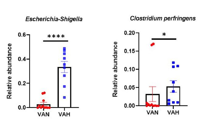
Pan et al.                                                                                                                             Vitamin A Regulates GVHD Risk

                       A                                         B                                           C

                       D                                                                   E                             F

 FIGURE 4 | Recipient mice fed a diet high in vitamin A show more severe lung GVHD after allogeneic BMT. (A) Lethally irradiated VAN and VAH mice were
 transplanted with BM (7–8 x 10⁶) plus 0.4 x 10⁶ purified T cells from B6 mice. Overall survival is depicted. Data are the cumulative results from 7 independent
 experiments (n = 21–22 per group). (B) Serum liver enzymes in VAN and VAH recipients 7 days after transplantation. Data are shown as Mean ± SEM and are
 cumulative results from three independent experiments (n = 9–11 per group). (C) Recipient mice were euthanized on day 14 after BMT and pathologic damage in the
 liver, lung, small intestine, and colon was examined. Data are derived from one representative experiment (n = 5 per group). (D) Representative photo micrographs of
 the lungs from VAN and VAH mice are shown. (E, F) The absolute numbers of donor H‐2b+CD4+ T cells (E) and donor H‐2b+CD8+ T cells (F) in the lungs of
 recipient mice were examined. Data are shown as Mean ± SEM and are cumulative results from two experiments (n = 6 per group). Statistics: *P ≤ .05.

                           A                                     B                                       C

                           D                                          E

 FIGURE 5 | Dietary vitamin A supplementation modulates recipient gut microbiota after allogeneic BMT. (A) Lethally irradiated VAN and VAH mice were transplanted
 with BM (7 x 10⁶) plus 0.3–0.4 x 10⁶ purified T cells from B6 mice. On Day 7 after BMT, fecal samples were collected and 16S rRNA sequencing was performed.
 (A) Shannon index of gut microbiota as a measure of alpha diversity; (B) Weighted UniFrac analysis of microbiota b-diversity. (n = 9–11 per group). Data are shown
 as Mean ± SEM and are cumulative results from 3 independent experiments. (C) Principle coordinate analysis (PCoA) using Weighted UniFrac distance of gut
 microbiota sourced from VAN and VAH mice reveal beta diversity differences; (D) Relative abundance of microbial community at the phylum level. (E) Relative
 abundance of microbial community at the family level. Statistics: *P ≤.05, **P ≤.01.

significantly different microbiota communities between VAN                             associated with more severe loss of gut microbiota diversity, a
and VAH recipient mice. The samples from VAH group                                    hallmark of GVHD. There were also noticeable differences in
clustered separately from those of VAN mice (Figure 5C).                              relative abundance of fecal bacteria between the two groups. At
These results indicated that vitamin A supplementation is                             the phyla level, VAH mice had increased abundance of the

Frontiers in Immunology | www.frontiersin.org                                     7                                       October 2021 | Volume 12 | Article 749002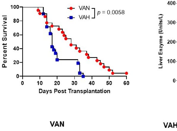
Pan et al.                                                                                                                          Vitamin A Regulates GVHD Risk

Proteobacteria phylum and decreased abundance of the                                 to modulating the integrity of intestinal barrier in patients
Firmicutes phylum (Figure 5D). At the family level,                                  undergoing allogeneic HSCT. Research efforts to identify these
Lactobacillaceae and Bacteroidaceae were decreased and                               host factors with the hope of targeting them to reduce GVHD are
Enterobacteriaceae was increased in VAH group (Figure 5E).                           of significant clinical relevance. We are interested in how
At the genus level, there was specific enrichment of microbiota of                    micronutrients, in particular vitamin A, regulates this process
VAN mice that are commonly associated with a healthier gut                           given the well-established beneficial effects of this molecule in
microenvironment. For example, Bacteroides and Lactobacillus                         maintaining mucosal barrier function. Indeed, recent studies
murinus were the top two enriched genera that were significantly                      have shown the crucial role of intestinal epithelial retinoid
increased in these animals (Figures 6A–C). In contrast, there                        signaling in regulating the survival of mice in the context of GI
was an expansion of opportunistic pathogens including                                infection (37). In this paper, we provide novel insights into how
Escherichia-Shigella and Clostridium perfringens in VAH mice                         retinoic acid signaling influences intestinal tight junctions and
(Figures 6A, D, E). Finally, functional pathway analysis by                          microbiota in the setting of allogeneic HSCT.
PICRUSt revealed distinct metabolic pathways between the                                 The intestinal epithelial barrier consists of a monolayer of
VAN and VAH groups. Specifically, carbohydrate metabolism                             epithelium and tight junctions that seal the paracellular pathway
with Bifidobacteria and metabolism of certain amino acids were                        and regulate permeability. Disrupting either of these two
increased in VAN mice, whereas pathways associated with fatty                        components can lead to a weakened barrier function
acid oxidation and formaldehyde assimilation were increased in                       manifested as “leaky gut”. Claudins are a family of proteins
VAH mice (Figure 6F). Thus, vitamin A supplementation is                             that are important in the formation of tight junctions (38).
associated with compositional and functional alterations of gut                      Among more than 10 Claudin family proteins, Claudin-2 is
microbiota after transplantation.                                                    unique in that it is a pore-forming protein with the potential of
                                                                                     increasing intestinal barrier permeability. Enhanced expression
                                                                                     of Claudin-2 has been found to be associated with intestinal
DISCUSSION                                                                           inflammatory disorders (35, 39). In our culture system, we found
                                                                                     that TNF-a and IL-6 enhanced Claudin-2 expression in Caco-2
There is growing interest in mitigating GVHD through                                 cells. Interestingly, RA appears to synergize with TNF-a and IL-6
protecting or restoring the gut mucosal barrier that is damaged                      to further enhance Claudin-2 expression. Thus, RA has the
after allogeneic HSCT (7–9). Such an approach is expected to                         potential of increasing permeability of the epithelial monolayer
lead to a maximal GVL response with significantly diminished                          in the presence of inflammatory cytokines. These results are
GVHD. It is conceivable that certain host factors may contribute                     different from other studies revealing a positive effect of RA on

                                     A                                               B                           C

                       D                          E                                      F

 FIGURE 6 | Vitamin A supplementation causes more profound gut dysbiosis with functional consequences in BMT recipients. (A) Lethally irradiated VAN and VAH
 mice were transplanted with BM (6–10 x 106) plus 0.3–0.4 x 10⁶ purified T cells from B6 mice. On Day 7 after BMT, fecal samples were collected and 16S rRNA
 sequencing was performed. (A) Taxonomic comparison at the genus level using linear discriminant analysis effect size analysis. (B–E) Comparison of relative
 abundance of taxa at the genus level between the two groups. (B) Bacteroides; (C) Lactobacillus murinus; (D) Escherichia-Shigella; (E) Clostridium perfringens;
 (n = 9–11 per group). Data are shown as Mean ± SEM and are cumulative results from 3 independent experiments. (F) Linear discriminant analysis effect size
 analysis for the predicted metabolic pathways (KEGG modules) from two groups. Top 5 pathways with an LDA score greater than 3 from each group are shown.
 Statistics: *P ≤.05, **P ≤.01, ****P ≤.0001.

Frontiers in Immunology | www.frontiersin.org                                    8                                      October 2021 | Volume 12 | Article 749002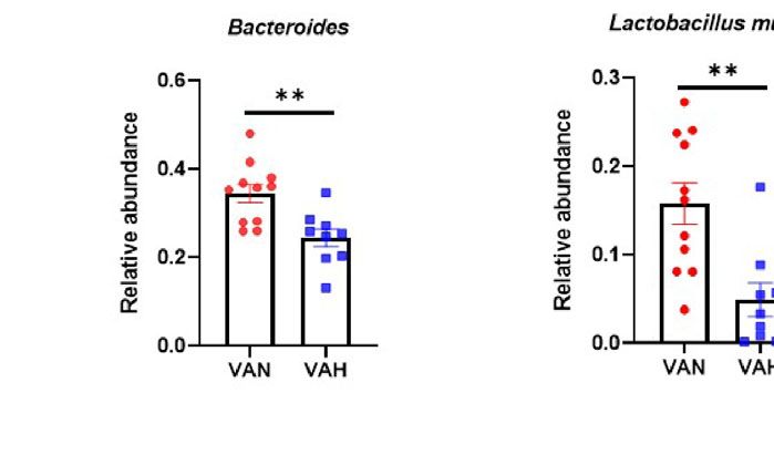
Pan et al.                                                                                                                        Vitamin A Regulates GVHD Risk

Caco-2 cells (40). The differences in experimental protocols,                      are associated with a significantly increased intestinal GVHD
including the concentration of RA as well as the timing and                        risk in pediatric patients undergoing allogeneic HSCT (46). In
duration of RA exposure may explain these different results. In                    another study, however, pre-transplant serum vitamin A levels
addition, the interplay between RA and inflammatory cytokine                        do not appear to affect the development of acute GVHD (47).
signaling may dictate the positive or negative impacts of RA on                    The discrepancy between the two studies may originate from
the permeability of Caco-2 cells.                                                  differences in study populations (pediatric vs. adult patients) and
    Exogenous RA has been shown to accelerate GVHD progression                     the timing of vitamin A measurement (post-transplant vs. pre-
in animal models (18–20). Its detrimental effect has largely been                  transplant). In addition, there is an interest in using vitamin A
attributed to increased Th1 cell differentiation and intestinal                    supplementation to reduce GVHD risk in the clinic (48).
migration or decreased myeloid-derived suppressor cells. We                        Therefore, it is imperative to test the applicability of this
found that exogenous RA can also enhance intestinal barrier                        strategy in animal models of GVHD. In this study, we use a
permeability early after HSCT, as shown by significantly                            clinically relevant dose of vitamin A supplementation to examine
increased FITC-dextran levels in RA treated mice. Thus, RA may                     how this approach influences the outcomes of allogeneic HSCT.
increase GVHD severity by acting on host nonimmune cells such as                   It has been estimated that over one third of US population are
intestinal epithelial cells. This observation is noteworthy given the              taking dietary supplements regularly, including vitamin A (49).
well-documented beneficial effects of vitamin A/RA in maintaining                   In addition, many types of foods are naturally fortified with
mucosal barrier integrity. It is worth noting that the complex effects             vitamin A. Thus, there is a legitimate concern of relatively high
of RA in vivo are often context dependent. For example, under                      prevalence of hypervitaminosis A in the population which may
steady state condition, RA acts as an anti-inflammatory molecule                    go unnoticed. Dietary vitamin A modification has been used
that dampens harmful immune responses in the GI tract by                           extensively in various mouse models of human diseases. Many
converting antigen-specific T cells into regulatory T cells (Tregs).                studies have used a vitamin A supplementation approach. In
However, in the presence of other inflammatory stimuli, such as IL-                 those experiments, a diet containing an excessive amount of
15, RA can act as an adjuvant that fuels T-cell mediated immune                    vitamin A was given to experimental animals. However, most of
responses in this tissue site (41). By the same token, it is possible that         those studies used aggressive vitamin A supplementation
RA promotes intestinal barrier integrity under homeostatic                         protocols that are not physiologically relevant; the vitamin A
condition, but it can do the opposite in a highly inflammatory                      excessive diet is often 50-100 fold higher than normal levels.
environment like the one observed in GVHD. An alternative                          According to Food and Nutrition Board of Institute of Medicine,
explanation is that supplementation of exogenous RA induces a                      the Recommended Dietary Allowance (RDA) of vitamin A for
negative feedback regulation of RA production in intestinal                        men is 900 ug/day and the Tolerable Upper Intake Level (UL) is
epithelial cells, resulting in a low RA signaling in local tissue                  set at 3,000 ug/day, a roughly 3-fold difference (50). In our study,
environment resembling vitamin A deficiency. It has been shown                      we used a 2.5-fold higher than normal vitamin A diet to mimic
that RA actively induces the activity of CYP26A1 and CYP26B1                       such a difference and ensured that our supplementation protocol
genes, which are responsible for RA breakdown in vivo (42).                        is within UL and is clinically relevant. Consistent with the results
    Vitamin A has been implicated in GVHD pathogenesis (36,                        from experiments using exogenous RA, VAH mice had increased
43–45). However, clinical studies have yielded some inconsistent                   serum LPS levels early after transplantation, indicating an
conclusions. It has been reported that lower levels of vitamin A                   increased epithelial barrier permeability. As discussed above,

 FIGURE 7 | Schematic summary of the study. Increased retinoic acid signaling or prolong and mild vitamin A supplementation may negatively impact intestinal
 barrier function during GVHD. Mild vitamin A supplementation is associated with increased lung damage and more profound gut dysbiosis in recipient mice.
 Micronutrients such as vitamin A could modulate complications of allogeneic HSCT, which may be mediated by shaping gut microbiota. Created with Biorender.com.

Frontiers in Immunology | www.frontiersin.org                                  9                                     October 2021 | Volume 12 | Article 749002
Pan et al.                                                                                                          Vitamin A Regulates GVHD Risk

the additional strong inflammatory signals after allogeneic HSCT             mice. Bifidobacteria are probiotics that can produce acetate, a
or a negative feedback mechanism with diminished local RA                   type of short chain fatty acid, to promote gut health (71).
signaling in VAH animals may partially explain such a weakened              In addition, pathway associated with lysine biosynthesis was
mucosal barrier function. However, the increased gene                       increased in VAN mice, consistent with results from other
expression of aldh1a1 and RAR-b in VAH mice does not                        studies (66). On the other hand, pathways associated with lipid
appear to support the latter possibility. Interestingly, gene               and carbohydrate metabolism were increased in VAH mice.
expression of Claudin-2 and apoptosis-associated molecules                  These data demonstrated that vitamin A supplementation can
was unaltered in gut epithelial cells of VAH mice, suggesting               cause more profound gut dysbiosis with functional consequences
that some other mechanisms may be responsible for                           in transplant recipients. The increased prevalence of pathogenic
the observation.                                                            bacteria together with the alternations in energy homeostasis are
    Donor T cells are the major pathogenic cells causing GVHD               also likely to contribute to the worse transplant outcomes of
(51). We found no significant differences in donor T cell                    VAH mice. It is worth noting that changes in gut microbiota
expansion and Treg reconstitution after BMT between VAN                     have been linked to the development of pulmonary
and VAH mice. Thus, vitamin A supplementation does not                      complications in patients undergoing allogeneic HSCT (72).
appear to directly affect the donor T cell compartment during               More studies focused on elucidating the role of micronutrients
GVHD. It is interesting that there was an increase in lung                  in regulating the interplay between intestinal epithelium, gut
pathologic scores and transplant-associated mortality of VAH                microbiota, and immune cells after allogeneic HSCT may lead to
mice. It is generally accepted that vitamin A plays a beneficial             the development of novel strategies to prevent and/or
role in maintaining lung homeostasis. However, it has also been             treat GVHD.
shown that high dose vitamin A supplementation is associated                    There are some limitations to the current study. First, this
an increased risk of lung cancer in humans, in particular among             study has not established a causal link between gut dysbiosis and
smokers (52). Thus, it appears that vitamin A could have                    increased lung pathology. Although there is clinical evidence
negative impacts on lung health in the presence of                          supporting the notion that gut microbiota can influence the
inflammation. In addition, vitamin A levels may also affect the              development of pulmonary complications in recipients of
integrity of lung epithelium, thus influencing lung pathologic               allogeneic HSCT (72), more studies in animal models are
damage in our model.                                                        needed to provide mechanistic insights. Second, it is unclear
    Recent preclinical and clinical studies have shown that                 whether similar gut dysbiosis will be observed in the absence of
GVHD causes significant intestinal dysbiosis in recipients of                alloimmune response. While it is tempting to speculate that there
allogeneic HSCT (53–60). There is also a strong interest in using           will be less changes of gut microbiota in both VAN and VAH
probiotics, prebiotics, or fecal microbiota transplantation (FMT)           recipient mice in the absence of allogeneic T cells, bone marrow
to ameliorate GVHD severity by modifying gut microbiota (61–                transplantations using TCD-BM alone will be needed to confirm
63). Diet is one of the most important factors that shape gut               this hypothesis. Third, we did not examine the microbial
microbiota (64, 65). Micronutrients, including vitamin A, have              metabolites in this study. It is of interest to study whether
been shown to modulate the gut microbiome community with                    alterations in gut microbiota diversity and composition lead to
functional consequences (66). In our study, we showed that a                different levels of bacterial products such as short chain fatty
mere 2.5-fold difference in dietary vitamin A levels is sufficient to        acids (acetate, butyrate, and propionate etc) that have been
induce drastic differences in gut microbiota community in                   shown to regulate GVHD severity.
recipient mice after allogeneic BMT. VAH mice showed a                          In summary, our studies showed that RA signaling
higher degree of gut dysbiosis as demonstrated by significant                participates in modulating host intestinal epithelial barrier
changes in microbiota alpha and beta diversities post-BMT. They             integrity and affects transplant outcomes (Figure 7). This effect
also had increased abundance of Proteobacteria and decreased                may be mediated by the changes in gut microbiota in a vitamin
abundance of Firmicutes, a characteristic dysbiosis frequently              A-dependent manner. In our model, it is possible that there is a
observed in patients after allogeneic HSCT or with inflammatory              reciprocal regulation between increased gut permeability and
bowel disease (67–69). Furthermore, opportunistic pathogens                 more profound gut dysbiosis in VAH recipient mice.
such as Escherichia-Shigella were increased in VAH mice (70).               The observation that VAH mice showed a significantly
On the other hand, Bacteroides and Lactobacillus, two bacterial             increased transplant-associated mortality in our model
genera that have the capacity to maintain intestinal homeostasis            indicates that some caution should be exercised when consider
and health, were significantly lower in these animals. Thus, mild            using vitamin A supplementation to prevent GVHD in the clinic.
vitamin A supplementation, as defined in this study, leads to the            However, we wanted to point out that we used a prolonged and
expansion of pathogenic microbiota in the microbiome after                  very mild vitamin A supplementation protocol in this study. It is
transplantation. We further confirmed that changes in the                    worth exploring whether a short-term, high dose vitamin A
diversity and composition of gut microbiota result in the                   supplementation can produce a different result. Dietary
alterations of bacterial metabolism. Functional pathway                     intervention of BMT recipients aimed at correcting dysbiosis
analysis revealed differences in amino acid, carbohydrate, and              and improving intestinal epithelial barrier function may
lipid metabolism between the two groups. Notably,                           represent a clinically applicable, simple, and cost-effective
Bifidobacterium shunt is among the top five pathways in VAN                   approach for managing GVHD after allogeneic HSCT.

Frontiers in Immunology | www.frontiersin.org                          10                                October 2021 | Volume 12 | Article 749002
Pan et al. Vitamin A Regulates GVHD Risk
DATA AVAILABILITY STATEMENT experimental mice. XC conceived and supervised the study,
designed experiments, analyzed data, and wrote the
The datasets presented in this study can be found in online manuscript. All authors contributed to the article and
repositories. The names of the repository/repositories and approved the submitted version.
accession number(s) can be found below: NCBI; PRJNA767073.
ACKNOWLEDGMENTS
ETHICS STATEMENT
The authors thank Dr. William R. Drobyski for critical reading of
All experiments and procedures were carried out under protocols the manuscript. The authors also thank the staff at the Center for
approved by the MCW Institutional Animal Care and Microbiome Research at the MCW for their expert assistance
Use Committee. with microbiota studies. Supported in part by the Flow
Cytometry Shared Resource at the Medical College of
Wisconsin Cancer Center. This research was supported in part
AUTHOR CONTRIBUTIONS by National Institutes of Health (NIH), National Institute of
Allergy and Infectious Diseases grant RO1 AI125334 and the
PP performed research, analyzed data, and wrote the manuscript. Amy Strelzer Manasevit Research Program which is funded
SA performed all bioinformatic analyses of microbiota through The Be The Match Foundation and the National
experiments. BT, HZ, DZ, and MM performed research. PF Marrow Donor Program (both to XC). The authors thank
and LS-W discussed results. CL performed pathological analyses Rachel H. Limpert for generating the schematic summary of
of GVHD target organs. IG performed hepatic function testing of the study.
REFERENCES 11. Swimm A, Giver CR, DeFilipp Z, Rangaraju S, Sharma A, Ulezko Antonova A,
et al. Indoles Derived From Intestinal Microbiota Act via Type I Interferon
1. Zeiser R, Blazar BR. Acute Graft-Versus-Host Disease - Biologic Process, Signaling to Limit Graft-Versus-Host Disease. Blood (2018) 132(23):2506–19.
Prevention, and Therapy. N Engl J Med (2017) 377(22):2167–79. doi: 10.1056/ doi: 10.1182/blood-2018-03-838193
NEJMra1609337 12. Coghill JM, Sarantopoulos S, Moran TP, Murphy WJ, Blazar BR, Serody JS.
2. Hill GR, Betts BC, Tkachev V, Kean LS, Blazar BR. Current Concepts and Effector CD4+ T Cells, the Cytokines They Generate, and GVHD: Something
Advances in Graft-Versus-Host Disease Immunology. Annu Rev Immunol Old and Something New. Blood (2011) 117(12):3268–76. doi: 10.1182/blood-
(2021) 39:19–49. doi: 10.1146/annurev-immunol-102119-073227 2010-12-290403
3. Hill GR, Ferrara JL. The Primacy of the Gastrointestinal Tract as a Target 13. Koyama M, Hill GR. The Primacy of Gastrointestinal Tract Antigen-
Organ of Acute Graft-Versus-Host Disease: Rationale for the Use of Cytokine Presenting Cells in Lethal Graft-Versus-Host Disease. Blood (2019) 134
Shields in Allogeneic Bone Marrow Transplantation. Blood (2000) 95 (24):2139–48. doi: 10.1182/blood.2019000823
(9):2754–9. doi: 10.1182/blood.V95.9.2754.009k25_2754_2759 14. Yu H, Tian Y, Wang Y, Mineishi S, Zhang Y. Dendritic Cell Regulation of
4. Castilla-Llorente C, Martin PJ, McDonald GB, Storer BE, Appelbaum FR, Graft-vs.-Host Disease: Immunostimulation and Tolerance. Front Immunol
Deeg HJ, et al. Prognostic Factors and Outcomes of Severe Gastrointestinal (2019) 10:93. doi: 10.3389/fimmu.2019.00093
GVHD After Allogeneic Hematopoietic Cell Transplantation. Bone Marrow 15. Stephensen CB. Vitamin a, Infection, and Immune Function. Annu Rev Nutr
Transplant (2014) 49(7):966–71. doi: 10.1038/bmt.2014.69 (2001) 21:167–92. doi: 10.1146/annurev.nutr.21.1.167
5. Ferrara JL, Smith CM, Sheets J, Reddy P, Serody JS. Altered Homeostatic 16. He C, Deng J, Hu X, Zhou S, Wu J, Xiao D, et al. Vitamin a Inhibits the Action
Regulation of Innate and Adaptive Immunity in Lower Gastrointestinal Tract of LPS on the Intestinal Epithelial Barrier Function and Tight Junction
GVHD Pathogenesis. J Clin Invest (2017) 127(7):2441–51. doi: 10.1172/ Proteins. Food Funct (2019) 10(2):1235–42. doi: 10.1039/c8fo01123k
JCI90592 17. McCullough FS, Northrop-Clewes CA, Thurnham DI. The Effect of Vitamin a
6. Cooke KR, Gerbitz A, Crawford JM, Teshima T, Hill GR, Tesolin A, et al. LPS on Epithelial Integrity. Proc Nutr Soc (1999) 58(2):289–93. doi: 10.1017/
Antagonism Reduces Graft-Versus-Host Disease and Preserves Graft-Versus- s0029665199000403
Leukemia Activity After Experimental Bone Marrow Transplantation. J Clin 18. Chen X, Dodge J, Komorowski R, Drobyski WR. A Critical Role for the
Invest (2001) 107(12):1581–9. doi: 10.1172/JCI12156 Retinoic Acid Signaling Pathway in the Pathophysiology of Gastrointestinal
7. Nalle SC, Kwak HA, Edelblum KL, Joseph NE, Singh G, Khramtsova GF, et al. Graft-Versus-Host Disease. Blood (2013) 121(19):3970–80. doi: 10.1182/
Recipient NK Cell Inactivation and Intestinal Barrier Loss Are Required for blood-2012-08-445130
MHC-Matched Graft-Versus-Host Disease. Sci Transl Med (2014) 6 19. Aoyama K, Saha A, Tolar J, Riddle MJ, Veenstra RG, Taylor PA, et al.
(243):243ra87. doi: 10.1126/scitranslmed.3008941 Inhibiting Retinoic Acid Signaling Ameliorates Graft-Versus-Host Disease by
8. Nalle SC, Zuo L, Ong MLDM, Singh G, Worthylake AM, Choi W, et al. Graft- Modifying T-Cell Differentiation and Intestinal Migration. Blood (2013) 122
Versus-Host Disease Propagation Depends on Increased Intestinal Epithelial (12):2125–34. doi: 10.1182/blood-2012-11-470252
Tight Junction Permeability. J Clin Invest (2019) 129(2):902–14. doi: 10.1172/ 20. Wang D, Yu Y, Haarberg K, Fu J, Kaosaard K, Nagaraj S, et al. Dynamic Change
JCI98554 and Impact of Myeloid-Derived Suppressor Cells in Allogeneic Bone Marrow
9. Mohammadpour H, Du W, O’Neill R, Khalili S, Qiu J, Repasky EA, et al. Transplantation in Mice. Biol Blood Marrow Transplant J Am Soc Blood Marrow
Host-Derived Serine Protease Inhibitor 6 Provides Granzyme B-Independent Transplant (2013) 19(5):692–702. doi: 10.1016/j.bbmt.2013.01.008
Protection of Intestinal Epithelial Cells in Murine Graft-Versus-Host Disease. 21. Thangavelu G, Lee Y-C, Loschi M, Schaechter KM, Feser CJ, Koehn BH, et al.
Biol Blood Marrow Transplant J Am Soc Blood Marrow Transplant (2018) 24 Dendritic Cell Expression of Retinal Aldehyde Dehydrogenase-2 Controls
(12):2397–408. doi: 10.1016/j.bbmt.2018.07.003 Graft-Versus-Host Disease Lethality. J Immunol Baltim Md 1950 (2019) 202
10. Peled JU, Hanash AM, Jenq RR. Role of the Intestinal Mucosa in Acute (9):2795–805. doi: 10.4049/jimmunol.1800899
Gastrointestinal GVHD. Blood (2016) 128(20):2395–402. doi: 10.1182/blood- 22. Zheng J, Taylor B, Dodge J, Stephans A, Zheng SG, Chen Q, et al. Radiation
2016-06-716738 and Host Retinoic Acid Signaling Promote the Induction of Gut-Homing
Frontiers in Immunology | www.frontiersin.org 11 October 2021 | Volume 12 | Article 749002Pan et al. Vitamin A Regulates GVHD Risk
Donor T Cells After Allogeneic Hematopoietic Stem Cell Transplantation. Am 43. Chen X, Mayne CG. The Role of Micronutrients in Graft-vs.-Host Disease:
J Transplant Off J Am Soc Transplant Am Soc Transpl Surg (2020) 20(1):64–74. Immunomodulatory Effects of Vitamins a and D. Front Immunol (2018)
doi: 10.1111/ajt.15501 9:2853. doi: 10.3389/fimmu.2018.02853
23. Chen X, Das R, Komorowski R, Beres A, Hessner MJ, Mihara M, et al. 44. Zheng J, Taylor B, Chen X. Role of Vitamin a in Modulating Graft-Versus-
Blockade of Interleukin-6 Signaling Augments Regulatory T-Cell Host Disease. J Immunol Res Ther (2018) 3(1):124–8.
Reconstitution and Attenuates the Severity of Graft-Versus-Host Disease. 45. Dodge J, Stephans A, Lai J, Drobyski WR, Chen X. Effects of Donor Vitamin a
Blood (2009) 114(4):891–900. doi: 10.1182/blood-2009-01-197178 Deficiency and Pharmacologic Modulation of Donor T Cell Retinoic Acid
24. Nik AM, Carlsson P. Separation of Intact Intestinal Epithelium From Pathway on the Severity of Experimental Graft-Versus-Host Disease. Biol
Mesenchyme. BioTechniques (2013) 55(1):42–4. doi: 10.2144/000114055 Blood Marrow Transplant J Am Soc Blood Marrow Transplant (2016) 22
25. Hill GR, Cooke KR, Teshima T, Crawford JM, Keith JC, Brinson YS, et al. (12):2141–8. doi: 10.1016/j.bbmt.2016.09.001
Interleukin-11 Promotes T Cell Polarization and Prevents Acute Graft- 46. Lounder DT, Khandelwal P, Dandoy CE, Jodele S, Grimley MS, Wallace G,
Versus-Host Disease After Allogeneic Bone Marrow Transplantation. J Clin et al. Lower Levels of Vitamin a Are Associated With Increased
Invest (1998) 102(1):115–23. doi: 10.1172/JCI3132 Gastrointestinal Graft-Versus-Host Disease in Children. Blood (2017) 129
26. Bolyen E, Rideout JR, Dillon MR, Bokulich NA, Abnet CC, Al-Ghalith GA, (20):2801–7. doi: 10.1182/blood-2017-02-765826
et al. Reproducible, Interactive, Scalable and Extensible Microbiome Data 47. Gjærde LK, Andersen NS, Friis LS, Kornblit B, Petersen SL, Schjødt I, et al.
Science Using QIIME 2. Nat Biotechnol (2019) 37(8):852–7. doi: 10.1038/ Pretransplantation Vitamin a Plasma Levels and Risk of Acute Graft-Versus-
s41587-019-0209-9 Host Disease Following Allogeneic Hematopoietic Stem Cell Transplantation.
27. Caporaso JG, Kuczynski J, Stombaugh J, Bittinger K, Bushman FD, Costello Bone Marrow Transplant (2020) 55(7):1457–9. doi: 10.1038/s41409-019-
EK, et al. QIIME Allows Analysis of High-Throughput Community 0760-5
Sequencing Data. Nat Methods (2010) 7(5):335–6. doi: 10.1038/nmeth.f.303 48. Carpenter PA. Vitamin a to Reduce Gut Leak and GVHD? Blood (2017) 129
28. Vá zquez-Baeza Y, Gonzalez A, Smarr L, McDonald D, Morton JT, Navas- (20):2715–7. doi: 10.1182/blood-2017-03-773226
Molina JA, et al. Bringing the Dynamic Microbiome to Life With Animations. 49. Bailey RL, Gahche JJ, Lentino CV, Dwyer JT, Engel JS, Thomas PR, et al.
Cell Host Microbe (2017) 21(1):7–10. doi: 10.1016/j.chom.2016.12.009 Dietary Supplement Use in the United States, 2003-2006. J Nutr (2011) 141
29. Segata N, Izard J, Waldron L, Gevers D, Miropolsky L, Garrett WS, et al. (2):261–6. doi: 10.3945/jn.110.133025
Metagenomic Biomarker Discovery and Explanation. Genome Biol (2011) 12 50. Trumbo P, Yates AA, Schlicker S, Poos M. Dietary Reference Intakes: Vitamin
(6):R60. doi: 10.1186/gb-2011-12-6-r60 a, Vitamin K, Arsenic, Boron, Chromium, Copper, Iodine, Iron, Manganese,
30. Czech L, Barbera P, Stamatakis A. Genesis and Gappa: Processing, Analyzing Molybdenum, Nickel, Silicon, Vanadium, and Zinc. J Am Diet Assoc (2001)
and Visualizing Phylogenetic (Placement) Data. Bioinforma Oxf Engl (2020) 101(3):294–301. doi: 10.1016/S0002-8223(01)00078-5
36(10):3263–5. doi: 10.1093/bioinformatics/btaa070 51. Piper C, Zhou V, Komorowski R, Szabo A, Vincent B, Serody J, et al.
31. Louca S, Doebeli M. Efficient Comparative Phylogenetics on Large Trees. Pathogenic Bhlhe40+ GM-CSF+ CD4+ T Cells Promote Indirect
Bioinforma Oxf Engl (2018) 34(6):1053–5. doi: 10.1093/bioinformatics/btx701 Alloantigen Presentation in the GI Tract During GVHD. Blood (2020) 135
32. Cui W, Li LX, Sun CM, Wen Y, Zhou Y, Dong YL, et al. Tumor Necrosis (8):568–81. doi: 10.1182/blood.2019001696
Factor Alpha Increases Epithelial Barrier Permeability by Disrupting Tight 52. Alpha-Tocopherol, Beta Carotene Cancer Prevention Study Group. The Effect
Junctions in Caco-2 Cells. Braz J Med Biol Res Rev Bras Pesqui Medicas E Biol of Vitamin E and Beta Carotene on the Incidence of Lung Cancer and Other
(2010) 43(4):330–7. doi: 10.1590/S0100-879X2010007500020 Cancers in Male Smokers. N Engl J Med (1994) 330(15):1029–35. doi: 10.1056/
33. Suzuki T, Yoshinaga N, Tanabe S. Interleukin-6 (IL-6) Regulates Claudin-2 NEJM199404143301501
Expression and Tight Junction Permeability in Intestinal Epithelium. J Biol 53. Rafei H, Jenq RR. Microbiome-Intestine Cross Talk During Acute Graft-
Chem (2011) 286(36):31263–71. doi: 10.1074/jbc.M111.238147 Versus-Host Disease. Blood (2020) 136(4):401–9. doi: 10.1182/
34. Günzel D, Yu ASL. Claudins and the Modulation of Tight Junction blood.2019000950
Permeability. Physiol Rev (2013) 93(2):525–69. doi: 10.1152/physrev. 54. Stein-Thoeringer CK, Nichols KB, Lazrak A, Docampo MD, Slingerland AE,
00019.2012 Slingerland JB, et al. Lactose Drives Enterococcus Expansion to Promote
35. Luettig J, Rosenthal R, Barmeyer C, Schulzke JD. Claudin-2 as a Mediator of Graft-Versus-Host Disease. Science (2019) 366(6469):1143–9. doi: 10.1126/
Leaky Gut Barrier During Intestinal Inflammation. Tissue Barriers (2015) 3(1- science.aax3760
2):e977176. doi: 10.4161/21688370.2014.977176 55. DeFilipp Z, Peled JU, Li S, Mahabamunuge J, Dagher Z, Slingerland AE, et al.
36. Koenecke C, Prinz I, Bubke A, Schreder A, Lee C-W, Pabst O, et al. Shift of Third-Party Fecal Microbiota Transplantation Following Allo-HCT
Graft-Versus-Host-Disease Target Organ Tropism by Dietary Vitamin a. PloS Reconstitutes Microbiome Diversity. Blood Adv (2018) 2(7):745–53.
One (2012) 7(5):e38252. doi: 10.1371/journal.pone.0038252 doi: 10.1182/bloodadvances.2018017731
37. Snyder LM, Arora J, Kennett MJ, Weaver V, Cantorna MT. Retinoid Signaling 56. Shono Y, van den Brink MRM. Gut Microbiota Injury in Allogeneic
in Intestinal Epithelial Cells Is Essential for Early Survival From Haematopoietic Stem Cell Transplantation. Nat Rev Cancer (2018) 18
Gastrointestinal Infection. Front Immunol (2020) 11:559635. doi: 10.3389/ (5):283–95. doi: 10.1038/nrc.2018.10
fimmu.2020.559635 57. Mathewson ND, Jenq R, Mathew AV, Koenigsknecht M, Hanash A, Toubai T,
38. Zeisel MB, Dhawan P, Baumert TF. Tight Junction Proteins in et al. Gut Microbiome-Derived Metabolites Modulate Intestinal Epithelial Cell
Gastrointestinal and Liver Disease. Gut (2019) 68(3):547–61. doi: 10.1136/ Damage and Mitigate Graft-Versus-Host Disease. Nat Immunol (2016) 17
gutjnl-2018-316906 (5):505–13. doi: 10.1038/ni.3400
39. Wang Y, Mumm JB, Herbst R, Kolbeck R, Wang Y. IL-22 Increases 58. Shono Y, Docampo MD, Peled JU, Perobelli SM, Jenq RR. Intestinal
Permeability of Intestinal Epithelial Tight Junctions by Enhancing Claudin- Microbiota-Related Effects on Graft-Versus-Host Disease. Int J Hematol
2 Expression. J Immunol Baltim Md 1950 (2017) 199(9):3316–25. (2015) 101(5):428–37. doi: 10.1007/s12185-015-1781-5
doi: 10.4049/jimmunol.1700152 59. Jenq RR, Ubeda C, Taur Y, Menezes CC, Khanin R, Dudakov JA, et al.
40. Li Y, Gao Y, Cui T, Yang T, Liu L, Li T, et al. Retinoic Acid Facilitates Toll- Regulation of Intestinal Inflammation by Microbiota Following Allogeneic
Like Receptor 4 Expression to Improve Intestinal Barrier Function Through Bone Marrow Transplantation. J Exp Med (2012) 209(5):903–11. doi: 10.1084/
Retinoic Acid Receptor Beta. Cell Physiol Biochem Int J Exp Cell Physiol jem.20112408
Biochem Pharmacol (2017) 42(4):1390–406. doi: 10.1159/000479203 60. Eriguchi Y, Takashima S, Oka H, Shimoji S, Nakamura K, Uryu H, et al. Graft-
41. DePaolo RW, Abadie V, Tang F, Fehlner-Peach H, Hall JA, Wang W, et al. Co- Versus-Host Disease Disrupts Intestinal Microbial Ecology by Inhibiting
Adjuvant Effects of Retinoic Acid and IL-15 Induce Inflammatory Immunity to Paneth Cell Production of a-Defensins. Blood (2012) 120(1):223–31.
Dietary Antigens. Nature (2011) 471(7337):220–4. doi: 10.1038/nature09849 doi: 10.1182/blood-2011-12-401166
42. Thatcher JE, Isoherranen N. The Role of CYP26 Enzymes in Retinoic Acid 61. Andermann TM, Rezvani A, Bhatt AS. Microbiota Manipulation With Prebiotics
Clearance. Expert Opin Drug Metab Toxicol (2009) 5(8):875–86. doi: 10.1517/ and Probiotics in Patients Undergoing Stem Cell Transplantation. Curr Hematol
17425250903032681 Malig Rep (2016) 11(1):19–28. doi: 10.1007/s11899-016-0302-9
Frontiers in Immunology | www.frontiersin.org 12 October 2021 | Volume 12 | Article 749002You can also read