Activated fibroblasts enhance cancer cell migration by microvesicles mediated transfer of Galectin 1 - Flore
←
→
Page content transcription
If your browser does not render page correctly, please read the page content below
Journal of Cell Communication and Signaling
https://doi.org/10.1007/s12079-021-00624-4

    ORIGINAL RESEARCH ARTICLE

Activated fibroblasts enhance cancer cell migration
by microvesicles‑mediated transfer of Galectin‑1
Alessandra Toti1 · Alice Santi1,2 · Elisa Pardella1 · Ilaria Nesi1 · Richard Tomasini3 · Tommaso Mello1 · Paolo Paoli1 ·
Anna Caselli1 · Paolo Cirri1

Received: 23 September 2020 / Accepted: 7 May 2021
© The Author(s) 2021

Abstract
Cancer-associated fibroblasts (CAFs) are one of the main components of the stromal compartment in the tumor microen-
vironment (TME) and the crosstalk between CAFs and cancer cells is essential for tumor progression and aggressiveness.
Cancer cells mediate an activation process, converting normal fibroblasts into CAFs, that are characterized by modified
expression of many proteins and increased production and release of microvesicles (MVs), extracellular vesicles generated
by outwards budding from the cell membrane. Recent evidence underlined that the uptake of CAF-derived MVs changes
the overall protein content of tumor cells. In this paper, we demonstrate that tumor activated fibroblasts overexpress Galec-
tin-1 (Gal-1) and consequently release MVs containing increased levels of this protein. The uptake of Gal-1 enriched MVs
by tumor cells leads to the upregulation of its intracellular concentration, that strongly affects cancer cell migration, while
neither proliferation nor adhesion are altered. Accordingly, tumor cells co-cultured with fibroblasts silenced for Gal-1 have
a reduced migratory ability. The present work reveals the key role of an exogenous protein, Gal-1, derived from activated
fibroblasts, in cancer progression, and contributes to clarify the importance of MVs-mediated protein trafficking in regulat-
ing tumor-stroma crosstalk.

Keywords Cancer associated fibroblasts · Galectin-1 · Microvesicles · Extracellular vesicles · Cell migration

Introduction                                                      all the crucial steps of tumorigenesis, by sustaining neo-
                                                                  angiogenesis (Orimo et al. 2005), the extracellular matrix
Tumor progression is not only dependent on cancer cell            (ECM) remodelling (Liu et al. 2019; Barbazán and Matic
genetic and epigenetic alterations but it is strongly sup-        Vignjevic 2019) and the epithelial-to-mesenchymal transi-
ported by the tumor reactive stroma (Valkenburg et al.            tion (EMT) (Giannoni et al. 2010; Yu et al. 2014; Laberna-
2018). Indeed, tumor mass is a complex network of cancer          die et al. 2017), thus promoting tumor cell migration and
and stromal cells, of whose fibroblasts represent the main        invasion, cancer stem cell (CSC) maintenance and metastatic
component. Upon stimulation by tumor cells, fibroblasts           dissemination (Santi et al. 2018). CAFs act also as a source
engage a trans-differentiation program converting them into       of immunosuppressive molecules, thereby contributing to
their activated form, known as cancer-associated fibroblasts      tumor immune escape (De Jaeghere et al. 2019). In addition,
(CAFs) (Kalluri 2016). Remarkably, CAFs are involved in           reciprocal metabolic symbiosis between CAFs and cancer
                                                                  cells supports invasive and drug resistant phenotypes in
                                                                  tumor cells and impairs antitumor immune responses, by
* Anna Caselli
  anna.caselli@unifi.it; sbsc@pec.unifi.it                        altering the overall nutrient composition within the tumor
                                                                  microenvironment (TME) (Morandi et al. 2016; Comito
1
     Dipartimento di Scienze Biomediche Sperimentali e            et al. 2020).
     Cliniche “Mario Serio”, Università degli Studi di Firenze,      The tumor-stroma crosstalk is mainly mediated by solu-
     Viale Morgagni 50, 50134 Firenze, Italy
                                                                  ble paracrine factors, cell–cell contacts and extracellular
2
     Cancer Research UK Beatson Institute, Glasgow, UK            vesicles (EVs) trafficking (van Niel et al. 2018). Interest-
3
     INSERM, U1068, Centre de Recherche en Cancérologie           ingly, EVs trafficking acts either in an autocrine or paracrine
     de Marseille, Institut Paoli‑Calmettes, CNRS, UMR7258,
     Université Aix-Marseille, Marseille, France

                                                                                                                      13
                                                                                                                 Vol.:(0123456789)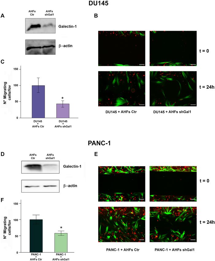
A. Toti et al.

manner within the TME, thereby representing a key form of         cells. Several CAFs proteins specifically transferred to can-
intercellular communication.                                      cer cells by this type of vehicle have been also identified.
   Although EVs comprise a great heterogeneous population         Among them, Galectin-1 (Gal-1) emerged as one of the most
of membrane vesicles, to date two types of EVs, with differ-      enriched protein in CAF-derived EVs (Santi et al. 2015).
ent size, biogenesis and composition, have been described:            Gal-1 is the best characterized member of the galectin
microvesicles (MVs)/ectosomes (diameter from 100 nm to            family (Johannes et al. 2018). Galectins are a phylogeneti-
1 μm) and exosomes (diameter from 30 to 100 nm) (Cocucci          cally conserved family of lectins and they are composed by
and Meldolesi 2015). With regard to EV biogenesis,                amino acid sequences of about 130 amino acids with the car-
exosomes are intraluminal vesicles (ILVs), that originate         bohydrate recognition domain responsible for β-galactoside
from inward budding of endosomal membrane during the              binding (Barondes et al. 1994). Gal-1 is generally localized
multivesicular endosome (MVE) maturation process, and are         in cell nuclei and cytoplasm, but it also translocates to
secreted as a consequence of MVEs fusion with cell surface.       the intracellular and extracellular side of cell membranes.
Conversely, MVs are generated by the outward budding and          Indeed, it displays the characteristics of typical cytoplasmic
fission of the plasma membrane and are then released in the       proteins, as well as an acetylated N-terminus and lack of gly-
extracellular space. EVs usually contain various cargoes,         cosylation. It is noteworthy that Gal-1 can also be secreted in
including proteins, lipids, and nucleic acids (DNA, mRNAs         the extracellular matrices of various normal and neoplastic
and miRNAs). The specific composition of the EV content           tissues (Cooper and Barondes 1990). Extracellular Gal-1 has
strictly depends on the cell type, the stimuli and molecular      been found altered in many cancer cell types (Thijssen et al.
mechanisms regulating their biogenesis and release, and the       2015), including melanoma (Yazawa et al. 2015), ovarian
physio-pathological state of the donor cells. Once released in    (Zhang et al. 2014) and prostate cancers (Laderach et al.
the extracellular space, EVs target recipient cells and deliver   2013). Moreover, Gal-1 is often overexpressed in the reac-
their content which then alters the phenotypical and func-        tive stromal cells in the TME (Valach et al. 2012). Increased
tional properties of the targeted cells (van Niel et al. 2018).   expression of Gal-1 correlates with a variety of processes in
   Several recent findings underlined that CAFs are able to       cancer progression, including the cellular aggregation/tumor
transfer EVs to tumor cells, providing significant evidence       formation, cancer metastatic spread, angiogenesis, and apop-
of EV-mediated back and forth exchange of factors between         tosis (Liu and Rabinovich 2005; Cousin and Cloninger 2016;
cancer and stromal cells within the TME (Shoucair et al.          Orozco et al. 2018).
2020). It is noteworthy that the uptake of CAF-derived EVs,           In the present work, we reveal that the intercellular trans-
usually loaded with specific miRNAs, lncRNAs and pro-             port of Gal-1 from CAFs to tumor cells, mediated by MVs
teins, by recipient tumor cells strongly supports invasion        trafficking, leads to the upregulation of its steady state con-
and metastasis (Josson et al. 2015; Ren et al. 2018; Miki         centration in the recipient cancer cells and contributes to
et al. 2018; Sun et al. 2019), and increases the proportion of    increase their migratory ability.
CSCs (Hu et al. 2015). In addition, it has been demonstrated
that patient-derived CAFs induce metabolic reprogramming
of prostate and pancreatic tumor cells following EV uptake,       Materials and methods
ultimately promoting cancer cell growth. In particular, CAF-
derived EVs supply nutrient-deprived tumor cells with vari-       Materials
ous metabolites, including amino acids, lipids and tricarbo-
xylic acid (TCA) cycle intermediates, to upregulate their         Unless otherwise specified all reagents are from Sigma-
central carbon metabolism (Zhao et al. 2016). Moreover,           Aldrich. Opti-MEM, FluoroBrite™ DMEM, Lipo-
recent findings underscored that miR-21, transferred from         fectamine® transfection reagent, DAPI, CellTrace™ CFSE
CAFs to recipient ovarian tumor cells through exosomes            Cell Proliferation Kit and CellTracker™ Orange Dye are
trafficking, suppresses tumor cell apoptosis and confers          from Invitrogen™ Life Technologies. siRNAs for Gal-
paclitaxel resistance (Au Yeung et al. 2016). Similarly, exo-     1, RNase-free DNase set are from Qiagen. Sh-RNA vec-
some-mediated transfer of miR-92a-3p from CAFs to colo-           tors are purchased from OriGene. RNA Nano Chip kit is
rectal cancer cells promotes metastasis and 5-fluorouracil        from Agilent. PrimeScript RT reagent kit, ProteaseMAX™
(5-FU)/ oxaliplatin (L-OHP) resistance (Hu et al. 2019).          Surfactant, Sequencing Grade Modified Trypsin are from
   Interestingly, a previous paper from our lab demonstrated      Promega. Polyvinylidene difluoride (PVDF) membrane is
that MVs transfer proteins and lipids essentially in a uni-       from Millipore. Transwells are purchased from Euroclone.
directional way, from CAFs to cancer cells. In particular,        Anti-Gal-1 antibody is from Cell Signaling, anti-β-actin
MV components have been found involved in increasing              and anti-Integrin-β1 antibodies are from Santa Cruz Bio-
melanoma and prostate cancer cell proliferation and in            technology, anti-CD81 antibody is from BD Biosciences.
inducing the reverse-Warburg phenotype in recipient tumor         The secondary antibodies enzyme horseradish peroxidase

13
Activated fibroblasts enhance cancer cell migration by microvesicles‑mediated transfer…
(HRP)–conjugated are from Santa Cruz Biotechnology. All 10.000 × g for 1 h obtaining a purified MVs fraction. To
materials for SDS-PAGE and Clarity™ Western ECL sub- isolate the exosomes fraction, the supernatant recovered
strate are from Biorad. Culture-inserts (Dish 35 mm, high) after the 10.000 × g centrifugation was further centrifuged
are from ibidi®. at 100.000 × g for 1 h and the pellet resuspended in PBS (Xu
et al. 2015).
Cell cultures
Western blot analysis
Human prostate (DU145 and LNCaP), pancreatic (PANC-
1), and melanoma (A375) cancer cells were purchased from For electrophoresis and western blot (WB) analysis,
European Collection of Cell Culture (ECACC). Normal MVs, exosomes or cell lysates were suspended in twofold
Human Fibroblasts (NHFs) used in our experiments were concentrated Laemmli electrophoresis buffer (without
Human Dermal Fibroblasts (HDFs, from Invitrogen™ Life β-mercaptoethanol and bromophenol blue) and assayed for
Technologies) and Human Prostate Fibroblasts (HPFs). protein content by the BCA method. Equal amount of total
HPFs were isolated from surgical explantation of patients protein (20–40 μg) from each sample were additioned with
who signed informed consent in accordance with the Ethics β-mercaptoethanol and separated by SDS-PAGE. Subse-
Committee of Azienda Ospedaliera Universitaria Careggi by quently, gels were electroblotted onto PVDF membranes.
Prof. Serni of Dipartimento di Medicina Sperimentale e Cli- The blots were incubated with the selected primary antibody
nica/Urology (Firenze, Italy) (Giannoni et al. 2010). No sig- in order to evaluate specific protein content as indicated in
nificant differences were actually determined in the behavior the figures. After incubation with secondary antibodies, the
of the two fibroblast lines. All cells were routinely cultured blotting was developed by using the ECL plus immunode-
in Dulbecco’s Modified Eagle’s Medium (DMEM)—high tection system.
glucose (4500 mg/L), except for LNCaP cells that were
cultured in RPMI medium. Both media were supplemented Quantitative real time polymerase chain reaction
with 10% fetal bovine serum (FBS, Euroclone), 2 mM glu- (qPCR)
tamine, 100 U/mL penicillin and 100 μg/mL streptomycin.
Cells were incubated at 37 °C in a humidified atmosphere Quantitative PCR reactions were performed by using GoTaq
of 5% CO2. qPCR master mix kit and the Mx3005P Stratagene system.
Differential expressions of transcripts of interest were cal-
Conditioned media preparation and fibroblast culated in relation to the h36B4 housekeeping transcript for
activation cDNA. The primers for GAL-1 were:
Tumor cells (DU145, A375, PANC-1) were incubated in Forward: 5’-TCG C CA G CA A CC T GA ATC T C-3’
growth medium with 1% of serum depleted by MVs and Reverse: 5’-GCACGAAGCTCTTAGCGTCA-3.
exosomes (EVs depleted FBS) for 24 h. Serum depletion was
performed by centrifugation at 10.000×g for 1 h and subse- Transwell co‑culture system
quently at 100.000 × g for 90 min. After 24 h of incubation
with tumor cells the medium was recovered, centrifuged at Co-culture experiments were performed by using Transwell
300 xg for 20 min to discard cell debris and used to culture permeable supports with pore sizes of 0.4 µm or 8 µm.
fibroblasts for 24 h, obtaining their activated forms (A-HDFs Tumor cells were seeded in 6-well plates, while NHFs were
and A-HPFs), superimposable to native CAFs (Giannoni plated on the upper compartment of the Transwells in a 2:1
et al. 2010). ratio. After 24 h of incubation, protein content of cancer
cells was analysed by western blot.
Purification of membrane vesicles secreted To verify that fibroblasts, seeded in the upper side of
by fibroblasts the 8 µm Transwells, do not traverse the filter and are not
collected with tumor cells, NHFs were stained with 10 µM
Normal Human Fibroblasts (NHFs: HPFs or HDFs) or Acti- CFDA-SE in Hank’s Balanced Salt Solution (HBSS) for
vated Human Fibroblasts (AHFs: A-HPFs or A-HDFs) were 15 min at 37 °C. Then, NHFs were incubated with com-
cultured for 24 h in growth medium supplemented with 1% plete DMEM for 1 h. After that, NHFs were harvested by
EVs depleted FBS. To isolate the MVs fraction, the medium trypsinization. Fibroblasts and tumor cells were co-cultured
was recovered and centrifuged at 300 xg for 10 min to dis- by using Transwell permeable supports with pore size of
card cells, at 2000 × g for 20 min to discard cell debris and 8.0 µm. Specifically, fibroblasts were seeded in the upper
finally at 10.000 × g for 1 h to pellet the MVs fraction. The side of the insert, while tumor cells were seeded in the lower
pellet was resuspended in PBS and centrifuged again at compartment. After 24 h of incubation, cells in the lower
13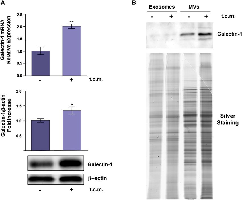
A. Toti et al.
compartment of the Transwell system were harvested by fluorescence microscopy and Gal-1 expression was evalu-
trypsinization, washed in PBS, and fixed in 3% paraform- ated by WB analysis. The vectors used were:
aldehyde. CFDA-SE fluorescence was analysed by flow
cytometry. Tumor cells not co-cultured with NHFs were #A AACCTGGAGGCCATCAACTACATGGCAGC
used as negative control. CFDA-SE-stained NHFs not co- #B TCTGGTCGCCAGCAACCTGAATCTCAAAC
cultured with cancer cells were used as positive control. #C GACGGTGACTTCAAGATCAAATGTGTGGC
#D CCTTCCAGCCTGGAAGTGTTGCAGAGGTG
siRNA cell transfection
The higher silencing has been found with vectors #A and
Silencing with siRNA plasmid was performed with Lipo- #B for both PANC-1 and NHFs. Results referred to PANC-1
fectamine™ 2000, following manufacturer’s instructions. and NHF cells silenced with vector #A.
Cells were plated at 70% confluence and, before transfec- The morphology of control and Gal-1 silenced PANC-1
tion, DMEM culture medium was removed and replaced cells was evaluated by taking photographs at randomly
with Optimem medium, lacking serum and antibiotics that chosen fields using the inverted microscope Nikon Eclipse
could interfere with liposome formation. Solution con- TS100.
taining siRNA was added to solution with Lipofectamine
and incubated at room temperature for 20 min, in order to Proliferation assay
promote liposome formation; then equal amounts of final
solution were added to each plate. Optimem medium was The proliferation of DU145 cells was evaluated using
removed after 4–6 h from transfection, as Lipofectamine CFDA-SE probe. Tumor cells, control or silenced for
could be slightly toxic for cells. Finally, cells were main- Gal-1, were labeled with the dye at the concentration of
tained in complete medium for 48 h and transfection effi- 2.5 μM. Then cells were cultured and after 24 and 48 h were
ciency was evaluated through immunoblotting assays. Gal-1 detached, fixed in 3% paraformaldehyde and analysed by
expression was silenced using four different pre-designed flow cytometry (BD FACSCanto II, BD Biosciences-US).
siRNAs directed against different regions of the Gal-1 The obtained fluorescence value was analysed by ModFit
transcript (Qiagen, Hilden, siRNA3 = Hs_LGALS1_3 software to estimate the proliferation index.
HP, Cat. No. SI00035924; siRNA5 = Hs_LGALS1_5 Cat. The proliferation of GFP positive PANC-1 cells, control
No. HP SI02628269; siRNA6 = Hs_LGALS1_6 HP, Cat. and silenced for Gal-1, was assessed by counting cells with
No. SI03033947; siRNA7 = Hs_LGALS1_7 HP, Cat. No. Bürker’s chamber three times per condition 24 and 48 h after
SI03085453). siRNA7 was more effective in terms of down- plating.
regulating Gal-1 expression on protein level compared to
the others. The morphology of control and Gal-1 silenced Adhesion assay
DU145 cells was evaluated by taking photographs at ran-
domly chosen fields using the inverted microscope Nikon 2 × 10⁵ cells were seeded in 35 mm dishes. After 0.5, 1, 2
Eclipse TS100. and 3 h, the number of adherent tumor cells was evaluated
with crystal violet staining for 5 min at 37 °C. Fixed cells
Sh‑RNA cell transfection were washed with PBS and solubilized with 0,1 M Sodium
Citrate, pH 4,2. The absorbance was evaluated at 595 nm.
Lentiviral particles were generated by transfecting 293 T
cells with a mix of 1/3 pGFP-C-shLenti construct (4 differ- Migration assay
ent vectors for Gal-1), 1/3 delta Helper (carries sequence
necessary for viral assembly of lentivirus) and 1/3 pVsVg PANC-1 migration was evaluated using Transwell systems,
(expresses the vesicular stomatitis virus envelop glycopro- equipped with 8 μm pore polyvinylpirrolidone-free polycar-
tein G pseudotype), using Lipofectamine™ 3000 Reagent bonate filters (6.5 mm diameter), coated with 1% fibronectin
and following manufacturer’s recommendations. 24 h post in 0.1% gelatin and stored at 4 °C. DU145 migration was
transfection, the medium was changed for fresh one. 24 h evaluated with the same Transwell system but without coat-
later, medium was changed again and viruses containing ing. 5 × 10⁴ cells resuspended in 150 μL of DMEM with 1%
medium was collected, filtered through a 0.2 μm filter, and EVs depleted FBS were seeded in the upper compartment of
added on 40% confluent PANC-1 and NHFs cells seeded Transwells placed into 24-well culture dishes. In the lower
in 6 multi-well plates. This step was repeated 24 h later to compartment, 500 μl of DMEM with 1% EVs depleted FBS
perform a second infection. Five days after infection, expres- for PANC-1 cells and DMEM with 10% EVs depleted FBS
sion of Green Fluorescent Protein (GFP) was verified by for DU145 cells was added. Then, PANC-1 and DU145 con-
trol cells were treated with 50 μL of DMEM with 1% EVs
13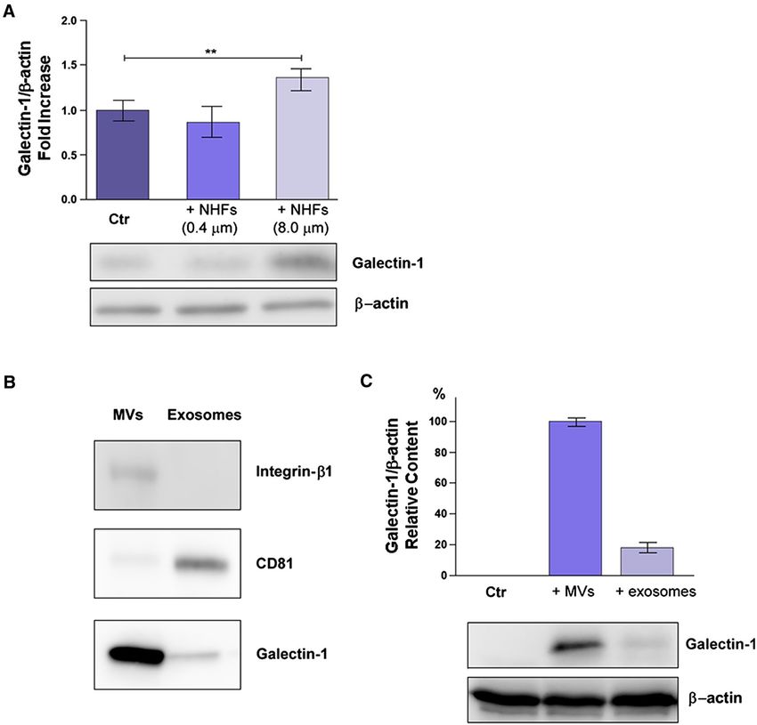
Activated fibroblasts enhance cancer cell migration by microvesicles‑mediated transfer…
depleted FBS. Besides, tumor cells were treated with MVs fibroblast activation is the increase in their ability to pro-
isolated from normal fibroblasts (HDFs or HPFs) or from duce and secrete MVs, which are then uploaded in cancer
activated fibroblasts (A-HDFs or A-HPFs). Specifically, cells, thereby playing an essential role in sustaining their
isolated MVs were resuspended in 50 μL of DMEM with proliferation. We previously identified various proteins that
1% EVs depleted FBS and then added to cell medium in the are specifically transferred from CAFs to cancer cells using
upper compartment of the Transwells. After 6 h for PANC-1 MVs as vehicle (Santi et al. 2015). Here we focused our
cells and 16 h for DU145 cells, while the insert was still attention on one of these proteins, Gal-1, that is both one of
moist, the non-migratory cells were mechanically removed the most present proteins in CAF-derived MVs and a protein
from the interior of the insert using a cotton swab. Migrated known to be involved in many aspects of cancer progression
cells were fixed in cold methanol, labeled with DAPI and (Liu and Rabinovich 2005).
photographed by fluorescence microscopy (Leica TCS SP5). Firstly, we found that NHFs upregulate both Gal-1 mRNA
Chemotaxis was evaluated by counting the cells migrated to and protein levels when treated for 24 h with tumor condi-
the lower surface of the filters as a mean of twenty randomly tioned medium (t.c.m.) from tumor cells (Fig. 1A). Treat-
chosen fields. ment with t.c.m. converts normal fibroblasts into their acti-
vated forms (AHFs), that express the same protein markers
Wound healing assay and share the same cellular properties of native CAFs iso-
lated from tumors (Giannoni et al. 2010; Santi et al. 2015;
PANC-1 and DU-145 cells were labeled with 5 μM Cell- Hu and Hu 2019).
Tracker™ Orange Dye and co-cultured with GFP-NHF cells All these experiments were performed using both Human
(control and silenced for Gal-1) in D ish35mm,high Culture- Prostate Fibroblasts (HPFs) and Human Dermal Fibroblasts
insert in ratio 1:1. The medium used for the co-culture was (HDFs), obtaining similar results. We then analysed the
the FluoroBrite™ DMEM that does not interfere with fluo- Gal-1 expression levels in EVs, i.e. MVs and exosomes,
rescence. The day after the insert was removed and pictures synthesized and secreted by normal and activated fibroblasts
at time 0 and 24 h were acquired with Leica FX 350 camera (Fig. 1B). Figure 1B clearly shows that Gal-1 is mostly pre-
in a Leica AM 600 microscope, using these parameters: L5: sent in MVs respect to exosomes and its expression in MVs
ex BP 480/40, dichroic 505, em BP 527–30; N2.1 ex BP is increased upon fibroblast activation.
515/560, dichroic 565, em LP 590. Overall, our data highlight that tumor secretome induces
Gal-1 overexpression in activated fibroblasts as well as in
Statistical analysis their secreted MVs.
In order to dissect the mechanism of Gal-1 transfer from
Data are presented in bar graphs as means ± SD from at least activated fibroblasts to cancer cells, we used Transwell sys-
three independent experiments (unless specified). Statistical tems with different pore sizes (0.4 or 8 μm). In this kind
analysis of the data was performed by Student’s /t/-test or of co-culture, we observe the rapid activation, within few
ANOVA followed by Tukey HSD test. /p/-values of ≤ 0.05 hours (data not shown), of fibroblasts into their activated
were considered statistically significant. Single asterisk counterpart due to cytokine-mediated crosstalk. Activated
indicates a significant difference at /p/-value < 0.05, double fibroblasts, in turn, increase their overall release of MVs
asterisks indicate a significant difference at /p/-value < 0.01 containing Gal-1.
and triple asterisks indicate a significant difference at Figure 2A shows that only when fibroblasts and
/p/value < 0.001. DU145 cancer cells were co-cultured in 8 μm Transwells
we observed an upregulation of Gal-1 in recipient tumor
cells. Hence, Gal-1 is not transferred by exosomes, that
Results easily pass through 0.4 μm filters, nor Gal-1 derives from
a de novo synthesis in cancer cells induced by fibroblast-
Gal‑1 is upregulated upon fibroblast activation derived cytokines for the same reason. In addition, we
and transferred to cancer cells using MVs as vehicles found that NHFs are not able to pass the 8.0 μm filters
and adhere to the lower compartment of Transwell sys-
The interplay between fibroblasts and cancer cells within tems where tumor cells are seeded, indicating that ana-
the TME induces remarkable changes in the phenotypic lysed cell lysates were specifically obtained from tumor
features of both cell types, favoring cancer progression cells. Overall these data underline that Gal-1 is trans-
(Kalluri 2016). In particular, upon stimulation with pro- ferred exclusively via MVs trafficking from fibroblasts
inflammatory cytokines secreted by cancer cells, fibroblasts to tumor cells. Analogous experiments were performed
trans-differentiate in their activated form (CAFs) (Giannoni on other tumor cell lines obtaining similar results (data
et al. 2010). One of the most important consequences of not shown).
13A. Toti et al.
Fig. 1 Gal-1 is overexpressed in activated fibroblasts and released DMEM supplemented with 1% EVs depleted FBS. Then, MVs and
into the extracellular milieu using MVs as vehicles. A Normal exosomes from NHFs and AHFs were isolated through differential
Human Fibroblasts (NHFs) were treated with t.c.m. from tumor centrifugation (see “Materials and methods”) and their total protein
cells for 24 h. qRT-PCR and WB analyses were performed to evalu- content analysed by SDS-PAGE and silver staining. The Gal-1 con-
ate Gal-1 mRNA and protein expression levels, respectively, in t.c.m. tent was evaluated by WB with anti-Gal-1 antibody. 30 µg aliquots of
activated NHFs (AHFs) with respect to NHFs. Data shown repre- proteins were loaded for each sample. The data shown are representa-
sent mean + / − SD from three independent experiments (*p < 0.05; tive of at least three experiments with similar results
**p < 0.01). B NHFs and AHFs were incubated for 24 h with fresh
Finally we purified and characterized MVs and Exogenous Gal‑1 improves tumor cell migration
exosomes from AHFs using integrin-β1 and CD81 as
specific markers of MVs and exosomes, respectively Gal-1 is widely reported as a protein able to influence sev-
(Fig. 2B). Purified MVs and exosomes were then incu- eral aspects of tumor progression (Cousin and Cloninger
bated for 4 h with LNCaP cells, a prostate cancer cell 2016). In order to assess its role in our experimental setting,
line characterized by extremely low expression levels of we silenced Gal-1 expression in both DU145 and PANC-1
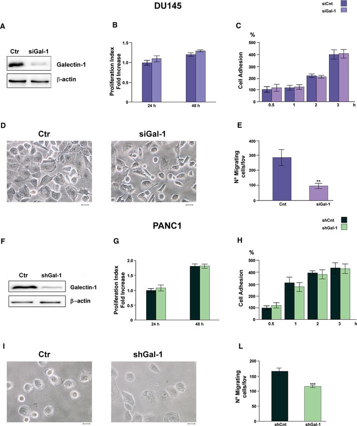
Gal-1. Interestingly, Gal-1 expression levels are strongly cells (Fig. 3A, F).
increased in cell lysates from LNCaP cells treated with Gal-1 silenced cells do not show differences nor in the
MVs, but only at a lower extent in tumor cells incubated cell proliferation rate (Fig. 3B, G) or in the adhesion proper-
with purified exosomes (Fig. 2C). ties (Fig. 3C, H), with respect to control cells. By contrast,
Gal-1 silenced DU145 and PANC-1 cells exhibit distinct
morphological traits when compared to their not-silenced
counterpart. In particular, control cells display a higher
13Activated fibroblasts enhance cancer cell migration by microvesicles‑mediated transfer…
Fig. 2 Gal-1 is transferred to tumor cells using MVs as vehicles. t.c.m. Then, fresh growth medium with 1% FBS (depleted of EVs)
A DU145 tumor prostate cells were seeded in a 6-well plate, while was added for the following 24 h in order to collect fibroblast-derived
fibroblasts (NHFs) were plated on Transwells of different pore sizes MVs and exosomes. WB of AHF-derived MVs and exosomes, quan-
(0.4 μm or 8 μm). The co-culture rapidly induces the fibroblasts acti- tified for protein content, was performed to characterize specific
vation. After 24 h cancer cells in the lower chamber were lysed and markers of MVs (integrin-β1) and exosomes (CD81). Equal amounts
subjected to WB analysis. Quantification plot of Gal-1 expression, (20 µg) of each sample were analysed. C LNCaP cells were treated
normalized using β-actin, was reported as fold increase respect to for 4 h with purified MVs or exosomes from A-HPF, resuspended
control (cancer cells in the lower chamber without fibroblasts plated in growth medium with 1% EVs depleted FBS. Then, the amount of
on the filter). Data represent mean + / − SD from four independ- Gal-1 in LNCaP cells was evaluated by WB analysis. The data shown
ent experiments (**p < 0.01). B NHFs were activated for 24 h with are representative of at least three experiments with similar results
number of cellular protrusion (Fig. 3D, I), that results in promoting DU145 and PANC-1 cell migration respectively
increased migratory potential. Indeed, Gal-1 silencing (Fig. 4B, F).
strongly impairs both DU145 and PANC-1 migratory abil- To demonstrate that fibroblast-derived Gal-1 is directly
ity (Fig. 3E, L). implicated in tumor cell migration, we set up a wound heal-
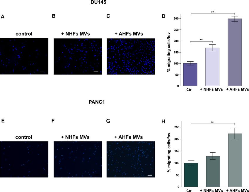
Moreover we observed that purified MVs derived ing assay in which PANC-1 and DU145 cells, labeled with
from AHFs (Fig. 4C, G) highly enhance both DU145 and CellTracker™ Orange Dye, were seeded in co-culture with
PANC-1 migration when compared to control cells treated AHFs, expressing GFP and stably silenced or not for Gal-1
with DMEM with 1% FBS depleted of EVs (Fig. 4A, E). (Fig. 5A, D). PANC-1 and DU145 cells co-cultured with
Interestingly, MVs purified from NHFs are less efficient in control AHFs result to have a greater migratory ability
13A. Toti et al. compared to cancer cells co-cultured with Gal-1 silenced considering cancer cells movement due to proliferation AHFs (Fig. 5B, E). To quantify the results of this experi- (Fig. 5C, F). ment, PANC-1 and DU145 cells present in the scratch at These findings evidence a novel mechanism exerted by 24 h were counted, excluding those placed at a distance CAFs to support tumor invasiveness through exogenous pro- less than 50 μm from the boundary of the scratch, to avoid tein transfer via MVs. 13
Activated fibroblasts enhance cancer cell migration by microvesicles‑mediated transfer…

◂Fig. 3  Gal-1 enhances cell migration but not proliferation nor adhe-       migration and tube formation (Tang et al. 2016). However,
 sion of DU145 and PANC-1 cells. Cell proliferation, cell adhe-              the molecular mechanisms by which high levels of exog-
 sion, morphology and cell migration were evaluated on DU145 and
 PANC-1 cells silenced or not for Gal-1 expression. A WB analysis
                                                                             enous Gal-1 in the stromal compartment affect cancer cell
 of Gal-1 expression of control and silenced DU145 cells. Silencing          aggressiveness have not yet been completely clarified. Inter-
 has been achieved by siRNA (see “Materials and methods”). B Pro-            estingly, in vitro and in vivo studies on pancreatic ductal
 liferation index of control and silenced DU145 cells using CFDA-SE          adenocarcinoma (PDAC) underlined that stromal Gal-1,
 assay (see “Materials and methods”). C Cell adhesion assay of con-
 trol and silenced DU145 cells. D Cell morphology of control and
                                                                             that is highly overexpressed by stromal fibroblasts and pan-
 Gal-1 silenced DU145 cells. Scale bar = 50 µm. E Migration assay of         creatic stellate cells, is directly secreted in the TME and
 control and silenced DU145 cells. F WB analysis of Gal-1 expression         consequently establishes paracrine crosstalk with epithelial
 of control and silenced PANC-1 cells. Silencing has been achieved           tumor cells to further trigger proliferation and invasion of
 by shRNA (see “Materials and methods”). G Proliferation index
 of control and silenced PANC-1 cells using Burker’s chamber cell
                                                                             cancer cells, enhance angiogenesis and inhibit immune cell
 count. H Cell adhesion assay of control and silenced PANC-1 cells.          infiltration (Xue et al. 2011; Martínez-Bosch et al. 2014;
 I Cell morphology of control and Gal-1 silenced PANC-1 cells. Scale         Orozco et al. 2018).
 bar = 50 µm. L Migration assay of control and silenced PANC-1 cells.            In the present paper, we demonstrate that Gal-1 expres-
 For all kind of tests data represent mean + / − SD from at least three
 independent experiments (**p < 0.01; ***p < 0.001)
                                                                             sion in various cancer cell types can be upregulated through
                                                                             its highly efficient transfer via-MVs from activated fibro-
                                                                             blasts to tumor cells, rather than being directly secreted in
 Discussion                                                                  the extracellular environment by stromal cells or being mod-
                                                                             ulated by the more common intercellular signaling mediated
 Solid tumors are composed by cancer cells and tumor reac-                   by paracrine factors.
 tive stroma (Wang et al. 2017). In the last decades the key                     It is noteworthy that protein transfer via EVs from CAFs
 role of stromal cells in promoting cancer aggressiveness                    to cancer cells has not been largely addressed, since the
 has been the focus of several studies. Fibroblasts are the                  majority of the studies focused on the role of EV-mediated
 major stromal component in the TME and, in this context,                    transfer of miRNAs within the TME (Shoucair et al. 2020).
 they switch into their activated phenotype, namely CAFs.                    Notably, we show that the conversion of normal fibroblasts
 Cancer progression is strongly influenced by CAFs, that                     into their activated counterparts, upon stimulation with
 support ECM remodeling, angiogenesis, and inflammatory                      cytokines in the tumor conditioned media from cancer
 cells recruitment, via secretion of cytokines, chemokines,                  cells, causes the upregulation of Gal-1 expression levels,
 growth factors and EVs (Kalluri 2016; Barbazán and Matic                    both in their intracellular compartment and in their secreted
 Vignjevic 2019; Choe et al. 2013).                                          MVs (Fig. 1). Indeed, the protein content of EVs is strictly
    Santi and co-workers recently discovered a horizontal                    dependent on the cell type they originate from, the biogen-
 unidirectional transfer of proteins and lipids from CAFs to                 esis and the stimuli driving their release (Zaborowski et al.
 cancer cells, mostly mediated by MVs. They demonstrated                     2015).
 that, even if structural proteins and glycolytic enzymes rep-                   Despite MVs trafficking is currently emerging as a criti-
 resent the 70% of total mass transferred from CAFs to cancer                cal mediator of intercellular communication in the context
 cells, the majority of these proteins are not enriched in tumor             of the tumor-stroma crosstalk (Muralidharan-Chari et al.
 cells. By contrast, among the CAF-derived proteins specifi-                 2010; Menck et al. 2020), its function is still poor defined.
 cally upregulated in recipient cells through MVs trafficking,               For instance, most studies concerning EVs transfer during
 Gal-1 resulted to be one of the most represented (Santi et al.              cancer progression focused on exosomes (Dai et al. 2020) or
 2015). Therefore, in this paper we focus our attention on                   mixed vesicle populations and it is still largely unclear which
 MV-mediated transfer of Gal-1 from CAFs to tumor cells                      sub-population of EVs is responsible for a given physio-
 in affecting the migratory abilities of recipient cancer cells.             pathological effect (van Niel et al. 2018; Han et al. 2019;
    Several studies investigated the role of stromal Gal-1                   Maacha et al. 2019). The reason for such a lack of informa-
 upregulation in supporting tumor progression and aggres-                    tion regarding MV trafficking is mainly due to the challenges
 siveness, thus revealing its possible application as a novel                encountered during the selective isolation of specific sub-
 therapeutic target for many types of cancer. In particular,                 species of vesicles. Indeed, although numerous reports have
 Gal-1 has been found overexpressed in cancer-associated                     been published on comparative methods for EV isolation,
 stromal cells of gastric adenocarcinoma and breast and pros-                including density gradient ultracentrifugation, size exclusion
 tate tumors, correlating with increased tumor invasiveness                  chromatography, precipitation via volume-excluding poly-
 and metastasis (van den Brûle et al. 2001; Jung et al. 2007;                mers, flow-cytometry, and high pressure liquid chromatogra-
 Bektas et al. 2010). Moreover, Tang and co-workers reported                 phy, we are still far from having pure isolation of exosomes
 that CAF-derived Gal-1 strongly promotes angiogenesis in                    and MVs (Konoshenko et al. 2018; Théry et al. 2018). In this
 gastric cancer by sustaining endothelial cell proliferation,                context, we checked the purity of EV separation, obtained

                                                                                                                                 13
A. Toti et al. through differential centrifugation, by using Integrin β1 settings, Gal-1 expression is upregulated in tumor and CD81 as markers of MVs and exosomes, respectively recipient cells (Fig. 2). In addition, LNCaP cells, which are (Fig. 2) (Santi et al. 2015). characterized by endogenous extremely low levels of Gal-1, Remarkably, we highlight that the specific intercellular treated with MVs purified from AHFs display an appreciable trafficking of MVs from activated fibroblasts to cancer cells increase in the expression levels of Gal-1, when compared mediates the transfer of Gal-1 to the latter. In fact, when to cells treated with purified exosomes from AHFs (Fig. 2). tumor cells are co-cultured with fibroblasts in Transwell These results further indicate that Gal-1 is transferred systems with 0.4 μm pore size, that allow the free passage from activated fibroblasts to cancer cells specifically via of exosomes and paracrine soluble factors released by MVs. This finding is consistent with our previous evidence fibroblasts but not the transfer of MVs, Gal-1 expression highlighting that MVs are more efficient than exosomes in levels are not increased in cancer cells. Conversely, when transferring proteins to recipient cells (Santi et al. 2015). using Transwell systems with 8 μm pore size, freely Along with Gal-1 upregulation in the tumor-associated permeable to MVs, in tumor cells/fibroblasts co-culture stromal compartment, Gal-1 has been found overexpressed Fig. 4 DU145 and PANC-1 migration is strongly enhanced by MVs of migrated cells respect to control. E–G PANC-1 cells cultured in derived from activated fibroblasts. Boyden chamber migration assay the presence of: E DMEM supplemented with 1% FBS depleted of was performed on DU145 and PANC-1 cells treated or not with MVs EVs (control); F MVs purified from NHFs; G MVs purified from derived from both normal fibroblasts and activated fibroblasts. A–C AHFs (see “Materials and methods”). H Quantification of PANC-1 DU145 cells cultured in the presence of: A DMEM supplemented cell migratory capacity expressed as fold increase of migrated cells with 1% FBS depleted of EVs (control); B MVs purified from NHFs; respect to control. Scale bar = 50 µm. Data represent mean + / − SD C MVs purified from AHFs (see “Materials and methods”). D Quan- from three independent experiments (**p < 0.01) tification of DU145 cell migratory capacity expressed as fold increase 13
Activated fibroblasts enhance cancer cell migration by microvesicles‑mediated transfer…

Fig. 5  Exogenous Gal-1 derived by activated fibroblasts promotes           distance of less than 50 μm from both sides of the scratch. E Wound
migration in tumor cells. A, D Stable Gal-1 silencing in AHFs was           healing assay of PANC-1 cells (labeled with Cell Tracker™ Orange
evaluated by WB analysis. β-actin was used for normalization. B             Dye) in co-culture with AHFs (GFP positive) silenced or not for
Wound healing assay of DU145 cells (labeled with Cell Tracker™              Gal-1 expression. Scale bar 200 µm. F Mean of PANC-1 cells pre-
Orange Dye) in co-culture with AHFs (GFP positive) silenced or not          sent in the wound after 24 h, without considering cells placed at a
for Gal-1 expression. Scale bar 200 µm. C Mean of DU145 cells pre-          distance of less than 50 μm from both sides of the scratch. Data rep-
sent in the wound after 24 h, without considering cells placed at a         resent mean + / − SD from three independent experiments (*p < 0.05)

                                                                                                                                     13
A. Toti et al.
also in cancer cells. Indeed, it has been reported that the counterpart (Fig. 1). This finding is consistent with our
overexpression of this protein in different tumor types corre- previous results, indicating that activated fibroblasts have
lates with several processes of cancer malignancy, including a greater ability to transfer bioactive molecules via MVs-
tumor cell proliferation, migration, invasion and T cell acti- trafficking to recipient tumor cells, when compared to their
vation (Spano et al. 2010; Kim et al. 2012; Noda et al. 2017), not-activated counterpart (Santi et al. 2015).
and it is associated with patient worse prognosis (Kim et al. Finally, we demonstrate that in a co-culture wound heal-
2013; Chen et al. 2014; Yazawa et al. 2015). Therefore, we ing assay, Gal-1 stably silenced AHFs are far less efficient
evaluate the biological effects of Gal-1 upregulation in our in promoting PANC-1 and DU145 cell migration than wild
cancer cell models. To this aim, we silenced Gal-1 expres- type AHFs, further substantiating the biological impor-
sion in two different cancer cell lines: DU145, a prostatic tance of exogenous Gal-1 in cancer cell migration (Fig. 5).
cancer cell line, and PANC-1, a pancreatic adenocarcinoma Accordingly, several studies highlight the key role of
cell line. This latter represents a kind of tumor characterized CAF-derived EVs in enhancing the migratory and invasive
by a high degree of stromal infiltration and fibrosis (von potential of recipient cancer cells. For example, Leca and
Ahrens et al. 2017). In DU145 as well as in PANC-1 cells, co-authors revealed an increase in the migratory abilities
Gal-1 downregulation does not affect cell proliferation nor of PDAC cells after the uptake of CAF-derived ANXA6
cell adhesion rate, while strongly impairs cell migration positive EVs (Leca et al. 2016). Similarly, EVs secreted
(Fig. 3). Accordingly, it has been demonstrated that Gal-1 by fibroblasts support colorectal cancer cell proliferation,
is highly overexpressed in castrate resistant prostate cancer probably through the transfer of amphiregulin (Oszvald
(CRPC) and its knockdown significantly decreases prostate et al. 2020), and FAK signaling in CAFs regulates the
cancer cell migration and invasion (Shih et al. 2018). Simi- abilities of CAF-derived exosomes to induce breast can-
larly, Gal-1 plays a pivotal role in PDAC progression, by cer cell migration and ultimately metastasis (Wu et al.
inducing tumor growth, immune evasion and angiogenesis 2020). Coherently, Luga and co-workers reported that
(Berberat et al. 2001; Roda et al. 2009; Martinez-Bosch et al. fibroblast-secreted exosomes drive breast cancer cell inva-
2018). Overall, besides the already described role of Gal-1 sion through Wnt-planar cell polarity autocrine signaling
overexpression in the stromal compartment of pancreatic (Luga et al. 2012). In addition, it has been demonstrated
and prostate tumors (van den Brûle et al. 2001; Orozco et al. that CAF-derived EVs promote the migration and invasion
2018), the upregulation of this protein in cancer cells is cru- of oral squamous cell carcinoma (Dourado et al. 2019).
cial for tumor malignancy. However, the importance of the specific transfer of pro-
MVs recently emerged as important vehicles for the teins via MVs in affecting migration of recipient cells has
transfer of bioactive molecules within the TME. Indeed, the not been largely addressed so far.
deliver of MV cargoes in tumor cells strongly alters their Overall, our work highlights the fundamental role of the
functional characteristics. However, the majority of these MVs-mediated trafficking of specific CAF proteins in the
studies investigated the effects of tumor-derived MVs in crosstalk, within TME, between cancer and stromal cells,
modulating the behavior of either tumor cells, via autologous thereby providing specific phenotypic advantages to tumor
cell–cell communication (Al-Nedawi et al. 2008; Arendt cells. In our case, exogenous Gal-1, derived from MVs
et al. 2014), or neighboring TME cells, including endothelial released by CAFs, increases cell motility in two different
cells (Kawamoto et al. 2012), fibroblasts (Jiang et al. 2019) tumor cell lines. To the best of our knowledge this is the
and immune cells (Baj-Krzyworzeka et al. 2007; Cui et al. first example showing that an exogenous protein trans-
2018), through heterologous cell–cell communication. ferred from CAFs to cancer cells using MVs as vehicles
Conversely, the effects of CAF-derived MVs in cancer may profoundly affect cancer progression.
progression still need to be elucidated. Since Gal-1 plays a
fundamental role in promoting migration of tumor cells (Zhu
et al. 2016; Orozco et al. 2018), we then investigated whether Author’s contribution A.T. Investigation, writing original draft, visu-
alization; A.S. Investigation, visualization; E.P. Investigation and
MV-derived Gal-1 released from activated fibroblasts affects reviewing the paper; I.N. Investigation, visualization; R.T. Supervision,
the migratory ability of DU145 and PANC-1 tumor cells. resources; T.M. Investigation; P.P. Investigation; A.C. Conceptualiza-
It is noteworthy that MVs purified from normal fibro- tion, supervision, funding acquisition, writing-reviewing and editing
blasts are weakly able to increase DU145 migration the paper; P.C. Conceptualization, supervision, funding acquisition,
writing original paper and editing. All authors contributed to the final
respect to control, while MVs purified from activated manuscript.
fibroblasts strongly increase the migratory ability of
tumor cells (Fig. 4). This effect correlates with enhanced Funding Open access funding provided by Università degli Studi di
Gal-1 expression in MVs derived from activated fibro- Firenze within the CRUI-CARE Agreement. This study was supported
blasts when compared to those derived from their normal by Università degli Studi di Firenze, “fondi di ateneo” to PC and AC
(2016–2018).
13Activated fibroblasts enhance cancer cell migration by microvesicles‑mediated transfer…
Declarations Comito G, Ippolito L, Chiarugi P, Cirri P (2020) Nutritional exchanges
within tumor microenvironment: impact for cancer aggressive-
ness. Front Oncol 10:396. https://doi.org/10.3389/fonc.2020.
Conflict of interest The authors declare that they have no conflict of
00396
interest.
Cooper DN, Barondes SH (1990) Evidence for export of a muscle
lectin from cytosol to extracellular matrix and for a novel secre-
Open Access This article is licensed under a Creative Commons Attri- tory mechanism. J Cell Biol 110:1681–1691. https://doi.org/10.
bution 4.0 International License, which permits use, sharing, adapta- 1083/jcb.110.5.1681
tion, distribution and reproduction in any medium or format, as long Cousin JM, Cloninger MJ (2016) The Role of Galectin-1 in Cancer
as you give appropriate credit to the original author(s) and the source, Progression, and Synthetic Multivalent Systems for the Study of
provide a link to the Creative Commons licence, and indicate if changes Galectin-1. Int J Mol Sci. https://doi.org/10.3390/ijms17091566
were made. The images or other third party material in this article are Cui J, Li Q, Luo M et al (2018) Leukemia cell-derived microvesicles
included in the article’s Creative Commons licence, unless indicated induce T cell exhaustion via miRNA delivery. Oncoimmunology
otherwise in a credit line to the material. If material is not included in 7:e1448330. https://doi.org/10.1080/2162402X.2018.1448330
the article’s Creative Commons licence and your intended use is not Dai J, Su Y, Zhong S et al (2020) Exosomes: key players in cancer
permitted by statutory regulation or exceeds the permitted use, you will and potential therapeutic strategy. Sig Transduct Target Ther
need to obtain permission directly from the copyright holder. To view a 5:145. https://doi.org/10.1038/s41392-020-00261-0
copy of this licence, visit http://creativecommons.org/licenses/by/4.0/. De Jaeghere EA, Denys HG, De Wever O (2019) Fibroblasts fuel
immune escape in the tumor microenvironment. Trends Cancer
5:704–723. https://doi.org/10.1016/j.trecan.2019.09.009
Dourado MR, Korvala J, Åström P et al (2019) Extracellular vesicles
derived from cancer-associated fibroblasts induce the migra-
References tion and invasion of oral squamous cell carcinoma. J Extracell
Vesicles 8:1578525. https://doi.org/10.1080/20013078.2019.
Al-Nedawi K, Meehan B, Micallef J et al (2008) Intercellular transfer 1578525
of the oncogenic receptor EGFRvIII by microvesicles derived Giannoni E, Bianchini F, Masieri L et al (2010) Reciprocal activation
from tumour cells. Nat Cell Biol 10:619–624. https://doi.org/10. of prostate cancer cells and cancer-associated fibroblasts stimu-
1038/ncb1725 lates epithelial-mesenchymal transition and cancer stemness.
Arendt BK, Walters DK, Wu X et al (2014) Multiple myeloma dell- Cancer Res 70:6945–6956. https://doi.org/10.1158/0008-5472.
derived microvesicles are enriched in CD147 expression and CAN-10-0785
enhance tumor cell proliferation. Oncotarget 5:5686–5699. https:// Han L, Lam EW-F, Sun Y (2019) Extracellular vesicles in the tumor
doi.org/10.18632/oncotarget.2159 microenvironment: old stories, but new tales. Mol Cancer 18:59.
Au Yeung CL, Co N-N, Tsuruga T et al (2016) Exosomal transfer https://doi.org/10.1186/s12943-019-0980-8
of stroma-derived miR21 confers paclitaxel resistance in ovarian Hu T, Hu J (2019) Melanoma-derived exosomes induce reprogram-
cancer cells through targeting APAF1. Nat Commun 7:11150. ming fibroblasts into cancer-associated fibroblasts via Gm26809
https://doi.org/10.1038/ncomms11150 delivery. Cell Cycle 18:3085–3094. https://d oi.o rg/1 0.1 080/1 5384
Baj-Krzyworzeka M, Szatanek R, Weglarczyk K et al (2007) Tumour- 101.2019.1669380
derived microvesicles modulate biological activity of human Hu Y, Yan C, Mu L et al (2015) Fibroblast-derived exosomes con-
monocytes. Immunol Lett 113:76–82. https://doi.org/10.1016/j. tribute to chemoresistance through priming cancer stem cells in
imlet.2007.07.014 colorectal cancer. PLoS ONE 10:e0125625. https://doi.org/10.
Barbazán J, Matic Vignjevic D (2019) Cancer associated fibroblasts: is 1371/journal.pone.0125625
the force the path to the dark side? Curr Opin Cell Biol 56:71–79. Hu JL, Wang W, Lan XL et al (2019) CAFs secreted exosomes
https://doi.org/10.1016/j.ceb.2018.09.002 promote metastasis and chemotherapy resistance by enhanc-
Barondes SH, Cooper DN, Gitt MA, Leffler H (1994) Galectins. Struc- ing cell stemness and epithelial-mesenchymal transition in
ture and function of a large family of animal lectins. J Biol Chem colorectal cancer. Mol Cancer 18:91. https://doi.org/10.1186/
269:20807–20810 s12943-019-1019-x
Bektas S, Bahadir B, Ucan BH, Ozdamar SO (2010) CD24 and galec- Jiang E, Xu Z, Wang M et al (2019) Tumoral microvesicle-activated
tin-1 expressions in gastric adenocarcinoma and clinicopathologic glycometabolic reprogramming in fibroblasts promotes the pro-
significance. Pathol Oncol Res 16:569–577. https://doi.org/10. gression of oral squamous cell carcinoma. FASEB J 33:5690–
1007/s12253-010-9248-8 5703. https://doi.org/10.1096/fj.201802226R
Berberat PO, Friess H, Wang L et al (2001) Comparative analysis of Johannes L, Jacob R, Leffler H (2018) Galectins at a glance. J Cell Sci.
galectins in primary tumors and tumor metastasis in human pan- https://doi.org/10.1242/jcs.208884
creatic cancer. J Histochem Cytochem 49:539–549. https://doi. Josson S, Gururajan M, Sung SY et al (2015) Stromal fibroblast-
org/10.1177/002215540104900414 derived miR-409 promotes epithelial-to-mesenchymal transition
Chen J, Tang D, Wang S et al (2014) High expressions of galectin-1 and prostate tumorigenesis. Oncogene 34:2690–2699. https://doi.
and VEGF are associated with poor prognosis in gastric cancer org/10.1038/onc.2014.212
patients. Tumour Biol 35:2513–2519. https://doi.org/10.1007/ Jung E-J, Moon H-G, Cho BI et al (2007) Galectin-1 expression in
s13277-013-1332-8 cancer-associated stromal cells correlates tumor invasiveness and
Choe C, Shin Y-S, Kim S-H et al (2013) Tumor-stromal interactions tumor progression in breast cancer. Int J Cancer 120:2331–2338.
with direct cell contacts enhance motility of non-small cell lung https://doi.org/10.1002/ijc.22434
cancer cells through the hedgehog signaling pathway. Anticancer Kalluri R (2016) The biology and function of fibroblasts in cancer.
Res 33:3715–3723 Nat Rev Cancer 16:582–598. https://doi.org/10.1038/nrc.2016.73
Cocucci E, Meldolesi J (2015) Ectosomes and exosomes: shedding Kawamoto T, Ohga N, Akiyama K et al (2012) Tumor-derived
the confusion between extracellular vesicles. Trends Cell Biol microvesicles induce proangiogenic phenotype in endothelial
25:364–372. https://doi.org/10.1016/j.tcb.2015.01.004 cells via endocytosis. PLoS ONE 7:e34045. https://doi.org/10.
1371/journal.pone.0034045
13A. Toti et al.
Kim H-J, Jeon H-K, Cho YJ et al (2012) High galectin-1 expression angiogenesis through elevated SDF-1/CXCL12 secretion. Cell
correlates with poor prognosis and is involved in epithelial ovarian 121:335–348. https://doi.org/10.1016/j.cell.2005.02.034
cancer proliferation and invasion. Eur J Cancer 48:1914–1921. Orozco CA, Martinez-Bosch N, Guerrero PE et al (2018) Targeting
https://doi.org/10.1016/j.ejca.2012.02.005 galectin-1 inhibits pancreatic cancer progression by modulating
Kim H-J, Do I-G, Jeon H-K et al (2013) Galectin 1 expression is asso- tumor-stroma crosstalk. Proc Natl Acad Sci USA 115:E3769–
ciated with tumor invasion and metastasis in stage IB to IIA cervi- E3778. https://doi.org/10.1073/pnas.1722434115
cal cancer. Hum Pathol 44:62–68. https://d oi.o rg/1 0.1 016/j.h umpa Oszvald Á, Szvicsek Z, Pápai M et al (2020) Fibroblast-derived extra-
th.2012.04.010 cellular vesicles induce colorectal cancer progression by transmit-
Konoshenko MY, Lekchnov EA, Vlassov AV, Laktionov PP (2018) ting amphiregulin. Front Cell Dev Biol 8:558. https://doi.org/10.
Isolation of extracellular vesicles: general methodologies and 3389/fcell.2020.00558
latest trends. Biomed Res Int 2018:8545347. https://doi.org/10. Ren J, Ding L, Zhang D et al (2018) Carcinoma-associated fibroblasts
1155/2018/8545347 promote the stemness and chemoresistance of colorectal cancer by
Labernadie A, Kato T, Brugués A et al (2017) A mechanically active transferring exosomal lncRNA H19. Theranostics 8:3932–3948.
heterotypic E-cadherin/N-cadherin adhesion enables fibroblasts https://doi.org/10.7150/thno.25541
to drive cancer cell invasion. Nat Cell Biol 19:224–237. https:// Roda O, Ortiz-Zapater E, Martínez-Bosch N et al (2009) Galectin-1
doi.org/10.1038/ncb3478 is a novel functional receptor for tissue plasminogen activator
Laderach DJ, Gentilini LD, Giribaldi L et al (2013) A unique galectin in pancreatic cancer. Gastroenterology 136(1379–1390):e1-5.
signature in human prostate cancer progression suggests galec- https://doi.org/10.1053/j.gastro.2008.12.039
tin-1 as a key target for treatment of advanced disease. Cancer Santi A, Caselli A, Ranaldi F et al (2015) Cancer associated fibro-
Res 73:86–96. https://doi.org/10.1158/0008-5472.CAN-12-1260 blasts transfer lipids and proteins to cancer cells through cargo
Leca J, Martinez S, Lac S et al (2016) Cancer-associated fibroblast- vesicles supporting tumor growth. Biochim Biophys Acta
derived annexin A6+ extracellular vesicles support pancreatic 1853:3211–3223. https://doi.org/10.1016/j.bbamcr.2015.09.013
cancer aggressiveness. J Clin Investig 126:4140–4156. https:// Santi A, Kugeratski FG, Zanivan S (2018) Cancer associated
doi.org/10.1172/JCI87734 fibroblasts: the architects of stroma remodeling. Proteomics
Liu F-T, Rabinovich GA (2005) Galectins as modulators of tumour 18:e1700167. https://doi.org/10.1002/pmic.201700167
progression. Nat Rev Cancer 5:29–41. https://doi.org/10.1038/ Shih T-C, Liu R, Wu C-T et al (2018) Targeting galectin-1 impairs
nrc1527 castration-resistant prostate cancer progression and invasion.
Liu T, Zhou L, Li D et al (2019) Cancer-associated fibroblasts build Clin Cancer Res 24:4319–4331. https://doi.org/10.1158/1078-
and secure the tumor microenvironment. Front Cell Dev Biol 7:60. 0432.CCR-18-0157
https://doi.org/10.3389/fcell.2019.00060 Shoucair I, Weber Mello F, Jabalee J et al (2020) The role of cancer-
Luga V, Zhang L, Viloria-Petit AM et al (2012) Exosomes mediate associated fibroblasts and extracellular vesicles in tumorigen-
stromal mobilization of autocrine Wnt-PCP signaling in breast esis. Int J Mol Sci. https://doi.org/10.3390/ijms21186837
cancer cell migration. Cell 151:1542–1556. https://doi.org/10. Spano D, Russo R, Di Maso V et al (2010) Galectin-1 and its
1016/j.cell.2012.11.024 involvement in hepatocellular carcinoma aggressiveness. Mol
Maacha S, Bhat AA, Jimenez L et al (2019) Extracellular vesicles- Med 16:102–115. https://doi.org/10.2119/molmed.2009.00119
mediated intercellular communication: roles in the tumor micro- Sun L-P, Xu K, Cui J et al (2019) Cancer-associated fibroblast-
environment and anti-cancer drug resistance. Mol Cancer 18:55. derived exosomal miR-382-5p promotes the migration and
https://doi.org/10.1186/s12943-019-0965-7 invasion of oral squamous cell carcinoma. Oncol Rep 42:1319–
Martínez-Bosch N, Fernández-Barrena MG, Moreno M et al (2014) 1328. https://doi.org/10.3892/or.2019.7255
Galectin-1 drives pancreatic carcinogenesis through stroma Tang D, Gao J, Wang S et al (2016) Cancer-associated fibroblasts
remodeling and Hedgehog signaling activation. Cancer Res promote angiogenesis in gastric cancer through galectin-1
74:3512–3524. https://d oi.org/1 0.1158/0008-5472.C AN-13-3013 expression. Tumour Biol 37:1889–1899. https://d oi.o rg/1 0.
Martinez-Bosch N, Barranco LE, Orozco CA et al (2018) Increased 1007/s13277-015-3942-9
plasma levels of galectin-1 in pancreatic cancer: potential use as Théry C, Witwer KW, Aikawa E et al (2018) Minimal information for
biomarker. Oncotarget 9:32984–32996. https://doi.org/10.18632/ studies of extracellular vesicles 2018 (MISEV2018): a position
oncotarget.26034 statement of the International Society for Extracellular Vesicles
Menck K, Sivaloganathan S, Bleckmann A, Binder C (2020) Microves- and update of the MISEV2014 guidelines. J Extracell Vesicles
icles in cancer: small size. Large Potential Int J Mol Sci. https:// 7:1535750. https://doi.org/10.1080/20013078.2018.1535750
doi.org/10.3390/ijms21155373 Thijssen VL, Heusschen R, Caers J, Griffioen AW (2015) Galec-
Miki Y, Yashiro M, Okuno T et al (2018) CD9-positive exosomes tin expression in cancer diagnosis and prognosis: a systematic
from cancer-associated fibroblasts stimulate the migration ability review. Biochim Biophys Acta 1855:235–247. https://doi.org/
of scirrhous-type gastric cancer cells. Br J Cancer 118:867–877. 10.1016/j.bbcan.2015.03.003
https://doi.org/10.1038/bjc.2017.487 Valach J, Fík Z, Strnad H et al (2012) Smooth muscle actin-express-
Morandi A, Giannoni E, Chiarugi P (2016) Nutrient exploitation within ing stromal fibroblasts in head and neck squamous cell carci-
the tumor-stroma metabolic crosstalk. Trends Cancer 2:736–746. noma: increased expression of galectin-1 and induction of poor
https://doi.org/10.1016/j.trecan.2016.11.001 prognosis factors. Int J Cancer 131:2499–2508. https://doi.org/
Muralidharan-Chari V, Clancy JW, Sedgwick A, D’Souza-Schorey C 10.1002/ijc.27550
(2010) Microvesicles: mediators of extracellular communication Valkenburg KC, de Groot AE, Pienta KC (2018) Targeting the
during cancer progression. J Cell Sci 123:1603–1611. https://doi. tumour stroma to improve cancer therapy. Nat Rev Clin Oncol
org/10.1242/jcs.064386 15:366. https://doi.org/10.1038/s41571-018-0007-1
Noda Y, Kishino M, Sato S et al (2017) Galectin-1 expression is associ- van den Brûle FA, Waltregny D, Castronovo V (2001) Increased
ated with tumour immunity and prognosis in gingival squamous expression of galectin-1 in carcinoma-associated stroma pre-
cell carcinoma. J Clin Pathol 70:126–133. https://d oi.o rg/1 0.1 136/ dicts poor outcome in prostate carcinoma patients. J Pathol
jclinpath-2016-203754 193:80–87. https://d oi.o rg/1 0.1 002/1 096-9 896(2000)9 999:
Orimo A, Gupta PB, Sgroi DC et al (2005) Stromal fibroblasts present 9999%3c::AID-PATH730%3e3.0.CO;2-2
in invasive human breast carcinomas promote tumor growth and
13You can also read